Introduction
Lung cancer is considered the most common type of
tumor and is a predominant cause of cancer-associated mortality in
China (1–3). Currently, more than 1.5 million cases
of mortality are reported from lung carcinomas annually, and the
5-year survival rate is only 17% (1,4). Early
stage symptoms of lung cancer are atypical and the majority of the
different types of lung cancer are not detected until advanced
stage. The survival rate for lung cancer at early stage is
approximately 50% (5). Timely
screening and effective treatment are fundamental for improving the
clinical outcomes of lung cancer (6,7). Biopsy
is used as the gold standard for the diagnosis of lung cancer due
to its minimal invasive risks; however, this technique poses
difficulties regarding accessibility to lung lesions, thus
resulting in potential misdiagnosis and subsequent complications
(8,9). Therefore, it is critical to identify
and develop novel therapeutic approaches for the diagnosis of lung
cancer, with minimal invasiveness. Furthermore, the current gold
standard treatment for lung cancer exhibits poor prognostic values
(10). In summary, the development
of novel therapeutic approaches for diagnosis, and screening novel
therapy targets are essential for improving clinical outcomes in
lung cancer.
Non-small cell lung cancer (NSCLC) accounts for 85%
of all lung cancer cases, and is considered the predominant subtype
of lung cancer (11). It is
beneficial to understand the pathogenesis of NSCLC, in order to
develop novel therapy targets and biomarkers to improve treatment
and diagnosis of lung cancer, respectively. Long non-coding RNAs
(lncRNAs) are RNA molecules with lengths exceeding 200 nucleotides,
which are transcribed from 90% of the genome and are involved in a
number of cellular processes (12).
LncRNAs are considered to act as pivotal players in the progression
of several types of disease, and have been reported to be
beneficial in the diagnosis and treatment of lung cancer. Thus,
aberrant expression of lncRNAs may be used as indicators for the
diagnosis of lung cancer, whereby the degree of lncRNA aberration
is considered more significant compared with miRNA expression
(13). Furthermore, understanding
the regulatory mechanisms of lncRNAs in lung cancer is beneficial
to the identification of underlying pathogenic molecular
mechanisms, and thus development of effective therapeutic
strategies.
Regarded as a notable regulator, lncRNA MEG3
possesses inhibitory effects on several types of cancer (14–17).
Furthermore, lncRNA MEG3 has been reported to act as a critical
regulatory player in lung cancer. Previous studies have
demonstrated that lncRNA MEG3 expression significantly decreases in
NSCLC, and low lncRNA MEG3 expression levels have been associated
with a poor prognosis in patients with lung cancer (18,19).
LncRNA MEG3 is also considered a key epigenetic regulator of
epithelial-to-mesenchymal transition (20). Given that lncRNA MEG3 is a vital
regulator in lung cancer, clarifying its underlying molecular
mechanism in lung cancer progression is beneficial in understanding
the pathological process, and thus identifying effective
therapeutic targets in lung cancer. A previous study indicated an
association between lncRNAs and proteins, which plays a vital role
in several types of physiological processes (21). Thus, the present study investigated
whether particular proteins may be involved in lung cancer
progression via interaction with lncRNA MEG3.
Dyskeratosis congenita 1 (DKC1) protein is a key
component of the telomerase complex and an essential structural
subunit of the telomerase ribonucleoprotein (RNP). DKC1 has the
ability to activate telomerase RNP activity, resulting in telomere
shortening, as well as promoting effects of lung cancer (22). DKC1 protein, which is reported to
play key roles in several types of cancer, is also aberrantly
expressed in lung cancer, whereby high DKC1 expression is
considered an unfavorable indicator of prognosis (23). Furthermore, DKC1 is speculated to be
the binding protein of lncRNA MEG3, via StarBase analysis. Thus,
the present study aimed to investigate the associations between
lncRNA MEG3 and DKC1 protein in lung cancer, and determine the
potential molecular mechanism involved in this interaction.
Materials and methods
Cell culture and transfection
HBE, A549, NCI-H1975 and SK-MES-1 cell lines were
purchased from the American Type Culture Collection, while the
PLA-801D cell line was purchased from Mingzhou Biotechnology Co.,
Ltd. All the cells were cultured in DMEM medium (Gibco)
supplemented with 10% FBS at 37°C in 5% CO2.
Overexpression-MEG3, overexpression-negative control (NC), short
hairpin (sh)RNA-DKC1 and shRNA-NC were purchased from Shanghai
GenePharma Co., Ltd. Transfection was performed using
Lipofectamine® 2000 reagent (Invitrogen; Thermo Fisher
Scientific, Inc.), according to the manufacturer's protocol. After
transfection for 48 h, the subsequent experimentations were
performed.
Western blotting
Total protein (5 mg/ml) was extracted from A549
cells using RIPA lysis buffer and the lysates were centrifuged at
15,000 × g for 20 min at 4°C. Total protein was quantified using
the bicinchoninic acid assay kit (Thermo Fisher Scientific, Inc.)
and the samples (30 µg) were separated via SDS-PAGE on a 15% gel.
The separated proteins were subsequently transferred onto
polyvinylidene difluoride membranes and blocked with 5% skim milk
overnight at 4°C. The membranes were incubated with primary
antibodies against DKC1 (cat. no. ab156877; Abcam; 1/1,000), Bax
(cat. no. ab32503; Abcam; 1: 150) and Bcl-2 (cat. no. ab185002;
Abcam; 1:500) overnight at 4°C. Following the primary incubation,
the membranes were incubated with a IgG-HRP secondary antibody
(cat. no. ab ab7090, Abcam, 1:3,000) for 1 h at room temperature.
GAPDH (cat. no. ab181602) served as the internal control. Protein
bands were visualized using an enhanced chemiluminescence detection
system (Thermo Fisher Scientific, Inc.) and quantified using
ImageQuant LAS500 (GE Healthcare).
Reverse transcription-quantitative
(RT-q) PCR
Total RNA was extracted from cells using
TRIzol® reagent and reverse transcribed into cDNA using
the QuantiNova Reverse Transcription kit (Qiagen GmbH). qPCR was
subsequently performed using a CFX96 Real-Time PCR Detection system
(Bio-Rad Laboratories, Inc.). The following thermocycling
conditions were used for RT-qPCR: Denaturation at 95°C for 5 sec;
45 cycles of 95°C for 5 sec, 60°C for 1 min, 95°C for 5 sec, 60°C
for 1 min. Relative mRNA levels were quantified using the
2−ΔΔCq method (24). The
primer sequences used for qPCR were as follows: DKC1 forward,
5′-CTCGGAAGTGGGGTTTAGGT-3′ and reverse, 5′-GAGGTGGTTGCTGAAGTGGT-3′;
lncRNA MEG3 forward, 5′-GCTATGCTCATACTTTGACTC-3′ and reverse,
5′-CATCATAAGGGTGATGACAG-3′; and β-actin forward,
5′-CATGTACGTTGCTATC-CAGGC-3′ and reverse,
5′-CCGTGCTGGGATTGGTCATA-3′. β-actin was used as the internal
control.
RNA immunoprecipitation (RIP)
assay
The binding between lncRNA MEG3 and DKC1 was
predicted using StarBase database (http://starbase.sysu.edu.cn/index.php). The RIP assay
was performed to verify binding between lncRNA MEG3 and DKC1, using
the EZ-Magna RIP kit (EMD Millipore), according to the
manufacturer's protocol. Briefly, the lysates of A549 cells were
incubated with the DKC1 antibody (cat. no. ab93777, Abcam) or
IgG-treated magnetic beads, following cell lysis in RIPA lysis
buffer overnight at 4°C. Following RNA purification, the levels of
lncRNA MEG3 in the magnetic beads treated with DKC1 antibody or IgG
were determined via RT-qPCR analysis.
Cell Counting Kit-8 (CCK-8) assay
The CCK-8 assay was performed to assess cell
viability within different groups. A549 cells were seeded into
96-well plates at a density of 1×10⁵. Following
treatment for 24, 48 and 72 h, 10 µl CCK-8 reagent was added to
each well and incubated in the dark for 2 h, according to the
manufacturer's protocol. Cell viability was subsequently analyzed
at a wavelength of 450 nm, using a microplate reader.
Migration assay
A total of 1×10⁵ A549 cells were plated
in the upper chambers of Transwell plates in serum-free medium for
24 h. Transwell membranes were precoated with Matrigel (Becton) at
37°C for 2 h. Medium supplemented with 20% FBS was plated in the
lower chambers. Cells in the upper chambers were discarded using
cotton swabs, while the migratory cells in the lower chambers were
fixed with 4% methanol, prior to staining with 0.1% crystal violet
(Sigma-Aldrich; Merck KGaA). Stained cells were counted in randomly
selected fields using a fluorescent microscope (Thermo Fisher
Scientific, Inc.; magnification, ×100).
Wound healing assay
Cells were seeded in 6-well plates until they
reached 90% confluency, and a pipette tip was used to scratch a
line across the cell monolayers. The cells were subsequently washed
with PBS. Following transfection, the cells were observed under a
microscope and the wound healing images were captured. The cells
were serum-starved during the wound healing assay. After incubation
for 24 h, cell migration was observed under a fluorescence
microscope (Thermo Fisher Scientific, Inc.; magnification,
×100).
Flow cytometric analysis of
apoptosis
Following transfection, the cells were centrifuged
at 200 × g for 5 min at room temperature and subsequently
resuspended in binding buffer (eBioscience; Thermo Fisher
Scientific, Inc.), and subsequently stained with annexin
V-fluorescein isothiocyanate (FITC) for 5 min at room temperature
and propidium iodide for 15 min at room temperature in the dark,
using the FITC Annexin V/PI Apoptosis Detection kit I (Guangzhou
RiboBio Co., Ltd.). Apoptotic cells were subsequently analyzed
using the FACScan flow cytometer.
Measurement of telomerase
activity
Telomerase activity was assessed using the
Quantitative TeloTAGGG Telomerase PCR-ELISA kit (Roche Diagnostics
GmbH), according to the manufacturer's protocol. Following
transfection, the cells were lysed and total protein was collected.
Subsequently, biotin-labeled primers were incubated with the cell
extracts. The telomere repeat sequences were loaded onto the primer
ends and submitted to PCR amplification. Following specific probes
detection, the PCR products were plated onto 96-wells plates coated
with streptavidin (5 pmol biotin/well; Thermo Fisher Scientific,
Inc.). Telomerase activity was subsequently analyzed at a
wavelength of 450 nm, using the STAT-FAX 3200 ELISA reader
(Awareness Technology, Inc.).
Statistical analysis
Statistical analysis was performed using SPSS
software (version 17.0) and GraphPad Prism software (version 5.0;
GraphPad Software, Inc.). Data are presented as the mean ± standard
deviation and experiments were performed in triplicate. One-way
analysis of variance followed by Tukey's post-hoc test was used to
compare differences between multiple groups. P<0.05 was
considered to indicate a statistically significant difference.
Results
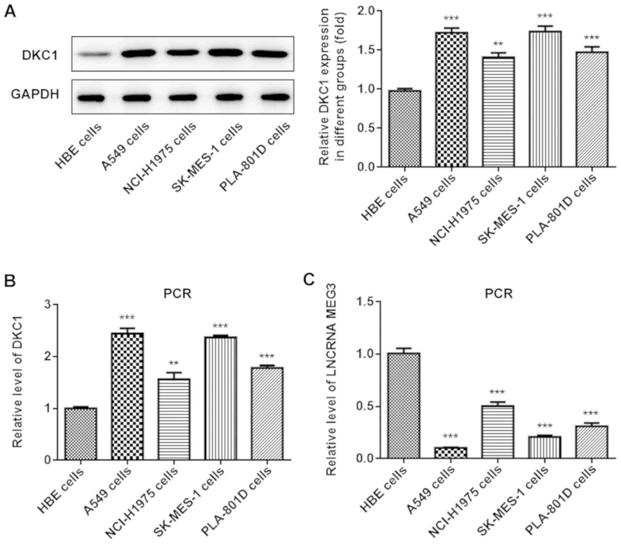
LncRNA MEG3 is downregulated and DKC1
is upregulated in lung cancer cells
The expression levels of lncRNA MEG3 and DKC1 were
assessed in different lung cancer cell lines and normal lung cell
lines, in order to determine their role in lung cancer progression.
Western blot and RT-qPCR analyses demonstrated that DKC1 expression
significantly increased in A549 cells compared with human bronchial
epithelial cells (Fig. 1A and B).
Furthermore, lncRNA MEG3 expression decreased in all the lung cell
lines, particularly in A549 cells (Fig.
1C); thus this cell line was selected for further
experimentation.
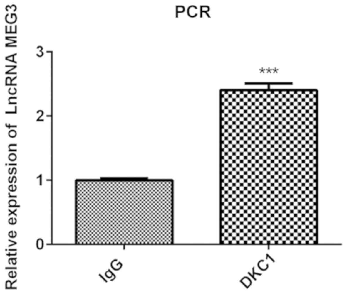
Verification of the binding between
lncRNA MEG3 and DKC1 protein
As predicted by the StarBase database, the results
of the RIP assay verified that DKC1 is the binding protein of
lncRNA MEG3. The results of the present study demonstrated that
lncRNA MEG3 expression increased in the DCK1 group compared with
the IgG group, confirming that lncRNA MEG3 binds to DK1 protein in
lung cancer cells (Fig. 2).
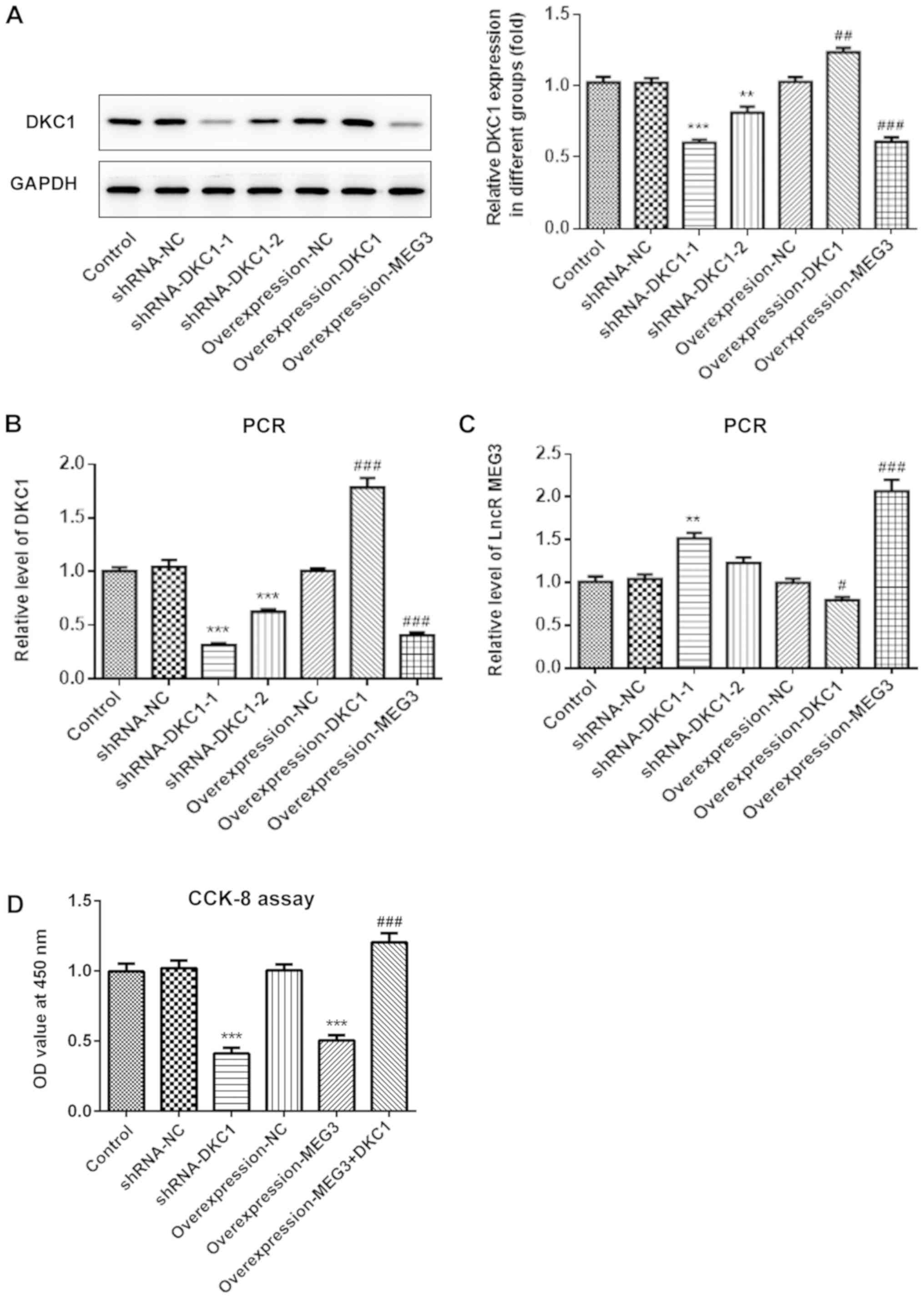
LncRNA MEG3 inhibits cell
proliferation by downregulating DKC1 in A549 cells
Due to the low expression of lncRNA MEG3 in A549
cells, the present study subsequently analyzed the effects of
overexpressing lncRNA MEG3, as well as DKC1 knockdown or
overexpression in lung cancer cells, in order to investigate the
effects of lncRNA MEG3/DKC1 in lung cancer progression. Both
RT-qPCR and western blot analyses demonstrated successful
overexpression of lncRNA MEG3, DKC1 knockdown and overexpression of
DKC1 (Fig. 3A-C). The shRNA-DKC1-1,
which demonstrated higher knockdown efficiency, was selected for
silencing of DKC1. The results demonstrated that overexpression of
lncRNA MEG3 or DKC1 knockdown significantly decreased cell
proliferation. Furthermore, overexpression of DKC1 reversed the
antiproliferative effects exhibited by overexpressing lncRNA MEG3
(Fig. 3D).
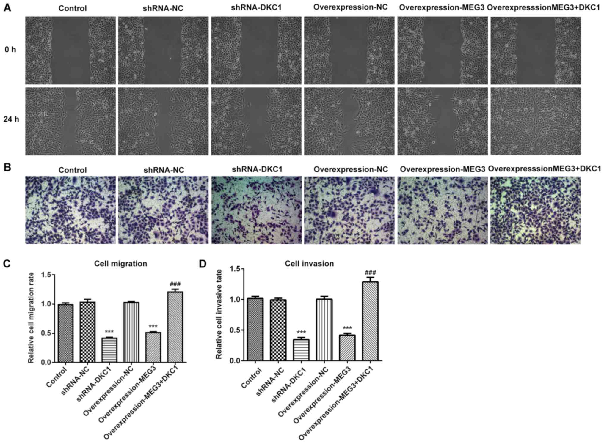
LncRNA MEG3 inhibits cell migration
and invasion by downregulating DKC1 in A549 cells
Both cell migration and invasion decreased following
overexpression of lncRNA MEG3 or DKC1 knockdown, compared with the
control group (Fig. 4A-D). Notably,
overexpression of DKC1 reversed the inhibitory effects exhibited by
overexpressing lncRNA MEG3. Taken together, these results suggest
that A549 cell migration and invasion may be inhibited by lncRNA
MEG3 via the downregulation of DKC1.
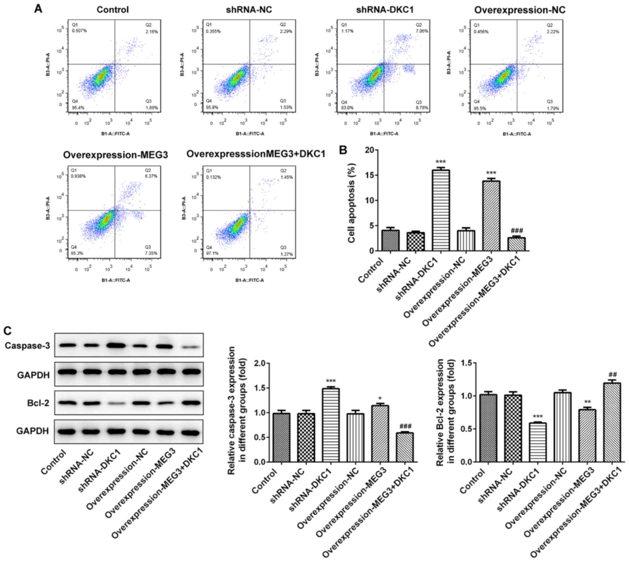
LncRNA MEG3 promotes cell apoptosis by
downregulating DKC1 in A549 cells
Overexpression of lncRNA MEG3 or DKC1 knockdown
increased cell apoptosis (Fig. 5A and
B). Assessment of the apoptosis-associated proteins
demonstrated that the expression level of pro-apoptosis
protein-caspase protein increased, while the expression level of
anti-apoptosis protein-bcl-2 decreased, following overexpression of
lncRNA MEG3 or DKC1 knockdown (Fig.
5C). Notably, overexpression of DKC1 reversed the proapoptotic
effects exhibited by overexpressing lncRNA MEG3, suggesting that
lncRNA MEG3 promotes cell apoptosis by downregulating DKC1.
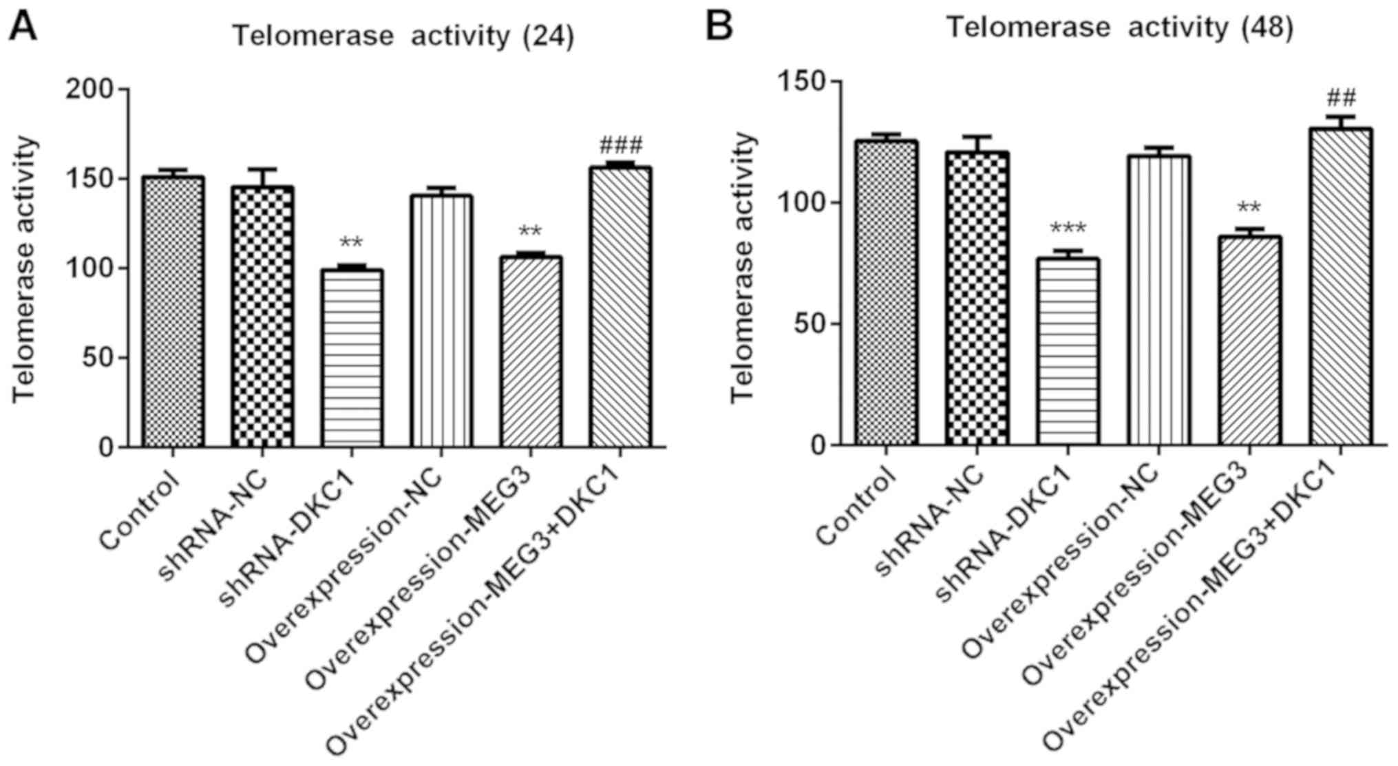
lncRNA MEG3 inhibits telomerase
activity by downregulating DKC1 in A549 cells
Regarded as a well-known tumor biomarker (25), telomerase activity was assessed in
several types of cancer cells. The results of the present study
demonstrated that overexpression of lncRNA MEG3 or DKC1 knockdown
remarkably decreased telomerase activity at 24 and 48 h, compared
with the control group (Fig. 6A and
B). Furthermore, the inhibitory effects of overexpressing
lncRNA MEG3 on cell apoptosis were abolished following
overexpression of DKC1, suggesting that the inhibitory effects of
lncRNA MEG3 on telomerase activity were achieved by downregulating
DKC1.
Discussion
Lung cancer is considered the leading cause of
mortality, thus critical measures are required for its quick and
efficient treatment. Dysregulation of lncRNAs have been confirmed
to play a key role in the progression of lung cancer. Several types
of lncRNAs are aberrantly expressed in lung cancer, including
lncRNA MEG3, which is considered a key RNA in lung cancer (13,26–28).
LncRNA MEG3 expression is significantly decreased in lung cancer,
which is associated with a poor prognosis for patients with lung
cancer (19). lncRNA MEG3 has been
reported to enhance the anticancer effects of vincristine and
cisplatin (19,29). Furthermore, lncRNA MEG3 has been
demonstrated to promote cell apoptosis, inhibit cell migration and
invasion, and induce cell apoptosis in lung cancer (30). However, the underlying molecular
mechanisms of lncRNA MEG3 in the progression of lung cancer remain
unclear.
Increasing evidence suggests that lncRNAs play a
regulatory role by binding to certain types of protein (21). The results of the present study
demonstrated that lncRNA MEG3 inhibited progression of NSCLC by
suppressing telomere function, by targeting DKC1 protein.
Furthermore, DKC1 was verified as a novel binding protein of lncRNA
MEG3 in the present study.
DKC1 has been reported to be upregulated in renal
cell carcinoma, resulting in enhanced cell proliferation, migration
and invasion (31). Furthermore,
overexpression of DKC1 is a prognostic indicator in liver cancer
(32). However, the effects of DKC1
expression in lung cancer remain to be determined. The results of
the present study indicated that DKC1 was upregulated in lung
cancer, suggesting its potential role in the progression of lung
cancer.
The present study revealed that DKC1 is the binding
protein of lncRNA MEG3. The role of lncRNA MEG3 binding to DKC1 in
lung cancer was further investigated. Vital cell processes,
including cell proliferation, apoptosis, invasion and migration
were investigated. The results demonstrated that cell
proliferation, invasion and migration were all inhibited, while
cell apoptosis increased following DKC1 knockdown. The effects of
DKC1 knockdown were in accordance with the effects of
overexpressing lncRNA MEG3 in lung cancer. Overexpression of DKC1
reversed the effects of overexpressing lncRNA MEG3 on cell
apoptosis, proliferation, invasion and migration, suggesting that
the effects of lncRNA MEG3 were achieved by downregulating
DKC1.
DKC1 serves as the encoding gene of the telomerase
subunit, dyskerin, and has been reported to be elevated in several
types of cancer cells (33).
Furthermore, DKC1 has the ability to induce telomerase activity,
resulting in an increase in length of the telomere (34). Its ability to enhance cell
proliferation in cancer has made telomerase the focus of research.
Maintaining the molecular mechanisms of telomere DNA is vital for
cell proliferation, without limitation in cancer. Targeting
telomerase activity is considered to be key in easing tumor
progression (35–37). Currently, DKC1 is highly expressed in
different types of cancer, including glioma and renal cell
carcinoma (31,38,39). In
the present study, telomerase activity was significantly inhibited
following DKC1 knockdown and overexpression of lncRNA MEG3, while
the effects of overexpressing lncRNA MEG3 on telomerase activity
were abolished by overexpressing DKC1, suggesting that the effects
of lncRNA MEG3 on telomerase activity was achieved by targeting and
silencing DKC1. Taken together, the results of the present study
support the hypothesis that the lncRNA MEG3/DKC1 regulatory axis is
a promising therapeutic target for lung cancer.
The present study is not without limitations. First,
in vitro analysis was performed only in A549 cells; thus,
prospective studies should focus on investigating the effects of
the lncRNA MEG3/DKC1 dual regulatory axis, in other lung cancer
cells or in vivo. DKC1 and lncRNA MEG3 are well-established
prognostic indicators in lung cancer, and the results of the
present study provided vital evidence for further study on DKC1, as
well as lncRNA MEG3 in the progression of lung cancer.
Overall, the present study identified a novel dual-
directional regulatory axis, the lncRNA MEG3/DKC1 axis.
Overexpression of lncRNA MEG3 inhibited cell proliferation,
migration, invasion and telomerase activity by sponging DKC1, thus
the lncRNA MEG3/DKC1 axis is a promising therapeutic target for
patients with lung cancer.
Acknowledgements
Not applicable.
Funding
No funding was received.
Availability of data and materials
The datasets used and/or analyzed during the study
are available from the corresponding author upon reasonable
request.
Authors' contributions
ZY designed the present study and drafted the
initial manuscript. ZY and ZW performed the experiments. YD
analyzed the experimental data. All authors read and approved the
final manuscript.
Ethics approval and consent to
participate
Not applicable.
Patient consent for publication
Not applicable.
Competing interests
The authors declare that they have no competing
interests.
References
|
1
|
Siegel RL, Miller KD and Jemal A: Cancer
statistics, 2018. CA Cancer J Clin. 68:7–30. 2018. View Article : Google Scholar : PubMed/NCBI
|
|
2
|
Shen M and Xu Z, Jiang K, Xu W, Chen Y and
Xu Z: Long noncoding nature brain-derived neurotrophic factor
antisense is associated with poor prognosis and functional
regulation in non-small cell lung caner. Tumor Biol.
39:10104283176959482017. View Article : Google Scholar
|
|
3
|
Hong QY, Wu GM, Qian GS, Hu CP, Zhou JY,
Chen LA, Li WM, Li SY, Wang K, Wang Q, et al: Prevention and
management of lung cancer in China. Cancer. 121 (Suppl
17):S3080–S3088. 2015. View Article : Google Scholar
|
|
4
|
Shu J, Wang L, Han F, Chen Y, Wang S and
Luo F: BTBD7 Downregulates E-cadherin and promotes
epithelial-mesenchymal transition in lung cancer. Biomed Res Int.
2019:59376352019. View Article : Google Scholar : PubMed/NCBI
|
|
5
|
Hoffman RM and Sanchez R: Lung cancer
screening. Med Clin North Am. 101:769–785. 2017. View Article : Google Scholar : PubMed/NCBI
|
|
6
|
O'Dowd EL and Baldwin DR: Early diagnosis
pivotal to survival in lung cancer. Practitioner. 258:21–24, 2-3.
2014.
|
|
7
|
Jacobsen MM, Silverstein SC, Quinn M,
Waterston LB, Thomas CA, Benneyan JC and Han PKJ: Timeliness of
access to lung cancer diagnosis and treatment: A scoping literature
review. Lung Cancer. 112:156–164. 2017. View Article : Google Scholar : PubMed/NCBI
|
|
8
|
Guibert N, Tsukada H, Hwang DH, Chambers
E, Cibas ES, Bale T, Supplee J, Ulrich B, Sholl LM, Paweletz CP and
Oxnard GR: Liquid biopsy of fine-needle aspiration supernatant for
lung cancer genotyping. Lung Cancer. 122:72–75. 2018. View Article : Google Scholar : PubMed/NCBI
|
|
9
|
Heerink WJ, de Bock GH, de Jonge GJ, Groen
HJ, Vliegenthart R and Oudkerk M: Complication rates of CT-guided
transthoracic lung biopsy: Meta-analysis. Eur Radiol. 27:138–148.
2017. View Article : Google Scholar : PubMed/NCBI
|
|
10
|
Woodard GA, Jones KD and Jablons DM: Lung
cancer staging and prognosis. Cancer Treat Res. 170:47–75. 2016.
View Article : Google Scholar : PubMed/NCBI
|
|
11
|
Shi YX, Sheng DQ, Cheng L and Song XY:
Current landscape of epigenetics in lung cancer: Focus on the
mechanism and application. J Oncol. 2019:81073182019. View Article : Google Scholar : PubMed/NCBI
|
|
12
|
Jathar S, Kumar V, Srivastava J and
Tripathi V: Technological developments in lncRNA biology. Adv Exp
Med Biol. 1008:283–323. 2017. View Article : Google Scholar : PubMed/NCBI
|
|
13
|
Chen R, Li WX, Sun Y, Duan Y, Li Q, Zhang
AX, Hu JL, Wang YM and Gao Y: Comprehensive analysis of lncRNA and
mRNA expression profiles in lung cancer. Clin Lab. 63:313–320.
2017. View Article : Google Scholar : PubMed/NCBI
|
|
14
|
Wei GH and Wang X: lncRNA MEG3 inhibit
proliferation and metastasis of gastric cancer via p53 signaling
pathway. Eur Rev Med Pharmacol Sci. 21:3850–3856. 2017.PubMed/NCBI
|
|
15
|
Zhou Y, Zhang X and Klibanski A: MEG3
noncoding RNA: A tumor suppressor. J Mol Endocrinol. 48:R45–R53.
2012. View Article : Google Scholar : PubMed/NCBI
|
|
16
|
Zhou Y, Yang H, Xia W, Cui L, Xu R, Lu H,
Xue D, Tian Z, Ding T, Cao Y, et al: LncRNA MEG3 inhibits the
progression of prostate cancer by facilitating H3K27 trimethylation
of EN2 through binding to EZH2. J Biochem. 167:295–301. 2019.
View Article : Google Scholar
|
|
17
|
Zhu X, Zhang HW, Chen HN, Deng XJ, Tu YX,
Jackson AO, Qing JN, Wang AP, Patel V and Yin K: Perivascular
adipose tissue dysfunction aggravates adventitial remodeling in
obese mini pigs via NLRP3 inflammasome/IL-1 signaling pathway. Acta
Pharmacol Sin. 40:46–54. 2019. View Article : Google Scholar : PubMed/NCBI
|
|
18
|
Wu JL, Meng FM and Li HJ: High expression
of lncRNA MEG3 participates in non-small cell lung cancer by
regulating microRNA-7-5p. Eur Rev Med Pharmacol Sci. 22:5938–5945.
2018.PubMed/NCBI
|
|
19
|
Zhang Z, Liu T, Wang K, Qu X, Pang Z, Liu
S, Liu Q and Du J: Down-regulation of long non-coding RNA MEG3
indicates an unfavorable prognosis in non-small cell lung cancer:
Evidence from the GEO database. Gene. 630:49–58. 2017. View Article : Google Scholar : PubMed/NCBI
|
|
20
|
Terashima M, Tange S, Ishimura A and
Suzuki T: MEG3 long noncoding RNA contributes to the epigenetic
regulation of epithelial-mesenchymal transition in lung cancer cell
lines. J Biol Chem. 292:82–99. 2017. View Article : Google Scholar : PubMed/NCBI
|
|
21
|
Ferrè F, Colantoni A and Helmer-Citterich
M: Revealing protein-lncRNA interaction. Brief Bioinform.
17:106–116. 2016. View Article : Google Scholar : PubMed/NCBI
|
|
22
|
Fernandez-Garcia I, Marcos T,
Muñoz-Barrutia A, Serrano D, Pio R, Montuenga LM and
Ortiz-de-Solorzano C: Multiscale in situ analysis of the role of
dyskerin in lung cancer cells. Integr Biol (Camb). 5:402–413. 2013.
View Article : Google Scholar : PubMed/NCBI
|
|
23
|
Penzo M, Ludovini V, Treré D, Siggillino
A, Vannucci J, Bellezza G, Crinò L and Montanaro L: Dyskerin and
TERC expression may condition survival in lung cancer patients.
Oncotarget. 6:21755–21760. 2015. View Article : Google Scholar : PubMed/NCBI
|
|
24
|
Livak KJ and Schmittgen TD: Analysis of
relative gene expression data using real-time quantitative PCR and
the 2(-Delta Delta C(T)) method. Methods. 25:402–408. 2001.
View Article : Google Scholar : PubMed/NCBI
|
|
25
|
Kulić A, Plavetić ND, Gamulin S,
Jakić-Razumović J, Vrbanec D and Sirotković-Skerlev M: Telomerase
activity in breast cancer patients: Association with poor prognosis
and more aggressive phenotype. Med Oncol. 33:232016. View Article : Google Scholar : PubMed/NCBI
|
|
26
|
Loewen G, Jayawickramarajah J, Zhuo Y and
Shan B: Functions of lncRNA HOTAIR in lung cancer. J Hematol Oncol.
7:902014. View Article : Google Scholar : PubMed/NCBI
|
|
27
|
Tan BS, Yang MC, Singh S, Chou YC, Chen
HY, Wang MY, Wang YC and Chen RH: LncRNA NORAD is repressed by the
YAP pathway and suppresses lung and breast cancer metastasis by
sequestering S100P. Oncogene. 38:5612–5626. 2019. View Article : Google Scholar : PubMed/NCBI
|
|
28
|
Zhao Y, Feng C, Li Y, Ma Y and Cai R:
LncRNA H19 promotes lung cancer proliferation and metastasis by
inhibiting miR-200a function. Mol Cell Biochem. 460:1–8. 2019.
View Article : Google Scholar : PubMed/NCBI
|
|
29
|
Xia H, Qu XL, Liu LY, Qian DH and Jing HY:
LncRNA MEG3 promotes the sensitivity of vincristine by inhibiting
autophagy in lung cancer chemotherapy. Eur Rev Med Pharmacol Sci.
22:1020–1027. 2018.PubMed/NCBI
|
|
30
|
Zhao Y, Zhu Z, Shi S, Wang J and Li N:
Long non-coding RNA MEG3 regulates migration and invasion of lung
cancer stem cells via miR-650/SLC34A2 axis. Biomed Pharmacother.
120:1094572019. View Article : Google Scholar : PubMed/NCBI
|
|
31
|
Zhang M, Pan Y, Jiang R, Hou P, Shan H,
Chen F, Jiang T, Bai J and Zheng J: DKC1 serves as a potential
prognostic biomarker for human clear cell renal cell carcinoma and
promotes its proliferation, migration and invasion via the NF-κB
pathway. Oncol Rep. 40:968–978. 2018.PubMed/NCBI
|
|
32
|
Liu B, Zhang J, Huang C and Liu H:
Dyskerin overexpression in human hepatocellular carcinoma is
associated with advanced clinical stage and poor patient prognosis.
PLoS One. 7:e431472012. View Article : Google Scholar : PubMed/NCBI
|
|
33
|
Nersisyan L, Hopp L, Loeffler-Wirth H,
Galle J, Loeffler M, Arakelyan A and Binder H: Telomere length
maintenance and its transcriptional regulation in lynch syndrome
and sporadic colorectal carcinoma. Front Oncol. 9:11722019.
View Article : Google Scholar : PubMed/NCBI
|
|
34
|
Richards LA, Kumari A, Knezevic K, Thoms
JA, von Jonquieres G, Napier CE, Ali Z, O'Brien R, Marks-Bluth J,
Maritz MF, et al: DKC1 is a transcriptional target of GATA1 and
drives upregulation of telomerase activity in normal human
erythroblasts. Haematologica. 105:1517–1526. 2020. View Article : Google Scholar : PubMed/NCBI
|
|
35
|
Bernardes de Jesus B and Blasco MA:
Telomerase at the intersection of cancer and aging. Trends Gene.
29:513–520. 2013. View Article : Google Scholar
|
|
36
|
Mizukoshi E and Kaneko S:
Telomerase-targeted cancer immunotherapy. Int J Mol Sci.
20:18232019. View Article : Google Scholar
|
|
37
|
Yuan X, Larsson C and Xu D: Mechanisms
underlying the activation of TERT transcription and telomerase
activity in human cancer: Old actors and new players. Oncogene.
38:6172–6183. 2019. View Article : Google Scholar : PubMed/NCBI
|
|
38
|
Miao FA, Chu K, Chen HR, Zhang M, Shi PC,
Bai J and You YP: Increased DKC1 expression in glioma and its
significance in tumor cell proliferation, migration and invasion.
Invest New Drugs. 37:1177–1186. 2019. View Article : Google Scholar : PubMed/NCBI
|
|
39
|
Penzo M, Casoli L, Ceccarelli C, Treré D,
Ludovini V, Crinò L and Montanaro L: DKC1 gene mutations in human
sporadic cancer. Histol Histopathol. 28:365–372. 2013.PubMed/NCBI
|