Introduction
Cervical cancer is a fast-growing malignancy
(1). Population statistics in 2018
reported that cervical cancer ranks fourth for both incidence and
mortality for different types of cancer among women worldwide
(2). A total of 570,000 new cases
are diagnosed and 311,000 cervical cancer-associated mortalities
occur globally each year (2).
Despite notable improvements in early screening, effective
diagnosis of cervical cancer remains difficult, with a 5-year
survival rate of 40–60% (3,4). This is due to the fact that recurrence
and metastasis occur in ~30% of patients following treatment
(3). The molecular mechanisms
involved in the progression of cervical cancer remain unclear.
Thus, the discovery of novel molecules and targets for cervical
cancer diagnosis and treatment is critical.
Chromatin assembly factor (CAF)-1 is a heterologous
complex purified from 293T cells and contains three subunits (p150,
p60 and p48), according to molecular weight (4). These subunits can form larger
structures and directly bind to the acetylated forms of histones H3
and H4 to promote the assembly of histone proteins on newly
synthesized DNA (5). CAF-1 promotes
nucleosome assembly on DNA undergoing replication and is involved
in the DNA repair process (6). CAF-1
expression is absent or very low in cells that are in the resting
state and is highly expressed in proliferating cells (6). CAF-1 expression is associated with the
proliferative state of cells and has previously been validated as a
useful proliferation marker (7). In
the mitotic phase of the cell cycle, CAF-1 accumulates in the
nucleolus and stabilizes the heterochromatin structure during
replication (8).
It has been demonstrated that CAF-1 can be used to
predict the clinical outcome of patients with cancer (9). CAF-1 protein is aberrantly expressed in
different tumor tissues and its high expression is significantly
associated with high histological grade in breast and renal cell
carcinomas, and with advanced disease stage in endometrial and
renal carcinomas (10,11). It has been reported that CAF-1
expression in cervical cancer tissues is positively associated with
Ki-67 expression and negatively associated with the overall
survival (OS) time of patients (12). OS defined as the time from
randomization to death from any cause, is a direct measure of
clinical benefit to a patient. Notably, the OS time of patients
with cervical cancer, with low CAF-1 expression, is prolonged
(12). This suggests that high CAF-1
expression levels may be used as a predictor of adverse clinical
outcomes in cervical carcinomas. However, the clinical significance
of p150 expression in cervical cancer tissues, and its role in
cervical cancer cells have not yet been fully investigated.
In the present study, immunohistochemical analysis
was used to detect the CAF-1/p150 expression in cervical cancer
tissues and to investigate the clinical significance of CAF-1/p150
in cervical cancer. An in vitro cell culture model including
HeLa and SiHa cervical cancer cell lines was used to silence
CAF-1/p150 expression and further investigate its function in
cervical cancer.
Materials and methods
Reagents
The recombinant anti-CAF-1/p150 antibody used for
immunohistochemistry (IHC; cat. no. ab109442) and western blot
(cat. no. ab126625) analyses was purchased from Abcam. The
anti-rabbit IgG horseradish peroxidase (HRP)-conjugated secondary
antibody (1:5,000; cat. no. BS10043) was purchased from Bioworld
Technology, Inc. The biotin-labeled anti-rabbit secondary antibody
(cat. no. A0277, 1:1,000) was purchased from Beyotime Institute of
Biotechnology, Inc. Matrigel (cat. no. 356234) was purchased from
BD Biosciences. The two-step immunohistochemical staining detection
kit (cat. no. PV-9000) and 3′-diaminobenzidine (DAB) substrate were
purchased from Beijing Zhongshan Golden Bridge Biotechnology Co.,
Ltd. The hematoxylin and eosin (H&E) staining kit (cat. no.
G1121) was purchased from Beijing Solarbio Science & Technology
Co., Ltd.
Patients and specimens
Paraffin specimens were collected from patients with
cervical cancer following biopsy or surgical resection at the Zunyi
Medical College Affiliated Hospital (Zunyi, China) between January
2013 and August 2014. A total of 214 cases met the screening
criteria, and 80 cases who did not undergo any anti-tumor
treatment, including surgery, radiotherapy and chemotherapy,
biological immunotherapy and traditional Chinese medical treatment,
were enrolled onto the present study (patient age range, 29–72;
mean age, 46.2 years). The clinicopathological characteristics of
these patients are listed in Table
I. Normal cervical tissue specimens (20 cases) were obtained
from 20 patients (patient age range, 35–47; mean age, 41.6 years)
who underwent total hysterectomy due to uterine fibroids, and the
absence of cervical lesions was confirmed via pathological analysis
by two pathologists (Zunyi Medical College Affiliated Hospital,
China). The tumor stage of each patient was determined according to
the International Federation of Gynecology and Obstetrics (FIGO,
2009) (13). The present study was
approved by the Human Ethics Committee of Zunyi Medical College
Affiliated Hospital [Zunyi, China, approval no. ZMC2012(841)]. The
experiments were performed in accordance with The Declaration of
Helsinki and in agreement with Chinese legislation. Written
informed consent was provided by all patients prior to the study
start.
 | Table I.Association between CAF-1/p150
expression and clinicopathological characteristics of patients with
cervical cancer (n=80). |
Table I.
Association between CAF-1/p150
expression and clinicopathological characteristics of patients with
cervical cancer (n=80).
|
|
| CAF-1/p150
expression |
|
|
|---|
|
|
|
|
|
|
|---|
| Characteristic | Patient number,
n | Low | High | χ2
value | P-value |
|---|
| Age, years |
|
|
| 2.064 | 0.151 |
|
<50 | 21 | 8 | 13 |
|
|
| ≥50 | 59 | 13 | 46 |
|
|
| Tumor size, cm |
|
|
| 1.214 | 0.271 |
|
<4 | 57 | 13 | 44 |
|
|
| ≥4 | 23 | 8 | 15 |
|
|
| Pathology |
|
|
| 0.231 | 0.631 |
| Squamous
cell carcinoma | 70 | 19 | 51 |
|
|
|
Adenocarcinoma | 10 | 2 | 8 |
|
|
| Vascular
invasion |
|
|
| 0.195 | 0.659 |
| No | 58 | 16 | 42 |
|
|
|
Yes | 22 | 5 | 17 |
|
|
| Lymphatic
metastasis |
|
|
| 0.016 | 0.898 |
| No | 58 | 15 | 43 |
|
|
|
Yes | 22 | 6 | 16 |
|
|
| FIGO stage |
|
|
| 5.230 | 0.022a |
|
I–II | 40 | 15 | 25 |
|
|
|
III–IV | 40 | 6 | 34 |
|
|
IHC
Tumor tissues were fixed in formalin (10%
formaldehyde) for 48 h at 25°C, embedded in paraffin and the
samples were cut into 4-µm-thick sections, which were mounted onto
slides for H&E staining or immunohistochemical analysis of
CAF-1/p150 expression. Briefly, the tissue sections were
deparaffinized by heating at 60°C for 2 h, washed three times with
xylene (for 5 min each) and rehydrated in a descending ethanol
series (100, 95, 90, 80 and 70% ethanol for 5 min each). Antigen
retrieval was achieved following incubation in 10 mM sodium citrate
(Sigma-Aldrich; Merck KGaA) buffer (pH 6.0) for 15 min at 98°C.
Subsequently, tissue sections were incubated with 3%
H2O2 for 20 min at 25°C to inhibit endogenous
peroxidase activity. The slides were blocked with 100% goat serum
(Dako; Agilent Technologies, Inc.) for 30 min 25°C and incubated
with rabbit anti-human CAF-1/p150 antibody (1:100) overnight at
4°C. Following the primary incubation, tissue sections were
subsequently incubated with biotin-labeled anti-rabbit secondary
antibody for 20 min and streptavidin-HRP for 2 min at 25°C. The
slides were stained with DAB substrate for 15 sec at 25°C and
counterstained with hematoxylin for 2 min at 25°C. Negative
controls were incubated with pre-immune serum instead of the
antibody overnight at 4°C.
CAF-1/p150 expression was evaluated by 2
pathologists above mentioned in a double-blind experimental design.
The staining intensity score was calculated as follows: 0,
completely absent; 1, light yellow; 2, light brown and 3, dark
brown. The area occupied by positive cells score was estimated as
follows: 0 points, ≤10%; 1 point, 11–25%; 2 points, 26–50% and 3
points, >50%. The area occupied by positive cells score is the
average number of positive cells in 5 randomly selected areas. The
total score was calculated as the sum of the staining intensity
score and the area occupied by positive cells score, as follows: 0
points, (−); 1–2 points, (+); 3–4 points, (++) and 5–6 points,
(+++). (−) and (+) are defined as low expression, and (++) and
(+++) are defined as high expression. The cut-off value is ≥3.0.
Images were captured using a CKX41 inverted light microscope
(magnification, ×100) and NIS-Elements F3.0 acquisition
software.
Follow-up information
All patients received standardized treatment
following diagnosis. Routine telephone follow-up was performed
every 3–6 months for the first 2 years and every 6–12 months for
the next 3–5 years from January 2013 to April 2016. The total
duration of follow-up was 40 months. The patients were assessed for
local recurrence, distant metastasis and OS. Local recurrence and
distant metastasis were confirmed via pathological analysis. OS was
defined as the first day of treatment until mortality caused by
cervical cancer or the last date of follow-up. A total of 80
patients were followed up for 8–40 months. The median follow-up
time was 25 months. A total of 16 patients exhibited local
recurrence, 12 patients presented with distant metastasis and 9
patients did not survive due to cervical cancer. The OS time of
patients with high CAF-1/p150 expression was between 8–37 months,
whereas the OS time of patients with low CAF-1/p150 expression was
between 10–40 months.
Cell lines and cell culture
The HeLa and SiHa cell lines were purchased from the
State Key Laboratory of Biotherapy, Sichuan University (passage
number, 5–20, Sichuan University, Sichuan, China). Cells were
maintained in Dulbecco's modified Eagle's medium (DMEM)
supplemented with 10% calf serum (CS), 100 U/ml penicillin and 100
µg/ml streptomycin (all purchased from Gibco; Thermo Fisher
Scientific, Inc.), at 37°C in 5% CO2.
RNA interference
CAF-1/p150 small interfering (si)RNA (forward,
5′-GCAUGUGCAUCACCCAAUUTT-3′ and reverse,
5′-AAUUGGGUGAUGCACAUGCTT-3′) and non-specific siRNA control
(forward, 5′-UUCUCCGAACGUGUCACGUTT-3′ and reverse,
5′-ACGUGACACGUUCGGAGAATT-3′) were designed and synthesized by
Shanghai GenePharma Co., Ltd., and transfected into the HeLa and
SiHa cells using 25 pmol/5×105 cells Lipofectamine 2000
reagent (Invitrogen; Thermo Fisher Scientific, Inc.) for 48 h,
according to the manufacturer's protocol. After transfection, the
HeLa and SiHa cells were cultured in fresh medium for next 24 h and
then used for the subsequent experiments.
Wound healing assay
Following transfection with siRNA, the HeLa and SiHa
cells (2×10⁴ cells/well) were seeded into 12-well plates
and scratched using a 10-µl micropipette tip once they reached ~90%
confluence. Cells were subsequently incubated with serum-free
medium for 48 h at 37°C in 5% CO2. Images were captured
at 0 and 48 h using a CKX41 inverted light microscope (Olympus
Corporation; magnification, ×100) and NIS-Elements F3.0 acquisition
software (Olympus Corporation). The migration rate was calculated
as the difference between the wound width at 0 and 48 h. The
migration rate=(W0 h-W48 h)/W0 h.
Data are presented as the fold-change relative to the non-specific
siRNA control cells.
Transwell assay
A Matrigel-coated Transwell chamber system (8-µm
pore size; EMD Millipore) was used to assess the migratory and
invasive abilities of cervical cancer cells in the absence or
presence of siRNA sequences. The Matrigel was precoated on the
membrane at 37°C for 30 min.
Following transfection with siRNA, the HeLa and SiHa
cells were digested by 0.25% Trypsin-EDTA (Gibico, Thermo Fisher
Scientific, Inc.) and plated in the upper chambers of Transwell
plates in serum-free DMEM medium (5×10⁴ cells/ml; 400
µl). DMEM Medium supplemented with 10% CS (600 µl) was added plated
in the lower chambers. Following incubation for 24 h at 37°C in 5%
CO2, cells that did not cross the membrane were removed
using cotton swabs, while cells that successfully crossed the
membrane were fixed with 100% methanol at 25°C for 20 min and
stained with H&E at 25°C for 20 min. Images were captured using
a CKX41 inverted light microscope (magnification, ×100) and
NIS-Elements F3.0 acquisition software.
Colony formation assay
The colony formation assay was performed using 0.7%
agarose (Sigma-Aldrich; Merck KGaA) as the bottom layer in 6-well
plates. A second layer containing agarose was also used. Following
transfection with siRNA, the Hela and SiHa cells were digested and
resuspended in DMEM medium containing 0.35% agarose, which was used
as the upper layer (1,000 cells/well). The gel was solidified, and
1 ml of DMEM medium (containing 10% CS, 100 U/ml penicillin and 100
µg/ml streptomycin) was added into the plates to prevent drying.
Cells were cultured in fresh DMEM medium (containing 10% CS, 100
U/ml penicillin and 100 µg/ml streptomycin) every 3 days. Following
incubation for 30 days at 37°C in 5% CO2, cell colonies
were stained with 0.005% crystal violet at 25°C for 1 min and
imaged using a D7200 digital color camera (Nikon Corporation). The
number of colonies were analyzed using ImageJ software (National
Institutes of Health). Colony formation was calculated as the
number colonies formed in the siRNA group divided by the number of
colonies in the non-specific siRNA group.
Western blotting
The HeLa and SiHa cells were lysed in modified RIPA
buffer (Beyotime Institute of Biotechnology), containing Tris-HCl
50 mM, pH: 7.4, 1% NP-40, 0.25% Na-deoxycholate, 150 mM NaCl, 1 mM
EDTA, 1 mM PMSF, 1 µg/ml Aprotinin, 1 µg/ml leupeptin, 1 µg/ml
pepstatin, 1 mM Na3VO4, 1 mM NaF) and
centrifuged at 13,000 × g for 30 min at 4°C. Protein concentration
was determined using a bicinchoninic acid protein assay. Equal
amounts of protein (50 µg) were separated via 12% SDS-PAGE and
transferred onto polyvinylidene fluoride membranes (EMD Millipore).
Membranes were blocked with 5% non-fat milk for 1 h at 37°C and
subsequently incubated with CAF-1/p150 antibody (1:5,000) overnight
at 4°C. Following the primary incubation, membranes were incubated
with anti-rabbit secondary antibody for 1 h at 37°C. Protein bands
were detected using the Horseradish Peroxidase Color Development
kit (cat. no. p0018s-1; Beyotime Institute of Biotechnology),
visualized using a chemiluminescence imaging system (Thermo Fisher
Scientific, Inc.) and evaluated using Image Lab software version
6.0 (Bio-Rad Laboratories).
Statistical analysis
Statistical analysis was performed using SPSS
software (version 21.0; IBM Corp.) Data are presented as the mean ±
standard deviation. In vitro experiments were repeated three
times. Student's t-test (unpaired) was used to compare differences
between two groups. χ2 test (unpaired) was used to
determine the association between CAF-1/p150 expression and
clinicopathological characteristics of patients with cervical
cancer. Univariate analysis by χ2 test was used to the
significant differences between CAF-1/p150 expression and variables
listed in Table I. Student's t-test,
χ2 test and univariate analysis were performed using
SPSS version 15.0 (SPSS Inc.). Kaplan-Meier survival analysis and
the Renyi test were performed to determine patient survival
according to CAF-1/p150 expression, using the program in SAS
software. P<0.05 was considered to indicate a statistically
significant difference.
Results
CAF-1/p150 expression in normal and
cervical cancer tissues
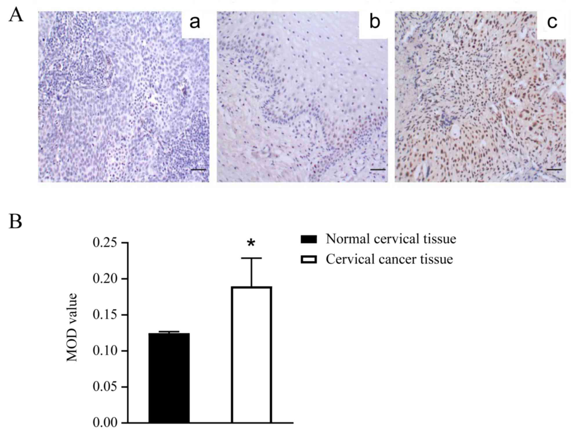
IHC analysis of CAF-1/p150 expression in normal and
cervical cancer tissues is presented in Fig. 1A (b) and (c), respectively. The
negative control, without CAF-1/p150 antibody in cervical cancer
tissues is presented in Fig. 1A (a).
CAF-1/p150 protein expression was localized in the nuclei of normal
and cervical cancer cells. Specifically, low CAF-1/p150 expression
(+) was observed in 13/20 cases of normal cervical tissues and in
21/80 cases of cervical cancer tissues. Moderate CAF-1/p150
expression (++) was observed in 7/20 cases of normal cervical
tissues and in 5/80 cases of cervical cancer tissues. High
CAF-1/p150 expression (+++) was observed in 54/80 cases of cervical
cancer tissues but not in normal cervical tissues. It was
demonstrated that CAF-1/p150 staining intensity was stronger in
cervical cancer tissues compared with normal cervical tissues.
Furthermore, a statistically significant difference of IHC analysis
in CAF-1/p150 expression which is represented by mean optical
density (MOD) was observed between normal cervical tissues
(0.1247±0.0021) and cervical cancer tissues (0.1896±0.0392,
P=0.001, Fig. 1A and B).
High CAF-1/p150 expression indicates
poor clinical outcomes in patients with cervical cancer
Univariate analysis demonstrated no significant
differences between CAF-1/p150 expression and age, tumor size,
pathological type, vascular invasion and lymph node metastasis
(Table I). Conversely, CAF-1/p150
expression was significantly associated with the different FIGO
stages of patients with cervical cancer (χ2=5.230;
P=0.022 Table I). Among the 59
patients with high CAF-1/p150 expression, eight patients did not
survive, 13 patients exhibited local recurrence and 10 patients
presented with distant metastasis. Among the patients with low
CAF-1/p150 expression, 1 patient did not survive, three patients
presented with local recurrence and two patients exhibited distant
metastasis. The results indicated that high CAF-1/p150 expression
was significantly associated with local recurrence and distant
metastasis (χ2=4.091; P=0.043; Table II), suggesting that high CAF-1/p150
expression is indicative of a poor prognosis of patients with
cervical cancer.
| Table II.Association between CAF-1/p150
expression and clinical outcomes in patients with cervical
cancer. |
Table II.
Association between CAF-1/p150
expression and clinical outcomes in patients with cervical
cancer.
|
| CAF-1/p150
expression |
|
|
|---|
|
|
|
|
|
|---|
| Follow-up | High | Low | χ2
value | P-value |
|---|
| Local
recurrence | 13 | 3 | 4.091 | 0.043 |
| Distant
metastasis | 10 | 2 |
|
|
| Death | 8 | 1 |
|
|
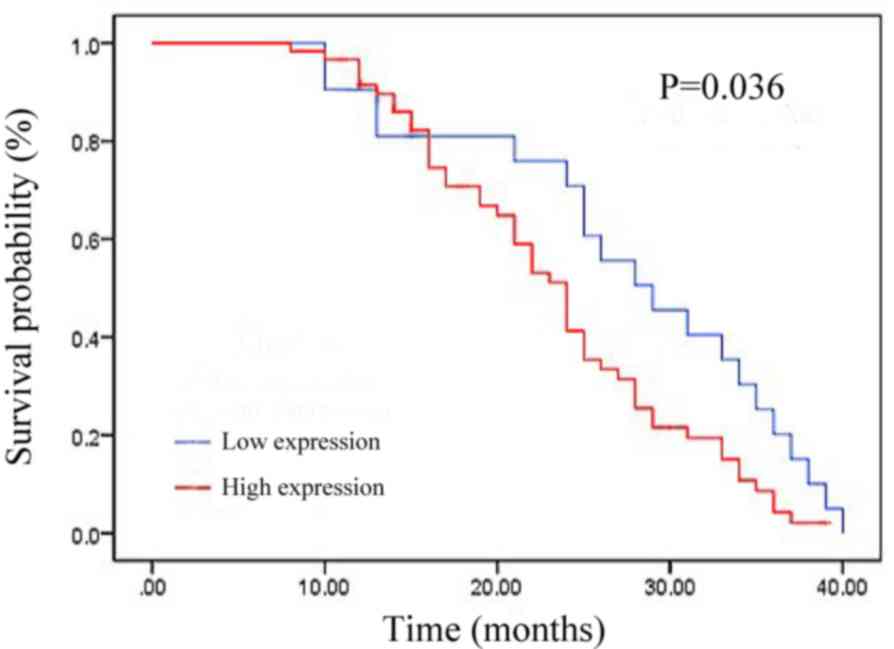
The association between CAF-1/p150 expression and OS
time of patients with cervical cancer was assessed. The OS time of
patients with high CAF-1/p150 expression was significantly lower
than that of patients with low CAF-1/p150 expression (P=0.036;
Fig. 2). These results suggest that
high CAF-1/p150 expression is associated with a low OS time of
patients with cervical cancer.
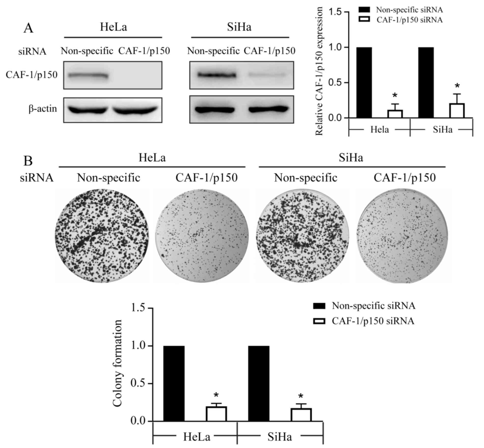
CAF-1/p150 knockdown impairs cervical
cancer cell proliferation
In vitro analysis of CAF-1/p150 was performed
using Hela and SiHa cervical cancer cells, and transfection
efficiency was assessed via western blot analysis. Cells
transfected with siRNA-CAF-1/p150 decreased the endogenous
expression of this protein compared with the non-specific siRNA
transfected cells (Fig. 3A). The
colony formation assay demonstrated that the number of cell
colonies significantly decreased in Hela and SiHa cells following
transfection with siRNA-CAF-1/p150 (Fig.
3B).
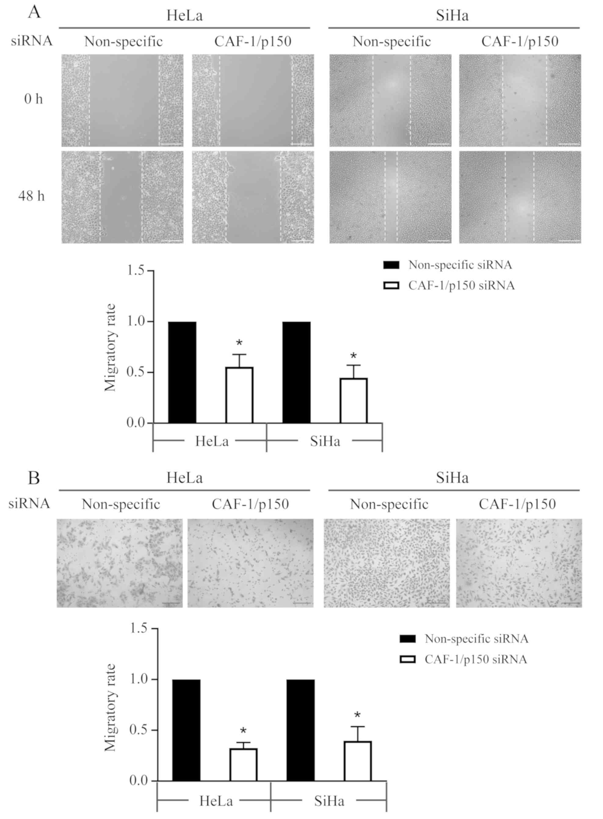
CAF-1/p150 knockdown suppresses the
migratory ability of cervical cancer cells
Wound healing and Transwell assays were performed to
assess the cellular lateral and chemotactic migratory abilities of
cervical cancer cells. The wound healing assay indicated that the
number of migratory cells decreased to 55.6±6.9 and 44.8±7.1% in
siRNA-CAF-1/p150 transfected Hela and SiHa cells, respectively
(Fig. 4A). Furthermore, the number
of migratory cells that penetrated the Transwell membrane
significantly decreased to 45.2±7.7 and 51±5.4% in Hela and SiHa
cells, respectively, following transfection with siRNA-CAF-1/p150
(Fig. 4B).
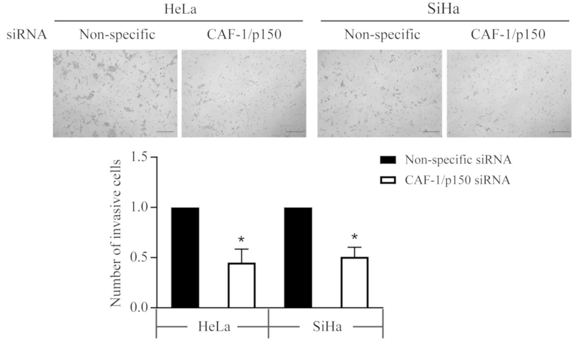
CAF-1/p150 knockdown suppresses the
invasive ability of cervical cancer cells
Transwell plates pre-coated with Matrigel were used
to assess the invasive ability of cervical cancer cells. Compared
with the invasive cells in the non-specific siRNA transfected
cells, the invasive cells in siRNA-CAF-1/p150 transfected HeLa and
SiHa cells reduced to 32.6±3.1 and 39.7±8.0%, respectively
(Fig. 5).
Discussion
In recent years, the early diagnosis of cervical
cancer has notably improved with the development of specific
screening methods (14). However,
the existing screening methods of cervical cancer, including
exfoliated cytology and high-risk HPV detection, lack sensitivity
and specificity (15). Thus, it is
necessary to identify novel prognostic markers that can describe
the degree of malignancy and provide information regarding the
occurrence and mortality of patients with cervical cancer.
Furthermore, the identification of these molecules can aid the
design of therapeutic agents for cervical cancer.
Previous studies have assessed the expression levels
of CAF-1 subunits via IHC analysis (16–19).
These findings may help improve the prognosis of different types of
solid human malignancies. In the present study, IHC analysis of
CAF-1/p150 expression was performed in 20 normal cervical tissue
slices and 80 cervical cancer tissue slices. The results
demonstrated that CAF-1/p150 expression was elevated in cervical
cancer tissues compared with normal cervical tissues. Although
three subunits (p150, p60 and p48) of CAF-1 have been identified,
their association with the clinicopathological characteristics of
the progression of cervical cancer requires further investigation.
CAF-1/p60 expression is associated with the histological grading of
breast cancer, cervical cancer and endometrial carcinoma, and with
the clinical stage of endometrial carcinoma (12). In the present study, the association
between CAF-1/p150 expression and prognosis of patients with
cervical cancer was assessed. The results indicated that the
expression levels of CAF-1/p150 protein in cervical cancer tissues
were closely associated with the FIGO stage, whereas no significant
associations were observed between CAF-1/p150 expression and age,
tumor size, pathological type, vascular infiltration and lymph node
metastasis. The CAF-1 subunit, p60 is considered a novel prognostic
marker in oral squamous cell carcinoma, prostate cancer, salivary
gland tumors and cutaneous melanoma (8,11).
Overexpression of CAF-1/p60 is significantly associated with the
biological aggressiveness of tumors and their metastatic phenotype
(8,11,20).
Similarly, the results of the present study demonstrated that
cervical cancer cases with high CAF-1/p150 expression were
significantly associated with tumor recurrence, metastasis and
shorter OS time.
Previous studies have predominantly focused on the
function of CAF-1 on cell proliferation and cell cycle distribution
(9,21–23).
CAF-1 is associated with maintaining appropriate nucleosome
assembly during DNA synthesis and DNA repair (9). Among the three subunits, p48 is mainly
responsible for acetylation/deacetylation of histones (21), whereas the p60 subunit acts
specifically on cell replication (22) and the p150 subunit induces high
activity by regulating the DNA double-strand break repair process
(23). Thus, CAF-1/p150 exerts a key
role in cell proliferation, most likely due to the high demand for
DNA replication and chromatin formation.
CAF-1/p150 appears to function upstream of Ki-67, as
it regulates the nucleolar localization and the peri-chromosomal
layer accumulation of Ki-67 during interphase and mitosis (24). Consistent with previous findings, the
results of the present study demonstrated that the
anchorage-independent proliferative ability, which leads to colony
formation in soft agar decreased when CAF-1/p150 protein was
knocked down in the Hela and SiHa cervical cancer cell lines.
Furthermore, the clinical observations from the cervical cancer
samples suggested that CAF-1/p150 exhibited additional functions
beyond DNA replication. The speculation that this protein may
possess a promoting role in driving metastasis of cervical cancer
was confirmed through the use of cell line models. CAF-1/p150
knockdown suppressed cervical cell migration and invasion, which
are the two necessary biological processes for tumor metastasis
(25). Thus, both in vitro
and in vivo analyses confirmed that CAF-1/p150 exerted a
positive function in cell proliferation, migration and invasion.
However, further studies are required to determine the detailed
molecular mechanisms by which CAF-1/p150 enhances cellular
migration and invasion, as well as its downstream signaling members
and it potential crosstalk with other molecular targets.
In conclusion, the results of the present study
demonstrated that CAF-1/p150 expression abnormally increased in
cervical cancer tissues compared with the corresponding normal
cervical tissues. Furthermore, CAF-1/p150 expression was closely
associated with the degree of cervical cancer malignancy and with a
poor prognosis of patients with cervical cancer. CAF-1/p150
knockdown significantly suppressed cell proliferation, migration
and invasion. Collectively, these results support the hypothesis
that CAF-1/p150 is a promising candidate and a reliable prognostic
marker for the identification of different types of human cancer
with aggressive phenotypes.
Acknowledgements
Not applicable.
Funding
The present study was funded by The Natural Science
Foundation of Guizhou Province of China [grant no. QKHJ-LKZ (2012)
49].
Availability of data and materials
The datasets used and/or analyzed in the present
study are available from the corresponding author upon reasonable
request.
Authors' contributions
SY designed the present study, performed the
literature analysis and drafted the initial manuscript. QL
collected and analyzed the data. MC performed in vitro
experiments. XL performed the statistical analysis and helped MC to
perform the siRNA transfection. HZ designed and conceptualized the
project and interpreted the data. All authors have read and
approved the manuscript.
Ethics approval and consent to
participate
The present study was approved by the Human Ethics
Committee of Zunyi Medical College Affiliated Hospital [Zunyi,
Guizhou, China, approval no. ZMC2012(841)]. The experiments were
performed in accordance with The Declaration of Helsinki and in
agreement with Chinese legislation. Written informed consent was
provided by all patients prior to the study start.
Patient consent for publication
Not applicable.
Competing interests
The authors declare that they have no competing
interests.
References
|
1
|
Shah UJ, Nasiruddin M, Dar SA, Khan MKA,
Akhter MR, Singh N, Rabaan AA and Haque S: Emerging biomarkers and
clinical significance of HPV genotyping in prevention and
management of cervical cancer. Microb Pathog. 143:1041312020.
View Article : Google Scholar : PubMed/NCBI
|
|
2
|
Bray F, Ferlay J, Soerjomataram I, Siegel
RL, Torre LA and Jemal A: Global cancer statistics 2018: GLOBOCAN
estimates of incidence and mortality worldwide for 36 cancers in
185 countries. CA Cancer J Clin. 68:394–424. 2018. View Article : Google Scholar : PubMed/NCBI
|
|
3
|
Banerjee R and Kamrava M: Brachytherapy in
the treatment of cervical cancer: A review. Int J Womens Health.
6:555–564. 2014.PubMed/NCBI
|
|
4
|
Smith S and Stillman B: Purification and
characterization of CAF-I, a human cell factor required for
chromatin assembly during DNA replication in vitro. Cell. 58:15–25.
1989. View Article : Google Scholar : PubMed/NCBI
|
|
5
|
Burgess RJ and Zhang Z: Histone chaperones
in nucleosome assembly and human disease. Nat Struct Mol Biol.
20:14–22. 2013. View Article : Google Scholar : PubMed/NCBI
|
|
6
|
Tchénio T, Casella JF and Heidmann T: A
truncated form of the human CAF-1 p150 subunit impairs the
maintenance of transcriptional gene silencing in mammalian cells.
Mol Cell Biol. 21:1953–1961. 2001. View Article : Google Scholar : PubMed/NCBI
|
|
7
|
Polo SE, Theocharis SE, Klijanienko J,
Savignoni A, Asselain B, Vielh P and Almouzni G: Chromatin assembly
factor-1, a marker of clinical value to distinguish quiescent from
proliferating cells. Cancer Res. 64:2371–2381. 2004. View Article : Google Scholar : PubMed/NCBI
|
|
8
|
Mascolo M, Ilardi G, Merolla F, Russo D,
Vecchione ML, de Rosa G and Staibano S: Tissue microarray-based
evaluation of chromatin assembly factor-1 (CAF-1)/p60 as tumour
prognostic marker. Int J Mol Sci. 13:11044–11062. 2012. View Article : Google Scholar : PubMed/NCBI
|
|
9
|
Volk A and Crispino JD: The role of the
chromatin assembly complex (CAF-1) and its p60 subunit (CHAF1b) in
homeostasis and disease. Biochim Biophys Acta. 1849:979–986. 2015.
View Article : Google Scholar : PubMed/NCBI
|
|
10
|
Poleshko A, Einarson MB, Shalginskikh N,
Zhang R, Adams PD, Skalka AM and Katz RA: Identification of a
functional network of human epigenetic silencing factors. J Biol
Chem. 285:422–433. 2010. View Article : Google Scholar : PubMed/NCBI
|
|
11
|
Mascolo M, Vecchione ML, Ilardi G,
Scalvenzi M, Molea G, Di Benedetto M, Nugnes L, Siano M, De Rosa G
and Staibano S: Overexpression of chromatin assembly factor-1/p60
helps to predict the prognosis of melanoma patients. BMC Cancer.
10:632010. View Article : Google Scholar : PubMed/NCBI
|
|
12
|
Polo SE, Theocharis SE, Grandin L,
Gambotti L, Antoni G, Savignoni A, Asselain B, Patsouris E and
Almouzni G: Clinical significance and prognostic value of chromatin
assembly factor-1 overexpression in human solid tumours.
Histopathology. 57:716–724. 2010. View Article : Google Scholar : PubMed/NCBI
|
|
13
|
Zhou J and Shan G: The prognostic role of
FIGO stage in patients with vulvar cancer: A systematic review and
meta-analysis. Curr Med Res Opin. 32:1121–1130. 2016. View Article : Google Scholar : PubMed/NCBI
|
|
14
|
Sawaya GF, Smith-McCune K and Kuppermann
M: Cervical cancer screening: More choices in 2019. JAMA.
321:2018–2019. 2019. View Article : Google Scholar : PubMed/NCBI
|
|
15
|
Wardak S: Human papillomavirus (HPV) and
cervical cancer. Med Dosw Mikrobiol. 68:73–84. 2016.PubMed/NCBI
|
|
16
|
Staibano S, Mignogna C, Lo Muzio L,
Mascolo M, Salvatore G, Di Benedetto M, Califano L, Rubini C and De
Rosa G: Chromatin assembly factor-1 (CAF-1)-mediated regulation of
cell proliferation and DNA repair: A link with the biological
behaviour of squamous cell carcinoma of the tongue? Histopathology.
50:911–919. 2007. View Article : Google Scholar : PubMed/NCBI
|
|
17
|
Staibano S, Mascolo M, Rocco A, Lo Muzio
L, Ilardi G, Siano M, Pannone G, Vecchione ML, Nugnes L, Califano
L, et al: The proliferation marker Chromatin Assembly Factor-1 is
of clinical value in predicting the biological behaviour of
salivary gland tumours. Oncol Rep. 25:13–22. 2011.PubMed/NCBI
|
|
18
|
Mesolella M, Iorio B, Landi M, Cimmino M,
Ilardi G, Iengo M and Mascolo M: Overexpression of chromatin
assembly factor-1/p60 predicts biological behaviour of laryngeal
carcinomas. Acta Otorhinolaryngol Ital. 37:17–24. 2017.PubMed/NCBI
|
|
19
|
Morra F, Merolla F, Picardi I, Russo D,
Ilardi G, Varricchio S, Liotti F, Pacelli R, Palazzo L, Mascolo M,
et al: CAF-1 subunits levels suggest combined treatments with
PARP-inhibitors and ionizing radiation in advanced HNSCC. Cancers
(Basel). 11:15822019. View Article : Google Scholar
|
|
20
|
Mascolo M, Ilardi G, Romano MF, Celetti A,
Siano M, Romano S, Luise C, Merolla F, Rocco A, Vecchione ML, et
al: Overexpression of chromatin assembly factor-1 p60,
poly(ADP-ribose) polymerase 1 and nestin predicts metastasizing
behaviour of oral cancer. Histopathology. 61:1089–1105. 2012.
View Article : Google Scholar : PubMed/NCBI
|
|
21
|
Taddei A, Roche D, Sibarita JB, Turner BM
and Almouzni G: Duplication and maintenance of heterochromatin
domains. J Cell Biol. 147:1153–1166. 1999. View Article : Google Scholar : PubMed/NCBI
|
|
22
|
Hoek M, Myers MP and Stillman B: An
analysis of CAF-1-interacting proteins reveals dynamic and direct
interactions with the KU complex and 14-3-3 proteins. J Biol Chem.
286:10876–10887. 2011. View Article : Google Scholar : PubMed/NCBI
|
|
23
|
Moggs JG, Grandi P, Quivy JP, Jónsson ZO,
Hübscher U, Becker PB and Almouzni G: A CAF-1-PCNA-mediated
chromatin assembly pathway triggered by sensing DNA damage. Mol
Cell Biol. 20:1206–1218. 2000. View Article : Google Scholar : PubMed/NCBI
|
|
24
|
Matheson TD and Kaufman PD: The p150N
domain of chromatin assembly factor-1 regulates Ki-67 accumulation
on the mitotic perichromosomal layer. Mol Biol Cell. 28:21–29.
2017. View Article : Google Scholar : PubMed/NCBI
|
|
25
|
Perlikos F, Harrington KJ and Syrigos KN:
Key molecular mechanisms in lung cancer invasion and metastasis: A
comprehensive review. Crit Rev Oncol Hematol. 87:1–11. 2013.
View Article : Google Scholar : PubMed/NCBI
|