Introduction
Colorectal cancer (CRC) is the third and second most
commonly diagnosed type of cancer in males and females,
respectively, and is the fourth leading cause of cancer-associated
mortalities worldwide (1).
Approximately 30% of patients with CRC have synchronous liver
metastases at the time of diagnosis (2,3), and
nearly 50% of patients acquire metachronous liver metastases
following radical resection of the primary lesion (4). Liver metastasis represents the most
common cause of mortality among patients with CRC (5). Therefore, in order to improve the
therapeutic outcome of patients with CRC, it is imperative to
target liver metastases.
The liver influences numerous immunological
processes and houses immune cells that serve important roles in
local and systemic host defense pathways (6). Regulatory T cells (Tregs) are a key
factor in antitumor immune responses, and tumor infiltration of
Tregs is associated with attenuated antitumor T-cell responses
(7). Moreover, it has previously
been revealed that there is a decreased number of CD4+
and CD8+ T cells, but an increase in the number of
CD4+CD25+ Tregs, relative to the total
CD4+ T cell number in metastatic liver tissues (8).
Hepatic stellate cells (HSCs), also known as Ito
cells or lipocytes, are vitamin A-storing cells located in the
Disse space between hepatocytes and sinusoidal endothelial cells,
and serve a major role in the tumorigenesis of hepatocellular
carcinoma (9,10). HSC activation is characterized by an
elevation of α-smooth muscle actin (α-SMA) levels (11) and the secretion of hepatocyte growth
factor (HGF), which suppresses the immune response and promotes
cancer cell proliferation and migration by binding to hepatocyte
growth factor receptor (c-Met) (12–14).
Inhibition of the HGF/c-Met signaling pathway may therefore
represent a novel therapeutic approach to the treatment of CRC
(15,16).
In the present study, a reproducible liver
metastases model was established by injecting colon carcinoma cells
into the spleen of immunocompetent mice and evaluating changes in
the number of CD4+ forkhead box p3 (Foxp3)+
Tregs and the HGF/c-Met pathway during liver metastases.
Materials and methods
Mice
Female Balb/c mice aged 6–8 weeks old with an
average weight of 20.52±0.83 g were obtained from the Laboratory
Animal Center of Sun Yat-sen University (Guangzhou, China). The
mice were housed in a special pathogen-free facility with free
access to drinking water and a pellet-based diet, and were
quarantined for 7 days prior to surgery. The mice were kept in a
room with controlled temperature (22±1°C) and humidity (50-70%),
and a 12-h light/dark cycle. All animal experiments were approved
by the Ethical Committee of Sun Yat-Sen University and were
conducted in accordance with guidelines of the Institutional Animal
Care and Use Committee of Sun Yat-Sen University and the Committee
for Animal Experiments of Sun Yat-Sen University.
On day 1, 20 mice were randomly assigned into
experimental and control groups, with 10 mice in each group. Mice
were anesthetized using an intraperitoneal injection of 4% chloral
hydrate (400 mg/kg). To verify anesthesia, it was ensured that the
mice were unresponsive when pinching the skin or passively opening
the eyes. After local disinfection of the skin, an abdominal
incision was made along the left subcostal margin, and
1×10⁵ CT-26 cells in 50 µl phosphate-buffered saline
(PBS) were injected into the spleen of the mice in the experimental
group using a 32-G needle. The control mice received the same
volume of sterile PBS. The abdominal wall was closed using a
two-layer technique and non-absorbable sutures. The mice were
monitored, with body weight and general health indicators being
recorded every 3 days over a period of 3 weeks. No mouse exhibited
signs of peritonitis in the present study following the use of
chloral hydrate. If a mouse could not ambulate or was unable to
intake food or water independently during the experiment, it would
be euthanized ahead of schedule; however, no mouse was euthanized
or found dead ahead of schedule in the present study. The mice were
sacrificed on day 22 using an intraperitoneal injection of 4%
chloral hydrate (400 mg/kg), followed by cervical dislocation. To
verify death, breath and heartbeat were observed. Briefly, the mice
exhibited no abdominal or thoracic respiration and no observable
throbbing in the precordium. After verification of death, the liver
and spleen were removed, and the number of macroscopic metastatic
nodules in the liver was counted. Liver tissues were fixed for 24 h
at room temperature using 10% neutral buffered formalin for
subsequent paraffin embedding, or stored in RNA stabilization
solution at −80°C (RNA later; Ambion; Thermo Fisher Scientific,
Inc.) for further analysis.
Cell lines
The undifferentiated murine colon adenocarcinoma
cell line, CT-26, was obtained from the Chinese Academy of Science
Cell Bank (Shanghai, China) and cultured in RPMI-1640 medium with
10% fetal calf serum and antibiotics (1% penicillin and
streptomycin) (all Gibco; Thermo Fisher Scientific, Inc.) at 37°C
in a humidified incubator (Thermo Fisher Scientific, Inc.) with 5%
CO2. The cultures were routinely tested once a week for
mycoplasma contamination using MycoProbe (R&D Systems, Inc.),
and the medium was regularly replaced every 2–3 days. The cells
were harvested using 0.25% trypsin (Gibco; Thermo Fisher
Scientific, Inc.) for 2 min at room temperature.
Flow cytometry
The spleen and liver were homogenized by trituration
(300 × g at 4°C for 10 min) and suspended in PBS, and single-cell
suspensions were prepared. After RBC lysis (cat. no. 555899; BD
Biosciences) with 0.17 M ammonium chloride at 4°C for 5 min, the
cells were washed in PBS and counted using the cellometer Auto T4
(BD Biosciences). Aliquots of 1×10⁶ cells were stained
using anti CD4-FITC antibody (1:10; cat. no. 561833; BD
Biosciences) and/or anti Foxp3-PE-Cy5 antibody (1:10; cat. no.
563101; BD Biosciences) at 4°C for 30 min, according to the
manufacturer's instructions. The stained cells were added into a
FACS canto flow cytometer (BD Biosciences), and the T-cell subsets
were analyzed in samples of 1×10⁴ cells each using Cell
Quest software (v5.1; BD Pharmingen). Samples from all mice were
analyzed using flow cytometry, which was repeated three times.
Histopathological evaluation and
immunohistochemistry
Tissues were fixed with 10% formalin at room
temperature for 24 h and embedded in 10% paraffin for 24 h, cut
into 4-µm thick sections and placed on salinized glass slides.
Hematoxylin and eosin staining was performed as per the standard
protocol. For immunohistochemistry, the sections were
deparaffinized with dimethylbenzene and rehydrated through an
alcohol gradient, followed by antigen retrieval in a hot sodium
citrate buffer at 96–98°C for 15 min. After quenching the
endogenous peroxidase activity with 0.3% hydrogen peroxide solution
and blocking using 5% goat serum (cat. no. 16210064; Gibco; Thermo
Fisher Scientific, Inc.) at room temperature for 30 min, the
sections were incubated overnight with anti-α-SMA (1:1,000; cat.
no. ab124964), anti-HGF (1:1,000; cat. no; ab83760) and anti-c-Met
(1:1,000; cat. no. ab51067) primary antibodies (all Abcam) at 4°C.
The sections were then incubated with a Qdot 655-conjugated goat
anti-chicken IgY (H+L) secondary antibody (1:50; cat. no. Q14421MP;
Invitrogen; Thermo Fisher Scientific, Inc.) at room temperature for
60 min, washed and stained with diaminobenzidine in an Envision
System (Dako), followed by counterstaining with hematoxylin at room
temperature for 2 min. Slides were observed under an inverted phase
contrast light microscope (magnification, ×200; Leica Microsystems
GmbH).
Statistical analysis
Sample size in each group was determined according
to the study by Syed Khaja et al (17) and the power test using G power
(t-test, difference between two independent means). Briefly, the
mean and standard deviation in each group was used to calculate the
effect size d, and the power was >90% for all tests using G
power software. All data are expressed as mean ± standard deviation
(n=3). The weight of the mice was compared using a
repeated-measures analysis of variance and the expression of T cell
markers was compared using an unpaired Student's t-test in SPSS v21
software (IBM Corp.). Two-sided P-values of <0.05 were
considered to indicate a statistically significant difference.
Results
Survival and general status of the
mice
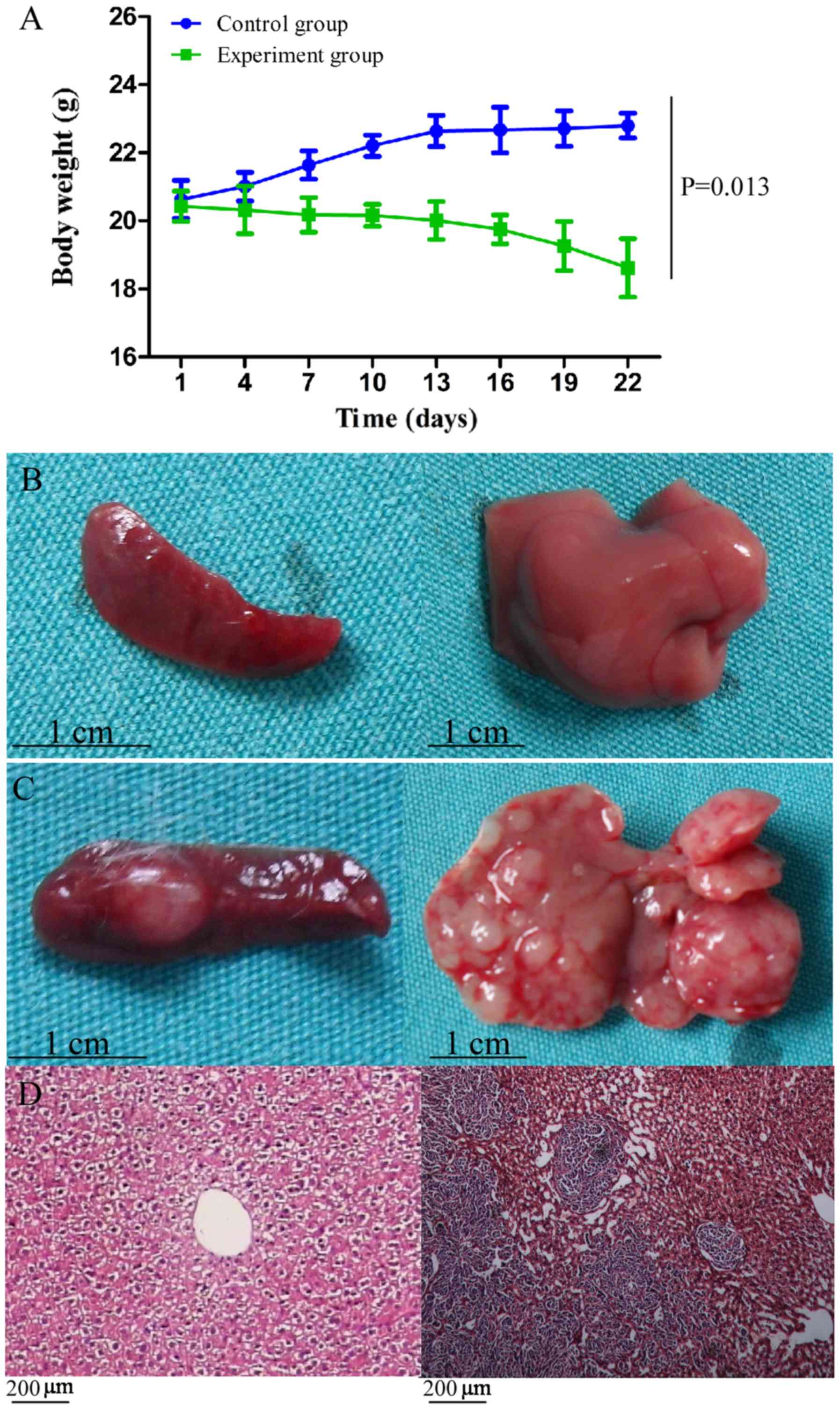
All tumor-bearing and control mice survived until
the end of the experiment. However, unlike the control mice, the
tumor-bearing mice exhibited signs of illness, such as reduced
activity, slow response, lackluster fur, loss of appetite,
emaciation and a distended abdomen, which were accompanied by
significant loss in body weight (P=0.013; Fig. 1A). The maximum percentage of body
weight loss was 8.86%.
Successful induction of liver
metastases of CT-26 cells
At 3 weeks post-tumor cell injection, the mice were
sacrificed. The liver and spleen of the control mice was soft and
smooth without any neoplasms (Fig.
1B). By contrast, both organs in the CT-26-injected mice had a
rough and uneven surface, while macroscopic tumors were also
observed (Fig. 1C). An average of
20.5 metastatic nodules was observed on the liver of the
CT-26-injected mice, and pathological evaluation using hematoxylin
and eosin staining confirmed the nodules as liver metastases of
colon carcinoma (Fig. 1D).
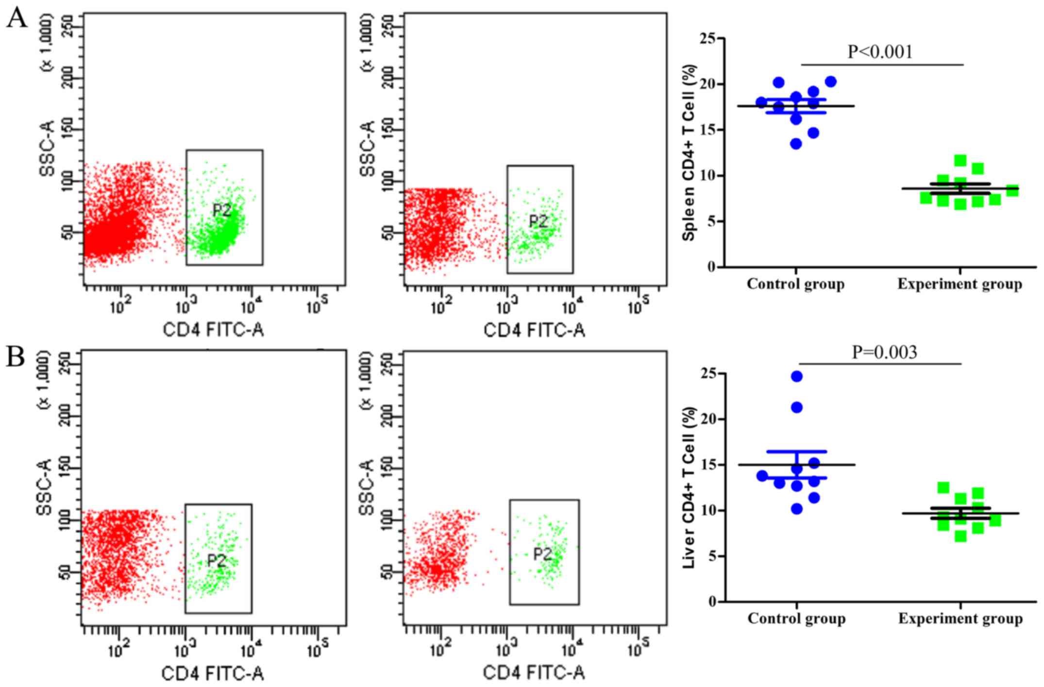
CD4+ T cells and
CD4+FOXP3+ Treg populations are influenced by
liver metastases
Single-cell suspensions of the spleen and liver were
prepared and analyzed using flow cytometry. As indicated in
Fig. 2, the numbers of
CD4+ T cells in the spleen and liver of the
tumor-bearing mice were significantly lower compared with those in
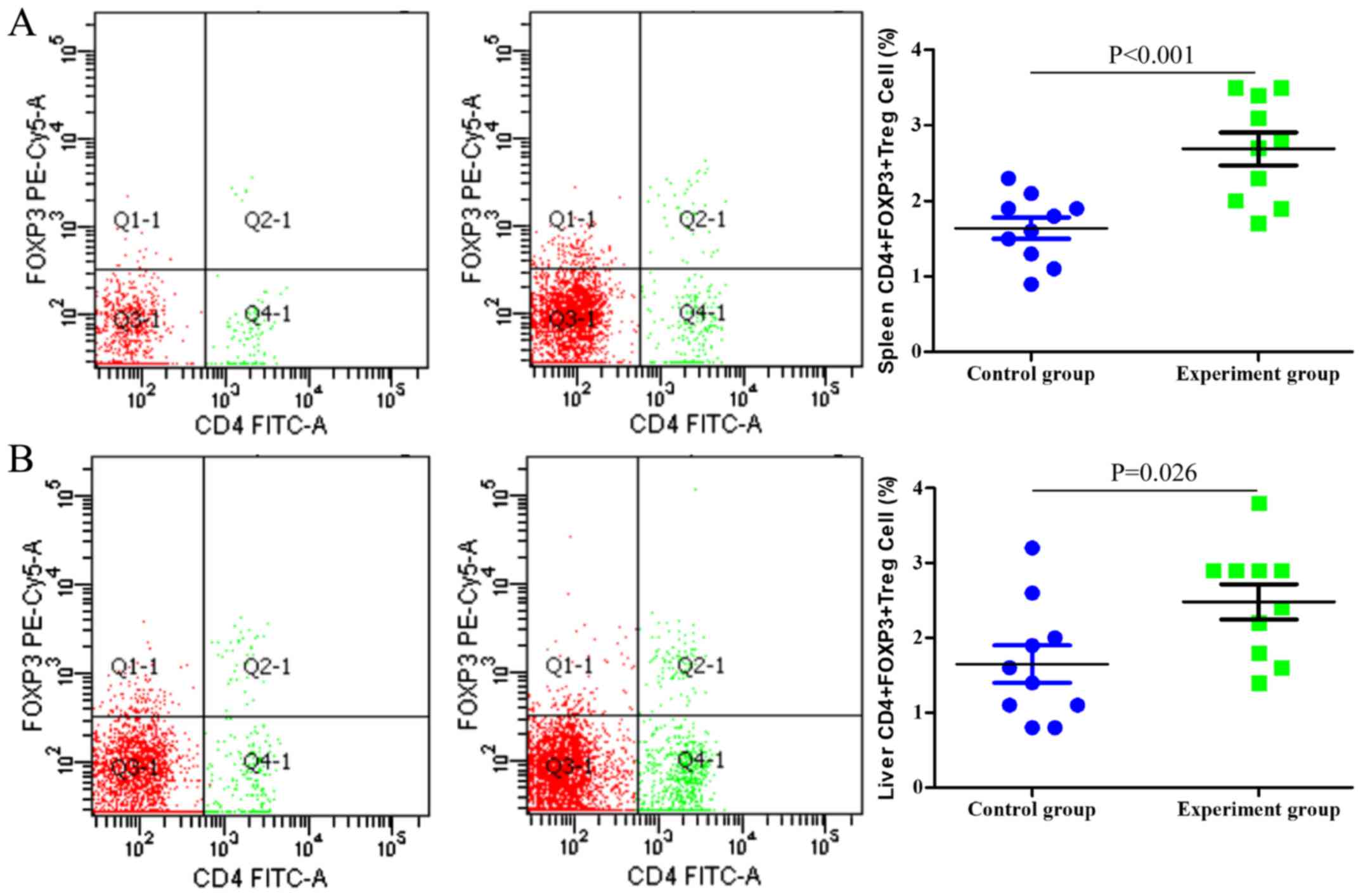
the control group (P<0.001 and P=0.003, respectively). By
contrast, the proportion of CD4+FOXP3+ Tregs
among the entire CD4+ T cell population was
significantly higher in the tumor-bearing group compared with that
in the control group (P<0.001 and P=0.026 in the spleen and
liver, respectively; Fig. 3).
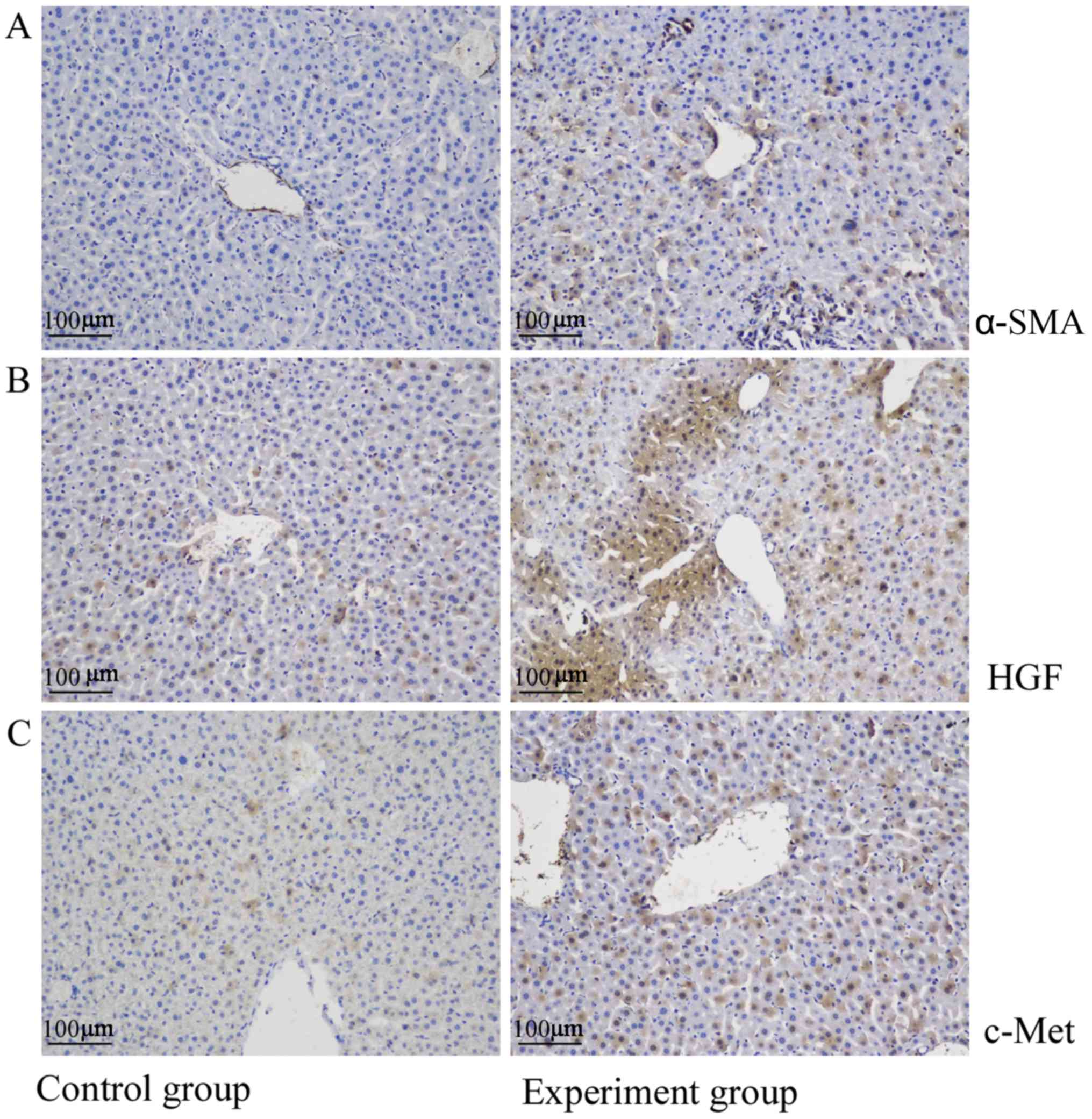
HGF/c-Met signaling pathway is
upregulated in liver metastases
The liver tissues of the control mice were negative
for α-SMA and expressed only low levels of HGF and c-Met. By
contrast, α-SMA, HGF and c-Met levels were significantly
upregulated in the liver metastatic nodules (Fig. 4).
Discussion
Liver metastasis is the primary cause of mortality
in patients with CRC. In the present study, a liver metastasis
model of colon carcinoma in mice was established via an injection
of CT-26 cells into the spleen (8,18), in
order to evaluate changes in CD4+Foxp3+ Treg
numbers and HGF/c-Met expression levels. All mice developed
metastatic growth in the liver and the survival rate was 100%
despite a lethargic appearance and loss of appetite. Macroscopic
tumors were observed on the liver surface, with an average of 20.5
metastatic nodules per mouse.
T cells serve an important role during
tumorigenesis, with cytotoxic T cells eliminating cancer cells
presenting major histocompatibility complex class I molecules,
while CD4+Foxp3+ Tregs promote tumor
progression via suppression of the antitumor immune response
(19,20). Tumor tissues are often infiltrated by
Tregs, and their depletion has been revealed to augment the
antitumor immune response in mice (21,22).
Tregs elicit an immunosuppressive effect by inhibiting
CD4+ and CD8+ effector T cells via T cell
receptor engagement and a cell contact-dependent mechanism
(23,24). In our previous study, the numbers of
CD4+ and CD8+ T cells were shown to decrease
in liver metastases (8), a fact
which was supported in the present study, along with an increase in
the proportion of CD4+Foxp3+ Tregs in the
spleen and liver. A higher proportion of Tregs may inhibit
cytotoxic T cells and promote metastasis of cancer cells from the
spleen to the liver.
The HGF/c-Met signaling pathway serves a key role in
metastasis (25) and influences the
proliferation, survival and invasive potential of colorectal,
gastric, gastro-esophageal and gastro-esophageal junction cancer
(26). In addition, HGF and c-Met
are upregulated in a number of cancer types, including CRC
(27), and are associated with tumor
recurrence and an unfavorable patient prognosis (28). Consistent with these findings,
HGF/c-Met expression was revealed to be significantly higher in
liver metastatic tissues in the current study, indicating that the
downregulation of HGF/c-Met expression may suppress tumor cell
invasion; however, further studies are required to confirm this
finding.
The role and potential mechanism of
CD4+FOXP3+ Tregs have also been investigated
in colitis-associated colon and colorectal cancer. Notably, Olguín
et al (29) discovered that
the percentage of CD4+FOXP3+ Tregs was
increased in colitis-associated colon cancer. Another study by Syed
Khaja et al (17) revealed
that numbers of CD4+FOXP3+ Tregs were
increased in colorectal cancer tissues. Although the present study
only obtained results and conclusions similar to those described in
previous papers, to the best of our knowledge, there is no
previously published study on this topic for the liver metastasis
of colorectal cancer. The results of the present study should
therefore supplement the existing knowledge in the field of
CD4+FOXP3+ Tregs and colorectal cancer.
In conclusion, liver metastasis of CRC is associated
with an increase in the number of CD4+FOXP3+
Tregs and upregulation of the HGF/c-Met signaling pathway,
indicating the presence of potential novel therapeutic targets for
liver metastasis.
Acknowledgements
Not applicable.
Funding
The present study was supported by a Medical Science
Research Grant from the Health Department of Guangdong Province
(grant no. A2018007).
Availability of data and materials
The datasets used and/or analyzed during the current
study are available from the corresponding author on reasonable
request.
Authors' contributions
XH and ZXC contributed equally to this work. XH, PL
and YW conceived and designed the study. XH, ZXC and NZ performed
the animal experiments and drafted the manuscript. CZ and XL
performed the cell experiments. JY and ZPC contributed to the
analysis and interpretation of data. All authors read and approved
the final manuscript.
Ethics approval and consent to
participate
All animal experiments were approved by the Ethical
Committee of Sun Yat-Sen University, and were conducted in
accordance with guidelines of the Institutional Animal Care and Use
Committee of Sun Yat-Sen University, and the Committee for Animal
Experiments.
Patient consent for publication
Not applicable.
Competing interests
The authors declare that they have no competing
interests.
Glossary
Abbreviations
Abbreviations:
|
CRC
|
colorectal cancer
|
|
Treg
|
regulatory T cell
|
|
HSC
|
hepatic stellate cell
|
|
α-SMA
|
α-smooth muscle actin
|
|
HGF
|
hepatocyte growth factor
|
References
|
1
|
Torre LA, Bray F, Siegel RL, Ferlay J,
Lortet-Tieulent J and Jemal A: Global cancer statistics, 2012. CA
Cancer J Clin. 65:87–108. 2015. View Article : Google Scholar : PubMed/NCBI
|
|
2
|
Bengmark S and Hafstrom L: The natural
history of primary and secondary malignant tumors of the liver. I.
The prognosis for patients with hepatic metastases from colonic and
rectal carcinoma by laparotomy. Cancer Am Cancer Soc. 23:198–202.
1969.
|
|
3
|
Manfredi S, Lepage C, Hatem C, Coatmeur O,
Faivre J and Bouvier AM: Epidemiology and management of liver
metastases from colorectal cancer. Ann Surg. 244:254–259. 2006.
View Article : Google Scholar : PubMed/NCBI
|
|
4
|
Konopke R, Roth J, Volk A, Pistorius S,
Folprecht G, Zöphel K, Schuetze C, Laniado M, Saeger HD and
Kersting S: Colorectal liver metastases: An update on palliative
treatment options. J Gastrointestin Liver Dis. 21:83–91.
2012.PubMed/NCBI
|
|
5
|
Lykoudis PM, O'Reilly D, Nastos K and
Fusai G: Systematic review of surgical management of synchronous
colorectal liver metastases. Br J Surg. 101:605–612. 2014.
View Article : Google Scholar : PubMed/NCBI
|
|
6
|
Robinson MW, Harmon C and O'Farrelly C:
Liver immunology and its role in inflammation and homeostasis. Cell
Mol Immunol. 13:267–276. 2016. View Article : Google Scholar : PubMed/NCBI
|
|
7
|
Nishikawa H and Sakaguchi S: Regulatory T
cells in cancer immunotherapy. Curr Opin Immunol. 27:1–7. 2014.
View Article : Google Scholar : PubMed/NCBI
|
|
8
|
Huang X, Zou Y, Lian L, Wu X, He X, He X,
Wu X, Huang Y and Lan P: Changes of T cells and cytokines TGF-β1
and IL-10 in mice during liver metastasis of colon carcinoma:
Implications for liver anti-tumor immunity. J Gastrointest Surg.
17:1283–1291. 2013. View Article : Google Scholar : PubMed/NCBI
|
|
9
|
Bárcena C, Stefanovic M, Tutusaus A,
Martinez-Nieto GA, Martinez L, García-Ruiz C, de Mingo A,
Caballeria J, Fernandez-Checa JC, Marí M and Morales A: Angiogenin
secretion from hepatoma cells activates hepatic stellate cells to
amplify a self-sustained cycle promoting liver cancer. Sci Rep.
5:79162015. View Article : Google Scholar : PubMed/NCBI
|
|
10
|
Gupta G, Khadem F and Uzonna JE: Role of
hepatic stellate cell (HSC)-derived cytokines in hepatic
inflammation and immunity. Cytokine. 124:1545422019. View Article : Google Scholar : PubMed/NCBI
|
|
11
|
Lu DH, Guo XY, Qin SY, Luo W, Huang XL,
Chen M, Wang JX, Ma SJ, Yang XW and Jiang HX: Interleukin-22
ameliorates liver fibrogenesis by attenuating hepatic stellate cell
activation and downregulating the levels of inflammatory cytokines.
World J Gastroenterol. 21:1531–1545. 2015. View Article : Google Scholar : PubMed/NCBI
|
|
12
|
Weiskirchen R and Tacke F: Cellular and
molecular functions of hepatic stellate cells in inflammatory
responses and liver immunology. Hepatobiliary Surg Nutr. 3:344–363.
2014.PubMed/NCBI
|
|
13
|
Patel MB, Pothula SP, Xu Z, Lee AK,
Goldstein D, Pirola RC, Apte MV and Wilson JS: The role of the
hepatocyte growth factor/c-MET pathway in pancreatic stellate
cell-endothelial cell interactions: Antiangiogenic implications in
pancreatic cancer. Carcinogenesis. 35:1891–1900. 2014. View Article : Google Scholar : PubMed/NCBI
|
|
14
|
Cai W, Rook SL, Jiang ZY, Takahara N and
Aiello LP: Mechanisms of hepatocyte growth factor-induced retinal
endothelial cell migration and growth. Invest Ophthalmol Vis Sci.
41:1885–1893. 2000.PubMed/NCBI
|
|
15
|
Pothula SP, Xu Z, Goldstein D, Biankin AV,
Pirola RC, Wilson JS and Apte MV: Hepatocyte growth factor
inhibition: A novel therapeutic approach in pancreatic cancer. Br J
Cancer. 114:269–280. 2016. View Article : Google Scholar : PubMed/NCBI
|
|
16
|
Parr C and Jiang WG: Expression of
hepatocyte growth factor/scatter factor, its activator, inhibitors
and the c-Met receptor in human cancer cells. Int J Oncol.
19:857–863. 2001.PubMed/NCBI
|
|
17
|
Syed Khaja A, Toor SM, El SH, Ali BR and
Elkord E: Intratumoral FoxP3+Helios+
regulatory T cells upregulating immunosuppressive molecules are
expanded in human colorectal cancer. Front Immunol. 8:6192017.
View Article : Google Scholar : PubMed/NCBI
|
|
18
|
Liu HY, Huang ZL, Yang GH, Lu WQ and Yu
NR: Inhibitory effect of modified citrus pectin on liver metastases
in a mouse colon cancer model. World J Gastroenterol. 14:7386–7391.
2008. View Article : Google Scholar : PubMed/NCBI
|
|
19
|
Takeuchi Y and Nishikawa H: Roles of
regulatory T cells in cancer immunity. Int Immunol. 28:401–409.
2016. View Article : Google Scholar : PubMed/NCBI
|
|
20
|
Fontenot JD, Gavin MA and Rudensky AY:
Foxp3 programs the development and function of
CD4+CD25+ regulatory T cells. Nat Immunol.
4:330–336. 2003. View
Article : Google Scholar : PubMed/NCBI
|
|
21
|
Shimizu J, Yamazaki S and Sakaguchi S:
Induction of tumor immunity by removing
CD25+CD4+ T cells: A common basis between
tumor immunity and autoimmunity. J Immunol. 163:5211–5218.
1999.PubMed/NCBI
|
|
22
|
Onizuka S, Tawara I, Shimizu J, Sakaguchi
S, Fujita T and Nakayama E: Tumor rejection by in vivo
administration of anti-CD25 (interleukin-2 receptor alpha)
monoclonal antibody. Cancer Res. 59:3128–3133. 1999.PubMed/NCBI
|
|
23
|
Almeida AR, Ciernik IF, Sallusto F and
Lanzavecchia A: CD4+ CD25+ Treg regulate the
contribution of CD8+ T-cell subsets in repopulation of
the lymphopenic environment. Eur J Immunol. 40:3478–3488. 2010.
View Article : Google Scholar : PubMed/NCBI
|
|
24
|
Zanin-Zhorov A, Ding Y, Kumari S, Attur M,
Hippen KL, Brown M, Blazar BR, Abramson SB, Lafaille JJ and Dustin
ML: Protein kinase C-theta mediates negative feedback on regulatory
T cell function. Science. 328:372–376. 2010. View Article : Google Scholar : PubMed/NCBI
|
|
25
|
Arlt F and Stein U: Colon cancer
metastasis: MACC1 and Met as metastatic pacemakers. Int J Biochem
Cell Biol. 41:2356–2359. 2009. View Article : Google Scholar : PubMed/NCBI
|
|
26
|
Bradley CA, Salto-Tellez M, Laurent-Puig
P, Bardelli A, Rolfo C, Tabernero J, Khawaja HA, Lawler M, Johnston
PG and Van Schaeybroeck S; MErCuRIC consortium, : Targeting c-MET
in gastrointestinal tumours: Rationale, opportunities and
challenges. Nat Rev Clin Oncol. 14:562–576. 2017. View Article : Google Scholar : PubMed/NCBI
|
|
27
|
Shojaei F, Simmons BH, Lee JH, Lappin PB
and Christensen JG: HGF/c-Met pathway is one of the mediators of
sunitinib-induced tumor cell type-dependent metastasis. Cancer
Lett. 320:48–55. 2012. View Article : Google Scholar : PubMed/NCBI
|
|
28
|
Osada S, Matsui S, Komori S, Yamada J,
Sanada Y, Ihawa A, Tanaka Y, Tokuyama Y, Okumura N, Nonaka K, et
al: Effect of hepatocyte growth factor on progression of liver
metastasis in colorectal cancer. Hepatogastroenterology. 57:76–80.
2010.PubMed/NCBI
|
|
29
|
Olguín JE, Medina-Andrade I, Molina E,
Vázquez A, Pacheco-Fernández T, Saavedra R, Pérez-Plasencia C,
Chirino YI, Vaca-Paniagua F, Arias-Romero LE, et al: Early and
partial reduction in CD4+Foxp3+ regulatory T
cells during Colitis-associated colon cancer induces
CD4+ and CD8+ T cell activation inhibiting
tumorigenesis. J Cancer. 9:239–249. 2018. View Article : Google Scholar : PubMed/NCBI
|