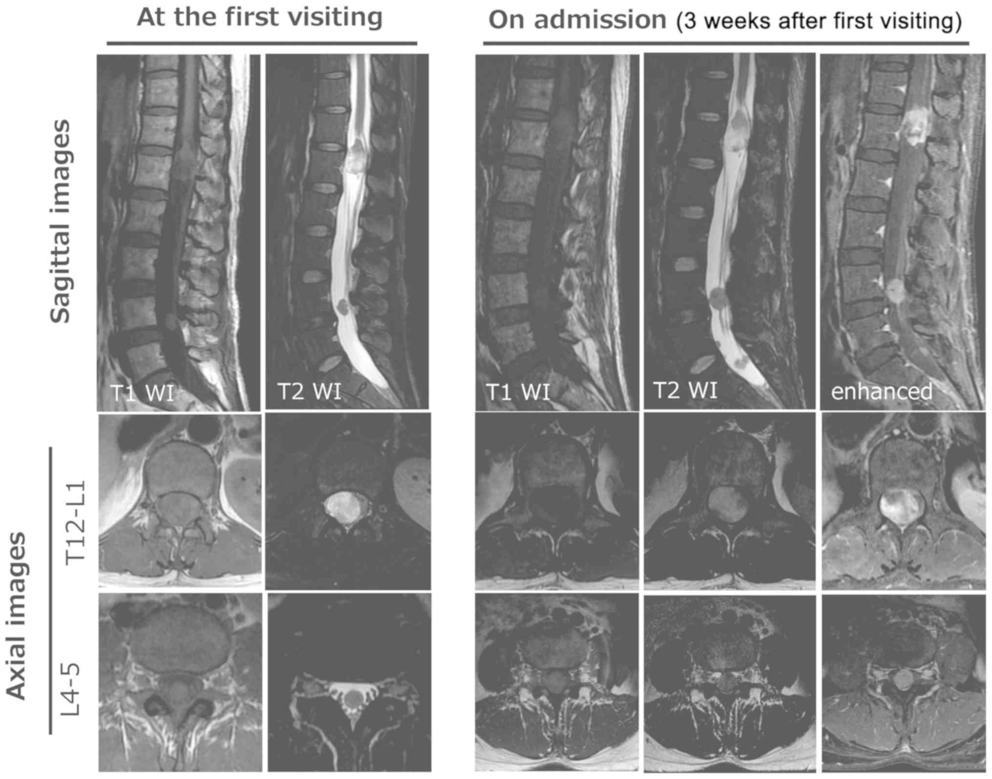
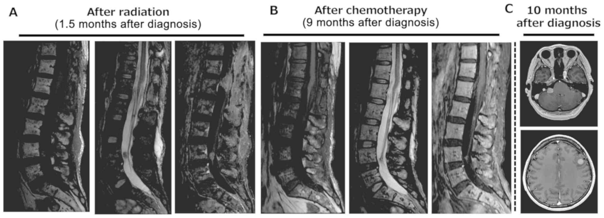
|
1
|
Van Goethem JW, van den Hauwe L, Ozsarlak
O, De Schepper AM and Parizel PM: Spinal tumors. Eur J Radiol.
50:159–176. 2004. View Article : Google Scholar : PubMed/NCBI
|
|
2
|
Harimaya K, Oda Y, Matsuda S, Tanaka K,
Chuman H and Iwamoto Y: Primitive neuroectodermal tumor and
extraskeletal Ewing sarcoma arising primarily around the spinal
column: Report of four cases and a review of the literature. Spine.
28:E408–E412. 2003. View Article : Google Scholar : PubMed/NCBI
|
|
3
|
Balamuth NJ and Womer RB: Ewings sarcoma.
Lancet Oncol. 11:184–192. 2010. View Article : Google Scholar : PubMed/NCBI
|
|
4
|
Gaspar N, Hawkins DS, Dirksen U, Lewis IJ,
Ferrari S, Le Deley MC, Kovar H, Grimer R, Whelan J, Claude L, et
al: Ewing sarcoma: Current management and future approaches through
collaboration. J Clin Oncol. 33:3036–3046. 2015. View Article : Google Scholar : PubMed/NCBI
|
|
5
|
Applebaum MA, Worch J, Matthay KK, Goldsby
R, Neuhaus J, West DC and Dubois SG: Clinical features and outcomes
in patients with extraskeletal Ewing sarcoma. Cancer.
117:3027–3032. 2011. View Article : Google Scholar : PubMed/NCBI
|
|
6
|
Hisaoka M, Hashimoto H and Murao T:
Peripheral primitive neuroectodermal tumour with
ganglioneuroma-like areas arising in the cauda equina. Virchows
Arch. 431:365–369. 1997. View Article : Google Scholar : PubMed/NCBI
|
|
7
|
Isotalo PA, Agbi C, Davidson B, Girard A,
Verma S and Robertson SJ: Primary primitive neuroectodermal tumor
of the cauda equina. Hum Pathol. 31:999–1001, 200.
View Article : Google Scholar : PubMed/NCBI
|
|
8
|
Uesaka T, Amano T, Inamura T, Ikezaki K,
Inoha S, Takamatsu M, Iwaki T and Fukui M: Intradural,
extramedullary spinal Ewings sarcoma in childhood. J Clin Neurosci.
10:122–125. 2003. View Article : Google Scholar : PubMed/NCBI
|
|
9
|
Akyüz M, Demiral AN, Gürer IE, Uçar T and
Tuncer R: Primary primitive neuro-ectodermal tumor of cauda equina
with intracranial seeding. Acta Neurochir (Wien). 146:525–528.
2004. View Article : Google Scholar : PubMed/NCBI
|
|
10
|
Mobley BC, Roulston D, Shah GV, Bijwaard
KE and McKeever PE: Peripheral primitive neuroectodermal
tumor/Ewings sarcoma of the craniospinal vault: Case reports and
review. Hum Pathol. 37:845–853. 2006. View Article : Google Scholar : PubMed/NCBI
|
|
11
|
Haresh KP, Chinikkatti SK, Prabhakar R,
Rishi A, Rath GK, Sharma DN and Julka PK: A rare case of intradural
extramedullary Ewings sarcoma with skip metastasis in the spine.
Spinal Cord. 46:582–584. 2008. View Article : Google Scholar : PubMed/NCBI
|
|
12
|
Kim SW and Shin H: Primary intradural
extraosseous Ewings sarcoma. J Korean Neurosurg Soc. 45:179–181.
2009. View Article : Google Scholar : PubMed/NCBI
|
|
13
|
Klimo P Jr, Codd PJ, Grier H and
Goumnerova LC: Primary pediatric intraspinal sarcomas. Report of 3
cases. J Neurosurg Pediatr. 4:222–229. 2009. View Article : Google Scholar : PubMed/NCBI
|
|
14
|
Yan Y, Xu T, Chen J, Hu G and Lu Y:
Intraspinal Ewings sarcoma/primitive neuroectodermal tumors. J Clin
Neurosci. 18:601–606. 2011. View Article : Google Scholar : PubMed/NCBI
|
|
15
|
Vincentelli F, Caruso G and
Figarella-Branger D: Primary intradural Ewings sarcoma of the cauda
equina presenting with acute bleeding. Acta Neurochir (Wien).
152:563–564. 2010. View Article : Google Scholar : PubMed/NCBI
|
|
16
|
Duan XH, Ban XH, Liu B, Zhong XM, Guo RM,
Zhang F, Liang BL and Shen J: Intraspinal primitive neuroectodermal
tumor: Imaging findings in six cases. Eur J Radiol. 80:426–431.
2011. View Article : Google Scholar : PubMed/NCBI
|
|
17
|
Karikari IO, Mehta AI, Nimjee S, Hodges
TR, Tibaleka J, Montgomery C, Simpson L, Cummings TJ and Bagley CA:
Primary intradural extraosseous Ewing sarcoma of the spine: Case
report and literature review. Neurosurgery. 69:E995–E999. 2011.
View Article : Google Scholar : PubMed/NCBI
|
|
18
|
Pancucci G, Simal-Julian JA, Plaza-Ramirez
E, García-Marcos R, Mayordomo-Aranda E and Botella-Asunción C:
Primary extraosseous intradural spinal Ewings sarcoma: Report of
two cases. Acta Neurochir (Wien). 155:1229–1234. 2013. View Article : Google Scholar : PubMed/NCBI
|
|
19
|
Khalatbari MR, Jalaeikhoo H and Moharamzad
Y: Primary intradural extraosseous Ewings sarcoma of the lumbar
spine presenting with acute bleeding. Br J Neurosurg. 27:840–841.
2013. View Article : Google Scholar : PubMed/NCBI
|
|
20
|
Bazzocchi A, Bacci A, Serchi E, Salerno A,
Salizzoni E and Leonardi M: Intradural extramedullary Ewings
sarcoma. Recurrence with acute clinical presentation and literature
review. Neuroradiol J. 26:476–481. 2013. View Article : Google Scholar : PubMed/NCBI
|
|
21
|
Gong HS, Huang QS, Liu GJ, Chen FH and
Zhao HB: Cervical primary Ewings sarcoma in intradural and
extramedullary location and skip metastasis to Cauda Equina. Turk
Neurosurg. 25:943–947. 2015.PubMed/NCBI
|
|
22
|
Lozupone E, Martucci M, Rigante L, Gaudino
S, Di Lella GM and Colosimo C: Magnetic resonance image findings of
primary intradural Ewing sarcoma of the cauda equina: Case report
and review of the literature. Spine J. 14:e7–e11. 2014. View Article : Google Scholar : PubMed/NCBI
|
|
23
|
Zhao M, Zhang B, Liang F and Zhang J:
Primary spinal intradural extraskeletal Ewing sarcoma mimicking a
giant nerve sheath tumor: Case report and review of the literature.
Int J Clin Exp Pathol. 7:9081–9085. 2014.PubMed/NCBI
|
|
24
|
Bostelmann R, Leimert M, Steiger HJ,
Gierga K and Petridis AK: The importance of surgery as part of
multimodal therapy in rapid progressive primary extraosseous Ewing
sarcoma of the cervical intra- and epidural space. Clin Pract.
6:8972016. View Article : Google Scholar : PubMed/NCBI
|
|
25
|
Kartal A and Akatlı A: Primary intradural
extraosseous Ewings sarcoma in a young child. Childs Nerv Syst.
32:409–410. 2016. View Article : Google Scholar : PubMed/NCBI
|
|
26
|
Chihak MA, Ahmed SK, Lachance DH,
Nageswara Rao AA and Laack NN: Patterns of failure and optimal
radiotherapy target volumes in primary intradural extramedullary
Ewing sarcoma. Acta Oncol. 55:1057–1061. 2016. View Article : Google Scholar : PubMed/NCBI
|
|
27
|
Scantland JT, Gondim MJ, Koivuniemi AS,
Fulkerson DH and Shih CS: Primary spinal intradural extraosseous
Ewing sarcoma in a pediatric patient: Case report and review of the
literature. Pediatr Neurosurg. 53:222–228. 2018. View Article : Google Scholar : PubMed/NCBI
|
|
28
|
Paterakis K, Brotis A, Tasiou A, Kotoula
V, Kapsalaki E and Vlychou M: Intradural extramedullary Ewings
sarcoma: A case report and review of the literature. Neurol
Neurochir Pol. 51:106–110. 2017. View Article : Google Scholar : PubMed/NCBI
|
|
29
|
Takami H, Kumar R, Brown DA and Krauss WE:
Histologic features and prognosis of spinal intradural
extramedullary ewing sarcoma: Case report, literature review, and
analysis of prognosis. World Neurosurg. 115:448–452.e2. 2018.
View Article : Google Scholar : PubMed/NCBI
|
|
30
|
Tan CH, Tan D, Phung TB and Lai LT:
Primary intradural extramedullary Ewing sarcoma of the cervical
spine: A case report and review of the literature. J Clin Neurosci.
66:280–284. 2019. View Article : Google Scholar : PubMed/NCBI
|
|
31
|
Fletcher CDM, Bridge JA, Hogendoorn PCW
and Mertens F: WHO Classification of Tumours of Soft Tissue and
Bone. 4th. IARC; Lyon, France: 2013
|
|
32
|
Zhang J, Huang Y, Sun Y, He A, Zhou Y, Hu
H, Yao Y and Shen Z: Impact of chemotherapy cycles and intervals on
outcomes of nonspinal Ewing sarcoma in adults: A real-world
experience. BMC Cancer. 19:11682019. View Article : Google Scholar : PubMed/NCBI
|
|
33
|
El Weshi A, Allam A, Ajarim D, Al Dayel F,
Pant R, Bazarbashi S and Memon M: Extraskeletal Ewings sarcoma
family of tumours in adults: Analysis of 57 patients from a single
institution. Clin Oncol (R Coll Radiol). 22:374–381. 2010.
View Article : Google Scholar : PubMed/NCBI
|