Introduction
Adenoid cystic carcinoma (ACC) of the external
auditory canal (EAC) is an extremely rare tumor characterized by an
aggressive clinical behavior and high risk of lymphatic spread
(1). Primary cancers of the EAC are
rare, and most are squamous cell carcinomas (80%), with ACC
accounting for 5% (2–5). The incidence of ACC of the EAC is low;
however, it has high recurrence and distant metastasis rates
(6–10).
The clinical symptoms of EAC carcinoma may be
non-specific and generally manifest as hearing loss, EAC masses,
otorrhea, and earache. Although early intermittent earache is the
most common type of complaint in patients with EAC carcinoma,
infection and otorrhea is not rare among some elderly patients
(1,11). Therefore, early diagnosis is often
missed, and the carcinoma may be misdiagnosed as chronic otitis
media, external otitis and benign neoplasms of the external ear
canal (12).
Surgery is the main treatment method for ACC of the
EAC, followed by radiotherapy. Nevertheless, surgery and
radiotherapy are associated with substantial morbidity (13,14) and
a high risk of recurrence in cases of incomplete excision (4). ACC of the EAC is associated with
perineural and bone invasion, and a high risk of direct
intracranial extension (4).
Recurrence may be observed several years after surgery, and
mortality has been mainly attributed to lung metastasis (4,15,16).
To the best of our knowledge, only a few reports
have included >3 patients with ACC of the EAC (4,15–17),
with other studies being reports of single cases (11,18–26). Due
to the rarity of the disease, the natural course and treatment of
ACC of the EAC remain poorly understood, and there are no
guidelines for the diagnosis and management of this malignancy.
Therefore, the aim of the present study was to review the clinical
features of patients with ACC of the EAC who were diagnosed and
managed at a single center. It was anticipated that the results may
improve the knowledge of this disease and guide diagnosis and
treatment strategies to improve patient outcomes.
Materials and methods
Study design and patients
The present retrospective study included patients
diagnosed with ACC of the EAC between January 2010 and September
2017 at the Department of Otorhinolaryngology–Head and Neck
Surgery, The First Affiliated Hospital of The Third Military
Medical University (Chongqing, China). The present study was
approved by the Ethics Committee of the Southwest Military Hospital
of the Military Army Medical University. The requirement of
informed consent was waived due to the retrospective nature of the
study.
Clinical information of the patients was retrieved
from the hospital databases. The patients were identified using
‘malignant tumors of the EAC’ as the search criterion. As ACC is
primarily diagnosed on the basis of pathological examination
(27), data retrieved using the
above search criterion were then manually screened to identify
patients who had a final diagnosis of ACC of the EAC. Patients
diagnosed with ACC originating in the EAC (excluding ACC invading
the EAC but originating from outside it) and those who were
diagnosed and treated at the aforementioned hospital were included
in the study. Patients with incomplete medical records were
excluded from the present study. All pathologic slides were
reviewed to confirm the diagnosis in all cases. Pathological
examination was performed via HE staining and immunohistochemistry
analysis was performed by pathologists from the Department of
Pathology, The First Affiliated Hospital of the Third Military
Medical University (Chongqing, China).
TNM staging
All patients were staged according to the Pittsburgh
staging criteria modified by Moody et al in 2000 (28). T1N1 tumors were considered as stage
3. T2-4N1 tumors were considered as stage 4. All stages were final
stages after surgery.
Treatment
Local resection, en bloc EAC resection or subtotal
temporal bone resection were performed for patients with
early-stage cancer. Subtotal temporal bone resection or extended
subtotal temporal bone resection were performed for patients with
advanced-stage cancer. A total parotidectomy was performed for
tumors that exhibited advanced invasion of the anterior wall of the
EAC. A superficial parotidectomy was performed if enhancing masses
were found in the parotid gland by enhanced CT. Temporomandibular
joint or mandibular branch resection was also performed if imaging
identified mandibular condyle or temporomandibular joint
involvement. For tumors involving the meninges, the lesion was
completely resected, and abdominal fat was used to fill the cavity.
The facial nerves were resected if facial nerve involvement was
identified during imaging or surgery. The patients received 75
mg/m2 docetaxel and 80 mg/m2 cisplatin on day
1 of each 21-day cycle. After two cycles, local radiotherapy (33
fractions) was performed using a dose of 70 Gy for the GTV (tumor
target area), 60 Gy for the CTV1 (high-risk clinical target area)
and 54 Gy for the CTV2 (low-risk clinical target area). The
patients received two cycles of chemotherapy after the end of
radiotherapy.
Follow-up
Routine follow-up was performed once a month in the
first year, once every 3 months in the second year, once every 6
months in the third year and once a year thereafter. The patients
were instructed to visit the hospital when they felt indisposed. A
biopsy was performed when a new lesion was found by imaging.
Recurrence was defined as biopsy results exhibiting ACC. Follow-up
was ceased on June 1, 2018.
Statistical analysis
Statistical analysis was performed using SPSS 19.0
software (IBM Corp.). The Kaplan-Meier method was used for survival
analysis. Continuous variables are presented as the mean ± standard
deviation. Categorical variables are presented as numbers and
percentages.
Results
Patient characteristics
Among 479 patients diagnosed with malignant tumors
of the EAC during the study period, 23 patients (4.8%) were
diagnosed with ACC of the EAC and included in the present analysis.
There were 12 males and 11 females. The follow-up duration ranged
between 2 and 81 months (median, 39 months). A total of 3 patients
were lost to follow-up (13.0%). The age at diagnosis ranged between
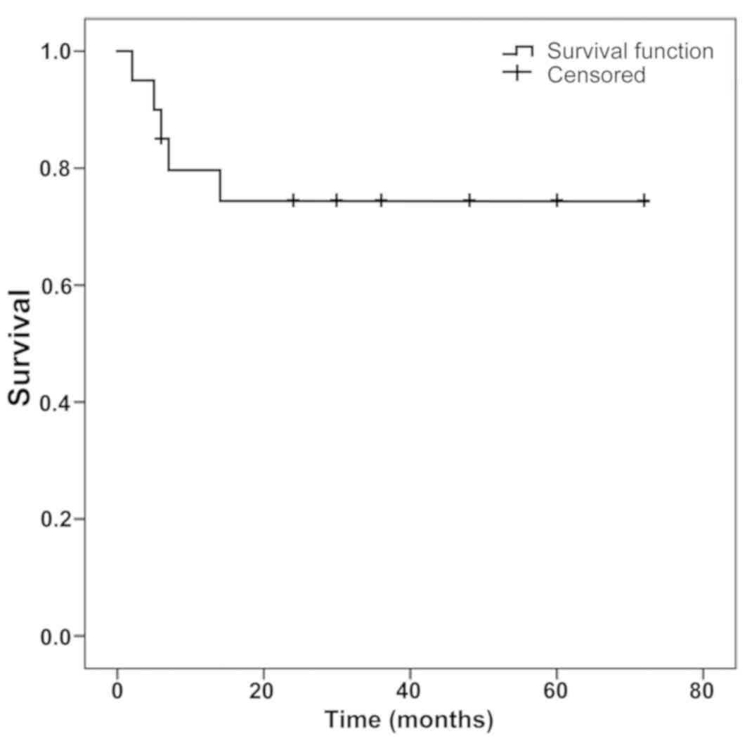
31 and 78 years (mean, 51.4±15.4 years). Fig. 1 illustrates overall survival. The
demographic and clinical characteristics of the patients are
presented in Table I. The initial
symptoms were earache in 16 patients, otorrhea in 7 patients, EAC
mass in 4 patients, hearing loss in 5 patients, and a feeling of
aural fullness in 2 patients (Table
II). The staging of all patients was determined based on CT and
MRI findings, intraoperative findings, and postoperative
pathological reports. The 23 ACC of the EAC cases were classified
according to the Pittsburgh staging system (6,10). There
were 13 (56.5%) cases with T1, 3 (13.0%) cases with T2, 3 (13.0%)
cases with T3 and 4 (17.4%) cases with T4 (Table III).
 | Table I.Detailed patient data. |
Table I.
Detailed patient data.
| Patient no. | Age
(years)/sex | Symptom | Pittsburgh
stage | Disease
coursea, months | Treatment | Follow-up and
outcome (months)b |
|---|
| 1 | 75/F | Earache | T3N0M0 | 1 | None | A (36) |
| 2 | 36/F | Fester, mass | T1N0M0 | 240 | En bloc
EACR+CT | A (24) |
| 3 | 40/F | Earache,
otorrhea | T3N0M0 | 1 | En bloc EACR | A (6) |
| 4 | 67/F | Earache,
otorrhea | T1N0M0 | 72 | Local EACR | LR (24); A
(30) |
| 5 | 34/M | Otorrhea | T1N0M0 | 6 | None | A (60) |
| 6 | 46/F | Mass | T1N0M0 | 120 | Local EACR | LR (30); A
(30) |
| 7 | 75/F | Earache | T2N0M0 | 24 | None | DWD (5) |
| 8 | 59/M | Earache,
otorrhea | T1N0M0 | 12 | None | A (30) |
| 9 | 60/F | Earache | T1N0M0 | 24 | None | DWD (2) |
| 10 | 51/M | Earache | T4N1M0 | 36 | None | DWD (6) |
| 11 | 50/F | Hearing loss,
aural | T4N0M0 | 6 | ETBR+TP+RT+CT | DM (12); |
|
|
| fullness |
|
|
| DWD (14) |
| 12 | 42/M | Earache | T4N0M0 | 24 | None | DWD (7) |
| 13 | 62/M | Mass, hearing
loss | T1N0M0 | 11 | Local EACR+RT | LFU (3) |
| 14 | 61/M | Earache, hearing
loss, otorrhea | T3N0M0 | 36 | None | LFU (3) |
| 15 | 32/F | Earache, hearing
loss | T1N0M0 | 84 | En bloc
EACR+RT+CT | A (48) |
| 16 | 31/M | Aural fullness | T1N0M0 | 2 | En bloc
EACR+RT | A (36) |
| 17 | 78/M | Otorrhea, earache,
mass | T2N0M0 | 120 | RM | A (48) |
| 18 | 42/F | Earache | T1N0M0 | 8 | En bloc
EACR+SP+RT+CT | A (18) |
| 19 | 37/F | Earache | T1N0M0 | 36 | En bloc
EACR+RT+CT | A (72) |
| 20 | 47/M | Earache,
facioplegia, vertigo | T4N0M0 | 84 | STBR+RT+CT | A (30) |
| 21 | 65/M | Hearing loss,
otorrhea | T2N0M0 | 24 | RM | LFU (3) |
| 22 | 40/M | Earache | T1N0M0 | 12 | En bloc EACR | A (60) |
| 23 | 52/M | Earache | T1N0M0 | 36 | STBR | A (24) |
| Table II.Clinical symptoms. |
Table II.
Clinical symptoms.
| Symptoms | Cases | Percentage |
|---|
| External auditory
canal masses | 4 | 11.1 |
| Earache | 16 | 44.4 |
| Otorrhea | 7 | 19.4 |
| Facioplegia | 1 |
2.7 |
| Hearing loss | 5 | 13.9 |
| Ear aural
fullness | 2 |
5.6 |
| Vertigo | 1 |
2.7 |
| Table III.Tumor staging. |
Table III.
Tumor staging.
| TNM staging | Cases | Percentage |
|---|
| Tumor size |
|
|
| T1 | 13 | 56.5 |
| T2 | 3 | 13.0 |
| T3 | 3 | 13.0 |
| T4 | 4 | 17.4 |
| Lymph node
metastasis |
|
|
| N0 | 22 | 95.7 |
| N1 | 1 | 4.3 |
| Distant
metastasis |
|
|
| M0 | 23 | 100.0 |
| M1 | 0 | 0 |
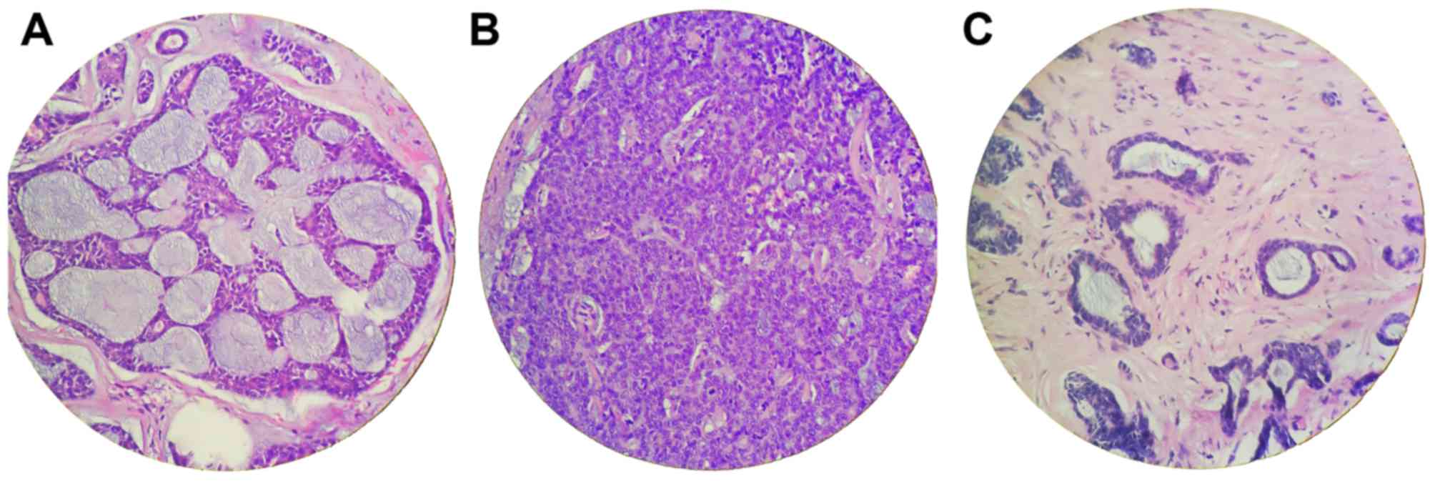
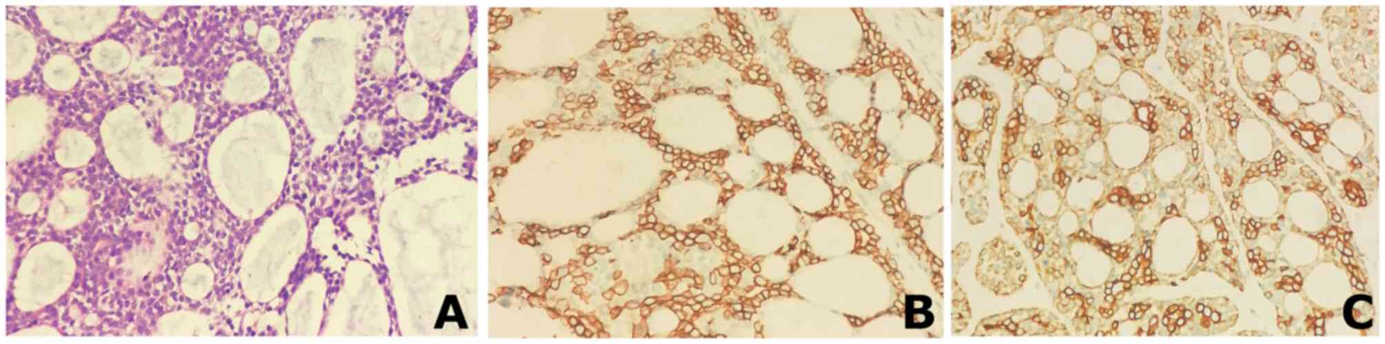
Fig. 2 presents the
pathology images of patient 2. Fig.
2A (H&E staining) shows the typical cribriform structure of
ACC. Most of the empty spaces were round or oval pseudoglandular
cavities; the lumens varied in diameter and were uniformly
basophilic. Fig. 2B and C illustrate
the immunohistochemistry results for CD117 and CK, respectively.
CD117 was localized on the glandular epithelial cell membrane,
whereas CK was localized in the cytoplasm. Both markers were
strongly positive. Fig. 3
illustrates representative images of the three subtypes of ACC. The
illustrated pathological images were obtained from the Department
of Pathology, The First Affiliated Hospital of the Third Military
Medical University (Chongqing, China).
Among the 23 patients, 8 patients declined surgery,
and the remaining 15 patients underwent surgery under general
anesthesia. Among these 15 patients, 1 patient had T4 disease and
underwent subtotal temporal bone resection, another patient with T4
underwent extended temporal bone resection, right parotidectomy,
right resection of middle cranial fossa tumors and right resection
of temporomandibular joint capsule. One patient with T3 underwent
en bloc EAC resection and 2 patients with T2 underwent radical
mastoidectomy. The remaining 10 patients had T1 disease, of which 5
patients underwent en bloc EAC resection, 3 underwent local EAC
resection, 1 underwent en bloc resection of EAC and superficial
parotidectomy, and 1 underwent subtotal temporal bone resection for
postoperative recurrence after treatment in another hospital. Among
these 15 surgical patients, 2 received postoperative radiotherapy
alone, 1 received postoperative chemotherapy alone, 5 received
postoperative chemo-radiotherapy and 7 did not receive
postoperative chemo-radiotherapy (Tables IV and V).
| Table IV.Treatment in relation to clinical
stage. |
Table IV.
Treatment in relation to clinical
stage.
| Stage | Surgery alone | Surgery +
radiotherapy | Surgery +
chemotherapy | Surgery +
chemo-radiotherapy | Conservative
treatment |
|---|
| T1 | 4 | 2 | 1 | 3 | 3 |
| T2 | 2 | 0 | 0 | 0 | 1 |
| T3 | 1 | 0 | 0 | 0 | 2 |
| T4 | 0 | 0 | 0 | 2 | 2 |
| Total | 7 | 2 | 1 | 5 | 8 |
| Table V.Surgical methods in relation to
clinical stage. |
Table V.
Surgical methods in relation to
clinical stage.
| Stage | RM | Local EACR | En bloc EACR | En bloc
EACR+SP | STBR | ETBR+TP |
|---|
| T1 | 0 | 3 | 5 | 1 | 1 | 0 |
| T2 | 2 | 0 | 0 | 0 | 0 | 0 |
| T3 | 0 | 0 | 1 | 0 | 0 | 0 |
| T4 | 0 | 0 | 0 | 0 | 1 | 1 |
| Total | 2 | 3 | 7 | 0 | 2 | 1 |
Of the 23 patients, 5 patients died due to the
primary disease, including 1 case with T1, 1 case with T2, and 3
cases with T4 disease. Among the living patients, 13 patients were
healthy and exhibited no signs of recurrence, 2 patients exhibited
recurrence but were alive. Furthermore, 3 patients were lost to
follow-up. The 3- and 5-year cumulative survival rate were
calculated via Kaplan-Meier survival curve analysis and were 47.8%
and 17.4%, respectively (Table I and
Fig. 1).
The present study demonstrated that the 5-year
cumulative survival rate was 40% in patients treated using a
combination of surgery and radio-chemotherapy, 14.3% in patients
treated with surgery alone, 0% in patients treated with a
combination of surgery and radiotherapy and 0% in patients treated
with a combination of surgery and chemotherapy (Fig. 1).
Recurrence and survival
Among the 23 patients, 2 (8.7%) had disease
recurrence, and both underwent local EACR without postoperative
chemo-radiotherapy. No further treatment was carried out after
recurrence, but the patients were still alive at the time of
writing. In the present study, 5/23 patients (21.7%) died: 1
patient with T1 disease, 1 with T2 disease and 3 with T4 disease.
The majority (4/5) of the patients who died had declined treatment
and died of their primary tumor after discharge. One patient who
underwent extended temporal bone resection, right parotidectomy,
right resection of middle cranial fossa tumors and right resection
of the temporomandibular joint capsule received postoperative
chemo-radiotherapy and died of systemic metastasis 14 months after
surgery (Fig. 1).
Associations among disease course,
lesion stage and curative effect
As presented in Table
I the disease course (time from symptom occurrence to
diagnosis) was 1–240 months (median, 24 months). Among the patients
with a disease course ≤24 months, there were 7 patients with T1
disease (5 survivors and 1 lost to follow-up and 1 dead), 2
patients with T2 disease (1 lost to follow-up and 1 dead), 2
patient with T3 disease (alive) and 2 patient with T4 disease
(dead). Among patients with a disease course >24 months, there
were 6 patients with T1 disease (alive), 1 patient with T2 disease
(alive), 1 patient with T3 disease (lost to follow-up) and 2
patients with T4 disease (1 dead and 1 alive). For patients with a
disease course >24 months, the initial symptoms were earache,
hearing loss and otorrhea, but these symptoms did not attract
attention or were misdiagnosed at other hospitals as benign masses
in the EAC and chronic otitis media, which delayed the diagnosis
and treatment.
Misdiagnosed cases
A total of 2 patients were misdiagnosed with otitis
media before surgery (initial symptoms of otorrhea and hearing
loss). Additionally, 2 patients were misdiagnosed preoperatively
with benign masses of the EAC; 1 patient had symptoms of earache
and itchy ears and 1 patient had EAC masses for >10 years. No
biopsy was performed prior to surgery, and radical mastoidectomy
and en bloc resection of the EAC masses were performed in the 2
patients. Postoperative pathology confirmed a diagnosis of ACC.
Further treatment was recommended, but none of the 4 patients
received surgical treatment or radiotherapy. At the end of the
follow-up period, 2 patients were still alive after en bloc mass
resection and the 2 patients misdiagnosed with otitis media were
lost to follow-up.
Discussion
The present case series describes the clinical
features and management of ACC of the EAC. Although several studies
have reported patients with ACC of the EAC, most of these have been
reports of single cases (11,18–26),
with only a few including more than three patients (4,15–17). The
present study included 23 patients from a single center and, to the
best of our knowledge, is the largest reported series to date.
ACC is a rare form of adenocarcinoma that arises
from glandular tissue. Although the origin of primary ACC of the
EAC remains controversial, it has been suggested that the
malignancy arises from eccrine sweat glands, ectopic salivary
glands, or ceruminous glands (11).
The differential diagnosis of ACC of the EAC includes adenoma;
papilloma; tuberculosis; and tumors such as squamous cell
carcinoma, adenocarcinoma, basal cell carcinoma, and mucoepidermoid
carcinoma (11).
Immunohistochemistry studies of salivary gland ACC demonstrated
that carcinoembryonic antigen (CEA) is expressed in some cells
(29), and p53 expression is
associated with poor outcomes (30).
However, immunohistochemistry data for p53 and CEA are limited for
ACC of the EAC.
The association between ACC and age or sex is
controversial. Triantafillidou et al (29) reported a female predilection for ACC,
whereas De Lucia et al (30)
reported a male predilection. The present study found that the
number of males with ACC of the EAC was slightly higher than the
number of females, and the average age of onset was approximately
50 years.
Fliss et al (31) suggested that earache is the most
common clinical symptom of ACC of the EAC. This is concurrent with
the findings of the present study, in which the most common initial
symptom was earache (reported by 43.2% of patients), followed by
otorrhea, hearing loss, and ear mass. Since these symptoms are
non-specific, more attention must be paid to them when they are
chronic, recurrent or aggravated, or if an ear mass exhibits
progressive growth.
Early diagnosis of the disease is crucial to improve
the survival of patients with ACC of the EAC; however, the
misdiagnosis rate is relatively high. In the present study, 4 out
of 23 patients (17.4%) were misdiagnosed. This high misdiagnosis
rate could be explained by the low disease incidence rate and a
resultant lack of awareness of the disease. Additionally, because
there are no typical symptoms, the atypical symptoms of ACC of the
EAC, such as earache, otorrhea, and hearing loss, are often
misdiagnosed as external otitis and otitis media. Furthermore,
although biopsy must be performed for suspected cases, awareness of
the necessity of a biopsy is low among physicians.
The surgical principle for the tumor is the same as
that for most malignant tumors, namely, complete curative
resection. The surgical methods include local EAC resection, en
bloc EAC resection, subtotal temporal bone resection, and temporal
bone resection. In the present study, two patients with local
resection of the EAC exhibited recurrence, indicating that even
early-stage disease can invade the surrounding tissues, and that
local mass resection does not guarantee a safe margin and can
result in recurrence. Garden et al (32) suggested that a positive margin is a
vital factor leading to poor prognosis. Due to the lack of
treatment guidelines, the recommendations of representative
literature, including case series, were mainly followed for the
treatment of patients in the present study. Some authors
recommended early extended resection (4,10). In
addition to removing local tumors, the surrounding tissues such as
the cartilaginous and osseous parts of the EAC, middle ear, and
even the whole temporal bone and parotid glands should be removed.
For ACC of the EAC, a large-area total resection of the EAC is
recommended to remove related cartilage, mastoid process, and
middle ear. If the tumor is observed to invade the middle ear and
temporal bone, or involve the facial nerve during preoperative
examinations or operative procedure, subtotal resection or total
resection of the temporal bone can be performed, and the
corresponding dura mater or the temporomandibular joint can be
removed if necessary. Some authors recommend the lateral resection
of the temporal bone for patients with T1 and T2 stages, and
subtotal resection of the temporal bone and infratemporal fossa for
those with T3 and T4 stages (28).
The range for lateral resection of the temporal bone includes
cartilaginous and osseous parts of the EAC, tympanic bone, tympanic
membrane, malleus, and incus. The medial side is limited to the
level of the incudostapedial joint and lateral facial nerve
(33). Subtotal resection of the
temporal bone is based on the lateral resection of the temporal
bone to expand the resection inward and remove the inner ear, but
retaining the apex of the petrous temporal bone (3). Although there are differences in the
treatment methods among studies, the consensus is that the
appropriate surgical methods should be based on the invaded sites
and stages of tumors of the EAC to achieve the goal of complete
tumor resection (28).
Additionally, the treatment of the parotid gland is
controversial. Some authors consider that superficial parotidectomy
is suitable for all patients with ACC of the EAC, even in the early
stages (12). The EAC and parotid
gland have a close histological association. Additionally, the
Santorini cracks provide a way for invading the parotid gland
(12). In the present series, there
was no difference in survival between patients with T1 and T4
disease. Nevertheless, it is recommended that patients with early
disease undergo superficial parotidectomy at the same time as
lesion resection because the lack of recurrence observed in the
present study may be caused by a combination of factors such as a
short follow-up and a small sample size. The parotid gland can be
partially removed to ensure safety. If the parotid gland is
markedly invaded, it can be completely removed (34).
Moffat et al (35) suggested that radiotherapy can destroy
subclinical tumor foci and increase surgical efficacy. Radiotherapy
may be particularly effective for advanced tumors when it is
difficult to achieve safe margins. Additionally, Chen et al
(36) suggested that postoperative
radiotherapy with >60 Gy radiation can effectively prevent
postoperative recurrence of ACC of the head and neck. Silverman
et al (37) suggested that
postoperative radiotherapy is required for patients with advanced
clinical disease, whereas patients with negative margins after the
first surgery and low clinical stage do not require radiotherapy.
It has been suggested that chemotherapy does not affect the
survival of patients with ACC (38).
The results of the present study indicate that the survival rate of
the combined treatment of surgery and chemoradiotherapy is higher
than surgery alone or postoperative isolated radiotherapy or
chemotherapy. Therefore, the survival rate was notably higher for
patients who received surgery and radiochemotherapy than for
patients who were not given all three treatment modalities.
In the present study, two patients exhibited
recurrence during follow-up and one patient with T4 disease died of
intracranial and pulmonary metastases 14 months after surgery.
Distant metastasis is more likely to occur in the lungs, followed
by bone, kidney, and brain (34).
Therefore, close attention should be paid to the lungs, brain,
kidneys, and other sites prone to metastasis during follow-up.
The present study has certain limitations. First,
the sample size was small due to the low incidence of ACC of the
EAC and because this was a single-center study. In addition, as all
cases were from a single center, this may lead to some bias based
on the diagnostic experience of the radiologists and pathologists,
as well as the experience of the surgeons and oncologists. Second,
there are three generally recognized histological patterns of ACC
(11); however, these patterns were
not consistently described in the pathology reports. Third, the p53
and CEA immunohistochemistry data were not available for the
patients in the present study. Thus, it was not possible to
evaluate these factors. Fourth, positron emission tomography
(PET)-CT scans were not performed during follow-up to detect the
occurrence of distant metastasis. This was partly due to PET-CT
scans being unaffordable for a number of the patients as they were
not covered by medical insurance. Therefore, it was not possible
for the current retrospective analysis to systematically evaluate
the occurrence of distant metastasis. Fifth, only routine follow-up
data were available for the current retrospective analysis, and 17%
of the patients were lost to follow-up. Patients may move
residence, die at another hospital, or simply refuse follow-up. A
prospective study would be difficult to carry out because of the
rarity of the disease, but a multicenter retrospective study could
be performed to refine the results of the present study.
Patients with a complaint of an EAC mass, EAC pain,
and otorrhea should be examined comprehensively with CT or MRI and
biopsy, if necessary. The initial surgical treatment should be
tumor resection with wide margins and superficial parotidectomy.
Frozen-section examination should be performed to ensure a negative
margin. The survival rate of patients after postoperative
chemoradiotherapy was higher than that of patients without
chemoradiotherapy. Early diagnosis may be the key to improving
survival. Nevertheless, it is noteworthy to highlight that a
75-year-old female patient with T3 disease refused surgery and
chemoradiotherapy and survived with the tumor for at least 51
months, since the patient was still alive when the present report
was written. Although this phenomenon has been observed before, the
specific reasons remain to be determined (39).
Therefore, early identification of symptoms,
performance of necessary imaging, and timely biopsy are key to
reducing misdiagnosis and improving the survival rate. It is
recommended to expand the tumor for the first operation and perform
parotidectomy for the superficial parotid gland. Pathological
examination must be performed during the surgery to ensure a
negative margin. For patients with doubtful negative margins,
postoperative radiotherapy and chemotherapy can be considered to
reduce recurrence and improve survival. The lack of unified staging
and treatment guidelines coupled with the low incidence of the
disease and few studies requires further investigations on the
specificity, biological behavior and optimal treatment plan of the
tumor.
Acknowledgements
Not applicable.
Funding
No funding was received.
Availability of data and materials
The datasets used and/or analyzed during the present
study are available from the corresponding author upon reasonable
request.
Authors' contributions
XJ participated in case collection, follow-up,
analysis and drafted the initial manuscript and was responsible for
the study concept and design. LJ participated in data analysis and
interpretation of the data. XZ and CZ made significant
contributions to the data acquisition, data analyses and
interpretation. FT and XC participated in patient follow-up. WY
performed the surgeries and participated in formulating the
treatment plan. WY also helped prepare the final version of the
manuscript to be submitted. All the authors made significant
contributions to the study design, data analysis, interpretation
and drafting of the paper. All the authors have read and approved
of the final manuscript for publication.
Ethics approval and consent to
participate
The present study was approved by the Ethics
Committee of The First Affiliated Hospital of The Third Military
Medical University (Chongqing, China; approval no. KY2020113). The
requirement for informed consent was waived due to the
retrospective nature of the study.
Patient consent for publication
Not applicable.
Competing interests
The authors declare that they have no competing
interests.
Glossary
Abbreviations
Abbreviations:
|
ACC
|
adenoid cystic carcinoma
|
|
EAC
|
external auditory canal
|
|
CK
|
cytokeratin
|
References
|
1
|
Gu FM, Chi FL, Dai CF, Chen B and Li HW:
Surgical outcomes of 43 cases with adenoid cystic carcinoma of the
external auditory canal. Am J Otolaryngol. 34:394–398. 2013.
View Article : Google Scholar : PubMed/NCBI
|
|
2
|
Conley J and Schuller DE: Malignancies of
the ear. Laryngoscope. 86:1147–1163. 1976. View Article : Google Scholar : PubMed/NCBI
|
|
3
|
Moore MG, Deschler DG, McKenna MJ,
Varvares MA and Lin DT: Management outcomes following lateral
temporal bone resection for ear and temporal bone malignancies.
Otolaryngol Head Neck Surg. 137:893–898. 2007. View Article : Google Scholar : PubMed/NCBI
|
|
4
|
Dong F, Gidley PW, Ho T, Luna MA, Ginsberg
LE and Sturgis EM: Adenoid cystic carcinoma of the external
auditory canal. Laryngoscope. 118:1591–1596. 2008. View Article : Google Scholar : PubMed/NCBI
|
|
5
|
Chang CH, Shu MT, Lee JC, Leu YS, Chen YC
and Lee KS: Treatments and outcomes of malignant tumors of external
auditory canal. Am J Otolaryngol. 30:44–48. 2009. View Article : Google Scholar : PubMed/NCBI
|
|
6
|
Arriaga M, Curtin H, Takahashi H, Hirsch
BE and Kamerer DB: Staging proposal for external auditory meatus
carcinoma based on preoperative clinical examination and computed
tomography findings. Ann Otol Rhinol Laryngol. 99:714–721. 1990.
View Article : Google Scholar : PubMed/NCBI
|
|
7
|
Shih L and Crabtree JA: Carcinoma of the
external auditory canal: An update. Laryngoscope. 100:1215–1218.
1990. View Article : Google Scholar : PubMed/NCBI
|
|
8
|
Kuhel WI, Hume CR and Selesnick SH: Cancer
of the external auditory canal and temporal bone. Otolaryngol Clin
North Am. 29:827–852. 1996. View Article : Google Scholar : PubMed/NCBI
|
|
9
|
Testa JR, Fukuda Y and Kowalski LP:
Prognostic factors in carcinoma of the external auditory canal.
Arch Otolaryngol Head Neck Surg. 123:720–724. 1997. View Article : Google Scholar : PubMed/NCBI
|
|
10
|
Nyrop M and Grontved A: Cancer of the
external auditory canal. Arch Otolaryngol Head Neck Surg.
128:834–837. 2002. View Article : Google Scholar : PubMed/NCBI
|
|
11
|
Liu SC, Kang BH, Nieh S, Chang JL and Wang
CH: Adenoid cystic carcinoma of the external auditory canal. J Chin
Med Assoc. 75:296–300. 2012. View Article : Google Scholar : PubMed/NCBI
|
|
12
|
Zhang T, Dai C and Wang Z: The
misdiagnosis of external auditory canal carcinoma. Eur Arch
Otorhinolaryngol. 270:1607–1613. 2013. View Article : Google Scholar : PubMed/NCBI
|
|
13
|
Kwok HC, Morton RP, Chaplin JM, McIvor NP
and Sillars HA: Quality of life after parotid and temporal bone
surgery for cancer. Laryngoscope. 112:820–833. 2002. View Article : Google Scholar : PubMed/NCBI
|
|
14
|
Wang CC: Radiation therapy in the
management of carcinoma of the external auditory canal, middle ear,
or mastoid. Radiology. 116:713–715. 1975. View Article : Google Scholar : PubMed/NCBI
|
|
15
|
Pulec JL, Parkhill EM and Devine KD:
Adenoid cystic carcinoma (Cylindroma) of the external auditory
canal. Trans Am Acad Ophthalmol Otolaryngol. 67:673–694.
1963.PubMed/NCBI
|
|
16
|
Perzin KH, Gullane P and Conley J: Adenoid
cystic carcinoma involving the external auditory canal. A
clinicopathologic study of 16 cases. Cancer. 50:2873–2883. 1982.
View Article : Google Scholar : PubMed/NCBI
|
|
17
|
Choi JY, Choi EC, Lee HK, Yoo JB, Kim SG
and Lee WS: Mode of parotid involvement in external auditory canal
carcinoma. J Laryngol Otol. 117:951–954. 2003. View Article : Google Scholar : PubMed/NCBI
|
|
18
|
O'Neill PB and Parker RA: Sweat gland
tumours (ceruminomata) of external auditory meatus. J Laryngol
Otol. 71:824–831. 1957. View Article : Google Scholar : PubMed/NCBI
|
|
19
|
Cankar V and Crowley H: Tumors of
Ceruminous Glands: A Clinicopathological Study of 7 Cases. Cancer.
17:67–75. 1964. View Article : Google Scholar : PubMed/NCBI
|
|
20
|
Batsakis JG, Hardy GC and Hishiyama RH:
Ceruminous gland tumors. Arch Otolaryngol. 86:66–69. 1967.
View Article : Google Scholar : PubMed/NCBI
|
|
21
|
Neldner KH: Ceruminoma. Arch Dermatol.
98:344–348. 1968. View Article : Google Scholar : PubMed/NCBI
|
|
22
|
Ahmed GM and Nath DK: Ceruminous gland
tumour (a case report). J Laryngol Otol. 102:346–349. 1988.
View Article : Google Scholar : PubMed/NCBI
|
|
23
|
Goldman NC: Adenoid cystic carcinoma of
the external auditory canal. Otolaryngol Head Neck Surg.
106:214–215. 1992. View Article : Google Scholar : PubMed/NCBI
|
|
24
|
Mohan H, Handa U, Amanjit, Kotwal SA and
Dass A: Adenoid cystic carcinoma of the external auditory canal. A
case report with diagnosis by fine needle aspiration. Acta Cytol.
47:792–794. 2003. View Article : Google Scholar : PubMed/NCBI
|
|
25
|
Buda-Nowak A, Swiader M, Puskulluoglu M,
Dyduch G, Krupinski M and Krzemieniecki K: Adenoid cystic carcinoma
of the external auditory canal with metastases to lymph nodes and
lungs-problematic diagnosis and treatment based on a case report.
Przegl Lek. 72:383–386. 2015.PubMed/NCBI
|
|
26
|
Nayak SP, Walke VA, Helwatkar SB and
Bobhate SK: Adenoid cystic carcinoma of the external auditory
canal: Report of two cases. Indian J Pathol Microbiol. 52:540–542.
2009. View Article : Google Scholar : PubMed/NCBI
|
|
27
|
Shi X and Dai C: Current status of
clinical diagnosis and treatment of temporal bone malignant tumors.
Chinese J Otol. 17:311–316. 2019.(In Chinese).
|
|
28
|
Moody SA, Hirsch BE and Myers EN: Squamous
cell carcinoma of the external auditory canal: An evaluation of a
staging system. Am J Otol. 21:582–588. 2000.PubMed/NCBI
|
|
29
|
Triantafillidou K, Dimitrakopoulos J,
Iordanidis F and Koufogiannis D: Management of adenoid cystic
carcinoma of minor salivary glands. J Oral Maxillofac Surg.
64:1114–1120. 2006. View Article : Google Scholar : PubMed/NCBI
|
|
30
|
De Lucia A, Gambardella T, Carra P and
Motta G: A case of highly aggressive adenoid cystic carcinoma of
the external auditory canal. Acta Otorhinolaryngol Ital.
24:354–356. 2004.PubMed/NCBI
|
|
31
|
Fliss DM, Kraus M and Tovi F: Adenoid
cystic carcinoma of the external auditory canal. Ear Nose Throat.
69:635, 638–639, 642, passim. 1990.
|
|
32
|
Garden AS, Weber RS, Morrison WH, Ang KK
and Peters LJ: The influence of positive margins and nerve invasion
in adenoid cystic carcinoma of the head and neck treated with
surgery and radiation. Int J Radiat Oncol Biol Phys. 32:619–626.
1995. View Article : Google Scholar : PubMed/NCBI
|
|
33
|
Yu YF, Zhang R and Dai CF: The study on
the en bloc resection of the external auditory canal to treat
external auditory canal carcinoma in the early stage. Lin Chung Er
Bi Yan Hou Tou Jing Wai Ke Za Zhi. 7:313–315. 2009.(In
Chinese).
|
|
34
|
Liu MB, Zhou QY, Wu WM, Wang JL, Liu LF
and Huang DL: A review of 24 cases of external auditory canal
adenoid cystic carcinoma. Chin J Otol. 7:2009.(In Chinese).
|
|
35
|
Moffat DA, Wagstaff SA and Hardy DG: The
outcome of radical surgery and postoperative radiotherapy for
squamous carcinoma of the temporal bone. Laryngoscope. 115:341–347.
2005. View Article : Google Scholar : PubMed/NCBI
|
|
36
|
Chen AM, Bucci MK, Weinberg V, Garcia J,
Quivey JM, Schechter NR, Phillips TL, Fu KK and Eisele DW: Adenoid
cystic carcinoma of the head and neck treated by surgery with or
without postoperative radiation therapy: Prognostic features of
recurrence. Int J Radiat Oncol Biol Phys. 66:152–159. 2006.
View Article : Google Scholar : PubMed/NCBI
|
|
37
|
Silverman DA, Carlson TP, Khuntia D,
Bergstrom RT, Saxton J and Esclamado RM: Role for postoperative
radiation therapy in adenoid cystic carcinoma of the head and neck.
Laryngoscope. 114:1194–1199. 2004. View Article : Google Scholar : PubMed/NCBI
|
|
38
|
Cristalli G, Manciocco V, Pichi B, Marucci
L, Arcangeli G, Telera S and Spriano G: Treatment and outcome of
advanced external auditory canal and middle ear squamous cell
carcinoma. J Craniofac Surg. 20:816–821. 2009. View Article : Google Scholar : PubMed/NCBI
|
|
39
|
Zhao FF, Wang JL, Wu WM, Huang DL, Dai P,
Yang SM, Han WJ and Han DY: Clinical analysis of adenoid cystic
carcinoma of external auditory canal. Zhonghua Er Bi Yan Hou Tou
Jing Wai Ke Za Zhi. 44:444–448. 2009.(In Chinese). PubMed/NCBI
|