Introduction
Intrahepatic cholangiocarcinoma (ICC) is the second
most common primary liver cancer, and its incidence is on the
increase. It is a painful disease that poses a great burden to
patients (1). Since ICC is resistant
to radiotherapy and chemotherapy, surgical resection is considered
to be the main treatment (2,3). However, the prognosis in patients with
ICC is poor, with an estimated median survival time of 18–39 months
and a median 5-year survival rate of 25–40% (2,3).
Over the past 20 years, the global burden of
diabetes mellitus (DM) has risen sharply and is expected to affect
>500 million adults by 2030, most of whom have type 2 diabetes
(4). Previous studies have
demonstrated that DM can significantly increase the risk of
hepatocellular carcinoma (HCC) and ICC (5,6). Some
retrospective studies and meta-analyses identified DM as an
independent predictor of poor prognosis in patients with HCC after
hepatectomy (7–10). ICC and HCC originate from different
cell types; however, obesity, diabetes, hepatitis B and C, drinking
and cirrhosis seem to be the main risk factors for both ICC and
HCC, suggesting that ICC and HCC share a common pathogenesis
mechanism (11). In addition,
whether DM is also a prognostic factor of ICC after hepatectomy
remains unclear.
Regardless of whether the supply of oxygen is
sufficient, rapidly dividing cells convert glucose into lactate to
produce ATP, which is known as aerobic glycolysis or the Warburg
effect (12,13). The Warburg effect is a widely
observed feature of human cancer; in numerous types of tumor,
including HCC and ICC, the presence of the Warburg effect is often
associated with tumor invasiveness and a poor prognosis (14–16). The
key rate limiting enzyme of the Warburg effect is pyruvate kinase
M2 (PKM2), a tumor-specific subtype of pyruvate kinase, which
catalyzes the synthesis of pyruvate and ATP with
phosphoenolpyruvate and ADP as substrates (17,18).
Compensatory hyperinsulinemia is an important feature of type 2 DM
and is one of the upstream regulators of PKM2 expression (19,20). The
effect of DM on PKM2 expression in patients with ICC remains
unknown.
The present study performed a retrospective analysis
of patients with ICC who underwent hepatectomy at the Second
Affiliated Hospital of Chongqing Medical University (Chongqing,
China). The aims were to determine whether DM affects the prognosis
in patients with ICC after hepatectomy and to provide a scientific
basis for further exploration of the specific molecular
mechanism.
Materials and methods
Patients and follow-up
A total of 157 patients who underwent hepatectomy
for ICC were eligible for inclusion in the present study at the
Second Affiliated Hospital of Chongqing Medical University between
January 2006 and January 2011. ICC was confirmed by histopathology
and the anatomic location of the tumor was determined by review of
histopathology, radiology and operation notes. After review, 47 of
the 157 patients with ICC were excluded (15 lacked integrated
clinical data, 13 lacked prospectively collected follow-up data and
19 had insufficient tissue for investigation). The remaining 110
patients (mean age, 53 years; age range, 28–79 years; 62 males and
48 females; 28 with type 2 DM and 82 without DM) were enrolled in
the study. Cancerous and paracancerous tissues were obtained for
both the DM and non-DM groups. Paracancerous tissue was defined as
normal liver tissue >5 cm away from the tumor.
DM was diagnosed as a fasting plasma glucose level
of >7.0 mmol/l (126 mg/dl), or a plasma glucose level of
>11.1 mmol/l (200 mg/dl) at 2 h in a 75-g oral glucose tolerance
test, or typical DM symptoms (polydipsia, polyuria, overeating,
emaciation, fatigue or obesity) together with a casual plasma
glucose level of >11.1 mmol/l (200 mg/dl; the normal fasting
plasma glucose level is 3.9–6.1 mmol/l) (21).
All the patients were followed up until death or the
end of the study in December 2016, with a median follow-up time of
55 months. After hepatectomy, all the patients were followed up
every 3 months for 1 year and every 6 months thereafter. Liver
function (based on the levels of alanine aminotransferase,
aspartate aminotransferase, total bilirubin and albumin),
prothrombin time (PT), abdominal ultrasound, chest film and
enhanced CT or MRI were collected to monitor the patients.
Diagnosis of tumor recurrence (intrahepatic and extrahepatic
recurrence) was based on typical imaging findings. Tumor tissues
were collected immediately upon resection from patients with ICC,
and were subsequently fixed and paraffin-embedded, as described
below, for immunohistochemistry.
The present retrospective study was conducted in
accordance with the Declaration of Helsinki 2013 edition and
national and international guidelines, and was approved by the
Ethical Review Committee of the Second Affiliated Hospital of
Chongqing Medical University. Written informed consent was provided
by all patients.
Immunohistochemical staining
The ICC tissues were fixed in 4% paraformaldehyde at
room temperature for 24 h and embedded in paraffin.
Immunohistochemical staining of paraffin sections was performed
using a two-step protocol. Briefly, the ICC sections (3-µm-thick)
were deparaffinized in xylene I for 15 min and xylene II for 15 min
at 37°C, and rehydrated in a graded ethanol series (100, 95, 80 and
75% ethanol for 5 min each). Subsequently, antigen retrieval was
performed in 10 mmol/l sodium citrate solution (pH 6.0) at 100°C
for 15 min, and the samples were cooled for 30 min at room
temperature. Endogenous peroxidase activity was inhibited using 3%
hydrogen peroxide for 30 min at 37°C, and 5% goat serum (Origene
Technologies, Inc.) was used to block non-specific binding for 15
min at 37°C, followed by incubation with a primary rabbit
monoclonal anti-PKM2 antibody (1:1,000; cat. no. ab137852; Abcam)
at 4°C overnight. Subsequently, the sections were incubated with a
secondary anti-rabbit biotin-labelled IgG antibody (1:100; cat. no.
SAP-9100; OriGene Technologies, Inc.) at 37°C for 30 min. After
washing with PBS, the visualization signal was detected using
3,3′-diaminobenzidine (Boster Biological Technology) and
counterstaining was performed using hematoxylin at room temperature
for 5 sec. To evaluate PKM2 expression, the slides were assessed
independently by two experienced pathologists with minimal
interobserver variability. The slides were assessed using an
orthotopic light microscope (magnification, ×100; Zeiss AG).
Scoring systems for
immunohistochemical staining
A semi-quantitative assessment method score was
used. Scoring parameters included the staining intensity (range,
0–3; 0, negative; 1, weak; 2, moderate; and 3, strong) and the
percentage of positive cells (range, 1–4; 0, negative or ≤5%; 1,
6–25%; 2, 26–50%; 3, 51–75%; and 4, 76–100%). The staining
intensity was based on the color of the positive markers: Light
yellow indicated weak staining, brown-yellow indicated moderate
staining and dark brown indicated strong staining. The percentage
of positive cells and the intensity scores were added to determine
the final staining scores. A total score <4 was defined as low
PKM2 expression, while a score ≥4 was defined as high PKM2
expression.
Statistical analysis
Statistical analyses were performed using SPSS 22.0
(IBM Corp.). The primary endpoint of the present study was overall
survival (OS) after hepatectomy. OS was recorded as the time from
the disease diagnosis to death due to any cause. Tumor
recurrence-free survival (RFS) was recorded as the time from tumor
resection to tumor recurrence. The significance of intergroup
differences in continuous data was assessed using an unpaired
Student's t-test to analyze the difference of PKM2 expression
between ICC patients with DM and without DM, while the significance
of differences in categorical data was assessed using the
χ2 test or Fisher's exact test (two-tailed). Survival
analysis was performed using the Kaplan-Meier method and the
log-rank test. Univariate and multivariate analyses were performed
using the Cox proportional hazards model to identify independent
prognostic factors (multivariate analysis was based on significant
results from the univariate analysis). The TNM staging system
(22) was used to determine the
tumor stage in the analysis of the clinicopathological features and
prognosis of patients. Categorical variables are expressed as
frequencies (%). The results of the survival analysis are described
as hazard ratios (HRs) and 95% CIs. P<0.05 (two-sided) was
considered to indicate a statistically significant difference.
Results
Clinicopathological features of
patients with and without DM
Between January 2006 and January 2011, 110 patients
(28 with type 2 DM and 82 without DM) were included in the present
study. The baseline characteristics were similar between the two
groups, except that patients in the DM group had a higher frequency
of vascular invasion (46.4 vs. 23.2%; P=0.019). There were no
significant differences in sex, age, TNM stage, tumor diameter, R0
resection, differentiation degree, lymph node metastasis,
intrahepatic metastasis, multiplicity, total bilirubin, alanine
aminotransferase, aspartate aminotransferase, albumin and PT
(Table I).
 | Table I.Statistical differences of
clinicopathological characteristics between patients with DM (n=28)
and without DM (n=82). |
Table I.
Statistical differences of
clinicopathological characteristics between patients with DM (n=28)
and without DM (n=82).
| Variables | DM, n (%) | Non-DM, n (%) | P-value |
|---|
| Sex (male) | 13 (46.4) | 49 (59.8) | 0.220 |
| Age (≥45 years) | 14 (50.0) | 49 (59.8) | 0.368 |
| TNM stage (I–II) | 5 (17.9) | 21 (25.6) | 0.404 |
| Tumor diameter (>5
cm) | 19 (67.9) | 51 (62.2) | 0.591 |
| R0 | 17 (60.7) | 47 (57.3) | 0.753 |
| Differentiation |
|
| 0.263 |
| Low | 1 (3.6) | 12 (14.6) |
|
|
Moderate | 23 (82.1) | 62 (75.6) |
|
|
High | 4 (14.3) | 8 (9.8) |
|
| Lymph node
metastasis | 10 (35.7) | 25 (30.5) | 0.608 |
| Intrahepatic
metastasis | 8 (28.6) | 15 (18.3) | 0.248 |
| Multiplicity | 14 (50.0) | 30 (36.6) | 0.211 |
| Vascular
invasion | 13 (46.4) | 19 (23.2) | 0.019a |
| Alanine
aminotransferase (>100 IU/l) | 2 (7.1) | 15 (18.3) | 0.229 |
| Aspartate
aminotransferase (>100 IU/l) | 5 (17.9) | 18 (22.0) | 0.646 |
| Total bilirubin
(>34 µmol/l) | 4 (14.3) | 20 (24.4) | 0.264 |
| Albumin (>35
g/dl) | 23 (82.1) | 72 (87.8) | 0.451 |
| Prothrombin time
(>14 sec) | 2 (7.1) | 3 (3.7) | 0.600 |
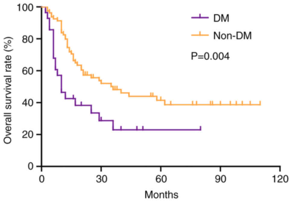
Association of DM and non-DM with OS
after hepatectomy in patients with ICC
At the end of the follow-up period, 61 (55.5%)
patients had died. The median follow-up time was 55 months for all
patients. The OS rate of patients with DM was significantly worse
than that of patients without DM (P=0.004; Fig. 1). The 1-, 3- and 5-year OS rates for
patients with DM were 42.6, 23.0 and 23.0%, which were lower than
83.8, 48.2 and 41.5% in patients without DM, and the median
survival time in patients with and without diabetes was 10 and 36
months, respectively (Fig. 1). As
shown in Table II, univariate
analysis revealed that advanced TNM stage (HR, 0.474; 95% CI,
0.245–0.918; P=0.027), >5 cm tumor diameter (HR, 0.460; 95% CI,
0.260–0.814; P=0.008), R1 resection (HR, 0.515; 95% CI,
0.310–0.857; P=0.011), lymph node metastasis (HR, 2.865; 95% CI,
1.658–4.952; P<0.001), intrahepatic metastasis (HR, 9.266; 95%
CI, 4.863–17.657; P<0.001), multiplicity (HR, 6.125; 95% CI,
3.482–10.774; P<0.001), vascular invasion (HR, 5.875; 95% CI,
3.252–10.614; P<0.001), high PKM2 expression (HR, 1.984; 95% CI,
1.177–3.344; P=0.010) and DM (HR, 2.152; 95% CI, 1.255–3.691;
P=0.005) were adverse prognostic factors that affected OS in
patients with ICC. In addition, multivariate analysis identified
the following factors as independent predictors for poor OS: DM
(HR, 1.989; 95% CI, 1.084–3.65; P=0.026), high PKM2 expression (HR,
1.364; 95% CI, 1.048–2.948; P=0.007), intrahepatic metastasis (HR,
2.826; 95% CI, 1.288–6.021; P=0.010), multiplicity (HR, 4.004; 95%
CI, 1.923–8.336; P<0.001) and vascular invasion (HR, 3.187; 95%
CI, 1.516–6.701; P=0.002).
| Table II.Univariate and multivariate analysis
of risk factors for overall survival. |
Table II.
Univariate and multivariate analysis
of risk factors for overall survival.
|
| Univariate
analysis | Multivariate
analysis |
|---|
|
|
|
|
|---|
| Variables | HR | 95% CI | P-value | HR | 95% CI | P-value |
|---|
| Sex
(male/female) | 1.026 | 0.617–1.706 | 0.922 |
|
|
|
| Age (<45/≥45
years) | 0.850 | 0.506–1.428 | 0.540 |
|
|
|
| TNM stage
(I–II/III–IV) | 0.474 | 0.245–0.918 | 0.027a | 1.003 | 0.462–2.176 | 0.995 |
| Tumor diameter
(≤5/>5 cm) | 0.460 | 0.260–0.814 | 0.008b | 0.728 | 0.348–1.527 | 0.402 |
| R0 (R0/R1) | 0.515 | 0.310–0.857 | 0.011a | 0.739 | 0.403–1.354 | 0.327 |
| Differentiation
(low) | 1.000 |
|
|
|
|
|
| Differentiation
(moderate) | 2.193 | 0.873–5.510 | 0.095 |
|
|
|
| Differentiation
(high) | 1.349 | 0.411–4.423 | 0.622 |
|
|
|
| Lymph node
metastasis (positive/negative) | 2.865 | 1.658–4.952 |
<0.001c | 0.919 | 0.472–1.788 | 0.803 |
| Intrahepatic
metastasis (positive/negative) | 9.266 | 4.863–17.657 |
<0.001c | 2.826 | 1.288–6.201 | 0.010a |
| Multiplicity
(positive/negative) | 6.125 | 3.482–10.774 |
<0.001c | 4.004 | 1.923–8.336 |
<0.001c |
| Vascular invasion
(positive/negative) | 5.875 | 3.252–10.614 |
<0.001c | 3.187 | 1.516–6.701 | 0.002b |
| Pyruvate kinase M2
expression (high/low) | 1.984 | 1.177–3.344 | 0.010a | 1.364 | 1.048–2.948 | 0.007b |
| Diabetes mellitus
(yes/no) | 2.152 | 1.255–3.691 | 0.005b | 1.989 | 1.084–3.650 | 0.026a |
| Total bilirubin
(>34/≤34 µmol/l) | 0.780 | 0.414–1.470 | 0.442 |
|
|
|
| Alanine
aminotransferase (>100/≤100 IU/l) | 0.940 | 0.477–1.855 | 0.859 |
|
|
|
| Prothrombin time
(>14/≤14 sec) | 1.533 | 0.556–4.232 | 0.409 |
|
|
|
| Albumin (>35/≤35
g/l) | 0.672 | 0.340–1.326 | 0.252 |
|
|
|
| Aspartate
aminotransferase (>100/≤100 IU/l) | 1.310 | 0.731–2.348 | 0.365 |
|
|
|
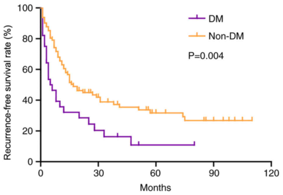
Association of DM and non-DM with RFS
after hepatectomy in patients with ICC
During follow-up, 78 (70.9%) patients experienced
tumor recurrence. The RFS rate in patients with DM was
significantly worse than that in patients without DM (P=0.004;
Fig. 2). The 1-, 3- and 5-year RFS
rates for patients with DM were 32.1, 16.3 and 10.9%, which were
lower than 60.5, 38.8 and 31.7% in patients without DM, and the
median RFS time in patients with and without diabetes was 5 and 17
months, respectively (Fig. 2). As
shown in Table III, univariate
analysis revealed that lymph node metastasis (HR, 2.664; 95% CI,
1.638–4.331; P<0.001), intrahepatic metastasis (HR, 5.640; 95%
CI, 3.143–10.123; P<0.001), multiplicity (HR, 4.427; 95% CI,
2.718–7.212; P<0.001), vascular invasion (HR, 5.100; 95% CI,
3.017–8.619; P<0.001), high PKM2 expression (HR, 2.048; 95% CI,
1.289–3.254; P=0.002) and DM (HR, 1.985; 95% CI, 1.222–3.225;
P=0.006) were adverse prognostic factors that affected RFS in
patients with ICC. In addition, multivariate analysis identified
the following factors as independent predictors for poor OS: DM
(HR, 1.784; 95% CI, 1.042–3.053; P=0.035), high PKM2 expression
(HR, 1.567; 95% CI, 1.057–3.012; P=0.006), multiplicity (HR, 2.898;
95% CI, 1.564–5.369; P=0.001) and vascular invasion (HR, 2.655; 95%
CI, 1.340–5.259; P=0.005).
| Table III.Univariate and multivariate analysis
of risk factors for recurrence-free survival. |
Table III.
Univariate and multivariate analysis
of risk factors for recurrence-free survival.
|
| Univariate
analysis | Multivariate
analysis |
|---|
|
|
|
|
|---|
| Variables | HR | 95% CI | P-value | HR | 95% CI | P-value |
|---|
| Sex
(male/female) | 0.922 | 0.591–1.440 | 0.722 |
|
|
|
| Age (<45/≥45
years) | 1.058 | 0.675–1.660 | 0.805 |
|
|
|
| TNM stage
(I–II/III–IV) | 0.644 | 0.378–1.099 | 0.107 |
|
|
|
| Tumor diameter
(≤5/>5 cm) | 0.699 | 0.438–1.118 | 0.135 |
|
|
|
| R0 (R0/R1) | 0.602 | 0.382–0.948 | 0.028 |
|
|
|
| Differentiation
(low) | 1.000 |
|
|
|
|
|
| Differentiation
(moderate) | 2.052 | 0.938–4.489 | 0.072 |
|
|
|
| Differentiation
(high) | 1.355 | 0.491–3.742 | 0.557 |
|
|
|
| Lymph node
metastasis (positive/negative) | 2.664 | 1.638–4.331 |
<0.001c | 1.065 | 0.582–1.948 | 0.839 |
| Intrahepatic
metastasis (positive/negative) | 5.640 | 3.143–10.123 |
<0.001c | 1.517 | 0.722–3.187 | 0.271 |
| Multiplicity
(positive/negative) | 4.427 | 2.718–7.212 |
<0.001c | 2.898 | 1.564–5.369 | 0.001b |
| Vascular invasion
(positive/negative) | 5.100 | 3.017–8.619 |
<0.001c | 2.655 | 1.340–5.259 | 0.005b |
| Pyruvate kinase M2
expression (high/low) | 2.048 | 1.289–3.254 | 0.002b | 1.567 | 1.057–3.012 | 0.006b |
| Diabetes mellitus
(yes/no) | 1.985 | 1.222–3.225 | 0.006b | 1.784 | 1.042–3.053 | 0.035a |
| Total bilirubin
(>34/≤34 µmol/l) | 0.750 | 0.426–1.321 | 0.319 |
|
|
|
| Alanine
aminotransferase (>100/≤100 IU/l) | 0.857 | 0.463–1.586 | 0.623 |
|
|
|
| Prothrombin time
(>14/≤14 sec) | 1.134 | 0.414–3.104 | 0.807 |
|
|
|
| Albumin (>35/≤35
g/l) | 0.627 | 0.345–1.139 | 0.126 |
|
|
|
| Aspartate
aminotransferase (>100/≤100 IU/l) | 1.198 | 0.706–2.032 | 0.503 |
|
|
|
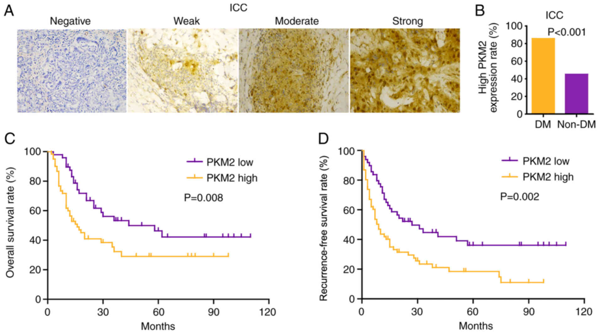
Association of PKM2 expression in
patients with ICC with and without DM
PKM2 expression was mainly concentrated in the
cytoplasm and nucleus. Representative images of immunohistochemical
staining are shown in Fig. 3A. High
PKM2 expression was observed in 61 (55.5%) patients with ICC; among
these patients, 85.7% patients had DM, while 45.1% patients did not
have DM (Fig. 3B). Similarly to HCC
(23), high PKM2 expression was
associated with poor OS and RFS (Fig. 3C
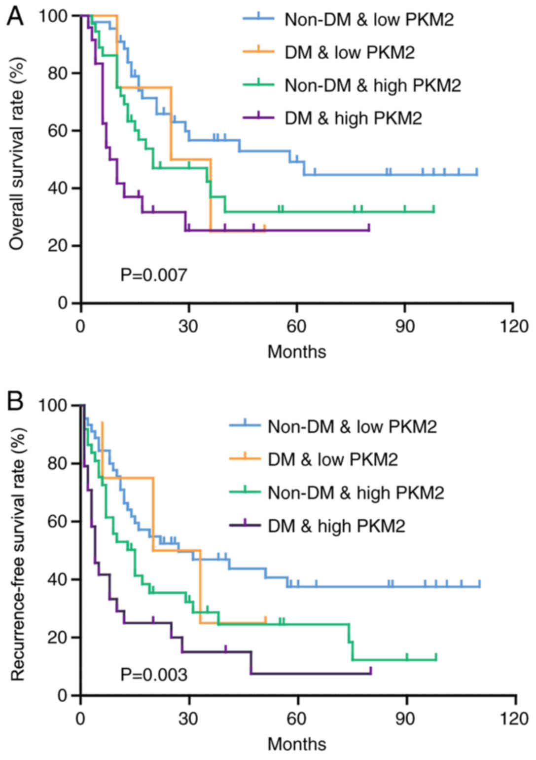
and D). Notably, the combination of low PKM2 expression and no
DM had a favorable prognostic value, while patients with high PKM2
expression and DM had the shortest OS ad RFS time (Fig. 4A and B).
Discussion
In the present retrospective study, it was revealed
that DM was an independent prognostic factor for survival that
significantly affected the OS and RFS rates of patients with ICC.
The impact of DM was independent of patient demographics. In
addition, patients with DM had a higher PKM2 expression rate than
patients without DM, but the mechanism by which DM may regulate
PKM2 expression remains to be uncovered. DM has been identified as
an independent risk factor for ICC in a number of countries, and
routine measurements for γ-glutamyl transferase and/or CA19-9 have
been recommended during follow-up for DM to detect ICC at an early
stage and expect a good OS (24);
however, the effect of DM on the outcome of ICC has been rarely
reported (24–27). A retrospective study by Endo et
al (28) revealed that DM was a
prognostic factor for patients undergoing surgery for ICC; however,
only 6 (6/81) patients with ICC had DM, affecting the reliability
of the results. In the present cohort, 28 (25.2%) patients had DM,
which is similar to a previously reported prevalence (4.9–33.1%) in
patients with ICC (29,30). The current multivariate analysis
revealed that DM was an independent risk factor for OS and RFS,
suggesting that a more rigorous follow-up strategy should be
adopted for patients with DM and suitable anti-diabetes treatments
may be efficacious in patients with ICC complicated with DM.
PKM2 is widely expressed in cancer and can promote
cancer cell proliferation through multiple biological mechanisms
(31). In breast cancer, PKM2-Y105D
phosphomimetic mutant increases MCF-10a cell colony formation and
CD44+/CD24− cancer stem cell population by
increasing YY1-associated protein 1 (YAP) nuclear localization
(32). ErbB2 is a strong inducer of
PKM2-Y105D phosphorylation, which promotes the nuclear localization
of YAP and increases the number of tumor stem cells (32). PKM2 binds directly to histone H3 and
phosphorylates histone H3 at threonine 11 when EGFR is activated;
this phosphorylation is required for the separation of histone
deacetylase 3 from cyclin D1 and Myc promoter regions, and
subsequent acetylation of histone H3 at lysine 9 (33). PKM2-dependent histone H3 modification
serves an important role in EGF-induced cyclin D1 and c-Myc
expression, glioma cell proliferation, cell cycle progression and
brain tumorigenesis (33). Further
analysis of the mechanism of PKM2 revealed that mitomycin 2 (MFN2),
a key regulator of mitochondrial fusion, interacted with PKM2,
promoted mitochondrial fusion and production of phosphorus oxide,
and suppressed glycolysis (34).
Additionally, mTOR increases the interaction between PKM2 and MFN2
through phosphorylation of MFN2, and it regulates the effects of
PKM2 and MFN2 on glycolysis, mitochondrial fusion and oxidative
phosphorylation in hepatocellular carcinoma and lung cancer cells
(34). Therefore, the mTOR-MFN2-PKM2
signal axis combines glycolysis with oxygen and phosphorus to
regulate the growth of hepatocellular carcinoma and lung cancer
cells (34). Insulin can increase
PKM2 expression in vitro, but this has not been verified in
the clinic (19,20). To the best of our knowledge, the
present data indicated for the first time in clinic samples that
PKM2 expression was higher in patients with DM than in those
without, consistent with previous in vitro results (23). Although the accurate underlying
mechanism remains unknown, the current results may partially
explain why patients with DM have a poorer survival outcome than
patients without DM.
The present study presents some limitations. First,
DM treatment can significantly affect long-term survival in
patients with HCC (35). Therefore,
the same phenomenon may be observed in patients with ICC. However,
the DM treatment strategy in the present study was unknown. Second,
all patients in the current cohort underwent surgical resection,
but these patients represented only a small proportion of all
patients with ICC (3). Whether DM
also indicates a poor prognosis in patients without surgery remains
to be explored.
In summary, the present data revealed that DM was
associated with a significantly lower OS rate in patients with ICC.
A potential cause may be associated with the abnormal glucose
metabolism mediated by PKM2, which should be further
investigated.
Acknowledgements
Not applicable.
Funding
The present study was supported by the National
Natural Science Foundation of China (grant no. 81802778).
Availability of data and materials
All data generated and/or analyzed during this study
are included in this published article.
Authors' contributions
KF, XY and HW wrote the manuscript. XY and HW were
involved in the statistical analysis. XY, HW and JG were involved
in clinical data collection. KF, XY, JG and XL were involved in the
study design, acquisition of data, financial support and
proofreading of the manuscript. All authors read and approved the
final manuscript.
Ethics approval and consent to
participate
All experiments were approved by the Ethics
Committee of the Second Affiliated Hospital of Chongqing Medical
University. Written informed consent was provided by all
patients.
Patient consent for publication
Not applicable.
Competing interests
The authors declare that they have no competing
interests.
References
|
1
|
Shaib YH, Davila JA, McGlynn K and
El-Serag HB: Rising incidence of intrahepatic cholangiocarcinoma in
the United States: A true increase? J Hepatol. 40:472–477. 2004.
View Article : Google Scholar : PubMed/NCBI
|
|
2
|
Spolverato G, Kim Y, Alexandrescu S,
Popescu I, Marques HP, Aldrighetti L, Clark Gamblin T, Miura J,
Maithel SK, Squires MH, et al: Is hepatic resection for large or
multifocal intrahepatic cholangiocarcinoma justified? Results from
a multi-institutional collaboration. Ann Surg Oncol. 22:2218–2225.
2015. View Article : Google Scholar : PubMed/NCBI
|
|
3
|
Raoof M, Dumitra S, Ituarte PHG, Melstrom
L, Warner SG, Fong Y and Singh G: Development and validation of a
prognostic score for intrahepatic cholangiocarcinoma. JAMA Surg.
152:e1701172017. View Article : Google Scholar : PubMed/NCBI
|
|
4
|
Whiting DR, Guariguata L, Weil C and Shaw
J: IDF diabetes atlas: Global estimates of the prevalence of
diabetes for 2011 and 2030. Diabetes Res Clin Pract. 94:311–321.
2011. View Article : Google Scholar : PubMed/NCBI
|
|
5
|
Yang WS, Shu XO, Gao J, Li HL, Cai H, Yang
G, Ji BT, Rothman N, Gao YT, Zheng W and Xiang YB: Prospective
evaluation of type 2 diabetes mellitus on the risk of primary liver
cancer in Chinese men and women. Ann Oncol. 24:1679–1685. 2013.
View Article : Google Scholar : PubMed/NCBI
|
|
6
|
Chaiteerakij R, Yang JD, Harmsen WS,
Slettedahl SW, Mettler TA, Fredericksen ZS, Kim WR, Gores GJ,
Roberts RO, Olson JE, et al: Risk factors for intrahepatic
cholangiocarcinoma: Association between metformin use and reduced
cancer risk. Hepatology. 57:648–655. 2013. View Article : Google Scholar : PubMed/NCBI
|
|
7
|
Komura T, Mizukoshi E, Kita Y, Sakurai M,
Takata Y, Arai K, Yamashita T, Ohta T, Shimizu K, Nakamoto Y, et
al: Impact of diabetes on recurrence of hepatocellular carcinoma
after surgical treatment in patients with viral hepatitis. Am J
Gastroenterol. 102:1939–1946. 2007. View Article : Google Scholar : PubMed/NCBI
|
|
8
|
Ting CT, Chen RC, Chen CC, Liu MH, Chu D
and Kuo NW: Diabetes worsens the surgical outcomes in cirrhotic
patients with hepatocellular carcinoma. Tohoku J Exp Med.
227:73–81. 2012. View Article : Google Scholar : PubMed/NCBI
|
|
9
|
Wang YG, Wang P, Wang B, Fu ZJ, Zhao WJ
and Yan SL: Diabetes mellitus and poorer prognosis in
hepatocellular carcinoma: A systematic review and meta-analysis.
PLoS One. 9:e954852014. View Article : Google Scholar : PubMed/NCBI
|
|
10
|
Wang YY, Huang S, Zhong JH, Ke Y, Guo Z,
Liu JQ, Ma L, Li H, Ou BN and Li LQ: Impact of diabetes mellitus on
the prognosis of patients with hepatocellular carcinoma after
curative hepatectomy. PLoS One. 9:e1138582014. View Article : Google Scholar : PubMed/NCBI
|
|
11
|
Palmer WC and Patel T: Are common factors
involved in the pathogenesis of primary liver cancers? A
meta-analysis of risk factors for intrahepatic cholangiocarcinoma.
J Hepatol. 57:69–76. 2012. View Article : Google Scholar : PubMed/NCBI
|
|
12
|
Vander Heiden MG, Cantley LC and Thompson
CB: Understanding the warburg effect: The metabolic requirements of
cell proliferation. Science. 324:1029–1033. 2009. View Article : Google Scholar : PubMed/NCBI
|
|
13
|
Cairns RA, Harris IS and Mak TW:
Regulation of cancer cell metabolism. Nat Rev Cancer. 11:85–95.
2011. View
Article : Google Scholar : PubMed/NCBI
|
|
14
|
Yu G, Yu W, Jin G, Xu D, Chen Y, Xia T, Yu
A, Fang W, Zhang X, Li Z and Xie K: PKM2 regulates neural invasion
of and predicts poor prognosis for human hilar cholangiocarcinoma.
Mol Cancer. 14:1932015. View Article : Google Scholar : PubMed/NCBI
|
|
15
|
Beyoglu D, Imbeaud S, Maurhofer O,
Bioulac-Sage P, Zucman-Rossi J, Dufour JF and Idle JR: Tissue
metabolomics of hepatocellular carcinoma: Tumor energy metabolism
and the role of transcriptomic classification. Hepatology.
58:229–238. 2013. View Article : Google Scholar : PubMed/NCBI
|
|
16
|
Beyoglu D and Idle JR: The metabolomic
window into hepatobiliary disease. J Hepatol. 59:842–858. 2013.
View Article : Google Scholar : PubMed/NCBI
|
|
17
|
Israelsen WJ, Dayton TL, Davidson SM,
Fiske BP, Hosios AM, Bellinger G, Li J, Yu Y, Sasaki M, Horner JW,
et al: PKM2 isoform-specific deletion reveals a differential
requirement for pyruvate kinase in tumor cells. Cell. 155:397–409.
2013. View Article : Google Scholar : PubMed/NCBI
|
|
18
|
Cortes-Cros M, Hemmerlin C, Ferretti S,
Zhang J, Gounarides JS, Yin H, Muller A, Haberkorn A, Chene P,
Sellers WR and Hofmann F: M2 isoform of pyruvate kinase is
dispensable for tumor maintenance and growth. Proc Natl Acad Sci
USA. 110:489–494. 2013. View Article : Google Scholar : PubMed/NCBI
|
|
19
|
Li Q, Liu X, Yin Y, Zheng JT, Jiang CF,
Wang J, Shen H, Li CY, Wang M, Liu LZ and Jiang BH: Insulin
regulates glucose consumption and lactate production through
reactive oxygen species and pyruvate kinase M2. Oxid Med Cell
Longev. 2014:5049532014. View Article : Google Scholar : PubMed/NCBI
|
|
20
|
Li W, Wang J, Chen QD, Qian X, Li Q, Yin
Y, Shi ZM, Wang L, Lin J, Liu LZ and Jiang BH: Insulin promotes
glucose consumption via regulation of miR-99a/mTOR/PKM2 pathway.
PLoS One. 8:e649242013. View Article : Google Scholar : PubMed/NCBI
|
|
21
|
American Diabetes Association: Standards
of medical care in diabetes-2013. Diabetes Care 36 Suppl. 1 (Suppl
1):S11–S66. 2013. View Article : Google Scholar
|
|
22
|
Edge SB, Byrd DR, Compton CC, Fritz AG,
Greene FL and Trotti A: AJCC Cancer Staging Manual. 7th. Springer;
New York, NY: 2010
|
|
23
|
Liu Y, Wu H, Mei Y, Ding X, Yang X, Li C,
Deng M and Gong J: Clinicopathological and prognostic significance
of PKM2 protein expression in cirrhotic hepatocellular carcinoma
and non-cirrhotic hepatocellular carcinoma. Sci Rep. 7:152942017.
View Article : Google Scholar : PubMed/NCBI
|
|
24
|
Nishioka T, Kubo S, Tanaka S, Wakasa K,
Takemura S, Kinoshita M, Hamano G, Kuwae Y, Shibata T and Suehiro
S: Outcomes of hepatic resection in intrahepatic cholangiocarcinoma
patients with diabetes, hypertension, and dyslipidemia:
Significance of routine follow-up. Liver Cancer. 5:107–120. 2016.
View Article : Google Scholar : PubMed/NCBI
|
|
25
|
Lee BS, Park EC, Park SW, Nam CM and Roh
J: Hepatitis B virus infection, diabetes mellitus, and their
synergism for cholangiocarcinoma development: A case-control study
in Korea. World J Gastroenterol. 21:502–510. 2015. View Article : Google Scholar : PubMed/NCBI
|
|
26
|
Wu Q, He XD, Yu L, Liu W and Tao LY: The
metabolic syndrome and risk factors for biliary tract cancer: A
case-control study in China. Asian Pac J Cancer Prev. 13:1963–1969.
2012. View Article : Google Scholar : PubMed/NCBI
|
|
27
|
Huang YJ, Wu AT, Chiou HY, Chuang MT, Meng
TC, Chien LN and Yen Y: Interactive role of diabetes mellitus and
female sex in the risk of cholangiocarcinoma: A population-based
nested case-control study. Oncotarget. 8:6642–6651. 2017.
View Article : Google Scholar : PubMed/NCBI
|
|
28
|
Endo I, Gonen M, Yopp AC, Dalal KM, Zhou
Q, Klimstra D, D'Angelica M, DeMatteo RP, Fong Y, Schwartz L, et
al: Intrahepatic cholangiocarcinoma: Rising frequency, improved
survival, and determinants of outcome after resection. Ann Surg.
248:84–96. 2008. View Article : Google Scholar : PubMed/NCBI
|
|
29
|
Jing W, Jin G, Zhou X, Zhou Y, Zhang Y,
Shao C, Liu R and Hu X: Diabetes mellitus and increased risk of
cholangiocarcinoma: A meta-analysis. Eur J Cancer Prev. 21:24–31.
2012. View Article : Google Scholar : PubMed/NCBI
|
|
30
|
Welzel TM, Graubard BI, El-Serag HB, Shaib
YH, Hsing AW, Davila JA and McGlynn KA: Risk factors for
intrahepatic and extrahepatic cholangiocarcinoma in the United
States: A population-based case-control study. Clin Gastroenterol
Hepatol. 5:1221–1228. 2007. View Article : Google Scholar : PubMed/NCBI
|
|
31
|
Dayton TL, Jacks T and Vander Heiden MG:
PKM2, cancer metabolism, and the road ahead. EMBO Rep.
17:1721–1730. 2016. View Article : Google Scholar : PubMed/NCBI
|
|
32
|
Zhou Z, Li M, Zhang L, Zhao H, Şahin Ö,
Chen J, Zhao JJ, Songyang Z and Yu D: Oncogenic kinase-induced PKM2
tyrosine 105 phosphorylation converts nononcogenic PKM2 to a tumor
promoter and induces cancer stem-like cells. Cancer Res.
78:2248–2261. 2018. View Article : Google Scholar : PubMed/NCBI
|
|
33
|
Yang W, Xia Y, Hawke D, Li X, Liang J,
Xing D, Aldape K, Hunter T, Alfred Yung WK and Lu Z: PKM2
phosphorylates histone H3 and promotes gene transcription and
tumorigenesis. Cell. 150:685–696. 2012. View Article : Google Scholar : PubMed/NCBI
|
|
34
|
Li T, Han J, Jia L, Hu X, Chen L and Wang
Y: PKM2 Coordinates glycolysis with mitochondrial fusion and
oxidative phosphorylation. Protein Cell. 10:583–594. 2019.
View Article : Google Scholar : PubMed/NCBI
|
|
35
|
Casadei Gardini A, Faloppi L, De Matteis
S, Foschi FG, Silvestris N, Tovoli F, Palmieri V, Marisi G,
Brunetti O, Vespasiani-Gentilucci U, et al: Metformin and insulin
impact on clinical outcome in patients with advanced hepatocellular
carcinoma receiving sorafenib: Validation study and biological
rationale. Eur J Cancer. 86:106–114. 2017. View Article : Google Scholar : PubMed/NCBI
|