Introduction
Breast magnetic resonance imaging (MRI) is widely
used in clinical practice. The methods of MRI examination include
the conventional plain scan, the dynamic contrast-enhanced (DCE)
scan and the diffusion-weighted imaging (DWI) scan. DCE-MRI plays
an important role in the diagnosis of breast lesions, with a
reported sensitivity of 95–99% in detecting invasive cancer and 80%
in detecting ductal carcinoma in situ, whereas its mean
reported specificity is 80% (range, 37–97%) (1–5).
Moreover, the curve of the DCE scan may indicate the enhancement
type of the lesions. DWI is a functional MRI technique that
characterizes tissues by their water diffusion properties. DWI and
the apparent diffusion coefficient (ADC) value have been used
successfully in the differential diagnosis between malignant and
benign breast diseases, and to determine tumor extension. It has
been reported that the combination of DCE-MRI with DWI has a
sensitivity of 95.7% and a specificity of 89.2%; therefore,
radiologists mainly base their diagnosis on the combination of
these two techniques (6).
As regards plain scans, the signal of the lesions is
relatively similar to that of normal tissue in the conventional T1
sequence and the T2 fat suppression sequence; therefore, some
lesions may be easily overlooked without DCE-MRI as radiologists
may not examine the plain scan more closely. On this note, recent
studies have suggested that T2-weighted images are a valuable
component of the MRI evaluation of edema (7,8). It has
been reported that peritumoral edema is a common imaging
manifestation of advanced or inflammatory breast cancer. The cause
of edema is associated with the poor drainage caused by lymphatic
obstruction. For dense breasts, it is often difficult to
distinguish edema from tumor lesions on mammography. However, the
sequence of the plain scan and enhanced scan may help distinguish
between tumors and edema.
The aim of the present study was to compare the
prognostic value of the MRI T2 fat suppression sequence with that
of the DCE-MRI and DWI scan in patients with mass-type breast
cancer exhibiting peritumoral edema. The associations between the
presence of edema and tumor stage, pathological classification,
immunohistochemical findings and axillary lymph node metastasis
were further investigated in order to determine the clinical
prognostic value of the MRI T2 fat suppression sequence in breast
cancer. In addition, the association between the improvement of
edema and tumor shrinkage in patients who underwent neoadjuvant
chemotherapy (NAC) was also investigated.
Materials and methods
Patients
A total of 80 patients (age range, 24–77 years; mean
age ± standard deviation, 45.1±11.1 years) with histologically
proven breast cancer who had undergone an MRI examination for tumor
staging and detection of axillary lymph node involvement between
September 2017 and December 2018 were included in the present
study. The study protocol was approved by the Ethics Committee of
the First Affiliated Hospital of Xi'an Jiao Tong University (no.:
XJTU1AF2017LSK-72), and all patients provided written informed
consent prior to their inclusion in the study. The MRI examination
was performed in the second week of the menstrual cycle in
premenopausal women. No breast implants were found in any of the
patients. Histopathological diagnosis was performed following
surgical resection in 27 cases and puncture biopsy in 53 cases.
Immunohistochemical staining
Breast tumor tissues were fixed in formalin (10%, 24
h, room temperature) embedded in paraffin and cut into 5-µm
sections. The sections were deparaffinized and immersed in citrate
buffer (pH 6.0) for 5 min to retrieve antigen, followed by 0.3%
hydrogen peroxide for 15 min to block endogenous peroxidase
activity. The sections were then blocked for 30 min using 10% goat
plasma (Invitrogen; Thermo Fisher Scientific, Inc.). Following
incubation with primary antibodies directed against estrogen
receptor (ER; ab32063; 1:1,000), progesterone receptor (PR;
ab16661; 1:400) or human epidermal growth factor receptor 2 (HER2;
ab214275; 1:4,000) for 1 h at 37°C, the slides were incubated with
an HRP-conjugated goat anti-rabbit secondary antibody (ab205718;
1:2,000) according to the manufacturer's recommendations. All
primary and secondary antibodies were purchased from Abcam.
Finally, the sections were visualized with diaminobenzidine and
counterstained with hematoxylin for 5 min at room temperature, then
dehydrated in alcohol and xylene and mounted onto glass slides. The
numbers of ER-, PR- or HER2-positive cells were counted from at
least 10 randomly selected fields of each sample (NIKON ECLIPSE
T1-S; Nikon Corporation) at a magnification of ×400. The tissue
samples were classified as positive for ER and PR when the
proportion of positively stained tumor cells was >1% (9). For HER2, the tumor was considered
positive if >10% of the tumor cells exhibited positively stained
nuclei (10).
MRI protocol
MRI examinations were performed on a 3.0 T MR device
(Discovery 750w, Cytiva) using a four-channel breast coil. The
protocol consisted of the following: Transverse T2 FSE-IDEAL ASSET
sequence [TR/TE=6020/80.7 msec, field of view (FOV)=320×320 mm (AP
× RL), matrix 256×256, 4-mm slice thickness and 1-mm gap];
transverse T1-weighted TSE [TR/TE=867/5.7 msec, FOV=320×320 mm (AP
× RL), matrix 256×256, 4-mm slice thickness and 1-mm gap]; 3D
dynamic, CE T1-weighted vibrant sequences [TR/TE=7.6/4.3 msec,
FOV=320×320 mm (AP × RL), matrix 125×125, 5-mm slice thickness
without gaps, 6 dynamic acquisitions, a dynamic data acquisition
time of 1 min and 30 sec, and a total sequence duration of 9 min];
gadobenate dimeglumine (MultiHance®), which is
considered as the most accurate contrast medium used in this field,
was intravenously injected at a dose of 0.1 mmol/kg body weight and
flow rate of 2 ml/sec followed by 10 ml of saline solution, as
previously reported (11); EPI-DWI
sequences were acquired in the transversal orientation using the
following parameters: TR/TE=4,000/69.6 msec, FOV=320×320 mm (AP ×
RL), matrix 128×140, 4-mm slice thickness with 1-mm gap.
Image analysis
All MRI data were transferred to and analyzed on a
diagnostic workstation equipped with dedicated software for MRI
examination (Advantage Workstation 4.6; Cytiva). Two radiologists
with 5 years of experience in the field of breast MRI who were
blinded to the clinical findings and pathological diagnosis
reviewed all the MRI films. In addition to the size, shape, signal,
enhancement curve and ADC value of the mass, the presence of edema
around the tumor mass, skin thickening and increase of subcutaneous
fibrous connective tissue on the MRI T2 fat suppression sequence
were more closely observed. Moreover, the presence of abnormal
enhancement on dynamic enhanced scanning and increased signal
intensity on DWI on these areas were also determined.
Statistical analysis
The χ2 test or Fisher's exact test were
used to analyze the association between the MRI T2 fat suppression
sequence findings around the mass and tumor stage, pathological
classification, luminal classification and axillary lymph node
metastasis. All calculations were performed using SPSS software,
version 25.0 (IBM Corp.).
Results
Imaging characteristics
In the present study, according to the Breast
Imaging Reporting and Data System Magnetic Resonance Imaging
(BI-RADS-MRI) by the American College of Radiology, 80 patients
were classified as type a, b, c and d (n=10, n=31, n=25 and n=14,
respectively) on the basis of breast composition, and as type a, b,
c and d (n=22, n=34, n=16 and n=8, respectively) on the basis of
background parenchymal enhancement. On the DCE-MRI, 3 patients
exhibited a type I curve, while 66 exhibited the type II and 11 the
type III curve. The ADC value of 16 cases was
>1.2×10⁻³ mm2/sec and that of 64 cases was
<1.2×10⁻³ mm2/sec. In total, 27 cases were
diagnosed with BI-RADS V categories and 53 cases were diagnosed
with BI-RADS VI categories.
Presence of edema
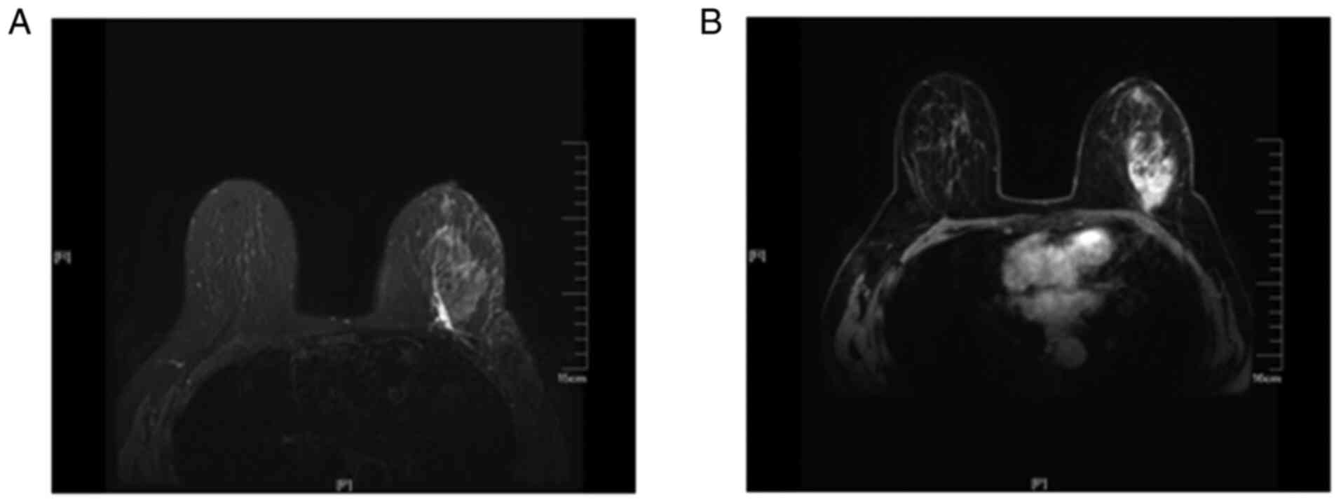
Of the 80 patients, 35 exhibited edema (Fig. 1) around the mass on the MRI T2 fat
suppression sequence (group I) and the remaining 45 cases did not
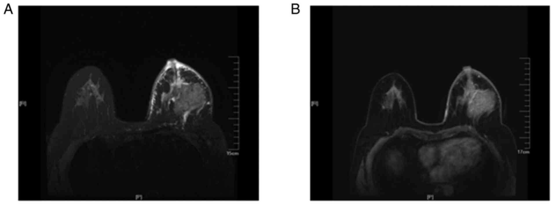
exhibit edema (group II). Furthermore, in group I, 26 cases
exhibited skin thickening and increased subcutaneous fibrous
connective tissue (Fig. 2) and 7
cases exhibited nipple retraction on the MRI T2 fat suppression
sequence. Compared with the edema observed on the plain scan, there
was no abnormal enhancement on the DCE and no abnormal signal on
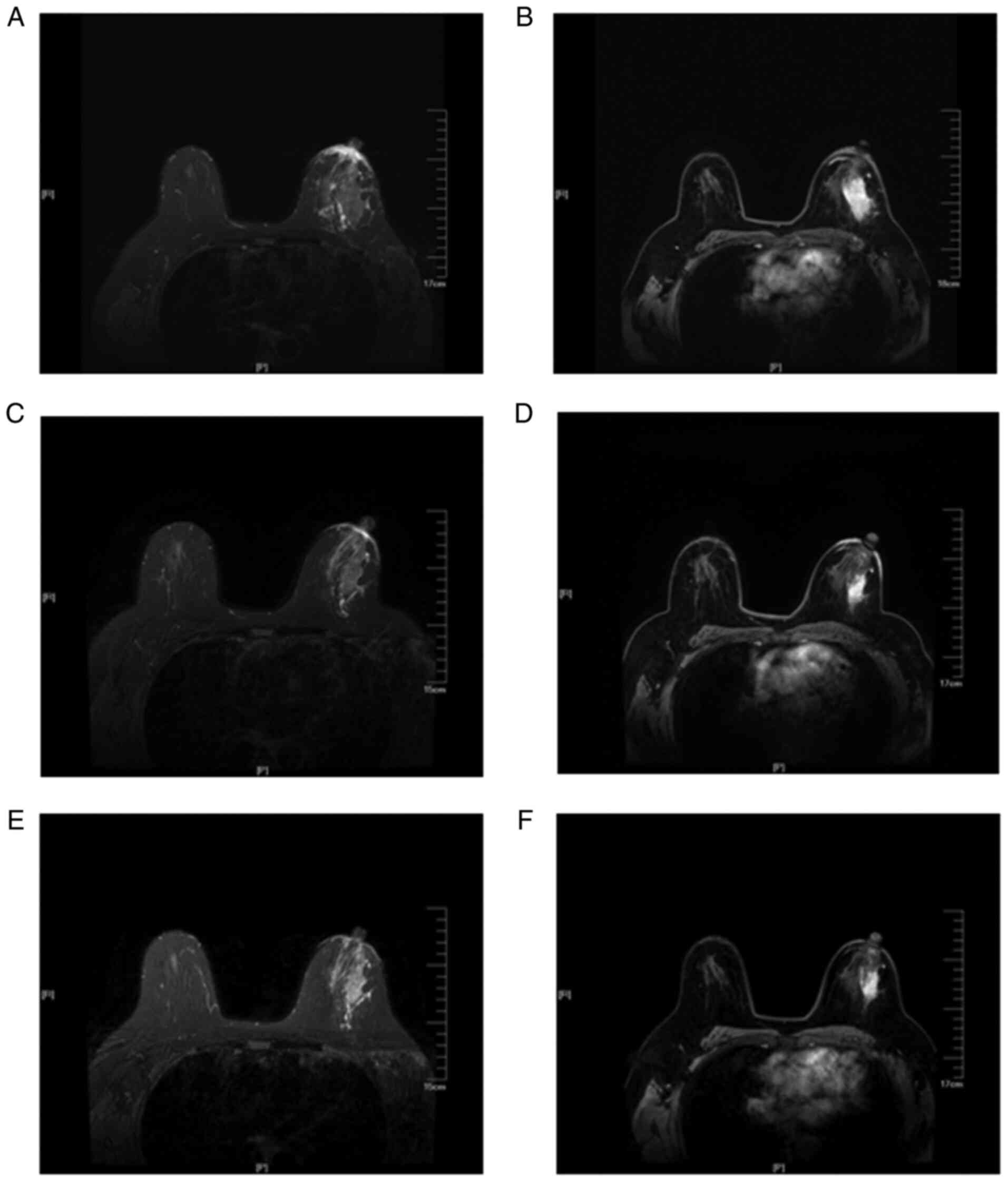
the DWI in the edema area. A total of 12 patients in group I and 3
patients in group II eventually received NAC. In the 12 cases in
group 1, following chemotherapy, the size of the tumors was smaller
compared with that prior to treatment, and the edema shadows around
the mass, thickened skin, as well as the increased subcutaneous
fibrous connective tissue on the MRI T2 fat suppression sequences
also improved (Fig. 3).
Pathological classification
Pathological results were obtained by needle
puncture or surgical biopsy in all 80 patients. In group I, 25
cases were non-specific invasive carcinoma and the remaining 10
cases were non-specific invasive carcinoma with high-grade ductal
carcinoma. In group II, 30 cases were non-specific invasive
carcinoma, 11 cases were non-specific invasive carcinoma with
high-grade ductal carcinoma, 3 cases were ductal carcinoma in
situ and 1 case was medullary carcinoma. There was no
significant difference in pathological classification between the
two groups (P>0.05).
Association between imaging findings
and stage
According to the TNM staging criteria of breast
cancer, the tumors were classified as stage I, II, III and IV. The
associations between the MRI T2 fat suppression sequence imaging
findings around the mass and tumor stage are presented in Table I. Compared with group II, the
patients in group I exhibited a significantly higher tumor stage
(P<0.05).
 | Table I.Correlation between the findings of
magnetic resonance imaging T2 fat suppression sequence around the
mass and tumor stage. |
Table I.
Correlation between the findings of
magnetic resonance imaging T2 fat suppression sequence around the
mass and tumor stage.
|
| Tumor stage |
|---|
|
|
|
|---|
| Groups | I | II | III | IV | P-value |
|---|
| Ia | 3 | 3 | 25 | 4 | 0.000 |
| IIb | 9 | 33 | 3 |
|
|
Association between imaging findings
and hormone receptor status
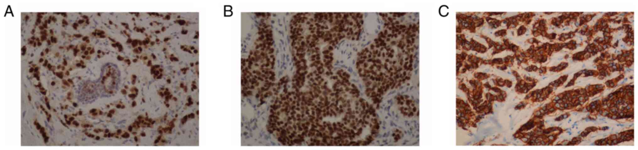
According to the expression of ER, PR and HER2 on
immunohistochemistry (Fig. 4), the
tumors were categorized as luminal A, luminal B, HER2+
and triple-negative. The associations between the MRI T2 fat
suppression sequence imaging findings around the mass and luminal
type are presented in Table II.
There was no significant difference in the expression of ER, PR or
HER2, as determined by immunohistochemistry, between the two groups
(P>0.05).
| Table II.Correlation between the findings of
magnetic resonance imaging T2 fat suppression sequence around the
mass and luminal type. |
Table II.
Correlation between the findings of
magnetic resonance imaging T2 fat suppression sequence around the
mass and luminal type.
|
| Luminal type |
|
|---|
|
|
|
|
|---|
| Groups | Luminal A | Luminal B | HER2+ | Triple-negative | P-value |
|---|
| Ia | 5 | 13 | 6 | 11 | 0.203 |
| IIb | 5 | 27 | 6 | 7 |
|
Association between imaging findings
and axillary lymph node metastasis
All 80 patients underwent axillary lymph node biopsy
or surgery. The associations between the MRI T2 fat suppression
sequence imaging findings around the mass and axillary lymph node
metastasis are presented in Table
III. Compared with group II, the patients in group I exhibited
a higher rate of axillary lymph node metastasis (P<0.05).
| Table III.Correlation between the findings of
magnetic resonance imaging T2 fat suppression sequence around the
mass and axillary lymph node metastasis. |
Table III.
Correlation between the findings of
magnetic resonance imaging T2 fat suppression sequence around the
mass and axillary lymph node metastasis.
|
| Axillary lymph node
metastasis |
|
|---|
|
|
|
|
|---|
| Groups | Metastasis | No metastasis | P-value |
|---|
| Ia | 17 | 18 | 0.043 |
| IIb | 12 | 33 |
|
Discussion
MRI is the most sensitive method for the detection
of breast diseases. The DCE-MRI and DWI techniques are hotspots in
current research and the main focus is the study of the tumor
per se. By contrast, for regular base sequences, the
research on the T1 plain scan and T2 fat suppression sequence has
been less extensive. In particular, in recent years, studies have
demonstrated that there is no significant difference between fast
magnetic resonance scanning and conventional scanning in the
diagnosis of lesions; the only exception is that the time required
for the former is markedly shorter (12–14).
Thus, the conventional T2 fat suppression sequence was deleted from
the fast magnetic resonance sequence. The present study aimed to
determine whether the T2 fat suppression sequence can provide other
information that may be valuable for the diagnosis of breast
cancer.
The clinical and imaging data of 80 cases of
mass-type breast cancer were collected for the present study. In 35
of those cases, grid or patchy blurred high signal shadows were
identified around the mass on the T2-weighted fat suppression
sequence, whereas there was no grid or patchy blurred high signal
shadows around the mass in the other 45 cases. However, no abnormal
enhancement or diffusion was found in these areas on DCE-MRI or
DWI.
A previous study indicated that the aforementioned
abnormal signal on the T2-weighted with fat suppression sequence
was edema (15). Mammary edema
refers to the swelling caused by the accumulation of fluid in the
tissues of the breast. The underlying causes are usually malignant,
including advanced breast cancer, inflammatory breast cancer,
lymphatic inflammatory metastasis and lymphoma. Pathologically,
edema caused by malignant lesions may be observed as lymphatic
obstruction and/or capillary and venule obstruction (16–18).
Previous studies demonstrated that peritumoral edema is likely to
occur due to tumor angiogenesis, which is one of the hallmarks of
cancer, and leads to increased vascular permeability and the
release of peritumoral cytokines (19,20).
On imaging, edema is characterized by increased
density on mammography or signal intensity on the T2-weighted with
fat suppression sequence, enlargement of fibrous trabeculae,
thickening of Cooper's ligament and cutaneous edema. In contrast to
T1-weighted sequences, which highlight anatomic detail and are used
on the enhanced scan, fluid-sensitive T2-weighted sequences depict
edema, hemorrhage, mucus, or cystic fluid (8). Therefore, T2-weighted images are a
valuable component of the MRI evaluation of edema (15,21).
Previous studies have demonstrated that the
prognosis of breast cancer is closely associated with tumor state,
luminal type and axillary lymph node metastasis (22,23). It
was previously demonstrated that the prognosis of patients with
luminal A type breast cancer was the superior to that of patients
with luminal B type breast cancer, whereas patients with
HER2-positive and triple-negative breast cancer had the worst
prognosis (24). Further research
found that patients with HER2-positive and luminal B breast cancer
were more prone to developing multiple lesions and axillary lymph
node metastasis, while the distant metastasis rate of patients with
triple-negative breast cancer was significantly higher compared
with that of patients with luminal A, luminal B and HER2-positive
breast cancer (25,26).
In the present study, the association of peritumoral
edema with clinical stage, pathological classification and axillary
lymph node metastasis was analyzed. The results revealed that the
35 patients with peritumoral edema had a more advanced clinical
stage and higher rate of axillary lymph node metastasis compared
with the remaining 45 patients; however, there was no significant
difference in pathological classification or the expression of ER,
PR and HER2, as determined by immunohistochemistry, between the two
groups. In addition, of the 35 cases with edema, 26 exhibited skin
thickening and increase of subcutaneous fibrous connective tissue,
and 7 exhibited nipple retraction.
It has been reported that the presence of
peritumoral edema may be a strong prognostic indicator for
lymphatic spread and the cancerous infiltration of lymph nodes
(27,28). Cheon et al demonstrated that
the presence of peritumoral edema was associated with the
characteristics of biologically aggressive tumors (29) and Uematsu et al demonstrated
that peritumoral edema was associated with prognostic factors
(30). The results of the present
study confirmed these previous conclusions, indicating the
prognostic value of peritumoral edema in breast cancer. More
importantly, in the present study, the shrinkage of the tumors in
patients receiving NAC was accompanied by the improvement of the
edema around the mass and the skin thickening, as well as a
decrease of the subcutaneous fibrous connective tissue on the MRI
T2 fat suppression sequences. To the best of our knowledge, few
studies to date have investigated the imaging changes associated
with the presence of edema around the mass following NAC. Based on
these findings, less or no edema post-NAC may be used as a
predictive indicator of a better prognosis.
In conclusion, the pathological basis of edema
around the breast cancer mass is the infiltration of lymphatic
vessels by tumor tissue, and the results of the present study
demonstrated that the MRI T2 may clearly demonstrate the signs of
peritumoral edema. The results further confirmed that the
occurrence of edema was closely associated with a higher tumor
stage and axillary lymph node metastasis. Moreover, it was observed
that, with the reduction of the tumor volume following NAC, edema
also improved. Therefore, the presence of peritumoral edema in the
MRI T2 may be used as a valuable imaging index for evaluating the
prognosis and treatment efficacy for patients with breast
cancer.
However, the present study had several limitations.
First, the number of cases in the study was insufficient and,
therefore, the association among the three parameters (tumor stage,
luminal type and axillary lymph node metastasis) was not analyzed.
Second, a proportion of the patients in the present study underwent
MRI following core-needle biopsy, and the peritumoral signals may
have thus been affected by the post-procedural changes. Third, this
study lacked further follow-up.
In conclusion, the T2-weighted images are
irreplaceable for evaluating edema, skin thickening and the
increase in subcutaneous fibrous connective tissue. Peritumoral
edema in the MRI T2 fat suppression sequence may predict a poor
prognosis in patients with mass-type breast cancer, whereas the
improvement of peritumoral edema post-NAC may predict a more
favorable prognosis.
Acknowledgements
Not applicable.
Funding
The present study was supported by grants from the
Shaanxi Research and Development Program (grant no. 2017SF-020) and
the Institutional Foundation of the First Affiliated Hospital of
Xi'an Jiaotong University (grant no. 2016MS-05).
Availability of data and materials
All data generated or analyzed during this study are
included in this published article.
Authors' contributions
TL analyzed and interpreted the experimental data
and contributed to the writing of the manuscript. BH performed the
immunohistochemical staining of the breast tumors. HD designed the
experiments and reviewed and revised the manuscript. YZ designed
the experiments and is the major contributor to writing the
manuscript. All the authors have read and approved the final
manuscript.
Ethics approval and consent to
participate
The study protocol was approved by the Ethics
Committee of the First Affiliated Hospital of Xi'an Jiao Tong
University (no.: XJTU1AF2017LSK-72), and all patients provided
written informed consent prior to their inclusion in the study.
Patient consent for publication
Not applicable.
Competing interests
All the authors declare that they have no competing
interests.
References
|
1
|
Kuhl CK: MRI of breast tumors. Eur Radiol.
10:46–58. 2000. View Article : Google Scholar : PubMed/NCBI
|
|
2
|
Huang W, Fisher PR, Dulaimy K, Tudoria LA,
O'Hea B and Button TM: Detection of breast malignancy: Diagnostic
MR protocol for improved specificity. Radiology. 232:585–591. 2004.
View Article : Google Scholar : PubMed/NCBI
|
|
3
|
Belli P, Costantini M, Bufi E, Magistrelli
A, La Torre G and Bonomo L: Diffusion-weighted imaging in breast
lesion evaluation. Radiol Med. 115:51–69. 2010.(In English and
Italian). View Article : Google Scholar : PubMed/NCBI
|
|
4
|
Peter NH, Rinkes IH, Zuithoff NP, Mali WP,
Moons KG and Peeters PH: Meta-Analysis of MR imaging in the
diagnosis of breast lesions. Radiology. 246:116–124. 2008.
View Article : Google Scholar : PubMed/NCBI
|
|
5
|
Moschetta M, Telegrafo M, Rella L,
Capolongo A, Ianora AA and Angelelli G: MR evaluation of breast
lesions obtained by diffusion-weighted imaging with background body
signal suppression(DWIBS) and correlations with histological
findings. Magn Reson Imaging. 32:605–609. 2014. View Article : Google Scholar : PubMed/NCBI
|
|
6
|
Kul S, Cansu A, Alhan E, Dinc H, Gunes G
and Reis A: Contribution of diffusion-weighted imaging to dynamic
contrast-enhanced MRI in the characterization of breast tumors. AJR
AM J Roentgenol. 196:210–217. 2011. View Article : Google Scholar : PubMed/NCBI
|
|
7
|
Guo Y, Tang WJ, Kong QC, Liang YY, Han XR,
Zheng BJ, Sun L, Wei XH, Jin Z and Liu CL: Can whole-tumor apparent
diffusion coefficient histogram analysis be helpful to evaluate
breast phyllode tumor grades? Eur J Radiol. 114:25–31. 2019.
View Article : Google Scholar : PubMed/NCBI
|
|
8
|
Westra C, Dialani V, Mehta TS and
Eisenberg RL: Using T2-weighted sequences to more accurately
characterize breast masses seen on MRI. AJR Am J Roentgenol.
202:W183–W190. 2014. View Article : Google Scholar : PubMed/NCBI
|
|
9
|
Hammond ME, Hayes DF, Dowsett M, Allred
DC, Hagerty KL, Badve S, Fitzgibbons PL, Francis G, Goldstein NS,
Hayes M, et al: American society of clinical oncology/college of
American pathologists guideline recommendations for
immunohistochemical testing of estrogen and progesterone receptors
in breast cancer. J Clin Oncol. 28:2784–2795. 2010. View Article : Google Scholar : PubMed/NCBI
|
|
10
|
Wolff AC, Hammond ME, Schwartz JN, Hagerty
KL, Allred DC, Cote RJ, Dowsett M, Fitzgibbons PL, Hanna WM, Langer
A, et al: American Society of clinical oncology/college of American
pathologists guideline recommendations for human epidermal growth
factor receptor 2 testing in breast cancer. Arch Pathol Lab Med.
131:18–43. 2007.PubMed/NCBI
|
|
11
|
Martincich L, Faivre-Pierret M, Zechmann
CM, Corcione S, van den Bosch HC, Peng WJ, Petrillo A, Siegmann KC,
Heverhagen JT, Panizza P, et al: Multicenter, double-blind,
randomized, intraindividual crossover comparison of gadobenate
dimeglumine and gadopentetate dimeglumine for Breast MR imaging
(DETECT Trial). Radiology. 258:396–408. 2011. View Article : Google Scholar : PubMed/NCBI
|
|
12
|
Jain M, Jain A, Hyzy MD and Werth G: FAST
MRI breast screening revisited. J Med Imaging Radiat Oncol.
61:24–28. 2017. View Article : Google Scholar : PubMed/NCBI
|
|
13
|
Mango VL, Morris EA, Dershaw DD, Abramson
A, Fry C, Moskowitz CS, Hughes M, Kaplan J and Jochelson MS:
Abbreviated protocol for breast MRI: Are multiple sequences needed
for cancer detection? Eur J Radiol. 84:65–70. 2015. View Article : Google Scholar : PubMed/NCBI
|
|
14
|
Heacock L, Melsaether AN, Heller SL, Gao
Y, Pysarenko KM, Babb JS, Kim SG and Moy L: Evaluation of a known
breast cancer using an abbreviated breast MRI protocol: Correlation
of imaging characteristics and pathology with lesion detection and
conspicuity. Eur J Radiol. 85:815–823. 2016. View Article : Google Scholar : PubMed/NCBI
|
|
15
|
Uematsu T: Focal breast edema associated
with malignancy on T2-weighted images of breast MRI: Peritumoral
edema, prepectoral edema, and subcutaneous edema. Breast Cancer.
22:66–70. 2015. View Article : Google Scholar : PubMed/NCBI
|
|
16
|
Kwak JY, Kim EK, Chung SY, You JK, Oh KK,
Lee YH, Kwon TH and Jung HK: Unilateral breast edema: Spectrum of
etiologies and imaging appearances. Yonsei Med J. 46:1–7. 2005.
View Article : Google Scholar : PubMed/NCBI
|
|
17
|
Tekbas G, Oguzkurt L, Gurel K, Ozkan U,
Gur S and Onder H: Is unilateral breast enlargement always a sign
of cancer? Hemodial Int. 15:553–538. 2011. View Article : Google Scholar : PubMed/NCBI
|
|
18
|
Bakırköy Tıp Dergisi: Radiologic findings
of breast edema and causes of unilateral edema. Med J Bakirköy.
1:1–6. 2006.
|
|
19
|
Baltzer PA, Yang F, Dietzel M, Herzog A,
Simon A, Vag T, Gajda M, Camara O and Kaiser WA: Sensitivity and
specificity of unilateral edema on T2w-TSE sequences in
MR-mammography considering 974 histologically verified lesions.
Breast J. 16:233–239. 2010. View Article : Google Scholar : PubMed/NCBI
|
|
20
|
Cheon H, Kim HJ, Kim TH, Ryeom HK, Lee J,
Kim GC, Yuk JS and Kim WH: Invasive breast cancer: Prognostic value
of peritumoral edema identified at preoperative MR imaging.
Radiology. 287:68–75. 2018. View Article : Google Scholar : PubMed/NCBI
|
|
21
|
Kuhl CK, Klaschik S, Mielcarek P, Gieseke
J, Wardelmann E and Schild HH: Do T2 weighted pulse sequences help
with the differential diagnosis of enhancing lesions in dynamic
breast MRI? J Magn Reson Imaging. 9:187–196. 1999. View Article : Google Scholar : PubMed/NCBI
|
|
22
|
Simon SD, Bines J, Werutsky G, Nunes JS,
Pacheco FC, Segalla JG, Gomes AJ, Van Eyll MH, Gimenes DL, Crocamo
S, et al: Characteristics and prognosis of stage I–III breast
cancer subtypes in Brazil: The AMAZONA retrospective cohort study.
Breast. 44:113–119. 2019. View Article : Google Scholar : PubMed/NCBI
|
|
23
|
Hashmi AA, Aijaz S, Khan SM, Mahboob R,
Irfan M, Zafar NI, Nisar M, Siddiqui M, Edhi MM, Faridi N and Khan
A: Prognostic parameters of luminal A and luminal B intrinsic
breast cancer subtypes of Pakistani patients. World J Surg Oncol.
16:12018. View Article : Google Scholar : PubMed/NCBI
|
|
24
|
Kast K, Link T, Friedrich K, Petzold A,
Niedostatek A, Schoffer O, Werner C, Klug SJ, Werner A, Gatzweiler
A, et al: Impact of breast cancer subtypes and patterns of
metastasis on outcome. Breast Cancer Res Treat. 150:621–629. 2015.
View Article : Google Scholar : PubMed/NCBI
|
|
25
|
Wu SY, Tan Y and Guan YS: Clinical
features and prognosis of patients with first-episode liver
metastasis of different molecular subtypes of breast cancer.
Zhonghua Gan Zang Bing Za Zhi. 24:422–428. 2016.(In Chinese).
PubMed/NCBI
|
|
26
|
O'Conor CJ, Chen T, González I, Cao D and
Peng Y: Cancer stem cells in triple-negative breast cancer: A
potential target and prognostic marker. Biomark Med. 12:813–820.
2018. View Article : Google Scholar : PubMed/NCBI
|
|
27
|
Kaiser CG, Herold M, Krammer J, Baltzer P,
Gajda M, Camara O, Schoenberg S, Kaiser WA and Dietzel M:
Prognostic value of ‘Prepectoral Edema’ in MR-mammography.
Anticancer Res. 37:1989–1995. 2017. View Article : Google Scholar : PubMed/NCBI
|
|
28
|
Baltzer PA, Dietzel M, Gajda, Camara O and
Kaiser WA: A systematic comparison of two pulse sequences for edema
assessment in MR-mammography. Eur J Radiol. 81:1500–1503. 2012.
View Article : Google Scholar : PubMed/NCBI
|
|
29
|
Cheon H, Kim HJ, Lee SM, Cho SH, Shin KM,
Kim GC, Park JY and Kim WH: Preoperative MRI features associated
with lymphovascular invasion in node-negative invasive breast
cancer: A propensity-matched analysis. J Magn Reson Imaging.
46:1037–1044. 2017. View Article : Google Scholar : PubMed/NCBI
|
|
30
|
Uematsu T, Kasami M and Watanabe J: Is
evaluation of the presence of prepectoral edema on T2-weighted with
fat-suppression 3 T breast MRI a simple and readily available
noninvasive technique for estimation of prognosis in patients with
breast cancer? Breast Cancer. 21:684–692. 2014. View Article : Google Scholar : PubMed/NCBI
|