Introduction
Osteosarcoma (OS) is an aggressive tumor of the bone
that has a high level of chromosomal instability and very complex
karyotypes (1,2). Although extensive studies have been
conducted on OS, the prognosis of OS remains unsatisfactory and OS
remains a leading cause of tumor-related death (3,4).
Statistical data between 1994 and 2013 show that the wordwide
5-year survial rate of OS patients wihout metastasis is ~70%, but
this rate in the patients with metastasis is <20% (5). In recent decades, the 5-year overall
survival rate of OS has improved significantly and a number of
patients have died from local relapse or distant metastasis
(6). Therefore, identifying a
sensitive and effective biomarker that is involved in the
progression of OS can help explore novel insights into the
treatment of OS. Currently, the role of microRNAs (miRNAs),
especially dysregulated miRNAs in the prognosis or diagnosis of
tumors or cancers has attracted growing attention (7).
miRNAs are small endogenous non-coding RNAs with
lengths of ~18–21 nucleotides (8).
Previously, miRNAs have been demonstrated to regulate the
expression of genes and mRNAs and the differentiation and apoptosis
of cells by binding to the 3′ untranslated regions of their targets
(9,10). An increasing amount of evidence has
revealed that miRNAs have functions in various cancers and tumors,
such as serving as tumor suppressors or promoters. For example,
miR-1285-5p acts as a tumor suppressor in breast cancer as it can
inhibit the cell proliferation of breast cancer cells (11). In gastric cancer, miR-19a serves as a
tumor promoter that facilitates cell growth and tumorigenesis
(12). Previous studies also have
investigated a variety of miRNAs that were associated with the
progression of OS (13–15). miR-21 can inhibit the progression of
OS by targeting reversion inducing cysteine rich protein with Kazal
motifs, which regulates the Wnt/β-catenin signaling pathway
(16). miR-206 is upregulated in
patients with OS compared to normal controls and induces the
proliferation and migration of OS cells (17).
miR-605 has been reported to serve roles in
non-small cell lung cancer, as cell proliferation and metastasis
are inhibited by the downregulation of miR-605 (18). In prostate cancer, miR-605 is
downregulated, which promotes the proliferation and invasion of
prostate cancer by targeting engrailed-2 (EN2) (19). As miR-605 is downregulated in OS, it
may be involved in the progression of OS (20). In the present study, the expression
of miR-605 was investigated in OS tissues and cells using reverse
transcription-quantitative (RT-q) PCR. The present study aimed to
investigate the function of miR-605 in the progression and
prognosis of OS to determine the potential therapeutic and
prognostic value of miR-605 in OS.
Materials and methods
Patients and tissue collection
A total of a 110 patients with OS that underwent
resection surgery in Weifang People's Hospital (Weifang, China)
from April 2009 to August 2014 were recruited in the present study.
OS tissues and adjacent normal tissues (5 cm from the edge of the
tumor) were collected during the surgery and immediately frozen in
liquid nitrogen at −80°C for the following experiments. The
inclusion criteria of patients were as follows: i) None of the
patients had received any antitumor therapy, such as radiotherapy
or chemotherapy; ii) patients had complete clinicopathological
data. The exclusion criteria were as follows: i) Patients with
severe organ dysfunction and injury; ii) patients who were not
willing to partipate in this study. The clinicopathological
features of the patients were summarized in Table I. There were 62 males and 48 females
in the OS patients, and the average age of patients was 19.84±12.69
years. The clinical stage of the tumor tissues was determined using
the criteria by the Musculoskeletal Tumor Society system (21), and 72 patients had I–IIA stage
tumors, whereas 38 patients had IIB-III stage tumors. A 5-year
follow-up survey was performed by telephone to obtain the survival
information of patients. The present study was approved by the
Ethics Committee of Weifang People's Hospital (Weifang, China) with
the approval no. WFRM01-082009, and all the patients signed
informed consent for tissue use and analysis.
 | Table I.Association between miR-605 expression
and clinical features in patients with OS. |
Table I.
Association between miR-605 expression
and clinical features in patients with OS.
| Parameters | No. of cases
(n=110) | miR-605 low
(n=60) | miR-605 high
(n=50) | P-value |
|---|
| Sex |
|
|
| 0.483 |
| Male | 62 | 32 | 30 |
|
|
Female | 48 | 28 | 20 |
|
| Age, years |
|
|
| 0.423 |
|
<20 | 57 | 29 | 28 |
|
|
≥20 | 53 | 31 | 22 |
|
| Anatomical
location |
|
|
| 0.175 |
|
Tibia/femur | 67 | 40 | 27 |
|
|
Elsewhere | 43 | 20 | 23 |
|
| Tumor size, cm |
|
|
| 0.117 |
|
<8 | 57 | 27 | 30 |
|
| ≥8 | 53 | 33 | 20 |
|
| Clinical stage |
|
|
| 0.012 |
|
I–IIA | 72 | 33 | 39 |
|
|
IIB-III | 38 | 27 | 11 |
|
| Distant
metatasis |
|
|
| 0.006 |
|
Negative | 78 | 36 | 42 |
|
|
Positive | 32 | 24 | 8 |
|
Cell lines and cell transfection
OS cell lines (MG63, U2OS, HOS, and SAOS-2) and the
normal cell line hFOB 1.19 were purchased from the American Type
Culture Collection. hFOB 1.19 cells were cultured in Dulbecco's
Modified Eagle's medium (DMEM)/F-12 (1:1; Invitrogen; Thermo Fisher
Scientific, Inc.) and osteosarcoma cells in DMEM (Invitrogen;
Thermo Fisher Scientific, Inc.). All cells were maintained at 37°C
in a humidified incubator with 5% CO2 in media
supplemented with 10% fetal bovine serum (FBS; Invitrogen; Thermo
Fisher Scientific, Inc.), 1% of 100 U/ml penicillin and
streptomycin (both Invitrogen; Thermo Fisher Scientific, Inc.) and
2 mM L-glutamine (Gibco, Thermo Fisher Scientific, Inc.).
Cells were transfected with miR-605 mimic (50 nM),
miR-605 inhibitor (100 nM), mimic negative control (mimic NC; 50
nM) and inhibitor negative control (inhibitor NC; 100 nM), in order
to regulate the expression of miR-605. The sequences for cell
transfection were as follows: miR-605 mimic
5′-UAAAUCCCAUGGUGCCUUCUCCU-3′; miR-605 inhibitor
5′-AGGAGAAGGCACCAUGGGAUUUA-3′; mimic NC 5′-UUCUCCGAACGUGUCACGU-3′;
and inhibitor NC 5′-CAGUACUUUUGUGUAGUACAA-3′. All the sequences
were synthesized by Shanghai GenePharma Co, Ltd. Cell transfection
was performed using Lipofectamine 2000® reagent
(Invitrogen; Thermo Fisher Scientific, Inc.) at 37°C for 6 h
according to the manufacturer's instructions. After that, the
culture medium was replaced, and the subsequent experiments were
carried out at 48 h post-transfection.
RNA isolation and reverse
transcription-quantitative PCR (RT-qPCR)
Total RNA was isolated from the collected tisues and
OS cell lines using TRIzol® reagent (Invitrogen; Thermo
Fisher Scientific, Inc.) and miRNeasy Mini kit (Qiagen, Inc.)
according to the manufacturer's instructions. Then cDNA was
synthesized using TaqMan MicroRNA Reverse Transcription kit
(Applied Biosystems; Thermo Fisher Scientific, Inc.) and the
following temperature protocol: 42°C for 30 min and 85°C for 5 sec.
RT-qPCR was performed using the SYBR-Green I Master Mix kit
(Invitrogen; Thermo Fisher Scientific, Inc.) with U6 as the
internal control. The thermocycling conditions were as follows:
95°C for 10 min, 95°C for 20 sec, 60°C for 15 sec, 72°C for 20 sec,
a total of 40 cycles. Following were the sequences of primers:
miR-605 forward 5′-GCCGAGTAAATCCCATGGTG-3′, and reverse
5′-CTCAACTGGTGTCGTGGA-3′; U6 forward 5′-CTCGCTTCGGCAGCACA-3′ and
reverse 5′-AACGCTTCACGAATTTGCGT-3′. The 2−ΔΔCq method
(22) was used to calculate the
relative quantitation of miRNA expression.
Cell counting kit-8 (CCK-8) assay
Cell proliferation of OS was assessed by the CCK-8
assay. Briefly, MG63 and U2OS cells with a density of
5×10³ cells/well were seeded into 96-well plates and
incubated for 0, 24, 48 and 72 h at 37°C. After the incubation, 10
µl of CCK-8 reagent (Sigma-Aldrich; Merck KGaA) was added to each
well for 4 h at 37°C with 5% CO2. The absorbance at 450
nm was measured to evaluate the proliferation of OS cells.
Transwell assay
Transwell insert chambers (Corning, Inc.) with 8-µm
pores were used to evaluate the migration and invasion of MG63 and
U2OS cells. A concentration of 1×10⁴ cell/well cells
were seeded into the upper chambers with serum-free DMEM and DMEM
with 10% FBS was contained in the lower chamber as chemoattractant.
Following 24 h of incubation at 37°C, cells migrated to the lower
chamber were fixed in 70% ethanol for 30 min and stained with 0.2%
crystal violet at room temperature for 10 min. For invasion assay,
the upper chambers were pre-coated with Matrigel (BD Biosciences)
for 6 h at 37°C. Stained cells were counted under a light
microscope using a ×200 magnification.
Statistical analysis
All data was collected from at least 3 independent
replicates and were represented as mean value ± standard deviation
(SD). Data analysis was performed using SPSS 20.0 software (IBM
Corp.) and GraphPad Prism 5.0 software (GraphPad Software, Inc.).
Differences between two groups were analyzed using the paired
Student's t-test and multiple groups by one-way ANOVA followed by
the post hoc Tukey's test. The association of miR-605 with the
clinicopathological features of the patients was assessed using
χ2 test. Kaplan-Meier analysis with the log-rank test
and multivariate Cox regression analysis were used to assess the
prognostic value of miR-605. P<0.05 was considered to indicate a
statistically significant difference.
Results
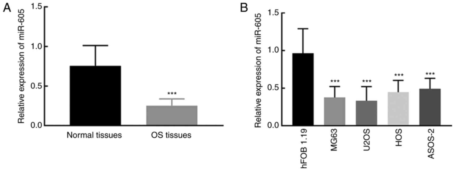
miR-605 expression is downregulated in
OS tissues and cell lines compared with normal tissues and
cells
miR-605 expression in tissues from patients with OS
and cell lines was detected by RT-qPCR. miR-605 was significantly
downregulated in OS tissues compared with adjacent normal tissues
(P<0.001; Fig. 1A). Similarly, in
OS cell lines, the expression of miR-605 was significantly lower
compared with that in the normal cell line hFOB 1.19 (P<0.001;
Fig. 1B). Additionally, MG63 and
U2OS were used for subsequent cell experimentation due to their
relatively lower miR-605 expression among the four OS cell
lines.
Relationship between miR-605
expression clinical features of patients with OS
Mean value of miR-605 mRNA expression (0.253) in
tissues from patients with OS was used to divided 110 patients into
the high miR-605 expression group (n=50) and low miR-605 expression
group (n=60). The expression of miR-605 demonstrated significant
association with the clinical stage (P=0.012) and distant
metastasis (P=0.006) of patients with OS, while the age, sex,
anatomical location and tumor size showed no significant
relationship with miR-605 expression (P>0.05; Table I).
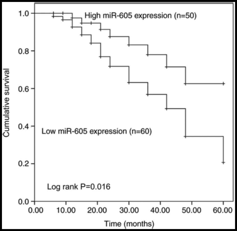
Prognostic value of miR-605
Kaplan-Meier curves demonstrated that patients with
low miR-605 expression had a lower overall survival rate compared
with patients with higher miR-605 expression (log rank P=0.016;
Fig. 2). In addition, Cox regression
analysis was used to evaluate the prognostic value of miR-605 and
the clinical features of patients with OS. miR-605 expression was
significantly associated with the overall survival rate of patients
with OS (Fig. 2). In addition, the
multivariate Cox regression analysis results indicated that miR-605
served as an independent factor in the prognosis of OS with the
hazard ratio (HR)=2.533; 95% confidence interval (CI)=1.150–6.107;
P= 0.029; Table II).
| Table II.Multivariate Cox regression analysis
of clinical features and miR-605 in patients with OS. |
Table II.
Multivariate Cox regression analysis
of clinical features and miR-605 in patients with OS.
| Indicators | HR | 95% CI | P-value |
|---|
| miR-605 | 2.533 | 1.150–6.107 | 0.029 |
| Age | 1.187 | 0.582–2.420 | 0.637 |
| Sex | 1.247 | 0.601–2.587 | 0.553 |
| Anatomical
location | 1.340 | 0.603–2.979 | 0.473 |
| Tumor size | 1.787 | 0.812–3.932 | 0.149 |
| Clinical stage | 1.896 | 1.055–3.885 | 0.040 |
| Distant
metastasis | 2.015 | 1.094–4.123 | 0.036 |
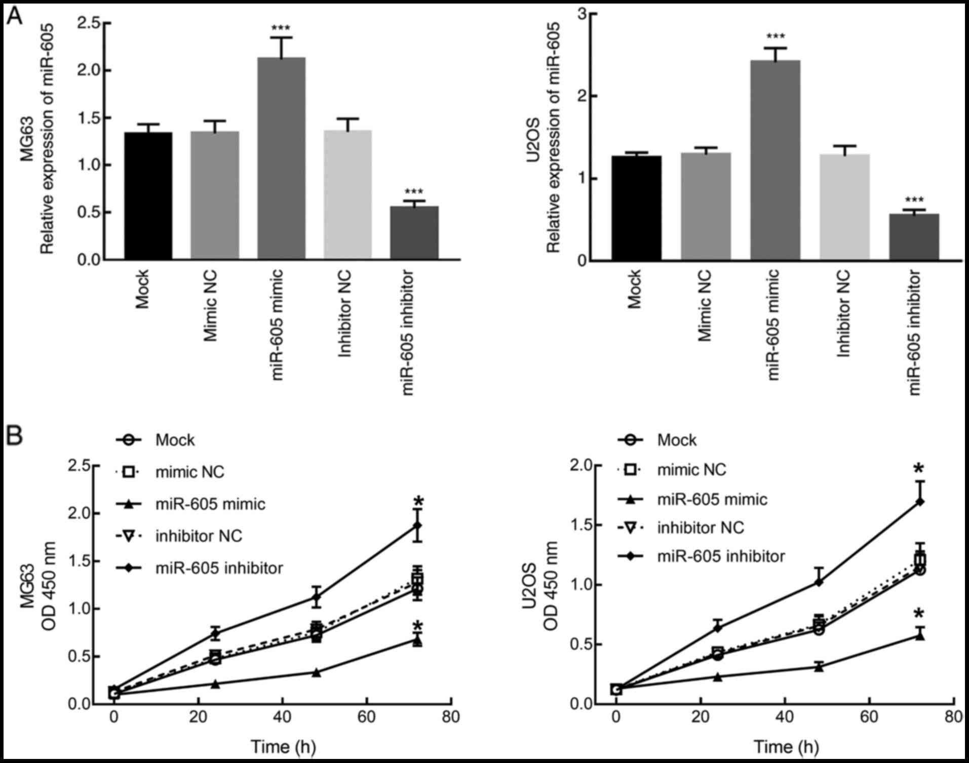
Downregulation of miR-605 promotes
cell proliferation in OS cells
MG63 and U2OS cells were transfected with miR-605
mimic or miR-605 inhibitor to overexpress or downregulate the
expression of miR-605. RT-qPCR results demonstrated that miR-605
was significantly upregulated after transfection with miR-605
mimic, while the transfection with miR-605 inhibitor significantly
downregulated the expression of miR-605 in both MG63 and U2OS cells
(P<0.001; Fig. 3A). The
proliferation of transfected cells was assessed using a CCK-8
assay. It was found that the downregulation of miR-605
significantly promoted the proliferation of MG63 and U2OS cells,
while the upregulation of miR-605 significantly inhibited the
proliferation of MG63 and U2OS cells (P<0.05; Fig. 3B).
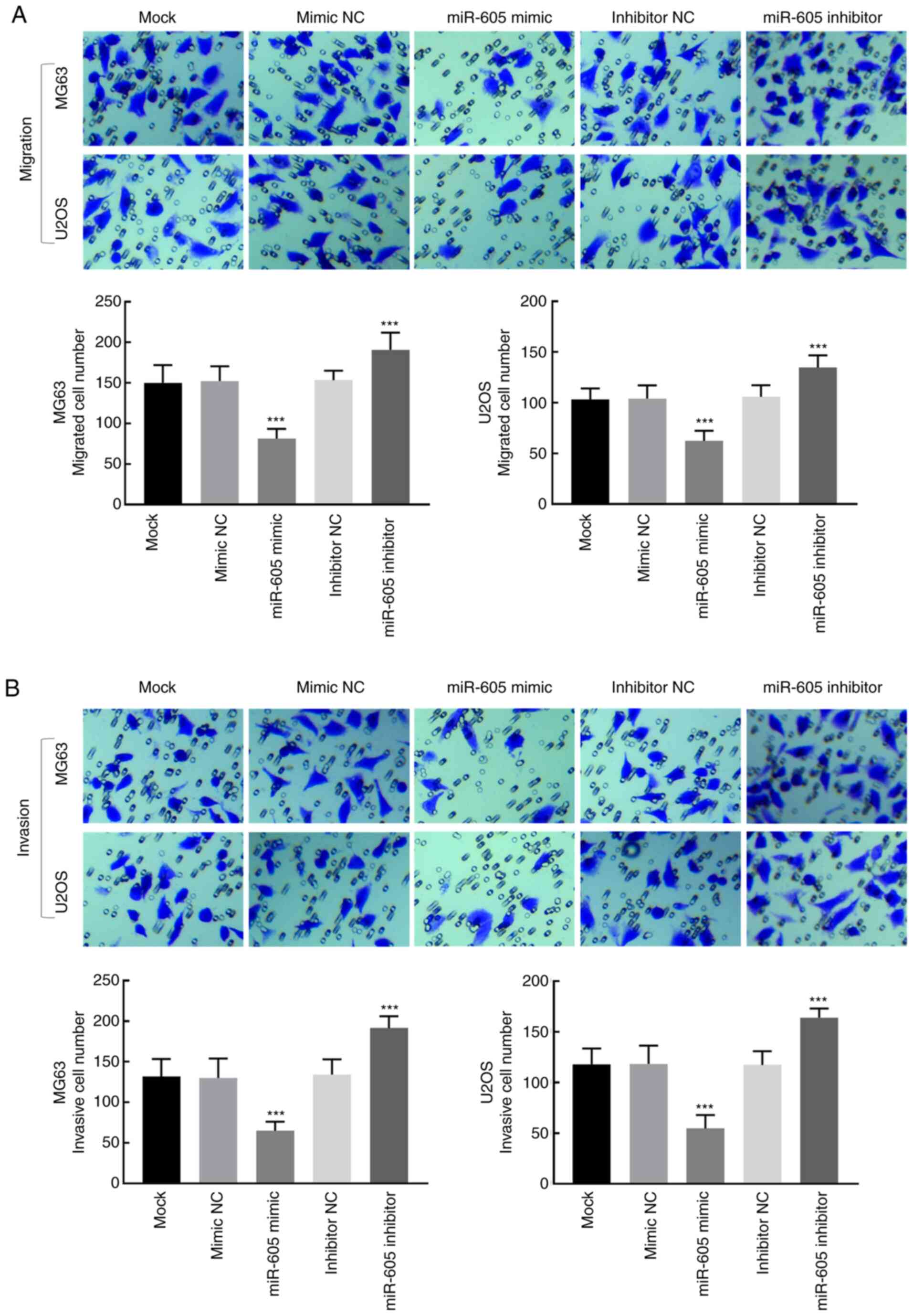
Effect of miR-605 on OS cell migration
and invasion
Transwell assays were used to evaluate the migration
and invasion abilities of MG63 and U2OS cells. The migration
ability of MG63 and U2OS cells was significantly inhibited by the
overexpression of miR-605 but promoted by the downregulation of
miR-605 (P<0.001; Fig. 4A). For
cell invasion, the knockdown of miR-605 significantly enhanced the
invasion of MG63 and U2OS cells, while the overexpression of
miR-605 inhibited OS cell invasion (P<0.001; Fig. 4B).
Discussion
With the development of molecular biology, an
increasing number of biomarkers, such as miRNAs, that play roles in
the progression of cancers and tumors have been identified, and the
therapy of different cancers, including OS, has improved rapidly
(23–25). miRNAs have been reported to play
vital biomarker roles in various cancers, such as lung cancer,
ovarian cancer, breast cancer, and gastric cancer (11,26–29). For
example, miR-593 can inhibit the migration and invasion of
non-small cell lung cancer cells by targeting SLUG-associated
signaling pathways (30). miR-486
can promote the cell proliferation and mobility of renal cell
carcinoma and inhibit its apoptosis (31). Several miRNAs have also been
identified as effective biomarkers for the prognosis and
progression of OS (32,33). miR-1225-5p serves as a tumor
suppressor in OS and inhibits the proliferation, migration and
invasion of OS cells by targeting Sox9 (32). miR-384 is downregulated in OS tissues
and cells compared to normal controls and upregulates stem loop
binding protein and promotes the growth and metastasis of OS
(33). A previous study reported the
downregulation of miR-605 in OS, which indicates that miR-605 may
play a role in the progression of OS (20).
The present study demonstrated that miR-605 was
significantly downregulated in OS tissues and cell lines compared
with adjacent normal tissues and cells. Additionally, miR-605
expression was found to be associated with the clinical stage and
distant metastasis of patients with OS in the present study. In
terms of the prognosis of OS, patients with high miR-605 expression
demonstrated an improved prognosis compared with patients with low
miR-605 expression. In the present study, miR-605 was found to be
an independent prognostic indicator of OS based on the results of
Cox regression analysis. These findings of the present study
revealed the potential prognostic biomarker role of miR-605 in OS,
which is consistent with a previous study (12).
miR-605 is reported to play roles in the progression
of numerous other cancers and tumors, where miR-605 is dysregulated
and affects the proliferation, migration, invasion and apoptosis of
tumor cells (34–36). For example, miR-605 inhibits the
progression of intrahepatic cholangiocarcinoma by suppressing
gankyrin (34). miR-605 also
functions as a tumor suppressor in melanoma by regulating the
expression of inositol polyphosphate-4-phosphatase type II B
(35). In addition, miR-605 was also
demonstrated to affect the proliferation, migration and invasion of
non-small cell lung cancer, prostate cancer, and breast cancer
(18,19,36). In
the present study, miR-605 was found to be a vital factor in the
progression of OS, as the downregulation of miR-605 significantly
promoted cell proliferation, migration, and invasion of OS, which
indicates that miR-605 may be involved in the progression and
development of OS.
In the present study, the expression and function of
miR-605 were investigated in OS tissues and cells in vitro,
however, the role of miR-605 in the progression and prognosis of OS
should be verified in in vivo experiments. On the other hand,
previous studies have provided some potential targets of miR-605,
such as tumor necrosis factor α-induced protein 3, EN2, enhancer of
zeste 2 (EZH2) and forkhead Box P1, which mediate the functional
effects of miR-605 in human malignancies (18,19,37,38).
EZH2 is a widely investigated oncogene in several human
malignancies, such as non-small cell lung cancer and bladder cancer
(39,40). In OS tissues, EZH2 has been found to
be upregulated and associated with aggressive tumor behavior and
poor prognosis and the knockdown of EZH2 could significantly
inhibit OS cell proliferation, migration and invasion (31). Thus, the functional role of miR-605
in OS progression may also be investigated by targeting EZH2. To
corroborate this deduction, further mechanical studies and in
vivo experiments are needed.
In conclusion, miR-605 is downregulated in OS and
this dysregulation is associated with clinical stage and distant
metastasis in patients with OS. The downregulation of miR-605
indicates a poor prognosis in terms of OS, which shows that miR-605
serves as an independent prognostic indicator for OS. In addition,
the downregulation of miR-605 promotes OS cell proliferation,
migration, and invasion, which suggests that miR-605 may be
involved in the progression of OS. The present study provides a
novel insight into the prognosis and progression of OS and provides
new therapeutic targets for the treatment of patients with OS.
Acknowledgements
Not applicable.
Funding
No funding was received.
Availability of data and material
All the data analyzed in this study are included in
this published article.
Authors' contributions
XY conducted the clinical study, analyzed clinical
data and wrote the manuscript. CL designed this research and
conducted the cell experiments. Both authors have read and approved
this manuscript.
Ethics approval and consent to
participate
The research was approved by the Ethics Committee of
Weifang People's Hospital (Weifang, China) with the approval no.
WFRM01-082009, and the participants provided signed informed
consent for tissue collection and analysis.
Consent for publication
Consent for publication was obtained from the
patients.
Competing interests
The authors declare that they have no competing
interests.
References
|
1
|
Selvarajah S, Yoshimoto M, Ludkovski O,
Park PC, Bayani J, Thorner P, Maire G, Squire JA and Zielenska M:
Genomic signatures of chromosomal instability and osteosarcoma
progression detected by high resolution array CGH and interphase
FISH. Cytogenet Genome Res. 122:5–15. 2008. View Article : Google Scholar : PubMed/NCBI
|
|
2
|
Selvarajah S, Yoshimoto M, Maire G,
Paderova J, Bayani J, Squire JA and Zielenska M: Identification of
cryptic microaberrations in osteosarcoma by high-definition
oligonucleotide array comparative genomic hybridization. Cancer
Genet Cytogenet. 179:52–61. 2007. View Article : Google Scholar : PubMed/NCBI
|
|
3
|
Wang J, Liu S, Shi J, Li J, Wang S, Liu H,
Zhao S, Duan K, Pan X and Yi Z: The role of miRNA in the diagnosis,
prognosis, and treatment of osteosarcoma. Cancer Biother
Radiopharm. 34:605–613. 2019. View Article : Google Scholar : PubMed/NCBI
|
|
4
|
Anderson ME: Update on survival in
osteosarcoma. Orthop Clin North Am. 47:283–292. 2016. View Article : Google Scholar : PubMed/NCBI
|
|
5
|
Song K, Song J, Lin K, Chen F, Ma X, Jiang
J and Li F: Survival analysis of patients with metastatic
osteosarcoma: A Surveillance, Epidemiology, and End Results
population-based study. Int Orthop. 43:1983–1991. 2019. View Article : Google Scholar : PubMed/NCBI
|
|
6
|
Ouyang L, Liu P, Yang S, Ye S, Xu W and
Liu X: A three-plasma miRNA signature serves as novel biomarkers
for osteosarcoma. Med Oncol. 30:3402013. View Article : Google Scholar : PubMed/NCBI
|
|
7
|
Zhang J, Yan YG, Wang C, Zhang SJ, Yu XH
and Wang WJ: MicroRNAs in osteosarcoma. Clin Chim Acta. 444:9–17.
2015. View Article : Google Scholar : PubMed/NCBI
|
|
8
|
Hesse E and Taipaleenmäki H: MicroRNAs in
bone metastasis. Curr Osteoporos Rep. 17:122–128. 2019. View Article : Google Scholar : PubMed/NCBI
|
|
9
|
Qu K, Wang Z, Lin XL, Zhang K, He XL and
Zhang H: MicroRNAs: Key regulators of endothelial progenitor cell
functions. Clin Chim Acta. 448:65–73. 2015. View Article : Google Scholar : PubMed/NCBI
|
|
10
|
Bartel DP: MicroRNAs: Genomics,
biogenesis, mechanism, and function. Cell. 116:281–297. 2004.
View Article : Google Scholar : PubMed/NCBI
|
|
11
|
Hironaka-Mitsuhashi A, Otsuka K,
Gailhouste L, Sanchez Calle A, Kumazaki M, Yamamoto Y, Fujiwara Y
and Ochiya T: miR-1285-5p/TMEM194A axis affects cell proliferation
in breast cancer. Cancer Sci. 111:395–405. 2020. View Article : Google Scholar : PubMed/NCBI
|
|
12
|
Qin S, Ai F, Ji WF, Rao W, Zhang HC and
Yao WJ: miR-19a promotes cell growth and tumorigenesis through
targeting SOCS1 in gastric cancer. Asian Pac J Cancer Prev.
14:835–840. 2013. View Article : Google Scholar : PubMed/NCBI
|
|
13
|
Chen J and Chen Z: Downregulation of
miR-19a inhibits the proliferation and promotes the apoptosis of
osteosarcoma cells by regulating the JAK2/STAT3 pathway. Oncol
Lett. 20:1732020. View Article : Google Scholar : PubMed/NCBI
|
|
14
|
Sun L, Wang L, Luan S, Jiang Y and Wang Q:
miR-429 inhibits osteosarcoma progression by targeting HOXA9
through suppressing Wnt/β-catenin signaling pathway. Oncol Lett.
20:2447–2455. 2020. View Article : Google Scholar : PubMed/NCBI
|
|
15
|
Yao J, Tan W, Wu W, Ye R, Li Y and Chen Y:
Effects of miR-432 and miR-548c-3p on the proliferation and
invasion of osteosarcoma cells. J BUON. 25:1562–1568.
2020.PubMed/NCBI
|
|
16
|
Zhou L, Lu Y, Liu JS, Long SZ, Liu HL,
Zhang J and Zhang T: The role of miR-21/RECK in the inhibition of
osteosarcoma by curcumin. Mol Cell Probes. 51:1015342020.
View Article : Google Scholar : PubMed/NCBI
|
|
17
|
Wang Y, Shi S, Zhang Q, Dong H and Zhang
J: MicroRNA-206 upregulation relieves circTCF25-induced
osteosarcoma cell proliferation and migration. J Cell Physiol
jcp.29570. 2020.
|
|
18
|
Liao Y, Cao L, Wang F and Pang R:
miR-605-5p promotes invasion and proliferation by targeting TNFAIP3
in non-small-cell lung cancer. J Cell Biochem. 121:779–787. 2020.
View Article : Google Scholar : PubMed/NCBI
|
|
19
|
Zhou YJ, Yang HQ, Xia W, Cui L, Xu RF, Lu
H, Xue Z, Zhang B, Tian ZN, Cao YJ, et al: Down-regulation of
miR-605 promotes the proliferation and invasion of prostate cancer
cells by up-regulating EN2. Life Sci. 190:7–14. 2017. View Article : Google Scholar : PubMed/NCBI
|
|
20
|
Zhang C, Wan J, Long F, Liu Q and He H:
Identification and validation of microRNAs and their targets
expressed in osteosarcoma. Oncol Lett. 18:5628–5636.
2019.PubMed/NCBI
|
|
21
|
Cates JM: Comparison of the AJCC, MSTS,
and modified spanier systems for clinical and pathologic staging of
osteosarcoma. Am J Surg Pathol. 41:405–413. 2017. View Article : Google Scholar : PubMed/NCBI
|
|
22
|
Livak KJ and Schmittgen TD: Analysis of
relative gene expression data using real-time quantitative PCR and
the 2(-Delta Delta C(T)) Method. Methods. 25:402–408. 2001.
View Article : Google Scholar : PubMed/NCBI
|
|
23
|
Jia G, Wang Y, Yu Y, Li Z and Wang X: Long
non coding RNA NR2F1 AS1 facilitates the osteosarcoma cell
malignant phenotype via the miR 485 5p/miR 218 5p/BIRC5 axis. Oncol
Rep. 44:1583–1595. 2020.PubMed/NCBI
|
|
24
|
Fan H, Liu T, Tian H and Zhang S: TUSC8
inhibits the development of osteosarcoma by sponging miR 197 3p and
targeting EHD2. Int J Mol Med. 46:1311–1320. 2020.PubMed/NCBI
|
|
25
|
Wang L, Jiang J, Sun G, Zhang P and Li Y:
Effects of lncRNA TUSC7 on the malignant biological behavior of
osteosarcoma cells via regulation of miR-375. Oncol Lett.
20:1332020. View Article : Google Scholar : PubMed/NCBI
|
|
26
|
Feliciano A, Garcia-Mayea Y, Jubierre L,
Mir C, Hummel M, Castellvi J, Hernández-Losa J, Paciucci R, Sansano
I, Sun Y, et al: miR-99a reveals two novel oncogenic proteins E2F2
and EMR2 and represses stemness in lung cancer. Cell Death Dis.
8:e31412017. View Article : Google Scholar : PubMed/NCBI
|
|
27
|
Zuberi M, Khan I, Mir R, Gandhi G, Ray PC
and Saxena A: Utility of serum miR-125b as a diagnostic and
prognostic indicator and its alliance with a panel of tumor
suppressor genes in epithelial ovarian cancer. PLoS One.
11:e01539022016. View Article : Google Scholar : PubMed/NCBI
|
|
28
|
Mousa H, Yuan M, Zhang X, Li X, Shopit A,
Almoiliqy M, Alshwmi M, Al-Dherasi A, Xu Y and Zuo Y: MicroRNA-4316
inhibits gastric cancer proliferation and migration via directly
targeting VEGF-A. Cancer Cell Int. 20:622020. View Article : Google Scholar : PubMed/NCBI
|
|
29
|
Huang D, Sun W, Zhou Y, Li P, Chen F, Chen
H, Xia D, Xu E, Lai M, Wu Y, et al: Mutations of key driver genes
in colorectal cancer progression and metastasis. Cancer Metastasis
Rev. 37:173–187. 2018. View Article : Google Scholar : PubMed/NCBI
|
|
30
|
Wei F, Wang M, Li Z, Wang Y and Zhou Y:
miR 593 inhibits proliferation and invasion and promotes apoptosis
in non small cell lung cancer cells by targeting SLUG associated
signaling pathways. Mol Med Rep. 20:5172–5182. 2019.PubMed/NCBI
|
|
31
|
Sun R, Shen J, Gao Y, Zhou Y, Yu Z,
Hornicek F, Kan Q and Duan Z: Overexpression of EZH2 is associated
with the poor prognosis in osteosarcoma and function analysis
indicates a therapeutic potential. Oncotarget. 7:38333–38346. 2016.
View Article : Google Scholar : PubMed/NCBI
|
|
32
|
Zhang W, Wei L, Sheng W, Kang B, Wang D
and Zeng H: miR-1225-5p Functions as a tumor suppressor in
osteosarcoma by targeting Sox9. DNA Cell Biol. 39:78–91. 2020.
View Article : Google Scholar : PubMed/NCBI
|
|
33
|
Wang Y, Huang H and Li Y: Knocking down
miR-384 promotes growth and metastasis of osteosarcoma MG63 cells
by targeting SLBP. Artif Cells Nanomed Biotechnol. 47:1458–1465.
2019. View Article : Google Scholar : PubMed/NCBI
|
|
34
|
Li J, Tian F, Li D, Chen J, Jiang P, Zheng
S, Li X and Wang S: miR-605 represses PSMD10/Gankyrin and inhibits
intrahepatic cholangiocarcinoma cell progression. FEBS Lett.
588:3491–3500. 2014. View Article : Google Scholar : PubMed/NCBI
|
|
35
|
Chen L and Cao Y, Rong D, Wang Y and Cao
Y: MicroRNA-605 functions as a tumor suppressor by targeting INPP4B
in melanoma. Oncol Rep. 38:1276–1286. 2017. View Article : Google Scholar : PubMed/NCBI
|
|
36
|
Danesh H, Hashemi M, Bizhani F, Hashemi SM
and Bahari G: Association study of miR-100, miR-124-1, miR-218-2,
miR-301b, miR-605, and miR-4293 polymorphisms and the risk of
breast cancer in a sample of Iranian population. Gene. 647:73–78.
2018. View Article : Google Scholar : PubMed/NCBI
|
|
37
|
Pan MZ, Song YL and Gao F: miR-605-3p
inhibits malignant progression of prostate cancer by up-regulating
EZH2. Eur Rev Med Pharmacol Sci. 23:8795–8805. 2019.PubMed/NCBI
|
|
38
|
Zhou W and Li R: MicroRNA-605 inhibits the
oncogenicity of non-small-cell lung cancer by directly targeting
Forkhead Box P1. OncoTargets Ther. 12:3765–3777. 2019. View Article : Google Scholar
|
|
39
|
Qiu C, Li S, Sun D and Yang S: lncRNA PVT1
accelerates progression of non-small cell lung cancer via targeting
miRNA-526b/EZH2 regulatory loop. Oncol Lett. 19:1267–1272.
2020.PubMed/NCBI
|
|
40
|
Chen Z, Du Y, Liu X, Chen H, Weng X, Guo
J, Wang M, Wang X and Wang L: EZH2 inhibition suppresses bladder
cancer cell growth and metastasis via the JAK2/STAT3 signaling
pathway. Oncol Lett. 18:907–915. 2019.PubMed/NCBI
|