Introduction
Head and neck squamous cell carcinoma (HNSCC) is the
sixth most common malignant tumor worldwide (1). Notably, the main types of HNSCC are
oral and oropharyngeal squamous cell carcinoma (OSCC/OPSCC), which
severely affect the quality of life of patients and endanger their
lives (2). Recent studies have
demonstrated that the development of OSCC/OPSCC is closely
associated with the changes of key genes (3–5).
Therefore, it is necessary to identify novel gene-level biomarkers
that predict the prognosis of OSCC/OPSCC.
Autophagy, a mechanism maintaining the stability of
the internal environment and the balance of protein metabolism in
cells, regulates the development and progression of various types
of cancer (6). In the early stage,
autophagy removes abnormal organelles and proteins in normal cells
to prevent the generation of the stress response, protect the genes
from being damaged and inhibit the occurrence of tumors; however,
autophagy also maintains the survival of tumor cells in the late
stage of tumor development under certain conditions, such as
hypoxia or nutrient deficiency, thereby sustaining proliferation
and progression of tumor cells (7).
By investigating autophagy-related molecules, the present study
aimed to identify novel biomarkers for prognostic prediction of
OSCC/OPSCC.
Abnormal expression of long non-coding RNAs
(lncRNAs) regulates the proliferation, apoptosis and migration of
tumor cells, implying that lncRNAs can potentially be used as
important biomarkers and therapeutic targets (8). Nonetheless, lncRNAs in OSCC/OPSCC
represent an important subject which has not been sufficiently
investigated. In the present study, The Cancer Genome Atlas (TCGA)
database was used to evaluate the potential value of
autophagy-related lncRNAs as a prognostic tool for patients with
OSCC/OPSCC. Through TCGA data analysis, nine autophagy-related
lncRNAs associated with the overall survival (OS) of patients with
OSCC/OPSCC were identified, and their independent associations were
further verified. Subsequently, the nine lncRNAs were integrated
into an independent signature, and it was revealed that their
predictive performance for the prognosis of patients with
OSCC/OPSCC was higher than that of clinicopathological features
(age, sex, grade, T stage, N stage and TNM comprehensive stage).
This demonstrated the potential of autophagy-related lncRNAs as
biomarkers to predict the prognosis of patients with
OSCC/OPSCC.
Materials and methods
Acquisition of autophagy-related
lncRNAs and clinicopathological data of patients with
OSCC/OPSCC
The level 3 RNA-seq and clinicopathological data
(project ID: TCGA-HNSC) of patients diagnosed with OSCC/OPSCC were
extracted from TCGA (https://portal.gdc.cancer.gov/), while the autophagy
gene set (accession number: M10281) was obtained from the Molecular
Signatures Database v7.0 (https://www.gsea-msigdb.org/gsea/msigdb/index.jsp).
Subsequently, the function of lncRNAs was explored based on the
hypothesis that co-expressed genes are more likely to be
functionally related (9). Spearman
correlation analysis was carried out on the expression of lncRNAs
and autophagy genes through the ‘limma’ (version 3.11; http://bioconductor.org/packages/limma/)
package of R software (version 3.6.1; R foundation) and lncRNAs
with high correlation (|correlation coefficient (cor)|>0.4;
P<0.001) with autophagy genes were identified as
autophagy-related lncRNAs.
Identification of a prognostic
multi-lncRNA signature
First, univariate Cox regression analysis was
applied to explore lncRNAs associated with the prognosis of
patients (P<0.01). Thereafter, Least Absolute Shrinkage and
Selection Operator (LASSO) Cox regression analysis was used to
optimize the prognostic multi-lncRNA signature (10). Afterwards, the present study
substituted the expression of lncRNAs in patients with OSCC/OSPCC
into the Cox model, and using the LASSO Cox regression coefficient,
the risk score of each patient was calculated. Subsequently, based
on the median risk score, the patients were subdivided into a
high-risk group and a low-risk group.
The Kaplan-Meier method and log-rank test were
performed to evaluate differences in OS between patients in the
high- and low-risk groups. The risk score distribution, survival
status of patients with OSCC/OPSCC and expression profiles of
prognostic lncRNAs were visualized through images. R software
(version 3.6.1; R foundation) was used for statistical calculations
and data plotting.
Independence of multi-lncRNA signature
in predicting prognosis of patients with OSCC/OPSCC
The independent associations of the multi-lncRNA
signature-based risk score and clinicopathological factors (age,
sex, TNM stage and pathological stage) with the prognosis of
patients with OSCC/OPSCC were determined through univariate and
multivariate Cox regression analysis using R software. Furthermore,
a receiver operating characteristic (ROC) curve was used to
evaluate the accuracy of these factors in predicting the prognosis
of OSCC/OPSCC.
Kruskal-Wallis with Dunn's post hoc test was used to
further explore the relationship between the expression levels of
the nine autophagy-related lncRNAs and the clinicopathological
characteristics of patients with OSCC/OPSCC.
Principal components analysis (PCA)
and gene set enrichment analysis (GSEA)
Furthermore, PCA was used to test the
differentiation of patients in low- and high-risk groups. GSEA
(https://www.gsea-msigdb.org/gsea/index.jsp) was used
to investigate the functions of autophagy-related genes in low- and
high-risk groups. Statistical calculations and data plotting were
done using R software (version 3.6.1; R foundation).
Validation of the clinical OSCC/OPSCC
specimens by reverse transcription-quantitative PCR (RT-qPCR)
To further validate the findings from TCGA, RT-qPCR
was performed to detect the expression levels of the nine
autophagy-related lncRNAs in OSCC/OPSCC samples (n=55). All
patients (30 men, 25 women; median age, 46 years; age range, 35–68
years) received lesion excision between January 2015 and October
2018 at the First Affiliated Hospital of Nanchang University
(Nanchang, China) where they were pathologically diagnosed with
OSCC/OPSCC. Patients with distant metastasis, multiple primary
cancers or non-first surgery were excluded from the present study.
The collection of the tumor samples from patients with OSCC/OPSCC
conformed to the Declaration of Helsinki and current legislation.
In addition, clinicopathological information of patients was
captured by an investigator through interviews and medical records.
The present study was approved by the Ethics Committee of First
Affiliated Hospital of Nanchang University (approval no.
2019B0017), and all patients provided written informed consent.
Total RNA was isolated from the tissue samples using
TRIzol® reagent (Invitrogen; Thermo Fisher Scientific,
Inc.). The GoScript Reverse Transcription kit (cat. no. PRA5000;
Promega Corporation) and GoTaq® qPCR Master Mix of Power
SYBR® Green (Promega Corporation) was used to synthesize
cDNA and for RT-qPCR detection. RT-qPCR was performed using the
GeneAmp® PCR System 9700 (Applied Biosystems; Thermo
Fisher Scientific, Inc.). Additionally, relative fold change
results were calculated using the 2−ΔΔCq method
(11). (The thermocycling conditions
were as follows: 95°C for 10 sec, followed by 40 cycles of 95°C for
5 sec and 60°C for 30 sec. RNA expression were normalized to GAPDH.
The following primer sequences were used: PTCSC2 forward
5′-CCCTAAGCCCACCGACTTTT-3′ and reverse, 5′-GGGTGCACTGGGTTTAGACA-3′;
AC099850.3 forward 5′-CGTCTTTCACCCAGCCTCTT-3′ and reverse,
5′-AAAGCAGGAACCCCTCTGTG-3′; LINC01963 forward
5′-CCCGGTGTAGGGTAAATGCA-3′ and reverse, 5′-ATTGGCCACTCCCGGATTTT-3′;
RTCA-AS1 forward 5′-CCGAGGTGCCGACTTTTAGA-3′ and reverse,
5′-CCACCCAGGTTCACATCTCA-3′; AP002884.1 forward
5′-TACGCTAAACTGCCTGGCAA-3′ and reverse, 5′-GTTGTGCAGCAGAGTTGTGG-3′;
UBAC2-AS1 forward 5′-TGAAACGATGGCGGTCAGAA-3′ and reverse,
5-TCAGGTCCTCAGGATGCAGA-3′; AL512274.1 forward
5′-AACACAGTGAGCGAGTCAGG-3′ and reverse, 5′-CAACCTCAGAGCAGAACCGT-3′;
MIR600HG forward 5′-GCCAGTCTGACGTGAACAGA-3′ and reverse,
5′-GCGCTTCTATCAGGCCATCT-3′; AL354733.3 forward
5′-TCCCCAGGCCTCAAAAATCC-3′ and reverse, 5′-TCTGCTGTCGACTTTCGCTT-3′;
GAPDH forward 5′-GGAAGCTTGTCATCAATGGAAATC-3′ and reverse,
5′-TGATGACCCTTTTGGCTCCC-3′.
Statistical analysis
Statistical differences were calculated using R
software (version 3.6.1; R foundation) and SPSS Statistics software
(version 20; IBM Corp.). All experiments were performed in
triplicate. For non-parametric analysis, Kruskal-Wallis with Dunn's
post hoc test was used. Survival curves were plotted using the
Kaplan-Meier method, and differences were analyzed via the log-rank
test. The regression analysis of univariate and multivariate Cox
proportional hazards analysis was completed using the ‘survival’
package (version 2.36-10, http://cran.r-project.org/web/packages/survival) of R
software. Results are presented in the form of hazard ratio and
corresponding 95% confidence interval (CI). P<0.05 was
considered to indicate a statistically significant difference.
Results
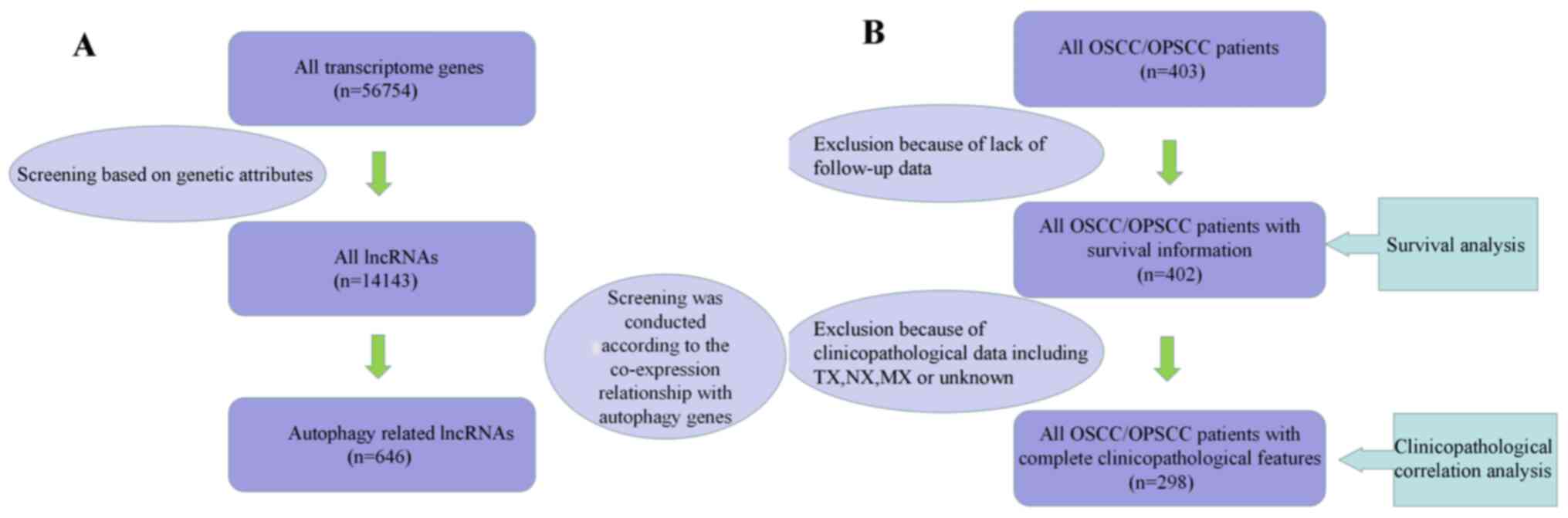
Autophagy-related lncRNAs and
clinicopathological data in patients with OSCC/OPSCC
A total of 646 autophagy-related lncRNAs, 402
patients with survival information and 298 patients with complete
clinicopathological information were identified (Fig. 1).
Identification of a prognostic
autophagy-related lncRNA signature in patients with OSCC/OPSCC
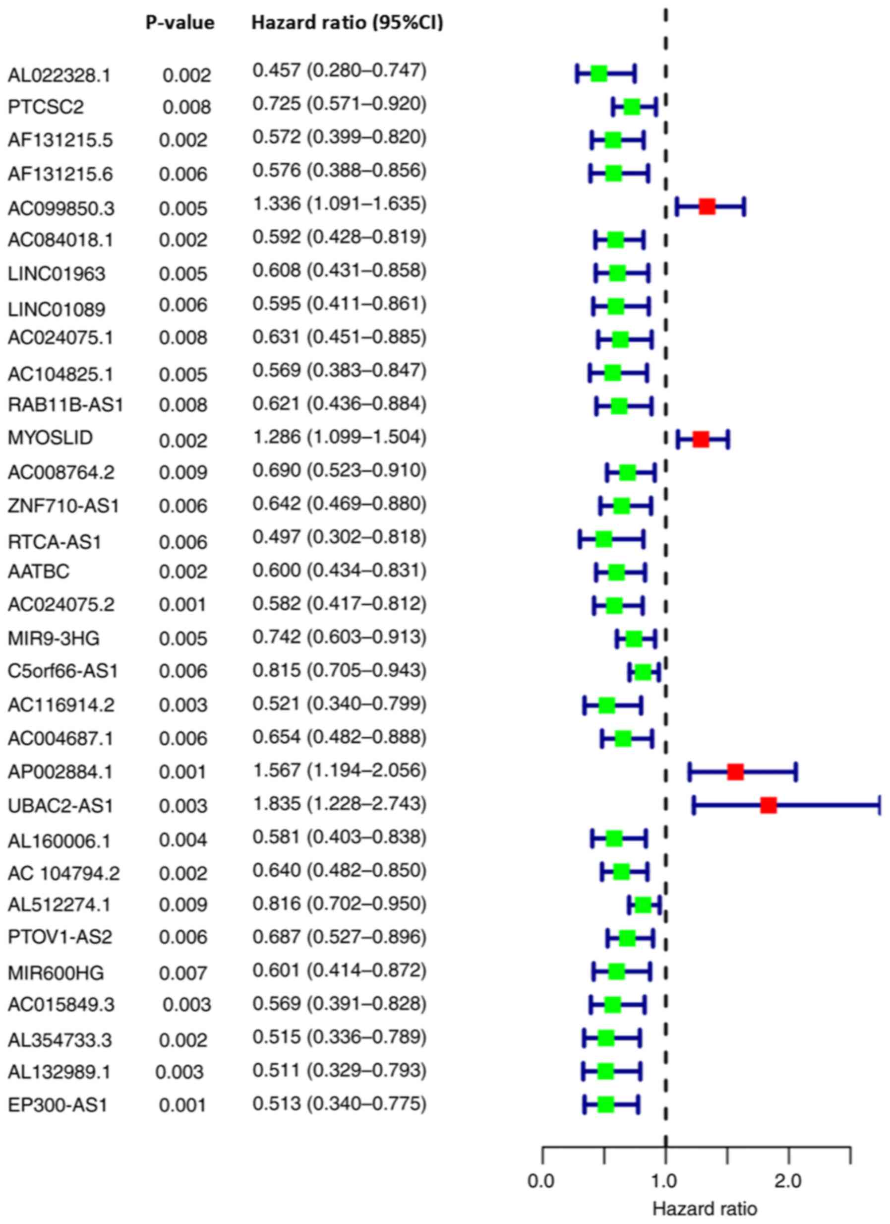
Using univariate Cox regression analysis, 32
autophagy-related lncRNAs associated with the OS of patients with
OSCC/OPSCC were identified (Fig.
2).
The 32 autophagy-related lncRNAs were further
screened using LASSO Cox regression analysis. Among these, nine
lncRNAs, including PTCSC2, AC099850.3, LINC01963, RTCA-AS1,
AP002884.1, UBAC2-AS1, AL512274.1, MIR600HG and AL354733.3, were
incorporated into the Cox model. Subsequently, the expression
levels of the aforementioned nine lncRNAs and the LASSO Cox
regression coefficients were integrated to establish the signature
(Table I). Based on the signature,
the risk scores of patients with OSCC/OPSCC (n=371; among the 402
patients, 31 patients with no detectable target lncRNA expression
were excluded) were calculated and subdivided into a high-risk
group (n=185) and a low-risk group (n=186) according to the median
value (1.057).
 | Table I.Least Absolute Shrinkage and
Selection Operator Cox proportional hazard model of the nine
autophagy-related lncRNAs. |
Table I.
Least Absolute Shrinkage and
Selection Operator Cox proportional hazard model of the nine
autophagy-related lncRNAs.
| lncRNA ID | Coefficient | Hazard ratio | 95% Confidence
interval | P-value |
|---|
| PTCSC2 | −0.310 | 0.733 | 0.557–0.966 | 0.027 |
| AC099850.3 | 0.270 | 1.309 | 1.064–1.611 | 0.011 |
| LINC01963 | −0.483 | 0.617 | 0.421–0.904 | 0.013 |
| RTCA-AS1 | −0.526 | 0.591 | 0.341–0.958 | 0.021 |
| AP002884.1 | 0.392 | 1.480 | 1.089–2.010 | 0.012 |
| UBAC2-AS1 | 0.387 | 1.473 | 1.035–2.312 | 0.022 |
| AL512274.1 | −0.178 | 0.837 | 0.710–0.986 | 0.034 |
| MIR600HG | 0.3798 | 1.462 | 1.057–2.334 | 0.030 |
| AL354733.3 | −0.777 | 0.460 | 0.277–0.762 | 0.002 |
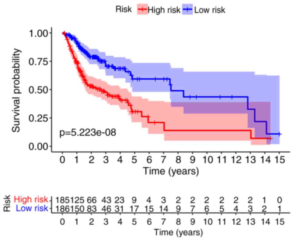
The Kaplan-Meier curve demonstrated that the OS of
the high-risk group was significantly poorer than that of low-risk
patients (P=5.223×10⁻⁸; Fig.
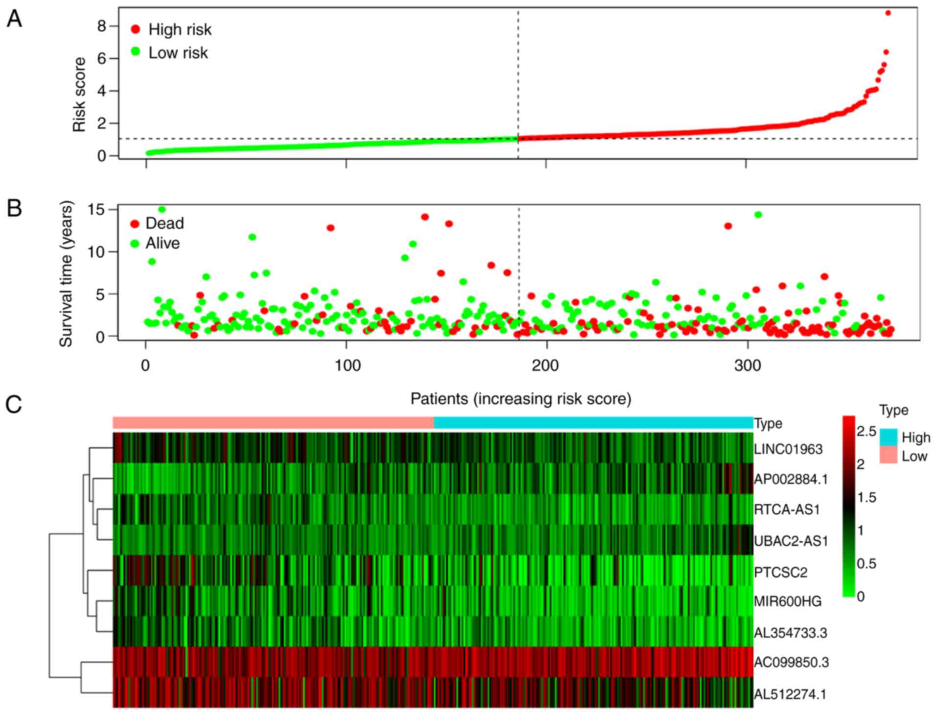
3). The risk score distribution, survival status of patients
with OSCC/OPSCC, and expression profiles of the nine prognostic
lncRNAs are shown in Fig. 4. It was
revealed that the rate of mortality among patients with high-risk
scores was higher compared with that in patients with low-risk
scores. The expression levels of AC099850.3, AP002884.1 and
UBAC2-AS1 were higher in high-risk patients compared with in
patients with low-risk. The expression levels of AL512274.1,
PTCSC2, LINC01963, RTCA-AS1, AL354733.3 and MIR600HG were lower in
high-risk patients compared with in patients with low-risk.
In order to determine the role of the nine lncRNAs,
we extracted and displayed the correlation analysis results between
these nine lncRNAs and autophagy-related genes (Fig. S1; Table
SI). All of the nine lncRNAs were positively correlated with
autophagy genes. PTCSC2 exhibited the highest correlation
coefficient (cor=0.614), while MIR600HG had the most co-expression
genes (n=20).
Association between the nine-lncRNA
signature and prognosis in patients with OSCC/OPSCC
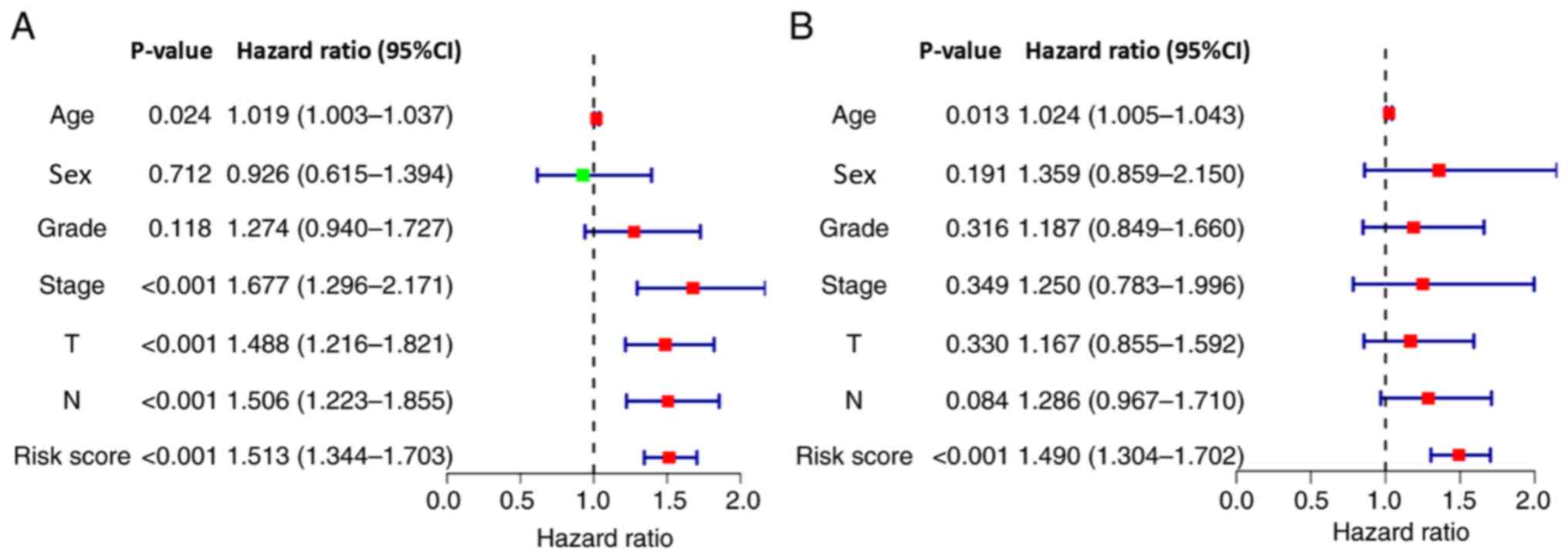
The results of univariate Cox regression analysis
demonstrated that the nine-lncRNA signature-based risk score, age,
T stage, N stage and TNM comprehensive stage were significantly
associated with patient survival (Fig.
5A). Furthermore, multivariate Cox regression analysis revealed
that the signature-based risk score and age were independent
factors associated with the OS of patients with OSCC/OPSCC
(Fig. 5B). Due to the absence of M1
in the case data, the M stage was not included in the analysis.
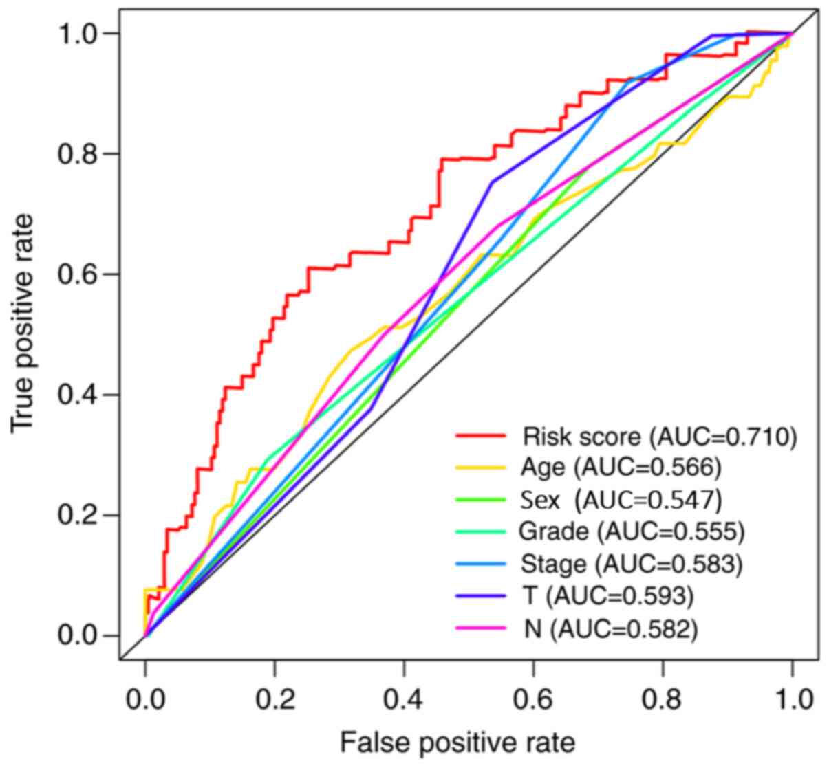
The prediction capability of the signature was
further analyzed using ROC curves, and the area under the curve
value was 0.710, which was higher than that of the other
clinicopathological characteristics (Fig. 6).
Relationship between nine
autophagy-related lncRNAs and clinicopathological factors in
patients with OSCC/OPSCC
A Kruskal-Wallis test was used to investigate the
association between the expression levels of the nine
autophagy-related lncRNAs and the clinicopathological
characteristics of the patients (Fig.
7). The results demonstrated that AC099850.3 (P<0.01),
AL512274.1 (P<0.001) and RTCA-AS1 (P<0.01) were significantly
associated with the pathological grade (Fig. 7A), and RTCA-AS1 was significantly
associated with T stage (P<0.05; Fig.
7B). However, none of the lncRNAs were significantly associated
with the clinical stage or N stage of the patients (Fig. 7C and D).
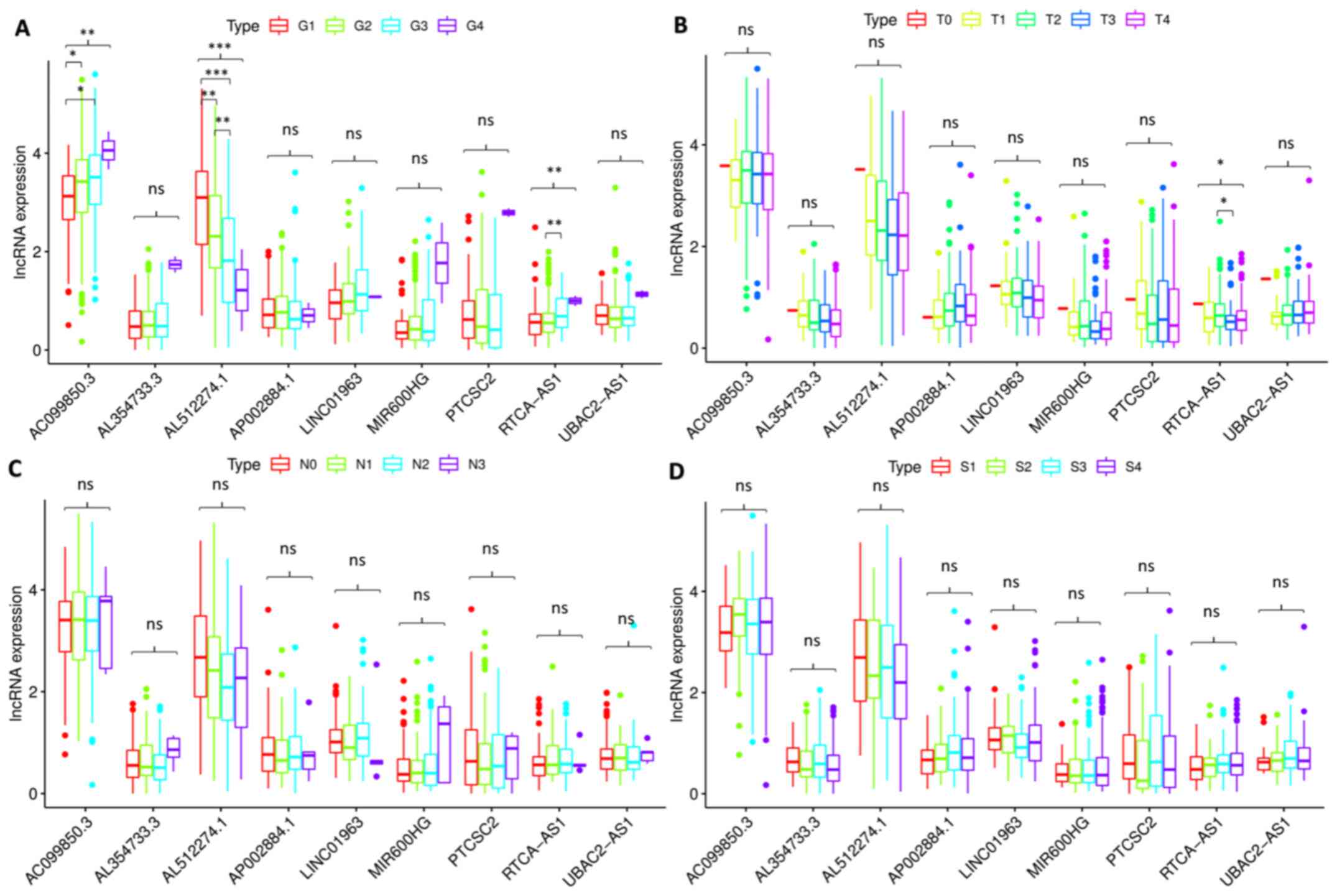 | Figure 7.Association between the expression
levels of the nine lncRNAs and clinicopathological characteristics
of the patients. The expression levels of (A) AC099850.3
(P<0.01), AL512274.1 (P<0.001) and RTCA-AS1 (P<0.01) were
significantly associated with the pathological grade. (B) RTCA-AS1
expression (P<0.05) was significantly associated with T stage.
None of the lncRNAs were significantly associated with the (C) N
stage and (D) clinical stage of the patients. *P<0.05,
**P<0.01 and ***P<0.001. lncRNA, long non-coding RNA; ns, not
significant; G1, well differentiated; G2, moderately
differentiated; G3, poorly differentiated; G4, undifferentiated; T,
T stage; N, N stage; S, TNM comprehensive stage; S1, stage I; S2,
stage II; S3, stage III; S4, stage IV. |
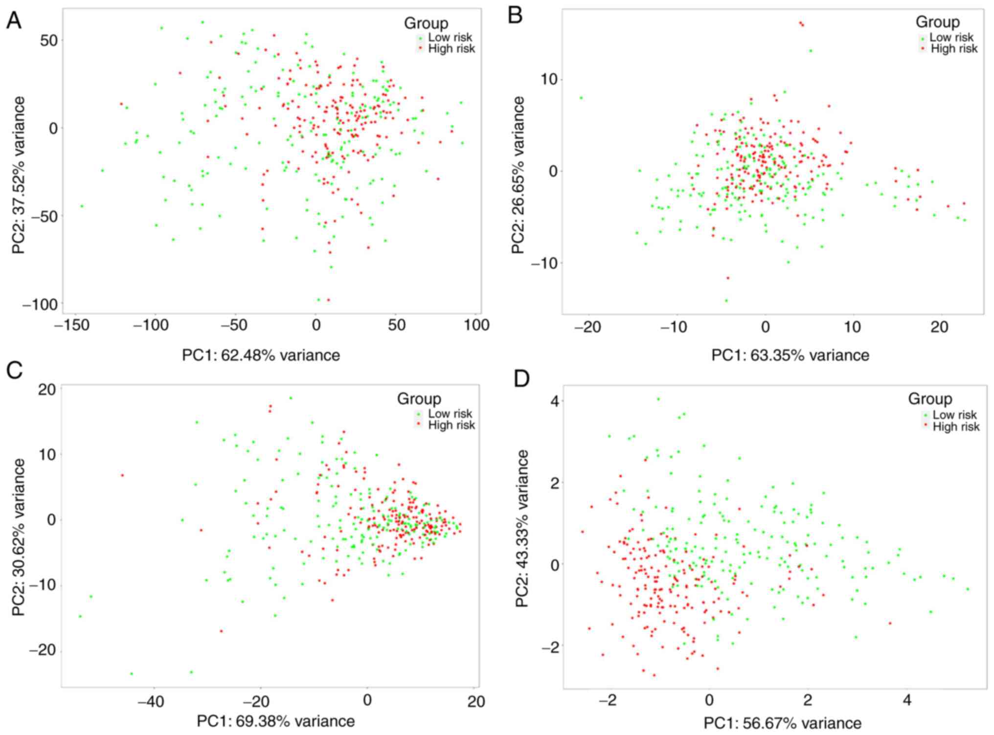
Autophagy status of low- and high-risk
groups
PCA was conducted to investigate differences in the
expression of the nine autophagy-related lncRNAs, autophagy-related
lncRNAs, autophagy-related genes and all genes between low- and
high-risk groups. The patients in the two groups were mixed when
the expression of all the genes was used as the spatial indicator.
The patients in the two groups were separated gradually when the
index was optimized gradually from all genes to autophagy-related
genes, autophagy-related lncRNAs and the nine autophagy-related
lncRNAs (Fig. 8), suggesting that
the signature established in the present study exhibited an
improved discriminative power for prognostic prediction of patients
with OSCC/OPSCC compared with other gene indices.
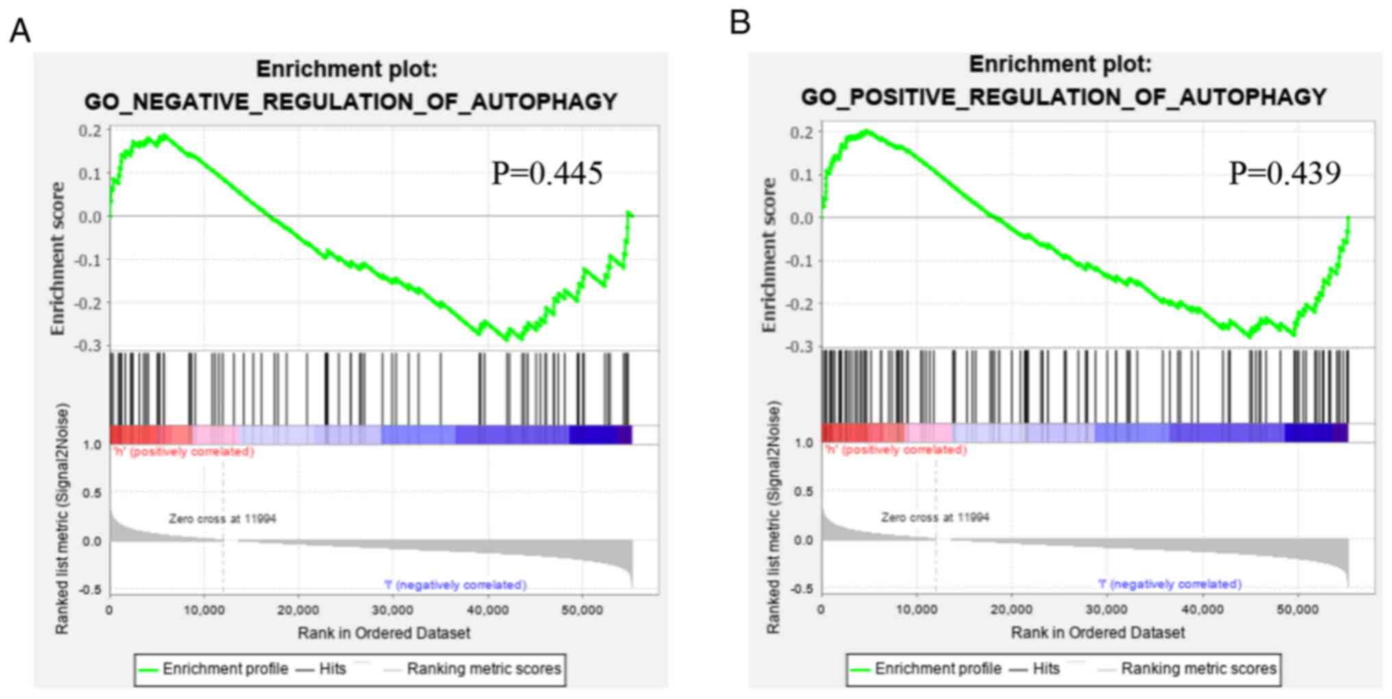
Functional annotation was further performed by GSEA,
and the results demonstrated that the difference in gene function
between low- and high-risk patients were not significantly enriched
in the positive or negative regulation gene sets of autophagy
(Fig. 9), indicating that, based on
the overall increase or decrease of autophagy, determining the
prognosis of the patients was not tenable.
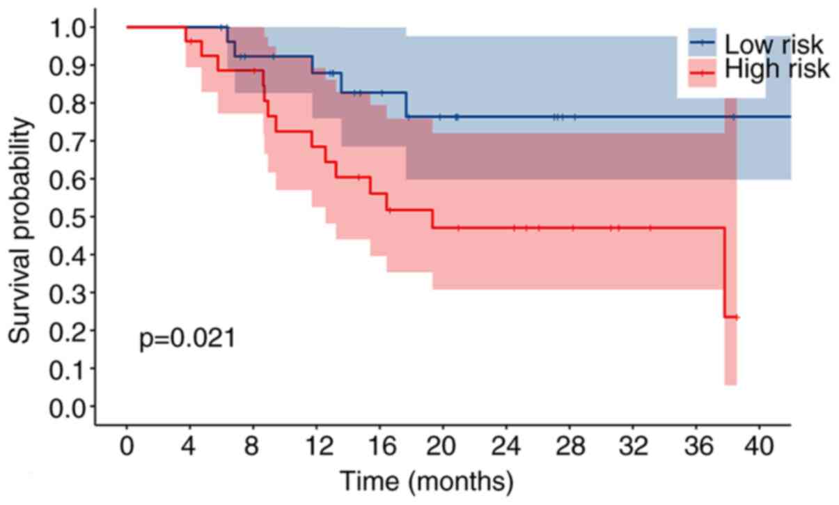
RT-qPCR validation
To assess the validity and reliability of the
bioinformatics results, the expression levels of the nine
autophagy-related lncRNAs were detected by RT-qPCR in 55 patients
with OSCC/OPSCC. Based on the results of RT-qPCR and risk scores of
the signature, the patients were divided into a high-risk group and
a low-risk group. Kaplan-Meier analysis revealed that the OS of the
high-risk group was significantly lower than that of the low-risk
group (P=0.021; Fig. 10), and
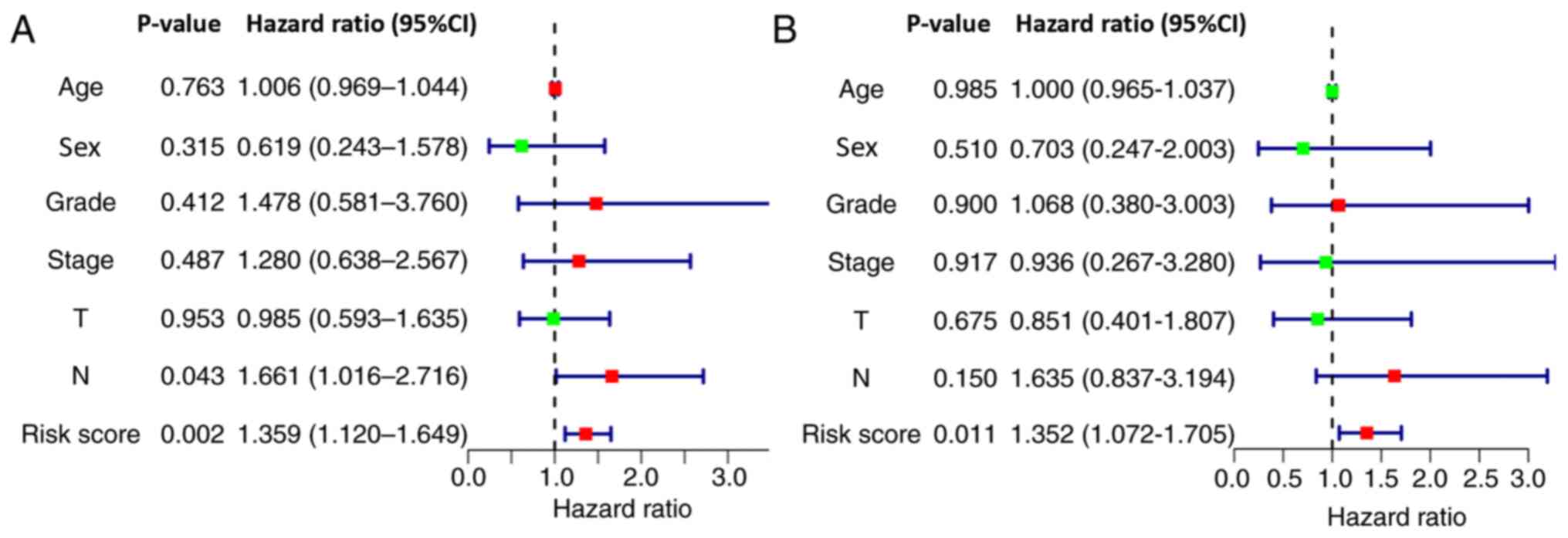
univariate and multivariate Cox regression analysis demonstrated
the potential of the signature as an independent prognostic factor
for patients with OSCC/OPSCC (Fig.
11), confirming the credibility of the signature.
Discussion
Autophagy is an important material catabolism
process in cells, is caused by hypoxia, peroxidation, drug and DNA
damage, and regulates cell self-renewal and homeostasis. According
to previous studies, autophagy can be a double-edged sword in tumor
development due to its ability to kill tumor cells during tumor
development (12). However, it also
protects tumor cells from being damaged (13).
At present, more than 40 autophagy-related genes
have been identified in yeast and mammals (14). Being highly conserved between
species, these genes regulate the occurrence and degree of
intracellular autophagy via complex regulatory networks, and they
are also implicated in the pathophysiology of diseases (15).
Targeted regulation of the autophagy level has
become a subject of research focus in the treatment of cancer and
other diseases (16). In the complex
regulatory network of intracellular genes, regulating the
expression of proteins associated with autophagy is a basic method
to regulate autophagy (17).
Previous studies have asserted that lncRNAs, such as
lncRNA-HOTAIR (18–21) and lncRNA-MALAT1 (22–25),
serve a pivotal role in regulating the expression of
autophagy-related proteins through positive or negative effects on
mRNAs. Therefore, understanding and utilizing the role of lncRNAs
in the regulation of autophagy will advance the clinical diagnosis
and treatment of tumors.
With the advent of high throughput RNA sequencing
and advanced computer technology, lncRNAs can continuously be
investigated. Nevertheless, due to wide expression, complexity of
the relationship and functional diversity, research on the
functions of most lncRNAs has not matured (26). Bioinformatics analysis is an
effective method for exploring the functions of lncRNAs, and
provides a basis for subsequent experimental studies (27,28).
Using Cox regression analysis and the Kaplan-Meier
method, the present study identified nine autophagy-related lncRNAs
which significantly influenced the prognosis of patients with
OSCC/OPSCC. Based on the aforementioned findings, a nine-lncRNA
signature with satisfactory performance was developed. As a result,
the present study further classified the patients with OSCC/OPSCC
into a high-risk group and a low-risk group with significantly
different prognosis.
By reviewing related literature and the genes
exhibiting co-expression relationships with IncRNAs, the functions
of the nine lncRNAs in the signature were explored.
Zhou et al (29) revealed that high expression levels of
AC099850.3 were closely associated with decreased survival of
patients diagnosed with tongue cancer. The present study revealed
that AC099850 exhibited the greatest positive co-expression
correlation with eukaryotic translation initiation factor 2 alpha
kinase 4 (EIF2AK4). Notably, EIF2AK4 belongs to the family of
protein kinases that phosphorylates the subunit of eukaryotic
translation initiation factor 2 (EIF2) in response to various
stress stimuli (30). To activate
autophagy-related gene expression in response to cellular stress,
activation of the EIF2AK4-EIF2A-activating transcription factor 4
signaling pathway is essential (31).
Zhu et al (32) reported that UBAC2-AS1 was a potential
therapeutic target and a prognostic biomarker of clear cell kidney
carcinoma. Furthermore, Chen et al (33) suggested that UBAC2-AS1 might be
implicated in adipogenesis by acting as a competing endogenous RNA
or being co-expressed with its targets. The present study
demonstrated that UBAC2-AS1 exhibited the greatest positive
co-expression correlation with a suppressor of Ty homolog-5, which
has been demonstrated to be a novel tumor-specific human telomerase
reverse transcriptase promoter-binding protein in colon cancer
cells (34).
According to Song et al (35), MIR600HG is a potential prognostic
biomarker in predicting the survival of patients diagnosed with
pancreatic ductal adenocarcinoma. The present study demonstrated a
positive correlation in the co-expression of MIR600HG and
ubiquitin-specific protease 30 (USP30). Furthermore, USP30
potentially reverses depolarization-induced PTEN induced kinase
1-parkin RBR E3 ubiquitin protein ligase-dependent mitophagy and
has rapidly emerged as a potential therapeutic target in
Parkinson's disease (36).
It was also identified that the PTCSC2, LINC01963,
AP002884.1, RTCA-AS1, AL512274.1 and AL354733.3 genes have not been
studied in detail. Additionally, a significant positive
co-expression relationship was identified between PTCSC2 and
Xeroderma pigmentosum group A (XPA). Notably, as a key subunit
implicated in the nucleotide excision repair (NER) system, XPA
protein is a central organizer in the NER signaling pathway which
identifies DNA damage and recruits other NER proteins to DNA
lesions (37). A study by Ge et
al (38) demonstrated that XPA
potentially promotes cell-protective autophagy in a DNA
repair-independent manner by enhancing the activation of
poly(ADP-ribose) polymerase 1 (PARP1) in melanoma cells resistant
to cisplatin.
It was revealed that LINC01963 harbored the greatest
positive co-expression correlation with tuberous sclerosis 2
(TSC2), while AL354733.3 had the greatest positive co-expression
correlation with tuberous sclerosis 1 (TSC1). By suppressing mTOR
signaling, studies have revealed that TSC1 and TSC2 potentially
induce autophagy (39,40).
AP002884.1 and fasciculation as well as elongation
zeta/zygin1 (FEZ1) had a significant positive co-expression
relationship. FEZ1 potentially acts as an adaptor of cargo
transport and may be a scaffold protein; the complexes of FEZ1
formed with unc-51 like autophagy activating kinase 1, short
coiled-coil protein, RAB3 GTPase activating protein catalytic
subunit 1 or RAB3 GTPase activating non-catalytic protein subunit 2
have been demonstrated to be associated with autophagy (41).
RTCA-AS1 was markedly positively correlated with
chromatin-modifying protein 4 (CHMP4)A. Notably, CHMP4A, CHMP4C and
CHMP2B belong to the family of chromatin-modifying protein/charged
multivesicular body protein. They are components of the endosomal
sorting complex required for transport III involved in the
formation of endocytic multivesicular bodies (42). CHMP4A expression is associated with
the recurrence of ovarian cancer (43), whereas CHMP4C regulates radiation
resistance in non-small cell lung cancer (44).
AL512274.1 was significantly positively co-expressed
with mitogen-activated protein kinase 3 (MAPK3). Previous studies
have identified MAPK3 to be specifically implicated in the control
of cell proliferation, differentiation and autophagy (45,46).
To the best of our knowledge, except for AC099850.3,
the other eight lncRNAs have not been reported in previous studies
on OSCC/OPSCC, implying that these lncRNAs represent a potential
target for the treatment of OSCC/OPSCC. In addition, their
biological roles in the autophagy of OSCC/OPSCC will be a focus in
future studies.
The present investigation had some limitations. For
instance, the present study was based on profiles of
high-throughput RNA-sequencing and data analysis, and therefore,
lacked validation in a large clinical sample. A multi-center, large
sample, longitudinal study with diverse clinical, radiographic and
histopathologic factors is required to further demonstrate the
reliability of the present study. Furthermore, the roles of the
nine autophagy-related lncRNAs deserve further in vitro and
in vivo investigation.
In conclusion, based on nine autophagy-related
lncRNAs, a signature that demonstrated the capability to predict
the prognosis of patients diagnosed with OSCC/OPSCC was developed.
Using this signature, patients with a higher risk of mortality can
be predicted, and therefore more priority in treatment should be
given to these patients.
Supplementary Material
Supporting Data
Acknowledgements
Not applicable.
Funding
The present study was supported by the National
Natural Science Foundation of China (grant no. 81860477), Jiangxi
Provincial Key R&D Plan (grant no. 20181ACG70009) and Special
Fund for Postgraduate Innovation in Jiangxi Province (grant no.
YC2020-B025).
Availability of data and materials
The datasets used can be obtained from TCGA
(https://portal.gdc.cancer.gov). The
RT-qPCR data generated in the present study is available from the
corresponding author upon reasonable request.
Authors' contributions
QJ and JQ conceived and designed the present study.
QJ performed the bioinformatics analysis. DX analyzed the data. QJ
and DX drafted the initial manuscript. QJ and FS performed tissue
tests and followed up patients. All authors have read and approved
the final manuscript.
Ethics approval and consent to
participate
For the use of human samples, the present study was
approved by the Institutional Ethics Committee of First Affiliated
Hospital of Nanchang University (Nanchang, China; approval no.
2019B0017), and written informed consent was provided by all
patients prior to the study start.
Patient consent for publication
Not applicable.
Competing interests
The authors declare that they have no competing
interests.
References
|
1
|
Siegel RL, Miller KD and Jemal A: Cancer
statistics, 2018. CA Cancer J Clin. 68:7–30. 2018. View Article : Google Scholar : PubMed/NCBI
|
|
2
|
Chaturvedi AK, Anderson WF,
Lortet-Tieulent J, Curado MP, Ferlay J, Franceschi S, Rosenberg PS,
Bray F and Gillison ML: Worldwide trends in incidence rates for
oral cavity and oropharyngeal cancers. J Clin Oncol. 31:4550–4559.
2013. View Article : Google Scholar : PubMed/NCBI
|
|
3
|
Falzone L, Lupo G, La Rosa GRM, Crimi S,
Anfuso CD, Salemi R, Rapisarda E, Libra M and Candido S:
Identification of novel MicroRNAs and their diagnostic and
prognostic significance in oral cancer. Cancers (Basel).
11:6102019. View Article : Google Scholar
|
|
4
|
Nakagaki T, Tamura M, Kobashi K, Koyama R,
Fukushima H, Ohashi T, Idogawa M, Ogi K, Hiratsuka H, Tokino T and
Sasaki Y: Profiling cancer-related gene mutations in oral squamous
cell carcinoma from Japanese patients by targeted amplicon
sequencing. Oncotarget. 8:59113–59122. 2017. View Article : Google Scholar : PubMed/NCBI
|
|
5
|
Bavle RM, Venugopal R, Konda P,
Muniswamappa S and Makarla S: Molecular classification of oral
squamous cell carcinoma. J Clin Diagn Res. 10:ZE18–ZE21.
2016.PubMed/NCBI
|
|
6
|
Wu WK, Coffelt SB, Cho CH, Wang XJ, Lee
CW, Chan FK, Yu J and Sung JJ: The autophagic paradox in cancer
therapy. Oncogene. 31:939–953. 2012. View Article : Google Scholar : PubMed/NCBI
|
|
7
|
Kimmelman AC: The dynamic nature of
autophagy in cancer. Genes Dev. 25:1999–2010. 2011. View Article : Google Scholar : PubMed/NCBI
|
|
8
|
Lu T, Yu C, Ni H, Liang W, Yan H and Jin
W: Expression of the long non-coding RNA H19 and MALAT-1 in growth
hormone-secreting pituitary adenomas and its relationship to tumor
behavior. Int J Dev Neurosci. 67:46–50. 2018. View Article : Google Scholar : PubMed/NCBI
|
|
9
|
Stuart JM, Segal E, Koller D and Kim SK: A
gene-coexpression network for global discovery of conserved genetic
modules. Science. 302:249–255. 2003. View Article : Google Scholar : PubMed/NCBI
|
|
10
|
Tibshirani R: Regression shrinkage and
selection via the lasso. J R Statist Soc B. 58:267–288. 1996.
|
|
11
|
Livak KJ and Schmittgen TD: Analysis of
relative gene expression data using real-time quantitative PCR and
the 2(-Delta Delta C(T)) method. Methods. 25:402–408. 2001.
View Article : Google Scholar : PubMed/NCBI
|
|
12
|
Nassour J, Radford R, Correia A, Fuste JM,
Schoell B, Jauch A, Shaw RJ and Karlseder J: Autophagic cell death
restricts chromosomal instability during replicative crisis.
Nature. 565:659–663. 2019. View Article : Google Scholar : PubMed/NCBI
|
|
13
|
Poillet-Perez L, Xie X, Zhan L, Yang Y,
Sharp DW, Hu ZS, Su X, Maganti A, Jiang C, Lu W, et al: Autophagy
maintains tumour growth through circulating arginine. Nature.
563:569–573. 2018. View Article : Google Scholar : PubMed/NCBI
|
|
14
|
Mizushima N, Yoshimori T and Ohsumi Y: The
role of Atg proteins in autophagosome formation. Annu Rev Cell Dev
Biol. 27:107–132. 2011. View Article : Google Scholar : PubMed/NCBI
|
|
15
|
Shibutani ST and Yoshimori T: A current
perspective of autophagosome biogenesis. Cell Res. 24:58–68. 2014.
View Article : Google Scholar : PubMed/NCBI
|
|
16
|
Levy JMM, Towers CG and Thorburn A:
Targeting autophagy in cancer. Nat Rev Cancer. 17:528–542. 2017.
View Article : Google Scholar : PubMed/NCBI
|
|
17
|
Chandra V, Bhagyaraj E, Parkesh R and
Gupta P: Transcription factors and cognate signalling cascades in
the regulation of autophagy. Biol Rev Camb Philos Soc. 91:429–451.
2016. View Article : Google Scholar : PubMed/NCBI
|
|
18
|
Bhan A, Hussain I, Ansari KI, Kasiri S,
Bashyal A and Mandal SS: Antisense transcript long noncoding RNA
(lncRNA) HOTAIR is transcriptionally induced by estradiol. J Mol
Biol. 425:3707–3722. 2013. View Article : Google Scholar : PubMed/NCBI
|
|
19
|
Yang L, Zhang X, Li H and Liu J: The long
noncoding RNA HOTAIR activates autophagy by upregulating ATG3 and
ATG7 in hepatocellular carcinoma. Mol Biosyst. 12:2605–2612. 2016.
View Article : Google Scholar : PubMed/NCBI
|
|
20
|
Yang Y, Jiang C, Yang Y, Guo L, Huang J,
Liu X, Wu C and Zou J: Silencing of lncRNA-HOTAIR decreases drug
resistance of non-small cell lung cancer cells by inactivating
autophagy via suppressing the phosphorylation of ULK1. Biochem
Biophys Res Commun. 497:1003–1010. 2018. View Article : Google Scholar : PubMed/NCBI
|
|
21
|
Bao X, Ren T, Huang Y, Sun K, Wang S, Liu
K, Zheng B and Guo W: Knockdown of long non-coding RNA HOTAIR
increases miR-454-3p by targeting Stat3 and Atg12 to inhibit
chondrosarcoma growth. Cell Death Dis. 8:e26052017. View Article : Google Scholar : PubMed/NCBI
|
|
22
|
YiRen H, YingCong Y, Sunwu Y, Keqin L,
Xiaochun T, Senrui C, Ende C, XiZhou L and Yanfan C: Long noncoding
RNA MALAT1 regulates autophagy associated chemoresistance via
miR-23b-3p sequestration in gastric cancer. Mol Cancer. 16:1742017.
View Article : Google Scholar : PubMed/NCBI
|
|
23
|
Guo D, Ma J, Yan L, Li T, Li Z, Han X and
Shui S: Down-Regulation of Lncrna MALAT1 Attenuates neuronal cell
death through suppressing Beclin1-Dependent autophagy by regulating
Mir-30a in cerebral ischemic stroke. Cell Physiol Biochem.
43:182–194. 2017. View Article : Google Scholar : PubMed/NCBI
|
|
24
|
Guo X, Wu X, Han Y, Tian E and Cheng J:
lncRNA MALAT1 protects cardiomyocytes from isoproterenol-induced
apoptosis through sponging miR-558 to enhance ULK1-mediated
protective autophagy. J Cell Physiol. 234:10842–10854. 2019.
View Article : Google Scholar : PubMed/NCBI
|
|
25
|
Wang S, Yu W, Luo X, Chen J and Deng F:
MALAT1/miR-204/LC3-II: A potential regulated axis of autophagy in
myocardial ischemia-reperfusion injury. Int J Cardiol. 277:2222019.
View Article : Google Scholar : PubMed/NCBI
|
|
26
|
Tang YH, He GL, Huang SZ, Zhong KB, Liao
H, Cai L, Gao Y, Peng ZW and Fu SJ: The long noncoding RNA AK002107
negatively modulates miR-140-5p and targets TGFBR1 to induce
epithelial-mesenchymal transition in hepatocellular carcinoma. Mol
Oncol. 13:1296–1310. 2019. View Article : Google Scholar : PubMed/NCBI
|
|
27
|
Kapusta A and Feschotte C: Volatile
evolution of long noncoding RNA repertoires: Mechanisms and
biological implications. Trends Genet. 30:439–452. 2014. View Article : Google Scholar : PubMed/NCBI
|
|
28
|
Zhao K, Wang M, Kang H and Wu A: A
prognostic five long-noncoding RNA signature for patients with
rectal cancer. J Cell Biochem. 2019.(Epub ahead of print).
|
|
29
|
Zhou RS, Zhang EX, Sun QF, Ye ZJ, Liu JW,
Zhou DH and Tang Y: Integrated analysis of lncRNA-miRNA-mRNA ceRNA
network in squamous cell carcinoma of tongue. BMC Cancer.
19:7792019. View Article : Google Scholar : PubMed/NCBI
|
|
30
|
Bretin A, Carriere J, Dalmasso G,
Bergougnoux A, B'Chir W, Maurin AC, Muller S, Seibold F, Barnich N,
Bruhat A, et al: Activation of the EIF2AK4-EIF2A/eIF2α-ATF4 pathway
triggers autophagy response to Crohn disease-associated
adherent-invasive Escherichia coli infection. Autophagy.
12:770–783. 2016. View Article : Google Scholar : PubMed/NCBI
|
|
31
|
B'Chir W, Maurin AC, Carraro V, Averous J,
Jousse C, Muranishi Y, Parry L, Stepien G, Fafournoux P and Bruhat
A: The eIF2α/ATF4 pathway is essential for stress-induced autophagy
gene expression. Nucleic Acids Res. 41:7683–7699. 2013. View Article : Google Scholar : PubMed/NCBI
|
|
32
|
Zhu H, Lu J, Zhao H, Chen Z, Cui Q, Lin Z,
Wang X, Wang J, Dong H, Wang S and Tan J: Functional long noncoding
RNAs (lncRNAs) in clear cell kidney carcinoma revealed by
reconstruction and comprehensive analysis of the lncRNA-miRNA-mRNA
regulatory network. Med Sci Monit. 24:8250–8263. 2018. View Article : Google Scholar : PubMed/NCBI
|
|
33
|
Chen K, Xie S and Jin W: Crucial lncRNAs
associated with adipocyte differentiation from human
adipose-derived stem cells based on co-expression and ceRNA network
analyses. Peerj. 7:e75442019. View Article : Google Scholar : PubMed/NCBI
|
|
34
|
Chen R, Zhu J, Dong Y, He C and Hu X:
Suppressor of Ty homolog-5, a novel tumor-specific human telomerase
reverse transcriptase promoter-binding protein and activator in
colon cancer cells. Oncotarget. 6:32841–32855. 2015. View Article : Google Scholar : PubMed/NCBI
|
|
35
|
Song J, Xu Q, Zhang H, Yin X, Zhu C, Zhao
K and Zhu J: Five key lncRNAs considered as prognostic targets for
predicting pancreatic ductal adenocarcinoma. J Cell Biochem.
119:4559–4569. 2018. View Article : Google Scholar : PubMed/NCBI
|
|
36
|
Kluge AF, Lagu BR, Maiti P, Jaleel M, Webb
M, Malhotra J, Mallat A, Srinivas PA and Thompson JE: Novel highly
selective inhibitors of ubiquitin specific protease 30 (USP30)
accelerate mitophagy. Bioorg Med Chem Lett. 28:2655–2659. 2018.
View Article : Google Scholar : PubMed/NCBI
|
|
37
|
Camenisch U, Dip R, Schumacher SB, Schuler
B and Naegeli H: Recognition of helical kinks by xeroderma
pigmentosum group A protein triggers DNA excision repair. Nat
Struct Mol Biol. 13:278–284. 2006. View Article : Google Scholar : PubMed/NCBI
|
|
38
|
Ge R, Liu L, Dai W, Zhang W, Yang Y, Wang
H, Shi Q, Guo S, Yi X, Wang G, et al: Xeroderma pigmentosum group A
promotes autophagy to facilitate cisplatin resistance in melanoma
cells through the activation of PARP1. J Invest Dermatol.
136:1219–1228. 2016. View Article : Google Scholar : PubMed/NCBI
|
|
39
|
Inoki K, Li Y, Zhu T, Wu J and Guan KL:
TSC2 is phosphorylated and inhibited by Akt and suppresses mTOR
signalling. Nat Cell Biol. 4:648–657. 2002. View Article : Google Scholar : PubMed/NCBI
|
|
40
|
Zhang J, Kim J, Alexander A, Cai S,
Tripathi DN, Dere R, Tee AR, Tait-Mulder J, Di Nardo A, Han JM, et
al: A tuberous sclerosis complex signalling node at the peroxisome
regulates mTORC1 and autophagy in response to ROS. Nat Cell Biol.
15:1186–1196. 2013. View Article : Google Scholar : PubMed/NCBI
|
|
41
|
Teixeira MB, Alborghetti MR and Kobarg J:
Fasciculation and elongation zeta proteins 1 and 2: From structural
flexibility to functional diversity. World J Biol Chem. 10:28–43.
2019. View Article : Google Scholar : PubMed/NCBI
|
|
42
|
Tsang HT, Connell JW, Brown SE, Thompson
A, Reid E and Sanderson CM: A systematic analysis of human CHMP
protein interactions: Additional MIT domain-containing proteins
bind to multiple components of the human ESCRT III complex.
Genomics. 88:333–346. 2006. View Article : Google Scholar : PubMed/NCBI
|
|
43
|
Barlin JN, Jelinic P, Olvera N, Bogomolniy
F, Bisogna M, Dao F, Barakat RR, Chi DS and Levine DA: Validated
gene targets associated with curatively treated advanced serous
ovarian carcinoma. Gynecol Oncol. 128:512–517. 2013. View Article : Google Scholar : PubMed/NCBI
|
|
44
|
Li K, Liu J, Tian M, Gao G, Qi X, Pan Y,
Ruan J, Liu C and Su X: CHMP4C disruption sensitizes the human lung
cancer cells to irradiation. Int J Mol Sci. 17:182015. View Article : Google Scholar
|
|
45
|
Cagnol S and Chambard JC: ERK and cell
death: Mechanisms of ERK-induced cell death-apoptosis, autophagy
and senescence. FEBS J. 277:2–21. 2010. View Article : Google Scholar : PubMed/NCBI
|
|
46
|
Ellington AA, Berhow MA and Singletary KW:
Inhibition of Akt signaling and enhanced ERK1/2 activity are
involved in induction of macroautophagy by triterpenoid B-group
soyasaponins in colon cancer cells. Carcinogenesis. 27:298–306.
2006. View Article : Google Scholar : PubMed/NCBI
|