Introduction
Although the incidence of gastric cancer has
declined in recent decades, gastric cancer remains one of the most
common cancers and leading causes of cancer-associated mortality
worldwide, accounting for 8.2% of the total number of deaths
(1). In 2015, there were 1.3 million
incident cases and 819,000 deaths worldwide (2,3).
Furthermore, the incidence of gastric cancer in men is twice higher
than in women (4). Cigarette
smoking, heavy alcohol consumption, obesity and high sodium intake
are considered as major risk factors for gastric cancer (5). The development of gastric cancer is a
multistep process involving numerous changes in key
growth-regulatory genes, such as oncogenes and tumor suppressors
(6,7). Tumor metastasis and recurrence are key
factors that affect the survival of patients (8). Despite improvements in gastric cancer
diagnosis and treatment, the overall survival rate of patients is
<40% and the clinical prognosis remains unfavorable (9). Determining gastric cancer associated
genes as therapeutic targets is therefore essential for improving
the prognosis of patients with gastric cancer.
MicroRNAs (miRNAs) are composed of 19–25 nucleotides
and are involved in the degradation and translation of target mRNAs
through binding mRNAs at the 3′ untranslated region with imperfect
complementarity (10,11). miRNAs have been demonstrated to
regulate the expression of important cancer-related genes, such as
β-catenin (12), and serve vital
roles in basic biological processes, such as development, cell
differentiation, cell proliferation and cell apoptosis (13). Due to their dysregulation in various
types of cancer, miRNAs can function as oncogenes or tumor
suppressors that regulate the development and progression of
cancers (14). In gastric cancer,
numerous aberrantly expressed miRNAs have been identified as key
factors responsible for the tumorigenesis and progression of
gastric cancer, and their aberrant expression was also correlated
with the prognosis of patients (15). For example, it was demonstrated that
miR-4766-5p is downregulated in gastric cancer tissues and cells
compared with normal tissues and cells. miR-4766-5 overexpression
can inhibit the proliferation and migratory and invasive abilities
of gastric cancer cells (16). In
addition, the upregulation of miR-130b-5p in gastric cancer cells
can promote tumor progression by targeting RAS protein activator
like 1 (17).
miR-1307-3p has been reported to be upregulated in
gastric cancer and its aberrant expression and function have been
well described in hepatocellular carcinoma, breast cancer and colon
cancer (18–20). The present study hypothesized that
the dysregulation of miR-1307-3p in gastric cancer tissues could be
correlated with the tumor progression and prognosis of patients
with gastric cancer. The expression of miR-1307-3p in gastric
cancer and its functional role in the progression of gastric cancer
were evaluated, and the findings from this study may allow the
identification of a novel biomarker for the progression and
prognosis of gastric cancer.
Materials and methods
Human tissue collection
The present study was approved by the Ethics
Committee of Qingdao Central Hospital (approval no. 201110). A
total of 117 paired samples of gastric cancer tissues and adjacent
normal tissues were surgically collected from patients diagnosed
with gastric cancer by histopathological examination at Qingdao
Central Hospital between January 2012 and December 2014. The
inclusion criteria were as follows: i) Patients diagnosed with
gastric cancer who underwent surgical operation; ii) patients who
had never received any chemotherapy or radiotherapy prior to
surgery; iii) patients without other simultaneous malignancies; iv)
comprehensive clinical records and postoperative pathological data
available; and v) signed informed consent. Collected tissues were
immediately frozen in liquid nitrogen and stored at −80°C. The
survival information of patients was obtained using a 5-year
follow-up survey. The clinicopathological characteristics of the
patients are summarized in Table
I.
 | Table I.Association between miR-1307-3p
expression and the clinicopathological characteristics of patients
with gastric cancer. |
Table I.
Association between miR-1307-3p
expression and the clinicopathological characteristics of patients
with gastric cancer.
|
|
| miR-1307-3p
expression level |
|
|---|
|
|
|
|
|
|---|
| Factors | Total (n=117) | Low (n=52) | High (n=65) | P-value |
|---|
| Age, years |
|
|
| 0.479 |
| ≤60 | 57 | 25 | 32 |
|
|
>60 | 60 | 27 | 33 |
|
| Sex |
|
|
| 0.429 |
| Male | 68 | 26 | 42 |
|
|
Female | 49 | 26 | 23 |
|
| Tumor size, cm |
|
|
| 0.244 |
| ≤3 | 52 | 22 | 30 |
|
|
>3 | 65 | 30 | 35 |
|
|
Differentiation |
|
|
| 0.279 |
| Well +
moderate | 73 | 41 | 32 |
|
|
Poor | 44 | 11 | 33 |
|
| Lymph node
metastasis |
|
|
| 0.145 |
|
Negative | 63 | 37 | 26 |
|
|
Positive | 54 | 15 | 39 |
|
| TNM stage |
|
|
| 0.027 |
|
I–II | 79 | 43 | 36 |
|
|
III–IV | 38 | 9 | 29 |
|
Cell culture and transfection
Four gastric cancer cell lines (AGS, SNU-1, HGC27
and MKN-74) and one normal gastric mucous membrane cell line
(GES-1) were selected for in vitro experiments. All cell lines were
obtained from the American Type Culture Collection. Cells were
cultured in RPMI-1640 medium with 10% FBS (Invitrogen; Thermo
Fisher Scientific, Inc.) and placed at 37°C in a humidified
incubator containing 5% CO2.
AGS and SNU-1 cell lines were transfected with
miR-1307-3p mimic (50 nM; 5′-ACUCGGCGUGGCGUCGGUCGUG-3′; Shanghai
GenePharma Co., Ltd.), miR-1307-3p inhibitor (50 nM;
5′-CACGACCGACGCCACGCCGAGU-3′; Shanghai GenePharma Co., Ltd.), mimic
NC (50 nM; 5′-UUCUCCGAACGUGUCACGUTT-3′; Shanghai GenePharma Co.,
Ltd.) or inhibitor NC (50 nM; 5′-UUUGUACUACACAAAAGUACUG-3′;
Shanghai GenePharma Co., Ltd.) with Lipofectamine® 2000
(Invitrogen; Thermo Fisher Scientific, Inc.) for 24 h at 37°C.
Cells were available for subsequent experiments following 48 h
transfection.
Reverse transcription quantitative
(RT-q)PCR
Total RNA was extracted form tissues and cell lines
using TRIzol reagent (Invitrogen; Thermo Fisher Scientific, Inc.).
cDNA was synthesized using a MicroRNA Reverse Transcription kit
(Thermo Fisher Scientific, Inc.) according to the manufacturers'
instructions. The relative miR-1307-3p expression level was
determined by RT-qPCR on the 7300 Real-Time PCR System (Applied
Biosystems; Thermo Fisher Scientific, Inc.) using the SYBR Green I
Master Mix kit (Invitrogen; Thermo Fisher Scientific, Inc.). The
reaction were performed as follows: 95°C for 2 min, 40 cycles of
94°C for 30 sec, and 60°C for 30 sec. The relative expression level
of miR-1307-3p was normalized to endogenous control U6 and was
expressed as 2−∆∆Cq (21). The sequences of the primers were as
follows: miR-1307-3p, forward 5′-TCGGCAGGACTCGGCGTGGCGT-3′, reverse
5′-CTCAACTGGTGTCGTGGA-3′; and U6, forward 5′-CTCGCTTCGGCAGCACA-3′
and reverse 5′-AACGCTTCACGAATTTGCGT-3′.
Cell proliferation, migration and
invasion assay
The proliferation of gastric cancer cells was
assessed using the Cell Counting Kit-8 (CCK-8; Dojindo Molecular
Technologies, Inc.) assay. Briefly, cells were seeded into 96-well
plates at the density of 5×10³ cells/well and incubated
for 0, 24, 48 and 72 h. Subsequently, 10 µl CCK8 reagent was added
into each well for 4 h at 37°C. The absorbance was read at 450 nm
using a microplate reader (Thermo Fisher Scientific, Inc.).
The Transwell assay was performed to detect the
migratory and invasive abilities of gastric cancer cells. For the
invasion assay, the upper chamber was precoated with Matrigel (BD
Biosciences). The migration assay was conducted without the
Matrigel coating. Cells were seeded into the 24-well chamber (8 µm
pore size; Multiskan MK3; Thermo Fisher Scientific, Inc.) at a cell
density of 1×10⁵ cells per well and cultured in
serum-free medium. Culture medium containing 20% FBS was added to
the bottom chamber as a chemoattractant. Following incubating for
48 h, migratory and invasive cells were stained with 0.1% crystal
violet and images using a light microscope (magnification, ×400).
The number of migrated and invasive cells was counted using ImageJ
software 1.49s (National Institutes of Health) in five random and
independent fields.
Dual-luciferase reporter assay
To determine the potential mechanism underlying the
role of miR-1307-3p, a dual-luciferase reporter assay was
performed. The potential target of miR-1307-3p was first explored
using TargetScan (http://www.targetscan.org) and then validated by the
dual-luciferase assay. The 3′UTR of DAB2 interacting protein
(DAB2IP) that contains the binding sites of miR-1307-3p was
amplified and inserted into the reporter vector pmirGLO (DAB2IP
Wt). Furthermore, its mutant vectors (DAB2IP Mut) were also
synthesized with point mutations in the binding sites by a
QuikChange Site-Directed Mutagenesis kit (Stratagene; Agilent).
miR-1307-3p mimic or inhibitor or corresponding negative controls
(mimic NC or inhibitor NC) were cotransfected with DAB2IP Wt or
DAB2IP Mut into AGS cells, and the luciferase activity levels were
detected by the Dual-Luciferase Reporter Assay Kit (Promega
Corporation) according to the manufacturer's protocol.
Statistical analysis
Data were presented as the means ± standard
deviation of the mean. Data analyses were performed with SPSS 20.0
software (IBM Corp.) and GraphPad Prism 7.0 software (GraphPad
Software, Inc.). The χ2 test was used to evaluate the
association between miR-1307-3p expression and the
clinicopathological characteristics of patients. Student's t-test
and one-way ANOVA followed by Tukey's post hoc test were used to
evaluate the difference between two groups or multiple groups,
respectively. The survival rate of patients was analyzed by
Kaplan-Meier analysis with the log-rank test, and the prognostic
value of miR-1307-3p was further evaluated by Cox regression
analysis. P<0.05 was considered to indicate a statistically
significant difference.
Results
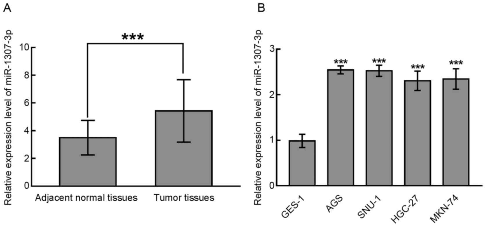
miR-1307-3p is upregulated in gastric
cancer tissues and cell lines
The expression of miR-1307-3p was analyzed in paired
samples of gastric cancer and matched normal tissues and gastric
cancer cell lines by RT-qPCR. The results demonstrated that
miR-1307-3p expression was significantly upregulated in gastric
cancer tissues compared with normal tissues (P<0.001; Fig. 1A). Furthermore, in the gastric cancer
cell lines AGS, SNU-1, HGC-27 and MKN-74, miR-1307-3p expression
was also significantly increased compared with the normal gastric
mucous membrane cell line GES-1 (P<0.001; Fig. 1B). The upregulation of miR-1307-3p
observed in gastric cancer tissues and cell lines suggests its
potential clinical significance and biological function in gastric
cancer.
miR-1307-3p expression is associated
with the TNM stage of patients with gastric cancer
According to the average expression level of
miR-1307-3p in gastric cancer tissues, patients were divided into
the low miR-1307-3p level group (n=52) and the high miR-1307-3p
level group (n=65). The results from χ2 test
demonstrated that miR-1307-3p expression was associated with the
TNM stage of patients (P=0.027), but not with the other
clinicopathological characteristics (P>0.05; Table I).
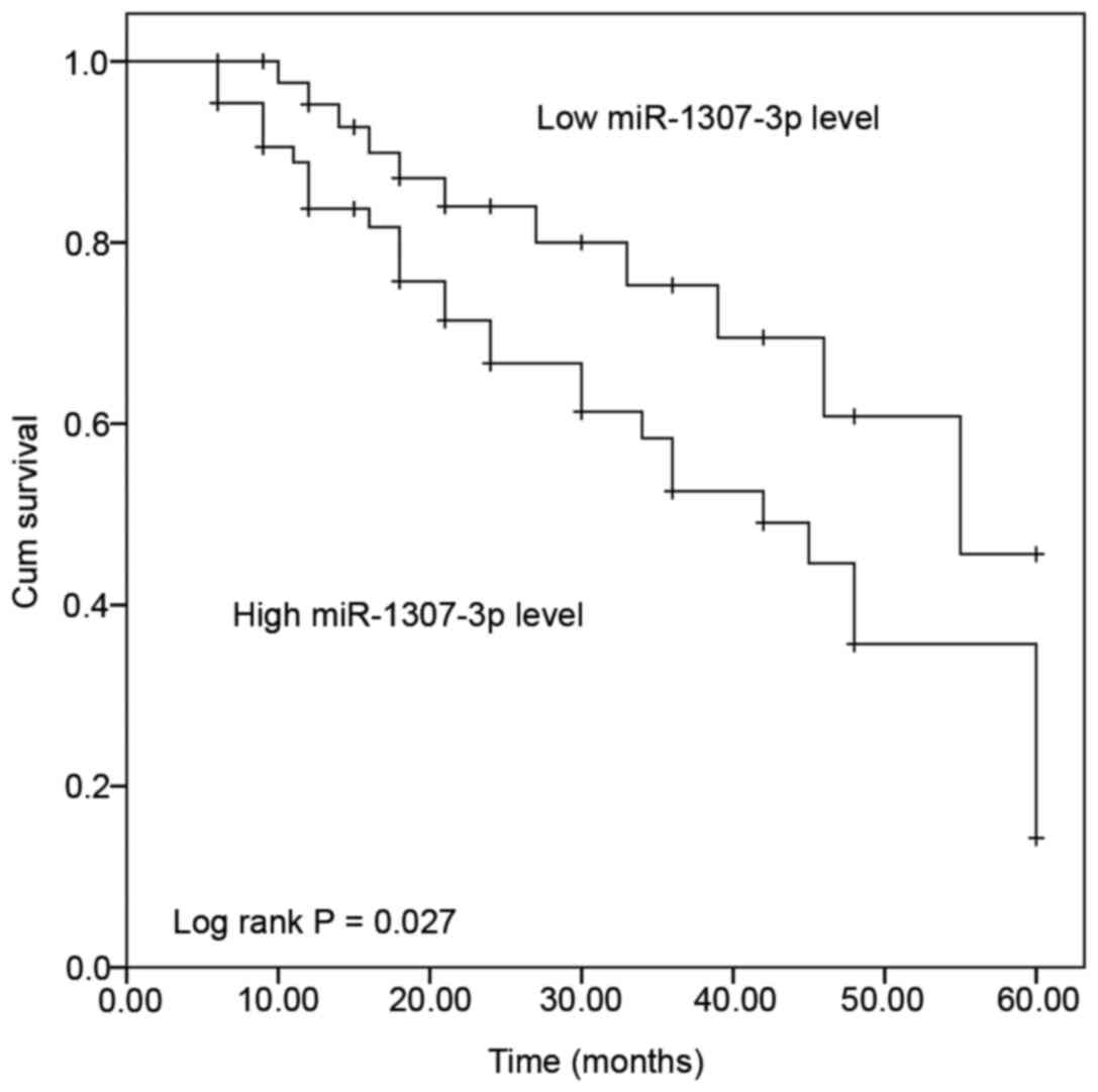
miR-1307-3p expression predicts a poor
prognosis and serves as an independent indicator for the survival
of patients with gastric cancer
The survival rate of patients with gastric cancer
according to miR-1307-3p expression was evaluated using
Kaplan-Meier curve. As presented in Fig.
2, patients with low miR-1307-3p expression had a significantly
longer survival than patients with high miR-1307-3p expression (log
rank P=0.027). The prognostic value of miR-1307-3p was further
evaluated by Cox regression analysis. The results suggested that
miR-1307-3p expression (HR=3.328; 95% CI=1.476–7.505; P=0.004) and
TNM stage (HR=2.348; 95% CI=1.205–4.576; P=0.012) may be considered
as independent predictors for the overall survival of patients with
gastric cancer (Table II).
| Table II.Prognostic value of miR-1307-3p in
gastric cancer evaluated by Cox regression analysis. |
Table II.
Prognostic value of miR-1307-3p in
gastric cancer evaluated by Cox regression analysis.
| Factors | Hazard ratio | 95% CI | P-value |
|---|
| miR-1307-3p | 3.328 | 1.476–7.505 | 0.004 |
| Age | 1.169 | 0.597–2.286 | 0.649 |
| Sex | 1.225 | 0.608–2.467 | 0.570 |
| Tumor size | 1.704 | 0.841–3.452 | 0.139 |
|
Differentiation | 1.709 | 0.860–3.397 | 0.126 |
| Lymph node
metastasis | 1.546 | 0.814–2.936 | 0.183 |
| TNM stage | 2.348 | 1.205–4.576 | 0.012 |
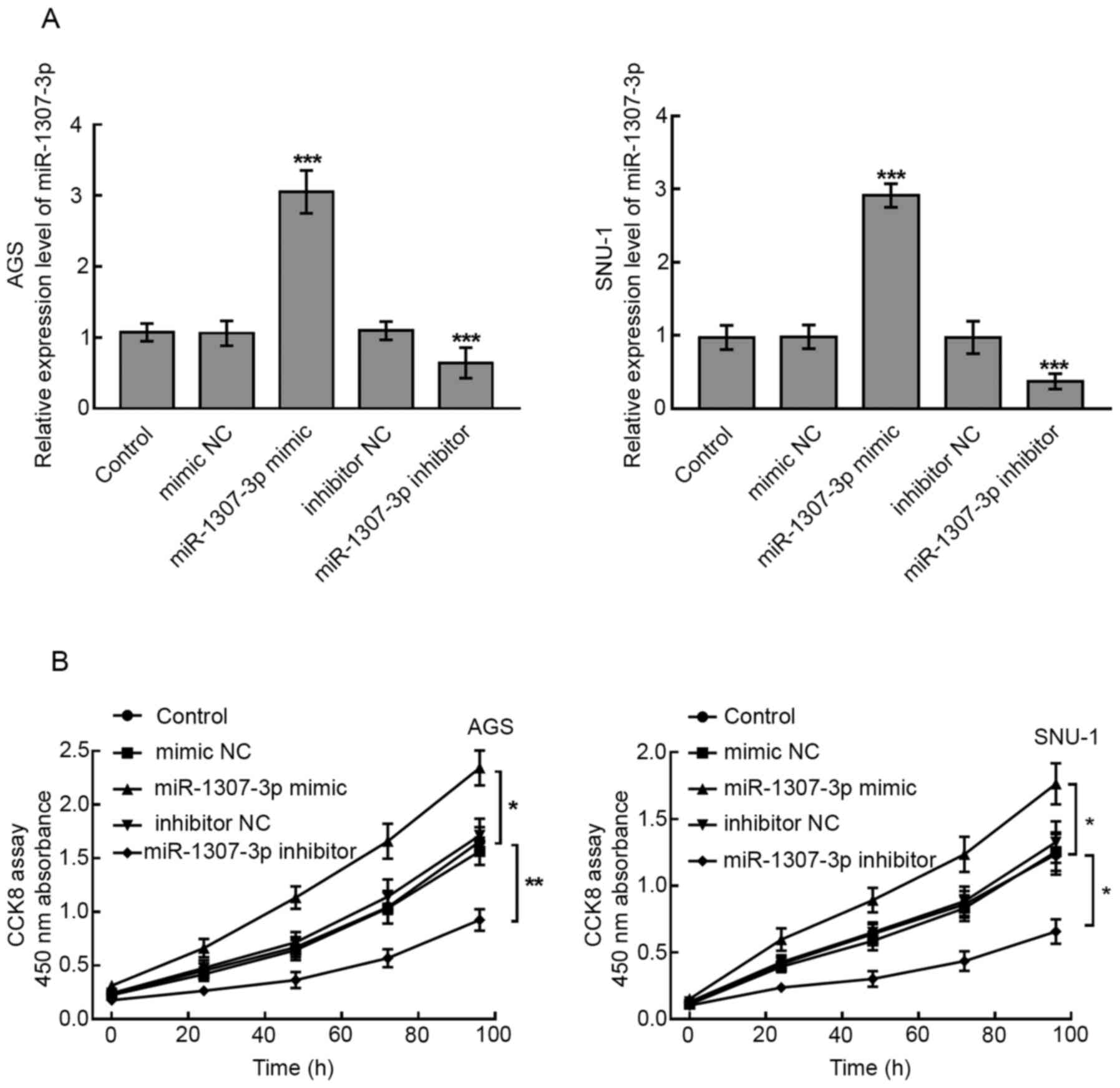
miR-1307-3p upregulation promotes the
proliferation and migratory and invasive abilities of gastric
cancer cells
Due to the high expression of miR-1307-3p in AGS and
SNU-1 cells, these two gastric cancer cell lines were used for
subsequent experiments and were transfected with miR-1307-3p mimic
or inhibitor to regulate the expression of miR-1307-3p. Cell
transfection with miR-1307-3p mimic significantly increased
miR-1307-3p expression, whereas transfection with miR-1307-3p
inhibitor significantly decreased miR-1307-3p expression
(P<0.001; Fig. 3A). The
proliferation of transfected cells was detected by the CCK-8 assay.
The results demonstrated that increased expression of miR-1307-3p
significantly promoted the proliferation of AGS and SNU-1 cells
(P<0.05; Fig. 3B). Furthermore,
miR-1307-3p downregulation significantly inhibited the
proliferation of AGS and SNU-1 cells (P<0.05 and P<0.01;
Fig. 3B).
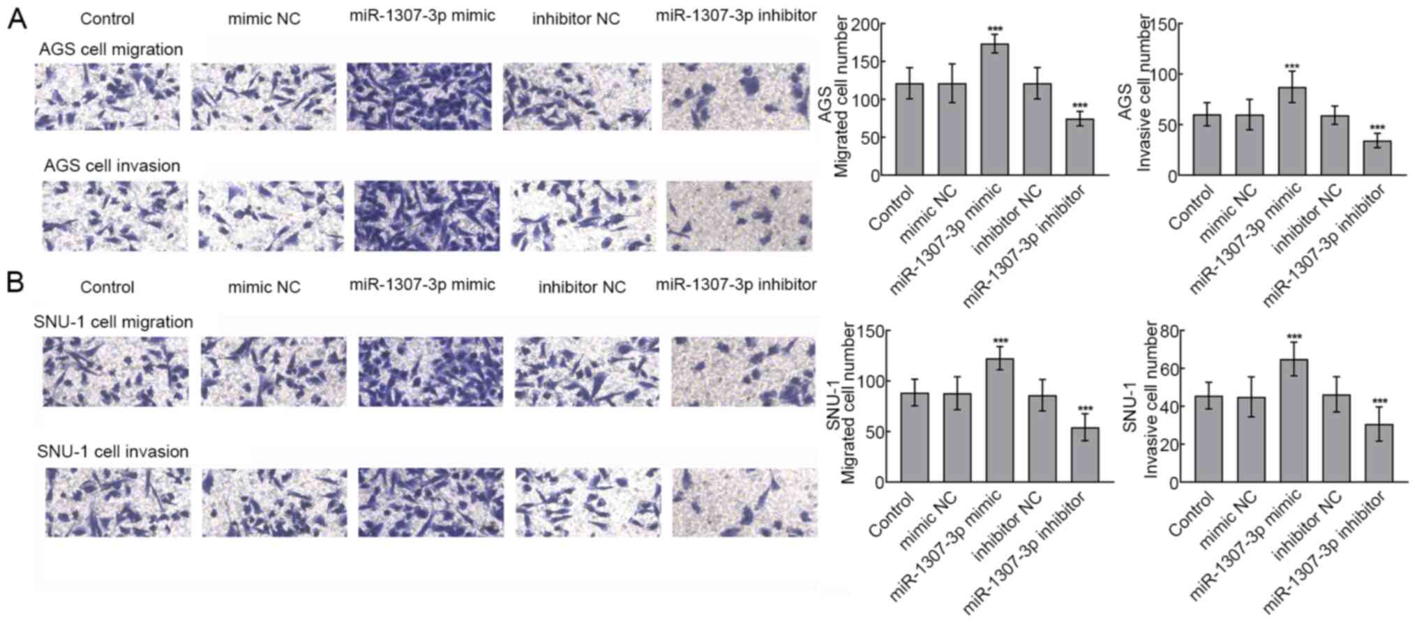
To further evaluate whether miR-1307-3p could be
involved in the migration and invasion processes of gastric cancer
cells, the Transwell assay was performed in transfected AGS and
SNU-1 cells. The upregulation of miR-1307-3p significantly promoted
the migratory and invasive abilities of AGS cells, whereas
miR-1307-3p silencing significantly inhibited AGS cell migratory
and invasive abilities (P<0.001; Fig.
4A). Similarly, SNU-1 cell migratory and invasive abilities
were significantly enhanced following miR-1307-3p overexpression
and inhibited following miR-1307-3p silencing, respectively
(P<0.001; Fig. 4B).
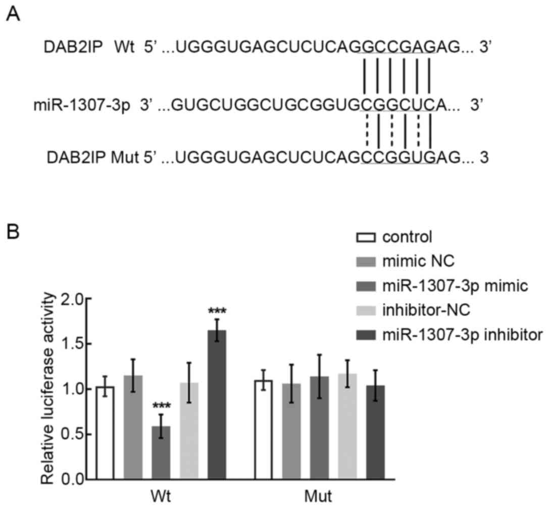
DAB2IP is identified as the direct
target of miR-1307-3p
DAB2IP was predicted as the potential target of
miR-1307-3p from online publicly available algorithms, and the
binding sites are presented in Fig.
5A. Subsequently, the dual-luciferase reporter assay was
performed to further validate the interaction between DAB2IP and
miR-1207-3p. The results demonstrated that the relative luciferase
activity of DAB2IP Wt was significantly suppressed following
transfection with miR-1307-3p mimic and increased by miR-1307-3p
inhibitor, suggesting that DAB2IP may be considered as a direct
target of miR-1307-3p (P<0.001; Fig.
5B).
Discussion
miRNAs are considered as novel prognostic and
therapeutic tools, and numerous studies have focused on the
function and underlying mechanism of miRNAs in cancer (22,23).
With the development of molecular biology, miRNA signatures of
gastric cancer have revealed that numerous dysregulated miRNAs are
involved in the progression and development of gastric cancer
(24). For example, miR-671-5p has
been identified as a downregulated miRNA that inhibits gastric
cancer cell proliferation by targeting brain derived neurotrophic
factors (25). Furthermore, miR-30a
has been identified as a tumor suppressor due to its inhibitory
effect on gastric cancer cell migratory ability and proliferation
(26).
miR-1307-3p has been considered as a biomarker that
could serve a vital role in the progression of various types of
cancer. For example, miR-1307-3p is downregulated in colon
adenocarcinoma, where it was found to inhibit the proliferation and
promote the apoptosis of colon cancer cells (20). Furthermore, the upregulation of
miR-1307-3p in hepatocellular carcinoma can facilitate the
proliferation, migration and invasion of hepatocellular carcinoma
cells (18). In the present study,
miR-1307-3p was significantly upregulated in gastric cancer tissues
and cells compared with normal tissues and cells, which was
consistent with the reported miRNA expression profile of gastric
cancer (27). In addition,
miR-1307-3p upregulation could stimulate the proliferation and
migratory and invasive abilities of gastric cancer cells,
suggesting a tumor promoter role of miR-1307-3p in the progression
of gastric cancer.
Previous studies demonstrated that the dysregulation
of miR-1307-3p in breast cancer and hepatocellular carcinoma is
significantly associated with a poor survival rate in patients
(18,19). Additional miRNAs have also been
reported to predict the prognosis of patients with gastric cancer
(15,28). Furthermore, the upregulation of
miR-199a-3p is associated with a poor prognosis in patients with
gastric cancer, indicating that it can serve as a prognostic
indicator (29). In the present
study, the clinical significance of miR-1307-3p in gastric cancer
was also evaluated. The upregulation of miR-1307-3p in gastric
cancer was closely associated with the TNM stage of patients,
suggesting that it may be considered as an independent indicator
for the prognosis of patients. These results indicated the
involvement of miR-1307-3p in gastric cancer development and
patients' prognosis.
In order to develop novel therapeutic strategies
against gastric cancer, it is crucial to determine the underlying
mechanism of miR-1307. A previous study reported that DAB2IP is a
direct target of miR-1307-3p, and that miR-1307-3p can promote
tumor growth and metastasis in hepatocellular carcinoma (18). In gastric cancer, DAB2IP is a
downregulated gene that regulates the proliferation and metastasis
of gastric cancer cells (30).
Furthermore, DAB2IP can regulate numerous cellular functions,
including cell proliferation, apoptosis and metastasis (31). In the present study, dual-luciferase
reporter assay identified DAB2IP as a direct target of miR-1307-3p.
It was therefore speculated that miR-1307-3p could participate in
gastric cancer progression by targeting DAB2IP.
This study presented a limitation. Due to the small
sample size, the association between miR-1307-3p expression and
certain clinicopathological characteristics of patients, such as
lymph node metastasis and differentiation, was not significant,
although these are important clinical factors (32–34). The
sample size should therefore be expanded in future studies in order
to obtain results that would better reflect the clinical
situation.
In conclusion, the present study demonstrated the
significant upregulation of miR-1307-3p in gastric cancer tissues
compared with normal tissues. The expression level of miR-1307-3p
was significantly associated with the TNM stage and poor prognosis
of patients. Subsequently, miR-1307-3p may serve as a tumor
promoter of gastric cancer, where its upregulation could
significantly promote the proliferation and migratory and invasive
abilities of gastric cancer cells. These results suggested that
miR-1307-3p inhibition may be considered as be a novel therapeutic
strategy for the management of gastric cancer.
Acknowledgements
Not applicable.
Funding
No funding was received.
Availability of data and materials
The datasets used and/or analyzed during the current
study are available from the corresponding author on reasonable
request.
Authors' contributions
YM, AZ and JS conceived and designed the project. YM
and AZ acquired the data and YM analyzed and interpreted the data.
YM, AZ and JS wrote the paper. All authors read and approved the
final version.
Ethics approval and consent to
participate
This study was approved by the Ethics Committee of
Qingdao Central Hospital. All patients signed informed consent
before tissue collection.
Patient consent for publication
Not applicable.
Competing interests
The authors declare that they have no competing
interests.
References
|
1
|
Siegel R, Ma J, Zou Z and Jemal A: Cancer
statistics, 2014. CA Cancer J Clin. 64:9–29. 2014. View Article : Google Scholar : PubMed/NCBI
|
|
2
|
Balakrishnan M, George R, Sharma A and
Graham DY: Changing Trends in Stomach Cancer Throughout the World.
Curr Gastroenterol Rep. 19:362017. View Article : Google Scholar : PubMed/NCBI
|
|
3
|
Global Burden of Disease Cancer
Collaboration, ; Fitzmaurice C, Allen C, Barber RM, Barregard L,
Bhutta ZA, Brenner H, Dicker DJ, Chimed-Orchir O, Dandona R,
Dandona L, et al: Global, regional, and national cancer incidence,
mortality, years of life lost, years lived with disability, and
disability-adjusted life-years for 32 cancer groups, 1990 to 2015:
A systematic analysis for the Global Burden of Disease Study. JAMA
Oncol. 3:524–548. 2017. View Article : Google Scholar : PubMed/NCBI
|
|
4
|
de Martel C, Ferlay J, Franceschi S,
Vignat J, Bray F, Forman D and Plummer M: Global burden of cancers
attributable to infections in 2008: A review and synthetic
analysis. Lancet Oncol. 13:607–615. 2012. View Article : Google Scholar : PubMed/NCBI
|
|
5
|
de Martel C, Forman D and Plummer M:
Gastric cancer: Epidemiology and risk factors. Gastroenterol Clin
North Am. 42:219–240. 2013. View Article : Google Scholar : PubMed/NCBI
|
|
6
|
Yasui W, Yokozaki H, Fujimoto J, Naka K,
Kuniyasu H and Tahara E: Genetic and epigenetic alterations in
multistep carcinogenesis of the stomach. J Gastroenterol. 35 (Suppl
12):111–115. 2000.PubMed/NCBI
|
|
7
|
Stock M and Otto F: Gene deregulation in
gastric cancer. Gene. 360:1–19. 2005. View Article : Google Scholar : PubMed/NCBI
|
|
8
|
Wang WC and Zhang XF, Peng J, Li XF, Wang
AL, Bie YQ, Shi LH, Lin MB and Zhang XF: Survival Mechanisms and
Influence Factors of Circulating Tumor Cells. BioMed Res Int.
2018:63047012018. View Article : Google Scholar : PubMed/NCBI
|
|
9
|
Karimi P, Islami F, Anandasabapathy S,
Freedman ND and Kamangar F: Gastric cancer: Descriptive
epidemiology, risk factors, screening, and prevention. Cancer
Epidemiol Biomarkers Prev. 23:700–713. 2014. View Article : Google Scholar : PubMed/NCBI
|
|
10
|
Bartel DP: MicroRNAs: Genomics,
biogenesis, mechanism, and function. Cell. 116:281–297. 2004.
View Article : Google Scholar : PubMed/NCBI
|
|
11
|
Hoffman Y, Bublik DR, Ugalde AP, Elkon R,
Biniashvili T, Agami R, Oren M and Pilpel Y: 3′UTR Shortening
Potentiates MicroRNA-Based Repression of Pro-differentiation Genes
in Proliferating Human Cells. PLoS Genet. 12:e10058792016.
View Article : Google Scholar : PubMed/NCBI
|
|
12
|
Kim S and Jeong S: Mutation hotspots in
the β-catenin gene: Lessons from the human cancer genome databases.
Mol Cells. 42:8–16. 2019.PubMed/NCBI
|
|
13
|
Bartel DP: MicroRNAs: Target recognition
and regulatory functions. Cell. 136:215–233. 2009. View Article : Google Scholar : PubMed/NCBI
|
|
14
|
Ahmed FE: Role of miRNA in carcinogenesis
and biomarker selection: A methodological view. Expert Rev Mol
Diagn. 7:569–603. 2007. View Article : Google Scholar : PubMed/NCBI
|
|
15
|
Brenner B, Hoshen MB, Purim O, David MB,
Ashkenazi K, Marshak G, Kundel Y, Brenner R, Morgenstern S, Halpern
M, et al: MicroRNAs as a potential prognostic factor in gastric
cancer. World J Gastroenterol. 17:3976–3985. 2011. View Article : Google Scholar : PubMed/NCBI
|
|
16
|
Wei Y, Wang Y, Zang A, Wang Z, Fang G and
Hong D: MiR-4766-5p inhibits the development and progression of
gastric cancer by targeting NKAP. OncoTargets Ther. 12:8525–8536.
2019. View Article : Google Scholar
|
|
17
|
Chen H, Yang Y, Wang J, Shen D, Zhao J and
Yu Q: miR-130b-5p promotes proliferation, migration and invasion of
gastric cancer cells via targeting RASAL1. Oncol Lett.
15:6361–6367. 2018.PubMed/NCBI
|
|
18
|
Chen S, Wang L, Yao B, Liu Q and Guo C:
miR-1307-3p promotes tumor growth and metastasis of hepatocellular
carcinoma by repressing DAB2 interacting protein. Biomed
Pharmacother. 117:1090552019. View Article : Google Scholar : PubMed/NCBI
|
|
19
|
Han S, Zou H, Lee JW, Han J, Kim HC, Cheol
JJ, Kim LS and Kim H: miR-1307-3p stimulates breast cancer
development and progression by targeting SMYD4. J Cancer.
10:441–448. 2019. View Article : Google Scholar : PubMed/NCBI
|
|
20
|
Zheng Y, Zheng Y, Lei W, Xiang L and Chen
M: miR-1307-3p overexpression inhibits cell proliferation and
promotes cell apoptosis by targeting ISM1 in colon cancer. Mol Cell
Probes. 48:1014452019. View Article : Google Scholar : PubMed/NCBI
|
|
21
|
Livak KJ and Schmittgen TD: Analysis of
relative gene expression data using real-time quantitative PCR and
the 2(-Delta Delta C(T)) Method. Methods. 25:402–408. 2001.
View Article : Google Scholar : PubMed/NCBI
|
|
22
|
Kalinowski FC, Brown RA, Ganda C, Giles
KM, Epis MR, Horsham J and Leedman PJ: microRNA-7: A tumor
suppressor miRNA with therapeutic potential. Int J Biochem Cell
Biol. 54:312–317. 2014. View Article : Google Scholar : PubMed/NCBI
|
|
23
|
Yadav S, Shekhawat M, Jahagirdar D and
Kumar Sharma N: Natural and artificial small RNAs: A promising
avenue of nucleic acid therapeutics for cancer. Cancer Biol Med.
14:242–253. 2017. View Article : Google Scholar : PubMed/NCBI
|
|
24
|
Shin VY and Chu KM: MiRNA as potential
biomarkers and therapeutic targets for gastric cancer. World J
Gastroenterol. 20:10432–10439. 2014. View Article : Google Scholar : PubMed/NCBI
|
|
25
|
Qiu T, Wang K, Li X and Jin J: miR-671-5p
inhibits gastric cancer cell proliferation and promotes cell
apoptosis by targeting URGCP. Exp Ther Med. 16:4753–4758.
2018.PubMed/NCBI
|
|
26
|
Yu T, Gong L, Li W, Zuo Q, Cai D, Mao H,
Wang L, Lin J and Xiao B: MiR-30a suppresses metastasis of gastric
adenocarcinoma via targeting FAPα. Cancer Biomark. 27:471–484.
2020. View Article : Google Scholar : PubMed/NCBI
|
|
27
|
Ding L, Zhang S, Xu M, Zhang R, Sui P and
Yang Q: MicroRNA-27a contributes to the malignant behavior of
gastric cancer cells by directly targeting PH domain and
leucine-rich repeat protein phosphatase 2. J Exp Clin Cancer Res.
36:452017. View Article : Google Scholar : PubMed/NCBI
|
|
28
|
Nakamura S, Kanda M and Kodera Y:
Incorporating molecular biomarkers into clinical practice for
gastric cancer. Expert Rev Anticancer Ther. 19:757–771. 2019.
View Article : Google Scholar : PubMed/NCBI
|
|
29
|
Li L, Mou YP, Wang YY, Wang HJ and Mou XZ:
miR-199a-3p targets ETNK1 to promote invasion and migration in
gastric cancer cells and is associated with poor prognosis. Pathol
Res Pract. 215:1525112019. View Article : Google Scholar : PubMed/NCBI
|
|
30
|
Sun L, Yao Y, Lu T, Shang Z, Zhan S, Shi
W, Pan G, Zhu X and He S: DAB2IP downregulation enhances the
proliferation and metastasis of human gastric cancer cells by
derepressing the ERK1/2 pathway. Gastroenterol Res Pract.
2018:29682522018. View Article : Google Scholar : PubMed/NCBI
|
|
31
|
Liu L, Xu C, Hsieh JT, Gong J and Xie D:
DAB2IP in cancer. Oncotarget. 7:3766–3776. 2016. View Article : Google Scholar : PubMed/NCBI
|
|
32
|
Nakamura R, Omori T, Mayanagi S, Irino T,
Wada N, Kawakubo H, Kameyama K and Kitagawa Y: Risk of lymph node
metastasis in undifferentiated-type mucosal gastric carcinoma.
World J Surg Oncol. 17:322019. View Article : Google Scholar : PubMed/NCBI
|
|
33
|
Mikami K, Hirano Y, Futami K and Maekawa
T: Expansion of lymph node metastasis in mixed-type submucosal
invasive gastric cancer. Asian J Surg. 41:462–466. 2018. View Article : Google Scholar : PubMed/NCBI
|
|
34
|
Bausys R, Bausys A, Vysniauskaite I,
Maneikis K, Klimas D, Luksta M, Strupas K and Stratilatovas E: Risk
factors for lymph node metastasis in early gastric cancer patients:
Report from Eastern Europe country- Lithuania. BMC Surg.
17:1082017. View Article : Google Scholar : PubMed/NCBI
|