Introduction
Hepatocellular carcinoma (HCC) is one of most
prevalent malignancies worldwide with a high mortality (1). According to the American
Epidemiological Survey of Liver Cancer, the adjusted incidence of
HCC increased at a rate of 4.5% annually between 2000 and 2009,
making it the fastest-growing cause of cancer-associated mortality
in the United States (2). HCC is
also a highly invasive tumor with frequent intrahepatic or
pulmonary metastasis, both of which account for the high disease
recurrence and poor survival following liver resection (3). HCC diagnosis and metastasis
identification are usually based on imaging and occasionally
verified using biopsy results (4).
Although advances in MRI and computed tomography have improved the
imaging of focal hypervascular masses consistent with HCC, these
procedures are costly and not suitable for daily practice (5). In addition, pathological biopsies are
the gold standard for HCC diagnosis, yet biopsy results are
associated with a high false negative rate (6), and the procedure can cause discomfort
or pain. The detailed molecular mechanisms that contribute to HCC
metastasis are largely unknown. Furthermore, effective targeted
therapeutic drugs for HCC remain unavailable. Therefore, the
identification of non-invasive diagnostic and predictive biomarkers
of HCC metastasis is essential.
Exosomes are microscopic vesicles that serve
important roles in cell-cell communication and signal transduction
(7). Exosomes transport biologically
active molecules (8) to target
cells, in order to initiate signaling and mediate intercellular
communication. Furthermore, the potential use of exosomes as
biomarkers has been demonstrated for other cancer types, such as
breast cancer and gastric cancer (9), which demonstrates that the use of
exosomes as a tumor marker for clinical diagnosis and prediction is
feasible. Li et al (10) have
highlighted the significance of exosomes in the development of HCC;
however, the role of exosomes in tumor growth and metastasis
remains a matter of debate. Therefore, the aim of the present study
was to identify exosomal molecules that may be used as
metastasis-related biomarkers in patients with HCC.
MicroRNAs (miRNAs/miRs) are a class of non-coding
RNAs ~22 nucleotides in length, which can modulate gene expression
(11). Tumor cells can secrete
miRNAs that mediate intracellular signaling cascades, which in turn
can affect various physiological processes, such as angiogenesis,
metabolic reprogramming and metastasis (12). A number of studies (13,14) have
indicated that secretory miRNAs, particularly exosomal miRNAs, are
involved in promoting metastasis (15), highlighting the potential for
exosomal miRNAs as promising biomarkers. For example, Lin et
al (16) identified an miRNA
classifier containing seven differentially expressed miRNAs that
allows early detection of HCC. However, the role of
metastasis-associated miRNAs in HCC has yet to be determined.
The aim of the present study was to identify a
comprehensive circulating exosomal miRNA profile in plasma from
patients with metastatic HCC. Furthermore, bioinformatics analysis
was used to screen target genes of differentially expressed miRNAs
and to evaluate their potential role in HCC metastasis.
Furthermore, the prognostic efficiency of candidate miRNAs, and
their association with overall survival (OS) were also assessed.
The study identified a panel consisting of three miRNAs that could
be used to discriminate metastatic HCC cases from non-metastatic
HCC cases. Overall, the present findings may provide insights into
the mechanisms that lead to metastasis and the role of exosomes in
this context. The present study also suggested that exosomal miRNAs
may represent potential biomarkers for HCC.
Materials and methods
Plasma and human tissue sample
collection
A total of 40 patients with HCC diagnosed by
clinical pathology at Nanfang Hospital (Guangzhou, China) between
January 2017 and April 2018 were recruited. Among them, 20 patients
with HCC (13 men and 7 women) with lung metastasis and 20
sex-matched primary cases were enrolled. The median age (age range,
50–80 years) of the metastatic group was 68 years and that of the
non-metastatic group (age range, 50–81 years) was 67.5 years. All
patients did not undergo radiotherapy and chemotherapy prior to
surgery. All patients were referred for extensive evaluation,
including measurement of α-fetoprotein levels and MRI before
surgical resection. The tumor was examined by postoperative
pathology. A 5-ml sample of peripheral venous blood was collected
from all patients into EDTA tubes, and then centrifuged at 2,500 ×
g for 10 min at room temperature. The supernatant was transferred
into RNase-free Eppendorf tubes and stored at −80°C until use. In
10 of the patients with metastatic HCC, lung metastasis biopsies
were also collected, and stored in the −80°C ultra-low temperature
refrigerator until further use. Informed consent was obtained from
each patient on the day of admission.
Isolation of exosomes
Exosomes were purified from the plasma of patients
with HCC by ultracentrifugation. The plasma was thawed on ice and
centrifuged at 500 × g for 10 min and the supernatant was
collected. The following steps were completed continuously and
without interruption as soon as possible. The supernatant was
ultracentrifuged using a W32Ti rotor (L-80XP; Beckman Coulter,
Inc.) at 16,800 × g for 30 min to pellet the exosomes.
Subsequently, the pellet was washed in PBS to eliminate
contaminating proteins, and centrifuged again at 110,000 × g for 70
min. PBS was subsequently removed and the exosomes were
re-suspended in 100 µl PBS. All centrifugation steps were performed
at 4°C. All samples were stored in a −80°C ultra-low temperature
refrigerator until use (within 1 month).
NTA
Exosome pellets were resuspended in PBS at a
concentration of 1×10⁷−109/ml, and then
examined using a NanoSight NS300 instrument (Malvern Panalytical)
to determine the size and concentration. Particle movement was
analyzed and recorded in a real-time video using the NTA software
(version 2.3; Malvern Panalytical). Exosome size and concentration
were determined using NTA v3.2 software (Malvern Panalytical).
Western blotting
Western blotting was performed as previously
described (17) Exosome lysates were
prepared using RIPA buffer (Nanjing KeyGen Biotech Co., Ltd.).
Protein concentrations were determined using a Bradford protein
assay (Nanjing KeyGen Biotech Co., Ltd.). The samples were resolved
using SDS-PAGE, transferred to PVDF membranes (EMD Millipore) and
incubated with primary antibodies overnight at 4°C, as previously
described (17). Primary antibodies
specific for 70 kilodalton heat shock protein (HSP70; cat. no.
ab194360; 1:1,000), CD63 (cat. no. ab59479; 1:1,000) and GAPDH
(cat. no. ab8245; 1:10,000) were purchased from Abcam. Following
primary antibody incubation, a HRP-conjugated secondary antibody
(cat. nos. FDM007 and FDR007; 1:10,000; Fdbio Science) was added.
The Fdbio-Femto ECL western blotting detection reagent (Fdbio
Science) was used to visualize protein bands.
RNA processing and miRNA
profiling
Exosomes from the plasma of 4 patients with primary
HCC and 4 patients with HCC lung metastasis were analyzed using
Affymetrix miRNA 4.0 arrays (Thermo Fisher Scientific, Inc.). The
RNA processing and miRNA profiling experiments were performed at
the laboratory of Gene-Cloud of Biotechnology Information (GCBI;
Shanghai, China).
RNA extraction and reverse
transcription-quantitative PCR (RT-qPCR)
The initial miRNA microarray results from the plasma
exosomes samples were validated using RT-qPCR. Exosome and tissue
miRNA purification and RT-qPCR were performed as described
previously (18). Briefly, total RNA
from stored plasma exosome and tissue samples was extracted using
TRIzol reagent (Thermo Fisher Scientific, Inc.) according to the
manufacturer's protocols. RT-qPCR was performed using SYBR Green
and detected using the Applied Biosystems Real-Time PCR system
(Thermo Fisher Scientific, Inc.). Each sample was analyzed in
triplicate. The primers used for RT-qPCR are listed in Table I. The U6 small nuclear RNA was used
as an internal control. The results were quantified using the
2−ΔΔCq method (19) and
normalized to U6 expression.
 | Table I.Primer sequences for reverse
transcription-quantitative PCR used in exosome and tissue miRNA
experiments. |
Table I.
Primer sequences for reverse
transcription-quantitative PCR used in exosome and tissue miRNA
experiments.
| Primer of
miRNA | Sequence
(5′-3′) |
|---|
| hsa-let-7e |
TGAGGTAGGAGGTTGTATAGTT |
| hsa-miR-18a |
CCCATCTAGTGCAGATAGAAA |
| hsa-miR-27a |
TTCACAGTGGCTAAGTTCCGC |
| hsa-miR-221 |
CTACATTGTCTGCTGGGTTTC |
| hsa-miR-185 |
GAGAGAAAGGCAGTTCCTGA |
| hsa-miR-361 |
TTATCAGAATCTCCAGGGGTA |
| hsa-miR-20b |
AGTGCTCATAGTGCAGGTAG |
| hsa-miR-652 |
CGCCACTAGGGTTGTGAAAA |
| hsa-miR-4454 |
CTGAGCGCTGCCAGTCAAA |
| hsa-miR-4720 |
CCCTGGCATATTTGGTATAAC |
| hsa-miR-5189 |
CAACCGTCAGAGCCCAGAA |
| U6-forward |
GGAACGATACAGAGAAGATTAGC |
| U6-reverse |
TGGAACGCTTCACGAATTTGCG |
Gene Expression Omnibus (GEO) miRNA
dataset and patient information
Datasets that met the following criteria were
selected for study: i) The patients did not undergo
chemoradiotherapy before surgery and only datasets containing
>20 samples were included; and ii) HCC with lung-metastasis and
without metastasis was confirmed by postoperative pathology and the
patients had complete follow-up data. The Cancer Genome Atlas
(https://www.cancer.gov/about-nci/organization/ccg/research/structural-genomics/tcga)
and GEO database were used to mined the data. A dataset containing
91 human HCC tumor samples without vascular invasion/metastasis and
81 samples with vascular invasion/metastasis was downloaded from
the GEO (https://www.ncbi.nlm.nih.gov/geo/) database [dataset
accession no. GSE67140 (20)].
Receiver operating characteristic (ROC) analysis was carried out on
these data using GraphPad Prism 8.0 software (GraphPad Software,
Inc.).
Survival data from the Kaplan-Meier
plotter database
The present study used the online Kaplan-Meier
plotter database (http://www.kmplot.com/analysis/index.php?p=service&cancer=liver_mirna)
tool to evaluate the prognostic values of candidate miRNAs
(21). Original data were downloaded
and Kaplan-Meier plots were generated to analyze potential
prognostic values of candidate miRNAs with the platform of RNA
sequencing. Analysis was performed by excluding patients who
survived >80 months because there might be unrecognized
confounding bias. In addition, the online plotter was used to
perform the survival analysis, and hazard ratios with 95%
confidence intervals and log rank P-values were calculated.
P<0.05 was considered to indicate a statistically significant
difference.
Bioinformatics analysis of
differentially expressed exosomal miRNAs
The miRanda (22)
(release miRanda-aug2010; http://www.microrna.org/) and TargetScan (release 7.0;
http://www.targetscan.org/vert_70/)
databases were used to predict the target genes of candidate
exosomal miRNAs. Gene Ontology (GO; release 2019-12; http://geneontology.org/) and Kyoto Encyclopedia of
Genes and Genomes (KEGG; release 92; http://www.kegg.jp/) analyses were performed to
identify the biological functions and pathways for which P<0.05
and false discovery rate <0.05. R language (version 3.6.0;
http://cran.r-project.org/) and ggplot2
(version 3.3.2) were used to generate the image (23,24). The
miRNA-mRNA networks were generated using GCBI (https://www.gcbi.com.cn/gclib/html/index) and
Cytoscape (version 3.6; http://www.cytoscape.org/).
Statistical analysis
Statistical analysis was performed using GraphPad
Prism (version 8.0; GraphPad Software, Inc.) or SPSS version 24.0
statistical software (SPSS, Inc.). Data are presented as the mean ±
standard deviation. Differences between two groups were analyzed
using a two-tailed unpaired Student's t-test. Pearson correlation
and residual analysis were used to measure and assess the
correlation between candidate plasma exosome miRNA levels, and
tissue ROC curves were established by plotting sensitivity against
1-specificity. The diagnostic values of candidate miRNAs and the
panel were evaluated using the area under the curve (AUC).
Comparisons of AUC values were based on Delong's test in SPSS using
R plug-in package pROC (R 4.0.3 package/pROC v1.16.2), and P-values
were adjusted using the Bonferroni strategy when multiple
comparisons were conducted (25).
Graphs were generated using GraphPad Prism 5.0. P<0.05 was
considered to indicate a statistically significant difference.
Results
Identification and characterization of
exosomes
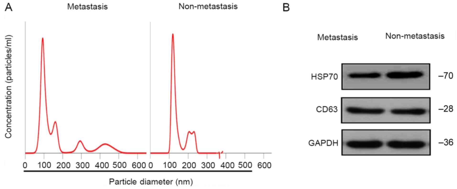
Using NTA, 30–150-nm vesicles were identified,
consistent with previously reported features of exosomes (26), suggesting that the isolated particles
were likely exosomes (Fig. 1A).
Additionally, western blot analysis indicated that the particles
were positive for exosome markers CD63 and HSP70 (Fig. 1B). These results demonstrated that
the particles isolated from the plasma of patients with HCC were
exosomes.
Exosomal miRNA profiles are altered
between patients with metastatic and non-metastatic HCC
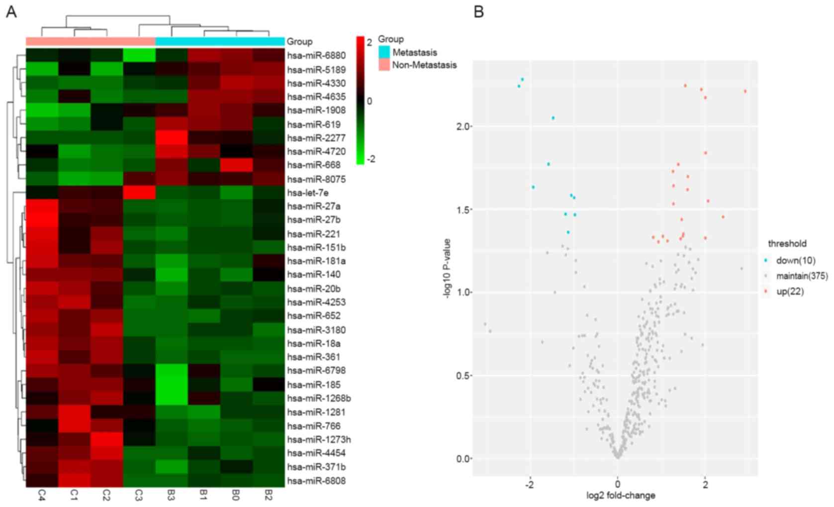
A total of 32 differentially expressed exosomal
miRNAs were detected in 4 patients with HCC lung metastasis and 4
sex- and age-matched patients with non-metastatic HCC (Table II). Among these, 18 miRNAs (let-7e,
miR-18a, miR-27a, miR-181a, miR-221, miR-27b, miR-185, miR-361,
miR-20b, miR-652, miR-140, miR-1280, miR-151b, miR-4253, miR-1268b,
miR-4454, miR-6798 and miR-1273h) were significantly upregulated
and 5 miRNAs (miR-4330, miR-2277, miR-4720, miR-5189 and miR-8075)
were significantly downregulated (fold change >1.2; P<0.05).
Group-specific signal intensities of the exosomal miRNA profile and
the volcano plot for the differentially expressed miRNAs are shown
in Fig. 2. These data indicated that
exosomal miRNAs were differentially expressed between patients with
HCC with lung metastasis and patients with non-metastatic HCC.
| Table II.Differentially expressed exosomal
microRNAs (n=32) in plasma from patients with hepatocellular
carcinoma with metastasis and without metastasis. |
Table II.
Differentially expressed exosomal
microRNAs (n=32) in plasma from patients with hepatocellular
carcinoma with metastasis and without metastasis.
| MicroRNA | Fold change | diffState | P-value |
|---|
| hsa-let-7e | 2.01425971 | Up | 0.01445168 |
| hsa-miR-18a | 1.91615029 | Up | 0.00600160 |
| hsa-miR-27a | 2.41141531 | Up | 0.03514587 |
| hsa-miR-181a | 1.44064685 | Up | 0.04755760 |
| hsa-miR-221 | 2.00841888 | Up | 0.04713177 |
| hsa-miR-27b | 1.49297559 | Up | 0.04580692 |
| hsa-miR-185 | 2.92150796 | Up | 0.00614653 |
| hsa-miR-361 | 2.00636979 | Up | 0.00671691 |
| hsa-miR-20b | 1.60817127 | Up | 0.02009393 |
| hsa-miR-652 | 1.59785688 | Up | 0.02407410 |
| hsa-miR-140 | 1.38821462 | Up | 0.01697033 |
| hsa-miR-1281 | 1.54732638 | Up | 0.00569455 |
| hsa-miR-1908 | −0.98002981 | Down | 0.03410698 |
| hsa-miR-151b | 1.27543975 | Up | 0.02927815 |
| hsa-miR-4253 | 1.50611955 | Up | 0.04447737 |
| hsa-miR-4330 | −1.47908532 | Down | 0.00890724 |
| hsa-miR-2277 | −1.93607770 | Down | 0.02324236 |
| hsa-miR-3180 | 0.93363816 | Up | 0.04959451 |
| hsa-miR-1268b | 1.46370203 | Up | 0.03639235 |
| hsa-miR-4454 | 2.07258859 | Up | 0.02816307 |
| hsa-miR-4635 | −1.13383056 | Down | 0.04346026 |
| hsa-miR-4720 | −2.26430974 | Down | 0.00572943 |
| hsa-miR-371b | 1.03442024 | Up | 0.04597321 |
| hsa-miR-766 | 0.81375899 | Up | 0.04659530 |
| hsa-miR-619 | −0.99689872 | Down | 0.02691538 |
| hsa-miR-668 | −1.06214814 | Down | 0.02606566 |
| hsa-miR-5189 | −2.18451380 | Down | 0.00521571 |
| hsa-miR-6798 | 1.27908232 | Up | 0.02282279 |
| hsa-miR-6808 | 1.13920150 | Up | 0.04899890 |
| hsa-miR-6880 | −1.19417658 | Down | 0.03378472 |
| hsa-miR-1273h | 1.26636581 | Up | 0.01870905 |
| hsa-miR-8075 | −1.58600573 | Down | 0.01690265 |
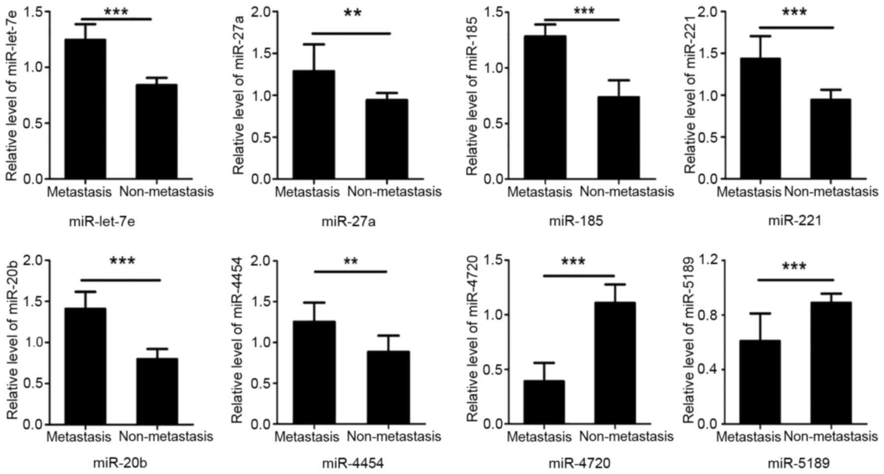
RT-qPCR validation of exosomal miRNA
expression profiles
Using RT-qPCR, the eight most dysregulated miRNAs
from the miRNA microarray were validated in plasma exosome samples
collected from 20 paired patients with HCC with lung metastasis and
without metastasis. A total of six exosomal miRNAs (let-7e,
miR-27a, miR-221, miR-185, miR-20b and miR-4454) were statistically
significantly upregulated in exosome samples from patients with HCC
with lung metastasis (P<0.05). The expression levels of the
remaining two miRNAs (miR-4720 and miR-5189) were significantly
downregulated in samples from patients with metastatic HCC compared
with in samples from patients with non-metastatic HCC (P<0.05;
Fig. 3). Therefore, the miRNA
microarray results were consistent with the results of RT-qPCR.
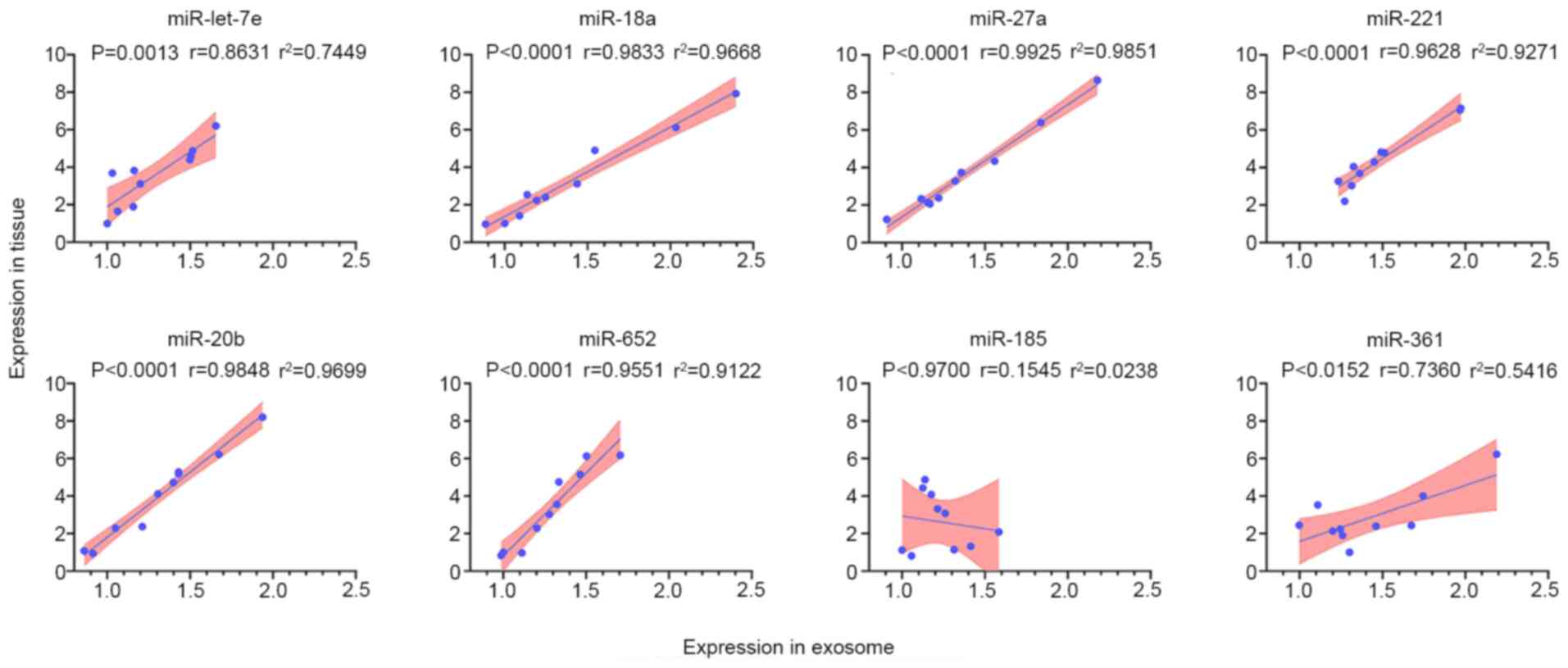
Correlation analysis of differentially
expressed miRNAs in exosomes and matched HCC tissue samples from
patients with lung metastasis
Several significant differentially expressed miRNAs
were randomly selected to verify the association between exocrine
expression and corresponding tissue expression. The expression
levels of the eight highly expressed miRNAs (let-7e, miR-18a,
miR-27a, miR-221, miR-185, miR-361, miR-20b and miR-652) in 10
matched plasma exosome samples and tissue samples from patients
with HCC with lung metastasis were analyzed by RT-qPCR. The results
demonstrated that the expression levels of let-7e, miR-18a,
miR-27a, miR-221, miR-20b, miR-361 and miR-652 were positively
correlated in exosome and tissue samples (Fig. 4), indicating consistency between
plasma and metastatic tissue expression of these miRNAs. However,
the levels of miR-185 were not correlated in exosome samples and
matched tissue samples. Residual analysis was performed when the
correlation was assessed, and these data are shown in Fig. S1. Additionally, a spline curve was
used to explore how it may fit the data, and this revealed that the
model was suitable (Fig. S2).
Therefore, the seven uniformly expressed miRNAs were selected as
candidate biomarkers for subsequent experiments.
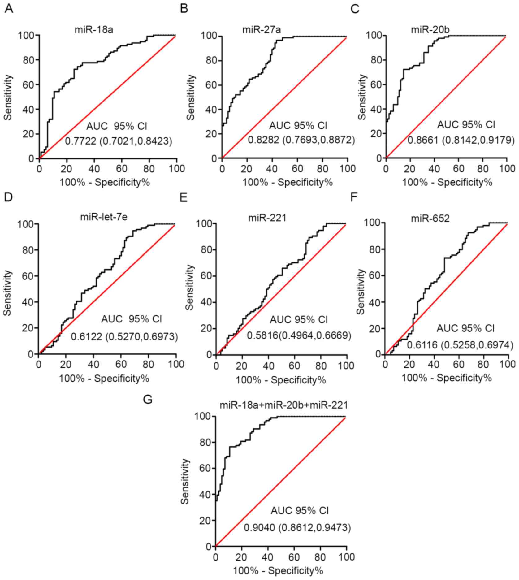
Candidate exosomal miRNAs and miRNA
panels predict HCC metastasis
The diagnostic value of each of the six plasma
miRNAs (let-7e, miR-18a, miR-27a, miR-221, miR-20b and miR-652) was
evaluated using ROC analysis of a GEO dataset. The GEO dataset
(accession no. GSE67140) included 172 patients with HCC with or
without vascular invasion/metastasis. miR-18a, miR-27a and miR-20b
had high AUC values (0.7722, 0.8282 and 0.8661, respectively;
Fig. 5A-C). However, the AUC values
for the other three miRNAs (let-7e, miR-221 and miR-652) suggested
weak classification accuracy (Fig.
5D-F).
Delong's test and Bonferroni correction were
performed on various combinations of the miRNA biomarkers. This
analysis demonstrated that the combined expression levels of
miR-18a, miR-20b and miR-221 exhibited the best diagnostic
performance (Fig. 5G). The
combination of these three miRNAs increased the AUC to 0.9040
(Tables III and SI). Overall, these findings, combined with
the consistent expression levels between plasma exosome samples and
tissue samples, demonstrated that the three exosomal miRNAs could
be used as a panel for improved detection of vascular
invasion/metastasis in patients with HCC.
| Table III.AUC, 95% CI and P-values of the
individual candidate microRNAs and the combined panel. |
Table III.
AUC, 95% CI and P-values of the
individual candidate microRNAs and the combined panel.
| Exosomal
miRNAs | AUC | 95% CI | P-value |
|---|
| let-7e | 0.6122 | 0.5270-0.6973 | 0.0101 |
| miR-18a | 0.7722 | 0.7021-0.8423 | <0.0001 |
| miR-27a | 0.8282 | 0.7963-0.8872 | <0.0001 |
| miR-221 | 0.5816 | 0.4964-0.6669 | 0.0612 |
| miR-20b | 0.8661 | 0.8142-0.9179 | <0.0001 |
| miR-652 | 0.6116 | 0.5258-0.6974 | 0.0105 |
| miR-18a + miR-20b +
miR-221 | 0.9040 | 0.8612-0.9473 | <0.0001 |
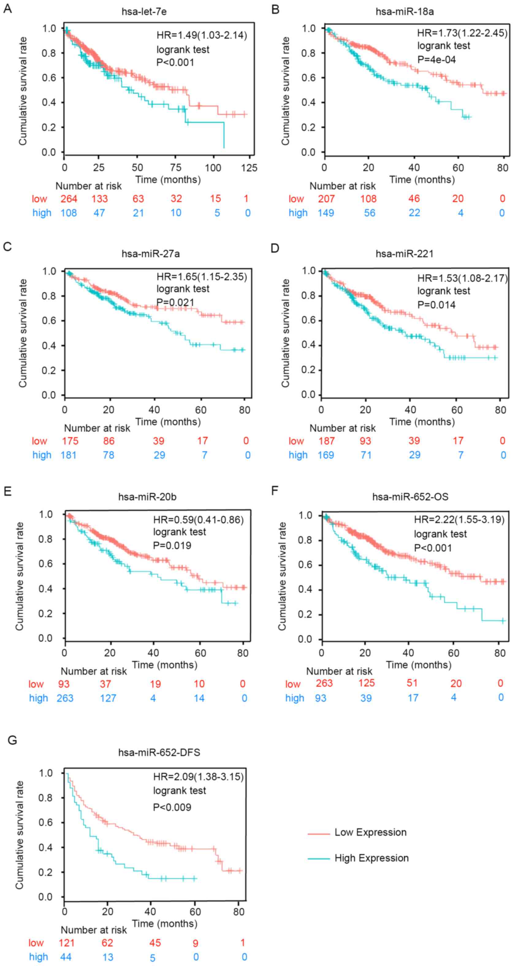
Candidate exosomal miRNAs are
associated with poor prognosis in patients with HCC
Kaplan-Meier analysis was used to determine whether
the candidate miRNAs were associated with OS or disease-free
survival (DFS) in patients with HCC using follow-up data collected
for 80 months. As shown in Fig. 6,
high expression levels of let-7e, miR-18a, miR-27a, miR-221,
miR-20b and miR-652 were significantly associated with poor OS
(Fig. 6A-F). Furthermore, high
expression levels of miR-652 were associated with shorter DFS
(Fig. 6G). However, the other miRNAs
did not exhibit significant associations with DFS based on the
results of the analysis using the Kaplan-Meier plotter tool (data
not shown). These results demonstrated that the upregulation of the
six exosomal miRNAs was significantly associated with poor survival
and increased risk of metastasis in HCC.
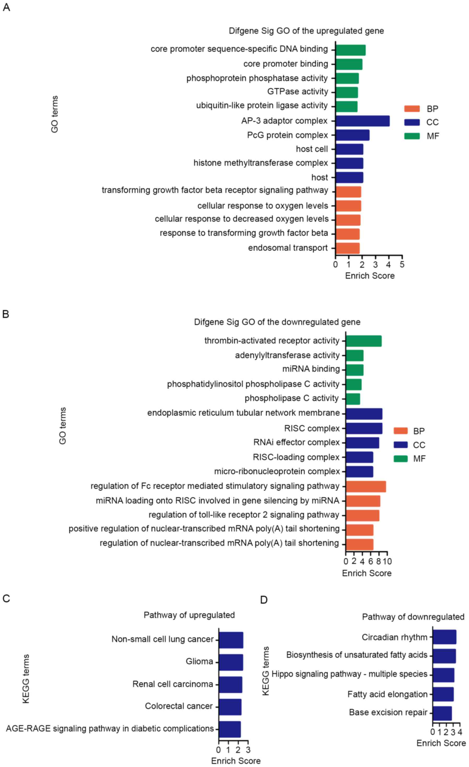
Bioinformatics prediction and
functional analysis of exosomal miRNAs in HCC metastasis
The target genes and signaling pathways associated
with the candidate miRNAs were identified using bioinformatics
tools. The results were summarized and shown in Tables SII and III). The GO analysis of target gene was
mainly classified into three functional groups: Cellular component
(CC), molecular function (MF) and biological process (BP). GO
enrichment analysis of upregulated miRNAs suggested that most
significantly predicted target genes were involved in ‘transforming
growth factor beta receptor signaling pathway (BP)’, ‘response to
transforming growth factor beta (BP)’, ‘AP-3 adaptor complex (CC)’,
‘PcG protein complex (CC)’ and ‘core promoter sequence-specific DNA
binding (MF)’, and these were among the top 5 BP, CC and MF terms
(Fig. 7A; Table SII).
KEGG pathway analysis of upregulated miRNAs revealed
that the top 5 enriched signaling pathways were ‘Non-small cell
lung cancer’, ‘Glioma’, ‘Renal cell carcinoma’ and ‘colorectal
cancer’ (Fig. 7C; Table SII).
Furthermore, GO enrichment analysis of downregulated
miRNAs revealed that most predicted target genes were involved in
‘regulation of the Fc receptor mediated stimulatory signaling
pathway’, ‘endoplasmic reticulum tubular network membrane’, ‘RISC
complex’, ‘thrombin-activated receptor activity’ and ‘miRNA loading
onto RISC involved in gene silencing by miRNA’ (Fig. 7B; Table
SIII).
In addition, KEGG pathway analysis of downregulated
miRNAs suggested that the top 5 enriched pathways were ‘Circadian
rhythm’, ‘Biosynthesis of unsaturated fatty acids’ and ‘Hippo
signaling pathway’ (Fig. 7D;
Table SIII). To study the
regulatory association between the miRNAs and target genes, a
miRNA-mRNA network was constructed for the upregulated miRNAs
(Fig. 8). Collectively, these
findings suggested that the six upregulated exosomal miRNAs were
involved in regulating physiological processes associated with
metastasis.
Discussion
In the present study, plasma exosomal miRNA
expression was evaluated in patients with HCC with and without
metastasis. A total of 32 exosomal miRNAs were differentially
expressed between patients with metastatic and non-metastatic HCC.
Among the upregulated miRNAs, the expression levels of six miRNAs
were consistent between plasma exosome samples and matched
metastatic tissue samples. When comparing the diagnostic value of
individual and combined biomarkers, different combination
strategies were considered and the combinations were compared with
each individual biomarker. Comparisons of AUC values were based on
Delong's test and the P-value was adjusted using the Bonferroni
strategy for multiple comparisons. The results demonstrated that
miR-18a, miR-27a and miR-20b had a high AUC, and the combination of
miR-18a, miR-20b and miR-221 exhibited improved performance
compared with single miRNA expression. The combined panel had an
AUC of 0.9040 in discriminating metastatic cases from
non-metastatic cases. Furthermore, high expression levels of
let-7e, miR-18a, miR-27a, miR-221, miR-20b and miR-652 were
associated with poor OS in patients with HCC.
The aforementioned process was the discovery step.
To verify the hypothesis with the help of the public databases, two
databases, namely, The Cancer Genome Atlas and GEO, were searched
to identify datasets with specific clinical information (patients
with HCC with lung-metastasis and without metastasis who did not
undergo chemoradiotherapy before surgery). Unfortunately, only the
GSE67140 dataset in the GEO database met the inclusion criteria.
The other datasets were either too small or scattered to utilize.
In the next step, large samples and corresponding follow-up data
will be collected to verify the results of OS, DFS and ROC curve
results, and to assess the possibility of plasma exosomal miRNA
markers for diagnosis or prognosis of patients with HCC.
Additionally, clinicopathologic variables, including tumor
diameter, number of tumor nodules, histopathologic classification,
vein invasion and clinical TNM classification, are also being
collected.
Let-7e expression has previously been reported to be
downregulated in several human cancer types, and acts as a tumor
suppressor that promotes apoptosis and inhibits proliferation,
migration and invasion (27,28). By contrast, in the present study, the
expression levels of let-7e were increased in plasma from patients
with metastatic HCC and high let-7e levels predicted shorter OS.
These findings are inconsistent with previous studies (29,30) and
require further study. The predicted target genes of let-7e were
identified using the miRanda database. High mobility group AT-hook
2 (HMGA2) was among the identified target genes. According to the
literature, HMGA2 mediates epithelial-mesenchymal transition (EMT)
and regulates transcription factors linking various signaling
pathways, such as the TGF-β and MAPK signaling pathways, to
regulators of tumor invasiveness and metastasis (31).
miR-18a regulates metastasis in gastric cancer
(32), colon cancer (33), breast cancer (34), HCC and nasopharyngeal carcinoma
(35,36). In the present study, miR-18a
demonstrated a good diagnostic value in discrimination of
metastasis. PTEN was among the predicted targets of miR-18a
according to the bioinformatics analysis. A previous study has
indicated that PTEN-dependent signaling pathways can drive EMT and
consequently increase migration, invasion and metastasis (37).
miR-27a influences tumorigenesis, tumor cell
proliferation, apoptosis, invasion, migration and angiogenesis by
regulating various target genes and could affect clinical therapy,
drug sensitivity of patients and patient prognosis (38). Additionally, miR-27a can act as a
promising biomarker in serum and is a potential therapeutic target
in various tumor types (39). In the
present study, miR-27a could accurately predict HCC metastasis.
Bioinformatics target prediction demonstrated miR-27a could target
SMAD4, a gene involved in the TGF-β signaling pathway. SMAD4/TGF-β
signaling is a pivotal regulator of tumor invasion and metastasis
following EMT (40). The present
study demonstrated that miR-27a served an important role in tumor
metastasis.
Dysregulated miR-20b levels are associated with
metastasis of breast cancer, colorectal cancer and gastric cancer
(41). However, to the best of our
knowledge, the potential of miR-20b as an HCC biomarker has not
been reported. In the present study, miR-20b could discriminate
metastatic HCC from non-metastatic HCC and could be used as a
predictive biomarker of HCC metastasis. Target prediction analysis
identified STAT3 as a miR-20b target. STAT3 signaling is central to
the regulation of metastasis via EMT (42).
miR-652 is defined as an oncomir and can promote
metastasis in endometrial cancer, pancreatic cancer, non-small cell
lung cancer and prostate cancer cells (43). In the present study, ROC analysis of
miR-652 did not demonstrate a high diagnostic accuracy. However,
the DFS and OS rates of patients with high miR-652 expression were
significantly worse than those of patients with low miR-652
expression.
Finally, bioinformatics tools were used to analyze
the possible mechanisms involved in HCC metastasis in the exosomes.
The results suggested that the dysregulated miRNAs were involved in
various signaling pathways, including ‘GTPase activity’ and ‘cell
adhesion molecule binding’, which constitute a complex regulatory
network that is relevant to metastasis. An miRNA-mRNA regulatory
network was also constructed to examine the associations among the
targets of the differentially expressed miRNAs. Nevertheless,
bioinformatics prediction can only provide clues for elucidating
the role of miRNAs in HCC metastasis and the possible underlying
mechanisms. Further experimental validation is still necessary to
clarify the detailed regulatory relationship between miRNAs and
their target genes in the future.
In conclusion, to the best of our knowledge, the
present study was the first to investigate metastasis-associated
miRNA profiles in plasma exosomes from patients with HCC.
Furthermore, comprehensive analysis of this profile, together with
target prediction and survival analysis provided novel insights
into the role of exosomes in HCC lung metastasis. A panel
consisting of three miRNAs that might serve as an accurate and
non-invasive tool for HCC metastasis prediction using plasma
exosomes was identified. The present findings suggested that
exosomal miRNAs served important roles in HCC metastasis and could
represent a complementary clinical tool for the assessment of HCC
prognosis.
Supplementary Material
Supporting Data
Acknowledgements
The authors would like to thank Professor Zhao Liang
and Professor Lin Jie (Southern Medical University, Guangzhou,
China) for linguistic advice and experimental assistance.
Funding
The present study was supported by grants from the
President Foundation of Nanfang Hospital, Southern Medical
University (grant no. 2017B005) and the Natural Science Foundation
of Guangdong Province, China (grant no. 2014A030310425).
Availability of data and materials
The datasets used and/or analyzed during the current
study are available from the corresponding author on reasonable
request.
Authors' contributions
BX, CH and ST conceived the study and were
responsible for confirming the authenticity of all raw data. CH and
ST performed outpatient service, obtained the informed consents,
preserved the samples, and performed analysis and interpretation of
data. Experiments were performed by BX, ST, CH and XL. BX and CH
drafted the manuscript. DS, XL and LL organized the tables and
prepared the figure. Data were analyzed by LL, YD and DS. All
authors read and approved the final manuscript.
Ethics approval and consent to
participate
The Ethics Committee of Southern Medical University
Nanfang Hospital (Guangzhou, China) approved the present study. All
patients provided written informed consent.
Patient consent for publication
Not applicable.
Competing interests
The authors declare that they have no competing
interests.
References
|
1
|
Bertuccio P, Turati F, Carioli G,
Rodriguez T, La Vecchia C, Malvezzi M and Negri E: Global trends
and predictions in hepatocellular carcinoma mortality. J Hepatol.
67:302–309. 2017. View Article : Google Scholar : PubMed/NCBI
|
|
2
|
Kulik L and El-Serag HB: Epidemiology and
management of hepatocellular carcinoma. Gastroenterology.
156:477–491.e1. 2019. View Article : Google Scholar : PubMed/NCBI
|
|
3
|
Lee EC, Kim SH, Park H, Lee SD, Lee SA and
Park SJ: Survival analysis after liver resection for hepatocellular
carcinoma: A consecutive cohort of 1002 patients. J Gastroenterol
Hepatol. 32:1055–1063. 2017. View Article : Google Scholar : PubMed/NCBI
|
|
4
|
Di Tommaso L, Spadaccini M, Donadon M,
Personeni N, Elamin A, Aghemo A and Lleo A: Role of liver biopsy in
hepatocellular carcinoma. World J Gastroenterol. 25:6041–6052.
2019. View Article : Google Scholar : PubMed/NCBI
|
|
5
|
Sersté T, Barrau V, Ozenne V, Vullierme
MP, Bedossa P, Farges O, Valla DC, Vilgrain V, Paradis V and Degos
F: Accuracy and disagreement of computed tomography and magnetic
resonance imaging for the diagnosis of small hepatocellular
carcinoma and dysplastic nodules: Role of biopsy. Hepatology.
55:800–806. 2012. View Article : Google Scholar : PubMed/NCBI
|
|
6
|
Russo FP, Imondi A, Lynch EN and Farinati
F: When and how should we perform a biopsy for HCC in patients with
liver cirrhosis in 2018? A review. Dig Liver Dis. 50:640–646. 2018.
View Article : Google Scholar : PubMed/NCBI
|
|
7
|
Mathieu M, Martin-Jaular L, Lavieu G and
Thèry C: Specificities of secretion and uptake of exosomes and
other extracellular vesicles for cell-to-cell communication. Nat
Cell Biol. 21:9–17. 2019. View Article : Google Scholar : PubMed/NCBI
|
|
8
|
Li I and Nabet BY: Exosomes in the tumor
microenvironment as mediators of cancer therapy resistance. Mol
Cancer. 18:322019. View Article : Google Scholar : PubMed/NCBI
|
|
9
|
Li S, Yi M, Dong B, Tan X, Luo S and Wu K:
The role of exosomes in liquid biopsy for cancer diagnosis and
prognosis prediction. Int J Cancer. Nov 12–2020.(Epub ahead of
print). doi: 10.1002/ijc.33386, 2020. View Article : Google Scholar
|
|
10
|
Li X, Li C, Zhang L, Wu M, Cao K, Jiang F,
Chen D, Li N and Li W: The significance of exosomes in the
development and treatment of hepatocellular carcinoma. Mol Cancer.
19:12020. View Article : Google Scholar : PubMed/NCBI
|
|
11
|
Bartel DP: Metazoan MicroRNAs. Cell.
173:20–51. 2018. View Article : Google Scholar : PubMed/NCBI
|
|
12
|
Yu X, Odenthal M and Fries JW: Exosomes as
miRNA carriers: Formation-function-future. Int J Mol Sci.
17:20282028. View Article : Google Scholar
|
|
13
|
Kai K, Dittmar RL and Sen S: Secretory
microRNAs as biomarkers of cancer. Semin Cell Dev Biol. 78:22–36.
2018. View Article : Google Scholar : PubMed/NCBI
|
|
14
|
Wortzel I, Dror S, Kenific CM and Lyden D:
Exosome-mediated metastasis: Communication from a distance. Dev
Cell. 49:347–360. 2019. View Article : Google Scholar : PubMed/NCBI
|
|
15
|
Kahroba H, Hejazi MS and Samadi N:
Exosomes: From carcinogenesis and metastasis to diagnosis and
treatment of gastric cancer. Cell Mol Life Sci. 76:1747–1758. 2019.
View Article : Google Scholar : PubMed/NCBI
|
|
16
|
Lin XJ, Chong Y, Guo ZW, Xie C, Yang XJ,
Zhang Q, Li SP, Xiong Y, Yuan Y, Min J, et al: A serum microRNA
classifier for early detection of hepatocellular carcinoma: A
multicentre, retrospective, longitudinal biomarker identification
study with a nested case-control study. Lancet Oncol. 16:804–815.
2015. View Article : Google Scholar : PubMed/NCBI
|
|
17
|
Zeng Z, Li Y, Pan Y, Lan X, Song F, Sun J,
Zhou K, Liu X, Ren X, Wang F, et al: Cancer-derived exosomal
miR-25-3p promotes pre-metastatic niche formation by inducing
vascular permeability and angiogenesis. Nat Commun. 9:53952018.
View Article : Google Scholar : PubMed/NCBI
|
|
18
|
Tian XP, Huang WJ, Huang HQ, Liu YH, Wang
L, Zhang X, Lin TY, Rao HL, Li M, Liu F, et al: Prognostic and
predictive value of a microRNA signature in adults with T-cell
lymphoblastic lymphoma. Leukemia. 33:454–2465. 2019. View Article : Google Scholar
|
|
19
|
Livak KJ and Schmittgen TD: Analysis of
relative gene expression data using real-time quantitative PCR and
the 2(-Delta Delta C(T)) method. Methods. 25:402–408. 2001.
View Article : Google Scholar : PubMed/NCBI
|
|
20
|
Lou W, Chen J, Ding B, Chen D, Zheng H,
Jiang D, Xu L, Bao C, Cao G and Fan W: Identification of
invasion-metastasis-associated microRNAs in hepatocellular
carcinoma based on bioinformatic analysis and experimental
validation. J Transl Med. 16:2662018. View Article : Google Scholar : PubMed/NCBI
|
|
21
|
Nagy Á, Lánczky A, Menyhárt O and Győrffy
B: Validation of miRNA prognostic power in hepatocellular carcinoma
using expression data of independent datasets. Sci Rep. 8:92272018.
View Article : Google Scholar : PubMed/NCBI
|
|
22
|
Yan S, Han X, Xue H, Zhang P, Guo X, Li T,
Guo X, Yuan G, Deng L and Li G: Let-7f Inhibits glioma cell
proliferation, migration, and invasion by targeting periostin. J
Cell Biochem. 116:1680–1692. 2015. View Article : Google Scholar : PubMed/NCBI
|
|
23
|
Ashburner M, Ball CA, Blake JA, Botstein
D, Butler H, Cherry JM, Davis AP, Dolinski K, Dwight SS, Eppig JT,
et al: Gene ontology: Tool for the unification of biology. The Gene
Ontology Consortium. Nat Genet. 25:25–29. 2000. View Article : Google Scholar : PubMed/NCBI
|
|
24
|
Chen BS and Wu CC: Systems biology as an
integrated platform for bioinformatics, systems synthetic biology,
and systems metabolic engineering. Cells. 2:635–688. 2013.
View Article : Google Scholar : PubMed/NCBI
|
|
25
|
Robin X, Turck N, Hainard A, Tiberti N,
Lisacek F, Sanchez JC and Müller M: pROC: An open-source package
for R and S+ to analyze and compare ROC curves. BMC Bioinformatics.
12:772011. View Article : Google Scholar : PubMed/NCBI
|
|
26
|
Zou W, Lai M, Zhang Y, Zheng L, Xing Z, Li
T, Zou Z, Song Q, Zhao X, Xia L, et al: Exosome release is
regulated by mTORC1. Adv Sci (Weinh). 6:18013132018. View Article : Google Scholar : PubMed/NCBI
|
|
27
|
Ding C, Yu H, Shi C, Shi T, Qin H and Cui
Y: MiR-let-7e inhibits invasion and magration and regulates HMGB1
expression in papillary thyroid carcinoma. Biomed Pharmacother.
110:528–536. 2019. View Article : Google Scholar : PubMed/NCBI
|
|
28
|
Wang S, Jin S, Liu MD, Pang P, Wu H, Qi
ZZ, Liu FY and Sun CF: Hsa-let-7e-5p Inhibits the proliferation and
metastasis of head and neck squamous cell carcinoma cells by
targeting chemokine receptor 7. J Cancer. 10:1941–1948. 2019.
View Article : Google Scholar : PubMed/NCBI
|
|
29
|
Liu Y, Yusheng Z, Qiang C, Zhen W and
Boshi S: Methyltransferase-like 1 (METTL1) served as a tumor
suppressor in colon cancer by activating 7-methyguanosine (m7G)
regulated let-7e miRNA/HMGA2 axis. Life Sci. 249:117482020.
View Article : Google Scholar
|
|
30
|
Bruna C, Laura G, Miyuki U, Isabela W,
Fernando A, Glauco B, Edmund C and Katia C: Let-7 miRNA's
expression profile and its potential prognostic role in uterine
leiomyosarcoma. Cells. 8:1452–1463. 2019. View Article : Google Scholar
|
|
31
|
Hou M, Bao X, Luo F, Chen X, Liu L and Wu
M: HMGA2 modulates the TGFβ/Smad, TGFβ/ERK and notch signaling
pathways in human lens epithelial-mesenchymal transition. Curr Mol
Med. 18:71–82. 2018. View Article : Google Scholar : PubMed/NCBI
|
|
32
|
Yuan J, Tan L, Yin Z, Zhu W, Tao K, Wang
G, Shi W and Gao J: MIR17HG-miR-18a/19a axis, regulated by
interferon regulatory factor-1, promotes gastric cancer metastasis
via Wnt/β-catenin signalling. Cell Death Dis. 10:4542019.
View Article : Google Scholar : PubMed/NCBI
|
|
33
|
Rammer M, Webersinke G, Haitchi-Petnehazy
S, Maier E, Hackl H, Charoentong P, Malli T, Steinmair M, Petzer AL
and Rumpold H: MicroRNAs and their role for T stage determination
and lymph node metastasis in early colon carcinoma. Clin Exp
Metastasis. 34:431–440. 2017. View Article : Google Scholar : PubMed/NCBI
|
|
34
|
Wang P, Yang Q, Du X, Chen Y and Zhang T:
Targeted regulation of Rell2 by microRNA-18a is implicated in the
anti-metastatic effect of polyphyllin VI in breast cancer cells.
Eur J Pharmacol. 851:161–173. 2019. View Article : Google Scholar : PubMed/NCBI
|
|
35
|
Liu L, Cai X, Liu E, Tian X and Tian C:
MicroRNA-18a promotes proliferation and metastasis in
hepatocellular carcinoma via targeting KLF4. Oncotarget.
8:68263–68269. 2017. View Article : Google Scholar : PubMed/NCBI
|
|
36
|
Chen X, Wang J, Cheng L and Lu MP: miR-18a
downregulates DICER1 and promotes proliferation and metastasis of
nasopharyngeal carcinoma. Int J Clin Exp Med. 7:847–855.
2014.PubMed/NCBI
|
|
37
|
Cai L, Ye Y, Jiang Q, Chen Y, Lyu X, Li J,
Wang S, Liu T, Cai H, Yao K, et al: Epstein-Barr virus-encoded
microRNA BART1 induces tumour metastasis by regulating
PTEN-dependent pathways in nasopharyngeal carcinoma. Nat Commun.
6:73532015. View Article : Google Scholar : PubMed/NCBI
|
|
38
|
Luo W, Zhang D, Ma S, Wang C, Zhang Q,
Wang H, He K and Liu Z: miR-27a is highly expressed in H1650 cancer
stem cells and regulates proliferation, migration, and invasion. J
Cancer Res Ther. 14 (Suppl 1):S1004–S1011. 2018. View Article : Google Scholar : PubMed/NCBI
|
|
39
|
Liu X, Pan B, Sun L, Chen X, Zeng K, Hu X,
Xu T, Xu M and Wang S: Circulating exosomal miR-27a and miR-130a
act as novel diagnostic and prognostic biomarkers of colorectal
cancer. Cancer Epidemiol Biomarkers Prev. 27:746–754. 2018.
View Article : Google Scholar : PubMed/NCBI
|
|
40
|
Siraj AK, Pratheeshkumar P, Divya SP,
Parvathareddy SK, Bu R, Masoodi T, Kong Y, Thangavel S, Al-Sanea N,
Ashari LH, et al: TGFβ-induced SMAD4-dependent apoptosis proceeded
by EMT in CRC. Mol Cancer Ther. 18:1312–1322. 2019. View Article : Google Scholar : PubMed/NCBI
|
|
41
|
Lu P, Gu Y, Li L, Wang F, Yang X and Yang
Y: Long noncoding RNA CAMTA1 promotes proliferation and mobility of
the human breast cancer cell line MDA-MB-231 via targeting miR-20b.
Oncol Res. 26:625–635. 2018. View Article : Google Scholar : PubMed/NCBI
|
|
42
|
Wendt MK, Balanis N, Carlin CR and
Schiemann WP: STAT3 and epithelial-mesenchymal transitions in
carcinomas. JAKSTAT. 3:e289752014.PubMed/NCBI
|
|
43
|
Yang W, Zhou C, Luo M, Shi X, Li Y, Sun Z,
Zhou F, Chen Z and He J: MiR-652-3p is upregulated in non-small
cell lung cancer and promotes proliferation and metastasis by
directly targeting Lgl1. Oncotarget. 7:16703–16715. 2016.
View Article : Google Scholar : PubMed/NCBI
|