Introduction
Esophageal cancer is a common malignancy of the
upper digestive tract, ranking 7th and 6th for mortality and
morbidity, respectively, among all types of cancer worldwide in
2018 (1). China has one of the
highest incidence rates of esophageal cancer globally (1), with a crude incidence rate of
17.87/100,000 individuals (2).
However, marked regional and ethnic differences exist in the
distribution of esophageal cancer in China (3). Xinjiang is among the regions with the
highest incidence of esophageal cancer (4), and has high morbidity and mortality
rates; in particular, the Kazakh population in Xinjiang has a
mortality rate of 68.88/100,000, which is extremely high for this
region (5,6). The early symptoms of esophageal cancer
are not easy to distinguish clinically and most patients are
diagnosed at a late stage, while esophageal cancer is prone to
metastasis at an early stage due to its extensive lymphatic
drainage network (7). At present,
the treatment of esophageal cancer mainly comprises surgery
combined with neoadjuvant radiotherapy and chemotherapy (8). However, the 5-year survival rate of
esophageal cancer is <20% (9),
and most patients present with recurrence or metastasis following
treatment. Additionally, >90% of cases present as esophageal
squamous cell carcinoma (ESCC) in Asian countries (3). Therefore, in order to develop more
effective treatment methods for advanced esophageal cancer, it
would be helpful to identify the genes associated with the
occurrence and development of ESCC, clarify their mechanism of
action and explore potential specific therapeutic targets.
Commonly occurring biological changes in the
pathogenesis of ESCC include the activation of oncogenes,
inactivation of tumor suppressor genes and changes in molecular
markers associated with the etiology of ESCC (10). Research into ESCC has increasingly
focused on tumor stem cells. Twist, Slug and Snail have been found
to endow tumor cells with stem cell characteristics, and play
important roles in epithelial-mesenchymal transition (EMT). EMT is
a complex process by which epithelial cells transform into stromal
cells, and is important in embryonic development, chronic
inflammation, tissue reconstruction, cancer invasion and
metastasis, and various fibrotic diseases. Its main manifestations
are the decreased expression of cell adhesion molecules such as
mucoproteins and E-cadherin, remodeling of the cytoskeleton with
vimentin enrichment, and the development of interstitial cell
characteristics. When EMT occurs, epithelial cells lose polarity,
become disconnected from the basement membrane and acquire
characteristics typical of interstitial cells, including high
migration and invasion activities, resistance to apoptosis and
degradation of the extracellular matrix (11). E-cadherin is a calcium-dependent
transmembrane protein, which is involved in the maintenance of cell
integrity and polarity; the loss of its expression results in
reduced cell adhesion and increased invasiveness (12). Classical EMT transcription factors,
including Twist, Slug and Snail, are able to downregulate the
expression of E-cadherin and induce EMT, thereby promoting the
invasiveness of tumors (13).
Twist is a transcription factor with a basic
helix-loop-helix protein structure (14). It plays an important role in the
development of the embryo, and also induces mesenchymal markers
during tumor progression and participates in the EMT of certain
epithelium-derived tumor cells. The underlying mechanism involves
the binding of Twist to the E-box element in the promoter region of
the E-cadherin gene, which inhibits the activity of the promoter
and leads to a reduction in the transcription of E-cadherin; this
then induces EMT, which increases the migration, invasion and
metastasis of tumor cells (15,16).
Twist is known to play a role in a variety of invasive cancers,
including breast (17), lung
(18) and prostate cancer (19).
Snail and Slug are members of the Snail family,
which consists of three members: Snail, Slug (Snail2) and Snail3.
The Snail family members are similar in structure, each comprising
a carboxyl end with a C2H2 zinc finger
structure (20). These conserved
zinc finger sequences are composed of two β-folds and an α-helix. A
specific sequence of the zinc finger can combine with the E-box
CACGTG sequence in the promoter region of a downstream target gene
so as to regulate the expression of the gene. As a key
transcriptional inhibitor of E-cadherin expression in EMT, Snail
plays important roles in embryonic development and tumor
progression. Snail imbues tumor cells with characteristics similar
to those of cancer stem cells, and promotes drug resistance, tumor
recurrence and metastasis (21). In
a variety of tumor types, the increased expression of Snail
positively correlates with tumor metastasis, as well as drug and
immune tolerance, and suggests a poor prognosis. Therefore, Snail
may be therapeutically targeted for the inhibition of tumor
progression (22,23). Slug is a classic EMT transcription
factor, which combines with promoter element (E-box) of the
downstream gene E-cadherin and directly inhibits E-cadherin
expression, thereby reducing cell adhesion and cell polarity
(24), inducing EMT and promoting
the development of tumors. Slug has been reported to be associated
with various biological functions of tumors, including invasion and
migration (25).
Twist, Slug and Snail may play important roles in
the development of ESCC. The aim of the present study was to detect
the expression of Twist, Slug and Snail in ESCC by
immunohistochemistry and reverse transcription-quantitative
polymerase chain reaction (RT-qPCR), analyze the associations
between Twist, Slug and Snail and clinicopathological parameters,
and determine their impact on the prognosis of ESCC, in order to
provide a scientific basis for improving the prognosis of patients
with ESCC.
Materials and methods
Patients
The paraffin-embedded ESCC tissues, matched normal
esophageal mucosal tissues and clinicopathological parameters of
229 cases of ESCC were collected from the Department of Pathology
of the First Affiliated Hospital of Xinjiang Medical University
(Xinjiang, China) between January 2014 and December 2018. Among
them, 115 were of Han ethnicity and 114 were of Kazakh ethnicity.
The age range of the cohort was 32–84 years (median age, 60 years).
The cases were assigned to the following groups: Age (≤60 or >60
years), sex (male or female), ethnicity (Han or Kazak), tumor
location (upper, middle or lower segment), tumor size (<3 or ≥3
cm), degree of differentiation (high, medium or low), depth of
invasion (mucosal, muscular or whole layer), lymph node metastasis
(yes or no), vascular invasion (yes or no), nerve invasion (yes or
no); and TNM stage (I, II, III or IV). A total of 29 matched pairs
of fresh frozen ESCC and normal esophageal mucosal tissues between
October 2007 and December 2018 were collected the specimen bank of
Xinjiang Medical University. The 229 patients with
paraffin-embedded ESCC tissues were followed up by telephone and
their details were included in the inpatient log. The start date of
follow-up was the date when the patient was diagnosed with ESCC,
and the end date was July 1, 2019. The 29 cases with fresh frozen
tissues were not followed up.
Immunohistochemical staining
The 229 paraffin-embedded ESCC and normal mucosal
tissues were made into tissue chips, sliced into 4-µm sections,
deparaffinized in xylene and rehydrated in 100, 95, 80 and 70%
ethanol. Following treatment with 3% hydrogen peroxide to block
endogenous peroxidase activity, the sections were heated with
citric acid (pH 6; Snail and Slug analysis) or EDTA (pH 9; Twist
analysis) in boiling water at 100°C for antigen retrieval. The
sections were then treated with goat serum (Blocking normal sheep
serum; cat. no. ZLI-9022; undiluted; Beijing Zhongshan Jinqiao
Biotechnology Co., Ltd.) at room temperature to block non-specific
antigens for 15 min. Subsequently, the sections were incubated with
rat anti-human Twist (1:50; cat. no. ab175430; Abcam) overnight at
4°C for 10 h, or rat anti-human Slug (1:100; cat. no. pb9439;
Boster Biological Technology) and goat anti-human Snail antibodies
(1:800; cat. no. ab53519; Abcam) at 37°C for 60 min. Next, the
tissue chips were washed with PBS and the sections were incubated
with secondary antibodies (universal kit, cat. no. PV-6000,
undiluted for Twist and Slug; goat two step detection kit, cat. no.
PV-9003, undiluted for Snail; both kits from Beijing Zhongshan
Jinqiao Biotechnology Co., Ltd.) for 30 min at 37°C. The slides
were subsequently stained with 3,3′-diaminobenzidine, dehydrated,
sealed and observed under a light microscope (DM300; Leica
Microsystems GmbH; magnifications, ×4, ×10 and ×20). The staining
strength was scored as follows: 0, no staining; 1, weak staining;
2, medium staining); and 3, strong staining. The percentage of
positive cells was scored as follows: 0, <5; 1, 5–25; 2, 26–50;
and 3, >50%. The staining index was calculated by multiplying
the score for the percentage of positive cells by the score for
staining strength. If the staining index calculation was <6, the
expression level was defined as negative expression, otherwise, it
was defined as positive expression. The final results were
interpreted by two senior pathologists. If there was a difference
in opinion, the results were judged by a third senior
pathologist.
RT-qPCR
The 29 fresh ESCC tissues and their matched normal
mucosa tissues were stored in a refrigerator at −80°C. Total RNA
was extracted by grinding 200 mg tissue in a liquid nitrogen
environment, adding 1 ml TRIzol® (Invitrogen; Thermo
Fisher Scientific, Inc.) and collecting the extract in a 1.5-ml
Eppendorf (EP) tube. Vortex oscillation was performed for 30 sec.
Following the addition of chloroform (0.2 ml), the tube was shaken
vigorously for 30 sec and kept at room temperature for 10 min.
Centrifugation was then performed at 12,000 × g at 4°C for 15 min.
The colorless aqueous upper layer (~0.5 ml) was transferred to
another EP tube, combined with an equal volume of isopropanol, kept
at room temperature for 10 min, and then centrifuged at 12,000 × g
and 4°C for 10 min. The supernatant was discarded, 1 ml 75% ethanol
was added to the RNA precipitate, and the mixture was shaken prior
to centrifugation at 7,500 × g and 4°C for 10 min. After discarding
the supernatant, the residual liquid was dried at room temperature
for 5–10 min. The purity of the resulting precipitate was
determined by calculation of the ratio of optical densities at 260
and 280 nm. RT was conducted using the RevertAid First Strand cDNA
Synthesis kit (cat. no. K1622; Thermo Fisher Scientific, Inc.) at
42°C for 60 min and then 72°C for 5 min. Following RT, the cDNA was
stored at −20°C until required for qPCR. SYBR Green dye (QuantiNova
SYBR Green PCR kit; cat. no. 208054; Qiagen China Co., Ltd.) and a
7500 Real-Time PCR system (Applied Biosystems; Thermo Fisher
Scientific, Inc.) were used to detect the expression of the target
genes. The primers used are shown in Table I. β-actin was used as the reference
gene. All reactions were carried out three times under the
following conditions: Activation of the polymerase for 2 min at
50°C, initial denaturation for 10 min at 94°C, followed by 40
cycles of denaturation for 15 sec at 94°C, and annealing and
elongation for 30 sec at 60°C. The mRNA expression of the target
gene was calculated using the 2−ΔΔCq method (26).
 | Table I.Quantitative polymerase chain
reaction primer pairs for marker molecule analysis. |
Table I.
Quantitative polymerase chain
reaction primer pairs for marker molecule analysis.
| Molecule | Primer sequence
(5′-3′) |
|---|
| Twist-F |
GTCCGCAGTCTTACGAGGAG |
| Twist-R |
GCTTGAGGGTCTGAATCGGGCT |
| Slug-F |
GCTACCCAATGGCCTCTCTC |
| Slug-R |
CTTCAATGGCATGGGGGTCT |
| Snail-F |
TCGGAAGCCTAACTACAGCGA |
| Snail-R |
AGATGAGCATTGGCAGCGAG |
| β-actin-F |
CATGTACGTTGCTATCCAGGC |
| β-actin-R |
CTCCTTAATGTCACGCACGAT |
Statistical analysis
SPSS 17.0 statistical software (SPSS, Inc.) was used
to analyze the associations between Twist, Snail and Slug and the
clinicopathological characteristics of the patients with ESCC using
χ2 and Fisher's exact tests. The mRNA expression levels
of Twist, Snail and Slug in ESCC and adjacent tissues were compared
using paired t-tests. The correlations among Twist, Snail, Slug and
their combinations were analyzed by Spearman's rank and Pearson's
correlation analyses. Overall and progression-free survival times
were used to evaluate the prognosis of the patients. The
progression-free survival time was defined as the time from the
diagnosis of esophageal cancer to the time of tumor progression or
death. The overall survival time was defined as the time from the
diagnosis of esophageal cancer to the time of death or final
follow-up (July 1, 2019). The Kaplan-Meier method (single-factor
analysis) and Cox risk proportion model (multi-factor analysis)
were used to analyze the survival and prognosis of the patients
with ESCC. The effects of Twist, Snail and Slug protein expression
and clinicopathological parameters on the prognosis of ESCC were
analyzed by Kaplan-Meier analysis. Based on the results of the
Kaplan-Meier analysis, the independent factors associated with the
prognosis of ESCC were identified and further analyzed using the
Cox proportional hazards model. P<0.05 was considered to
indicate a statistically significant result.
Results
Expression of Twist, Snail and Slug in
ESCC and their association with clinicopathological parameters
Twist, Snail and Slug are important transcription
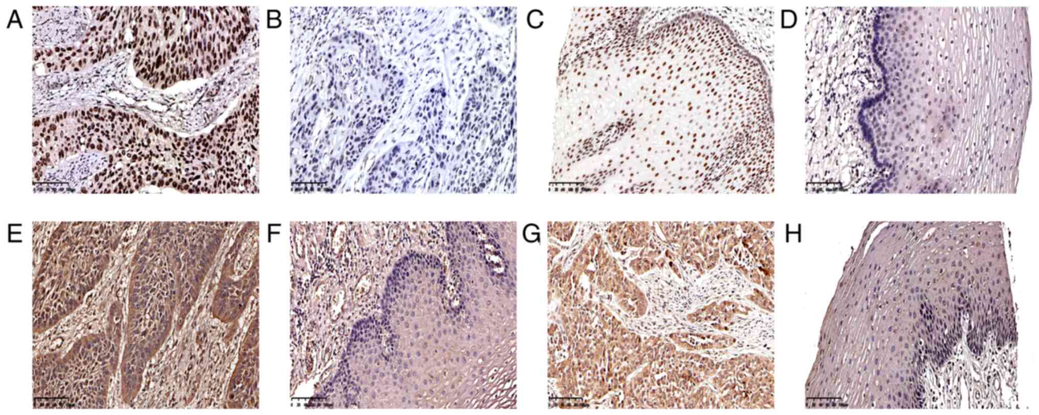
factors in tumors. In ESCC, Twist is expressed in the nucleus,
Snail in the cytoplasm or nucleus, and Slug in the cytoplasm
(Fig. 1). In the 229 cases of ESCC,
135 tumor tissues were positive for Twist (59.0%; Table II and Fig. 1A) and 94 were negative (41.0%;
Table II and Fig. 1B). In the normal esophageal mucosa,
36 cases (15.7%; Table II and
Fig. 1C) were positive for Twist and
193 (84.3%) were negative (Table II
and Fig. 1D). A total of 132 ESCC
specimens were positive for Snail (57.6%; Table II and Fig. 1E), and 97 were negative (42.4%); 112
were positive for Slug (48.9%; Table
II and Fig. 1G), and 117 were
negative (51.1%; Table II). Twist
and Snail were highly expressed in ESCC, but Slug expression was
positive in <50% of the ESCC samples. The expression of Snail
(Table II and Fig. 1F) and Slug (Table II and Fig. 1H) in all specimens of normal
esophageal mucosa was negative, and the frequency of Twist
expression in the ESCC and normal mucosal tissues was significantly
different (P<0.001; Table
II).
| Table II.Expression rates of Twist, Snail and
Slug in ESCC and esophageal normal mucosa. |
Table II.
Expression rates of Twist, Snail and
Slug in ESCC and esophageal normal mucosa.
| A, Twist |
|---|
|
|---|
|
| ESCC |
|
|
|---|
|
|
|
|
|
|---|
| Mucosa | Positive
(n=135) | Negative
(n=94) | P-value | χ2 |
|---|
| Positive
(n=36) | 32 (23.7) | 4 (4.3) | <0.001 | 15.82 |
| Negative
(n=193) | 103 (76.3) | 90 (95.7) |
|
|
|
| B,
Snail |
|
|
| ESCC |
|
|
|
|
|
|
|
| Mucosa | Positive
(n=132) | Negative
(n=97) | P-value |
χ2 |
|
| Positive (n=0) | 0 | 0 | – | – |
| Negative
(n=229) | 132 (57.6) | 97 (42.4) |
|
|
|
| C, Slug |
|
|
| ESCC |
|
|
|
|
|
|
|
| Mucosa | Positive
(n=112) | Negative
(n=117) | P-value |
χ2 |
|
| Positive (n=0) | 0 | 0 | – | – |
| Negative
(n=229) | 112 (48.9) | 117 (51.1) |
|
|
The 229 cases of ESCC included 115 Han and 114
Kazakh individuals. The χ2 test was used to analyze the
associations of Han and Kazakh patients with clinicopathological
parameters (Table III). The
results showed that there were statistically significant
differences in age (P=0.05), tumor location (P=0.039) and vascular
invasion between patients of different ethnicities (P=0.019). The
proportion of Kazakh patients aged ≤60 years was higher than that
of Han. In Kazakh patients, ESCC was more likely to be located in
the middle and lower part of the esophagus, but less likely to be
associated with vascular invasion.
| Table III.Characteristics of 229 patients with
esophageal squamous cell carcinoma. |
Table III.
Characteristics of 229 patients with
esophageal squamous cell carcinoma.
|
|
| N (%) |
|
|
|---|
|
|
|
|
|
|
|---|
| Clinicopathological
parameters | No. | Han | Kazakh | P-value | χ2 |
|---|
| Total | 229 | 115 | 114 |
|
|
| Age, years |
|
|
| 0.050 | 7.799 |
|
≤60 | 84 | 32 (38.1) | 52 (61.9) |
|
|
|
>60 | 145 | 83 (57.2) | 62 (42.8) |
|
|
| Sex |
|
|
| 0.133 | 2.253 |
|
Male | 163 | 87 (53.4) | 76 (46.6) |
|
|
|
Female | 66 | 28 (42.4) | 38 (57.6) |
|
|
| Tumor location |
|
|
| 0.039 | 6.471 |
|
Upper | 11 | 9
(81.8) | 2
(18.2) |
|
|
|
Middle | 136 | 71 (52.2) | 65 (47.8) |
|
|
|
Lower | 82 | 35 (42.7) | 47 (57.3) |
|
|
| Tumor size |
|
|
| 0.918 | 0.011 |
| <3
cm | 67 | 34 (50.7) | 33 (49.3) |
|
|
| ≥3
cm | 162 | 81 (50.0) | 81 (50.0) |
|
|
|
Differentiation |
|
|
| 0.376 | 1.958 |
|
Low | 47 | 25 (53.2) | 22 (46.8) |
|
|
|
Medium | 123 | 65 (52.8) | 58 (47.2) |
|
|
|
High | 59 | 25 (42.4) | 34 (57.6) |
|
|
| Depth of
invasion |
|
|
| 0.242 | 2.840 |
|
Mucosa | 6 | 5
(83.3) | 1
(16.7) |
|
|
|
Muscle | 94 | 45 (47.9) | 49 (52.1) |
|
|
|
Full | 129 | 65 (50.4) | 64 (49.6) |
|
|
| TNM stage |
|
|
| 0.564 | 2.039 |
| I | 16 | 8
(50.0) | 8
(50.0) |
|
|
| II | 143 | 67 (46.9) | 76 (53.1) |
|
|
|
III | 48 | 27 (56.3) | 21 (43.7) |
|
|
| IV | 22 | 13 (59.1) | 9
(40.9) |
|
|
| Lymph node
metastasis |
|
|
| 0.076 | 3.138 |
| No | 152 | 70 (46.1) | 82 (53.9) |
|
|
|
Yes | 77 | 45 (58.4) | 32 (41.6) |
|
|
| Vascular
invasion |
|
|
| 0.019 | 0.891 |
| No | 186 | 93 (50.0) | 93 (50.0) |
|
|
|
Yes | 43 | 22 (51.2) | 21 (48.8) |
|
|
| Nerve invasion |
|
|
| 0.500 | 0.456 |
| No | 179 | 92 (51.4) | 87 (48.6) |
|
|
|
Yes | 50 | 23 (46.0) | 27 (54.0) |
|
|
| Radiotherapy or
chemotherapy |
|
|
| 0.961 | 0.002 |
| No | 149 | 75 (50.3) | 74 (49.7) |
|
|
|
Yes | 80 | 40 (50.0) | 40 (50.0) |
|
|
The χ2 test was used to analyze the
association the expression of Twist, Snail and Slug with the
clinicopathological parameters of the 229 cases of ESCC (Table IV). The results showed that the
expression of Twist was associated with sex (P=0.040). The positive
expression rate of Twist was 63.2% in male patients, which was
higher than that in female patients (48.5%). Slug was associated
with the degree of differentiation (P=0.048), TNM stage (P=0.050)
and vascular invasion (P=0.017). The positive expression rate of
Slug in poorly differentiated ESCC was 44.6%, in ESCC of medium
differentiation was 43.9% and highly differentiated ESCC was 62.7%.
This indicated Slug positivity was most frequent in cases with
highly differentiated ESCC. With regards to TNM stage, the positive
expression rate of Slug was 81.3% in stage I, 48.3% in stage II,
43.8% in stage III and 40.9% in stage IV, indicating that Slug
positivity was most frequent in patients with ESCC at an early TNM
stage. The positive expression rate of Slug in ESCC without
vascular invasion was 52.7%, which was significantly higher than
that with vascular invasion (32.6%). However, no significant
association was detected between the positive expression of Snail
and any of the clinicopathological parameters (Table IV).
| Table IV.Associations between Twist, Snail and
Slug expression and clinicopathological parameters in esophageal
squamous cell carcinoma. |
Table IV.
Associations between Twist, Snail and
Slug expression and clinicopathological parameters in esophageal
squamous cell carcinoma.
|
|
| Twist, n (%) |
|
| Snail, n (%) |
|
| Slug, n (%) |
|
|
|---|
|
|
|
|
|
|
|
|
|
|
|
|
|---|
| Clinicopathological
parameters | No. | Positive | Negative | P-value | χ2 | Positive | Negative | P-value | χ2 | Positive | Negative | P-value | χ2 |
|---|
| Total | 229 | 135 (59.0) | 94 (41.0) |
|
| 132 (57.6) | 97 (42.4) |
|
| 112 (49.0) | 117 (51.0) |
|
|
| Age, years |
|
|
| 0.208 | 1.587 |
|
| 0.661 | 0.192 |
|
| 0.801 | 0.063 |
|
≤60 | 84 | 45 (53.6) | 39 (46.4) |
|
| 50 (59.5) | 34 (40.5) |
|
| 42 (50.0) | 42 (50.0) |
|
|
|
>60 | 145 | 90 (62.1) | 55 (37.9) |
|
| 82 (56.6) | 63 (43.4) |
|
| 70 (48.3) | 75 (51.7) |
|
|
| Sex |
|
|
| 0.040 | 4.198 |
|
| 0.99 | <0.001 |
|
| 0.616 | 0.252 |
|
Male | 163 | 103 (63.2) | 60 (36.8) |
|
| 94 (57.7) | 69 (42.3) |
|
| 78 (47.9) | 85 (52.1) |
|
|
|
Female | 66 | 32 (48.5) | 34 (51.5) |
|
| 38 (57.6) | 28 (42.4) |
|
| 34 (51.5) | 32 (48.5) |
|
|
| Ethnicity |
|
|
| 0.389 | 0.742 |
|
| 0.849 | 0.036 |
|
| 0.466 | 0.531 |
|
Han | 115 | 71 (61.7) | 44 (38.3) |
|
| 67 (58.3) | 48 (41.7) |
|
| 59 (51.3) | 56 (48.7) |
|
|
|
Kazakh | 114 | 64 (56.1) | 50 (43.9) |
|
| 65 (57.0) | 49 (43.0) |
|
| 53 (46.5) | 61 (53.6) |
|
|
| Tumor location |
|
|
| 0.616 | 0.968 |
|
| 0.785 | 0.484 |
|
| 0.269 | 2.625 |
|
Upper | 11 | 5 (45.5) | 6 (54.5) |
|
| 7 (63.6) | 4 (36.4) |
|
| 8 (72.7) | 3 (27.3) |
|
|
|
Middle | 136 | 80 (58.8) | 56 (41.2) |
|
| 76 (55.9) | 60 (44.1) |
|
| 65 (47.5) | 71 (52.2) |
|
|
|
Lower | 82 | 50 (61.0) | 32 (39.0) |
|
| 49 (59.8) | 33 (40.2) |
|
| 39 (47.6) | 43 (52.4) |
|
|
| Tumor size |
|
|
| 0.302 | 1.067 |
|
| 0.441 | 0.593 |
|
| 0.607 | 0.264 |
| <3
cm | 67 | 36 (53.7) | 31 (46.3) |
|
| 36 (53.7) | 31 (46.3) |
|
| 31 (46.3) | 36 (53.7) |
|
|
| ≥3
cm | 162 | 99 (61.1) | 63 (38.9) |
|
| 96 (59.3) | 66 (40.7) |
|
| 81 (50.0) | 81 (50.0) |
|
|
|
Differentiation |
|
|
| 0.652 | 0.856 |
|
| 0.562 | 1.153 |
|
| 0.048 | 6.068 |
|
Low | 47 | 25 (53.2) | 22 (46.8) |
|
| 24 (51.1) | 23 (48.9) |
|
| 21 (44.7) | 26 (55.3) |
|
|
|
Medium | 123 | 75 (61.0) | 48 (39.0) |
|
| 74 (60.2) | 49 (39.8) |
|
| 54 (43.9) | 69 (56.1) |
|
|
|
High | 59 | 35 (59.3) | 24 (40.7) |
|
| 34 (57.6) | 25 (42.4) |
|
| 37 (62.7) | 22 (37.3) |
|
|
| Depth of
invasion |
|
|
| 0.369 | 1.994 |
|
| 0.452 | 1.59 |
|
| 0.197 | 3.248 |
|
Mucosal | 6 | 2 (33.3) | 4 (66.7) |
|
| 3 (50.0) | 3 (50.0) |
|
| 5 (83.3) | 1 (16.7) |
|
|
|
Muscle | 94 | 54 (57.4) | 40 (42.6) |
|
| 50 (53.2) | 44 (46.8) |
|
| 43 (45.7) | 51 (54.3) |
|
|
|
Full | 129 | 79 (61.2) | 50 (38.8) |
|
| 79 (61.2) | 50 (38.8) |
|
| 64 (49.6) | 65 (50.4) |
|
|
| TNM stage |
|
|
| 0.577 | 1.98 |
|
| 0.968 | 0.256 |
|
| 0.050 | 7.797 |
| I | 16 | 7 (43.8) | 9 (56.2) |
|
| 10 (62.5) | 6 (37.5) |
|
| 13 (81.3) | 3 (18.7) |
|
|
| II | 143 | 84 (58.7) | 59 (41.3) |
|
| 82 (57.3) | 61 (42.7) |
|
| 69 (48.3) | 74 (51.7) |
|
|
|
III | 48 | 30 (62.5) | 18 (37.5) |
|
| 28 (58.3) | 20 (41.7) |
|
| 21 (43.8) | 27 (56.2) |
|
|
| IV | 22 | 14 (63.6) | 8 (36.4) |
|
| 12 (52.4) | 10 (47.6) |
|
| 9 (40.9) | 13 (59.1) |
|
|
| Lymph node
metastasis |
|
|
| 0.863 | 0.03 |
|
| 0.695 | 0.154 |
|
| 0.306 | 1.049 |
| No | 152 | 89 (58.6) | 63 (41.4) |
|
| 89 (58.6) | 63 (41.4) |
|
| 78 (51.3) | 74 (48.7) |
|
|
|
Yes | 77 | 46 (59.7) | 31 (40.3) |
|
| 43 (55.8) | 34 (44.2) |
|
| 34 (44.2) | 43 (55.8) |
|
|
| Vascular
invasion |
|
|
| 0.362 | 0.831 |
|
| 0.678 | 0.173 |
|
| 0.017 | 5.664 |
| No | 186 | 107 (57.5) | 79 (42.5) |
|
| 106 (57.0) | 80 (43.0) |
|
| 98 (52.7) | 88 (47.3) |
|
|
|
Yes | 43 | 28 (65.1) | 15 (34.9) |
|
| 26 (60.5) | 17 (39.5) |
|
| 14 (32.6) | 29 (67.3) |
|
|
| Nerve invasion |
|
|
| 0.62 | 0.246 |
|
| 0.481 | 0.498 |
|
| 0.432 | 0.617 |
| No | 179 | 104 (58.1) | 75 (41.9) |
|
| 101 (56.4) | 78 (43.6) |
|
| 90 (50.3) | 89 (49.7) |
|
|
|
Yes | 50 | 31 (62.0) | 19 (38.0) |
|
| 31 (62.0) | 19 (38.0) |
|
| 22 (44.0) | 28 (56.0) |
|
|
| Radiotherapy or
chemotherapy |
|
|
| 0.964 | 0.002 |
|
| 0.151 | 2.057 |
|
| 0.253 | 1.309 |
| No | 149 | 88 (59.1) | 61 (40.9) |
|
| 91 (61.1) | 58 (38.9) |
|
| 77 (51.7) | 72 (48.3) |
|
|
|
Yes | 80 | 47 (58.8) | 33 (41.0) |
|
| 41 (51.3) | 39 (48.7) |
|
| 35 (43.8) | 45 (56.2) |
|
|
Analysis of the associations between
protein and mRNA expression of Twist, Snail and Slug
According to the χ2 test analysis of 229
cases of esophageal squamous cell carcinoma, the positive
expression rate of Twist in patients with Snail positive expression
was 64.4%. In patients with negative Twist expression, the negative
expression rate of Snail was 52.1%, suggesting that there was a
significant difference in protein expression between them in ESCC
(P=0.013; Table V). In addition, in
patients with positive Snail expression, the positive expression
rate of Slug was 62.9%. When Snail was negative, the negative
expression rate of Slug was 70.1%. The results revealed that there
was a difference in the expression of Snail and Slug protein in
ESCC (P<0.001; Table V). However,
there was no significant difference in protein expression between
Twist and Slug (P=0.994; Table
V).
| Table V.Associations between Twist, Snail and
Slug protein expression in esophageal squamous cell carcinoma. |
Table V.
Associations between Twist, Snail and
Slug protein expression in esophageal squamous cell carcinoma.
| A, Twist and
Snail/Slug |
|---|
|
|---|
|
| Twist, n (%) |
|
|
|---|
|
|
|
|
|
|---|
| Protein | Positive | Negative | P-value | χ2 |
|---|
| Snail |
|
| 0.013 | 6.223 |
|
Positive | 87 (64.4) | 45 (47.9) |
|
|
|
Negative | 48 (35.6) | 49 (52.1) |
|
|
| Slug |
|
| 0.994 | 0.001 |
|
Positive | 66 (48.9) | 46 (48.9) |
|
|
|
Negative | 69 (51.1) | 48 (51.1) |
|
|
|
| B, Snail and
Slug |
|
|
| Snail, n
(%) |
|
|
|
|
|
|
|
| Protein |
Positive |
Negative | P-value |
χ2 |
|
| Slug |
|
| <0.001 | 24.340 |
|
Positive | 83 (62.9) | 29 (29.9) |
|
|
|
Negative | 49 (37.1) | 68 (70.1) |
|
|
The results of the analysis of Twist, Snail and Slug
mRNA expression levels in fresh frozen tissues using paired samples
t-tests are shown in Table VI. The
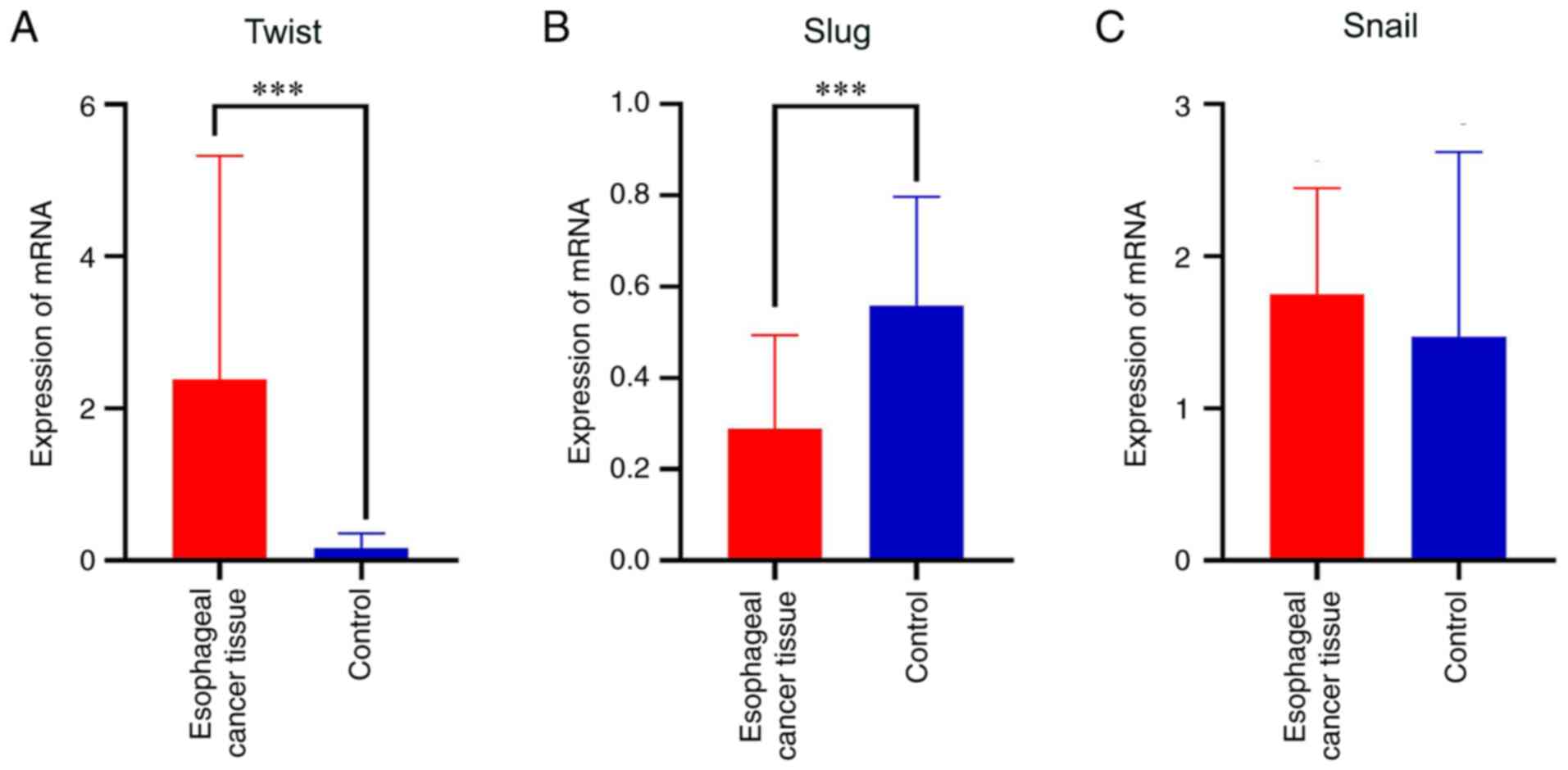
ESCC and adjacent tissues significantly differed in the mRNA
expression of Twist (t=4.044, P<0.001; Table VI and Fig. 2A). The expression of Twist mRNA in
ESCC was significantly higher than that in the adjacent normal
mucosa. The difference in the mRNA expression of transcription
factor Slug was also statistically significant in ESCC (t=−5.687,
P<0.001; Table VI and Fig. 2B); however, the expression of Slug in
esophageal normal mucosa was higher than that in ESCC tissue. No
significant difference in the mRNA expression of Snail was detected
between the ESCC and normal mucosal tissues (t=1.092, P=0.284;
Table VI and Fig. 2C).
| Table VI.Differential expression of Twist,
Snail and Slug in esophageal squamous cell carcinoma. |
Table VI.
Differential expression of Twist,
Snail and Slug in esophageal squamous cell carcinoma.
| Protein | Mean | SD | SE | 95% CI | t | P-value |
|---|
| Twist | 2.220 | 2.957 | 5.491 | (3.345, 1.096) | 4.044 | <0.001 |
| Slug | −2.698 | 2.555 | 4.745 | (−1.726,
−3.670) | −5.687 | <0.001 |
| Snail | 2.799 | 1.380 | 2.563 | (8.050,
−2.451) | 1.092 |
0.284 |
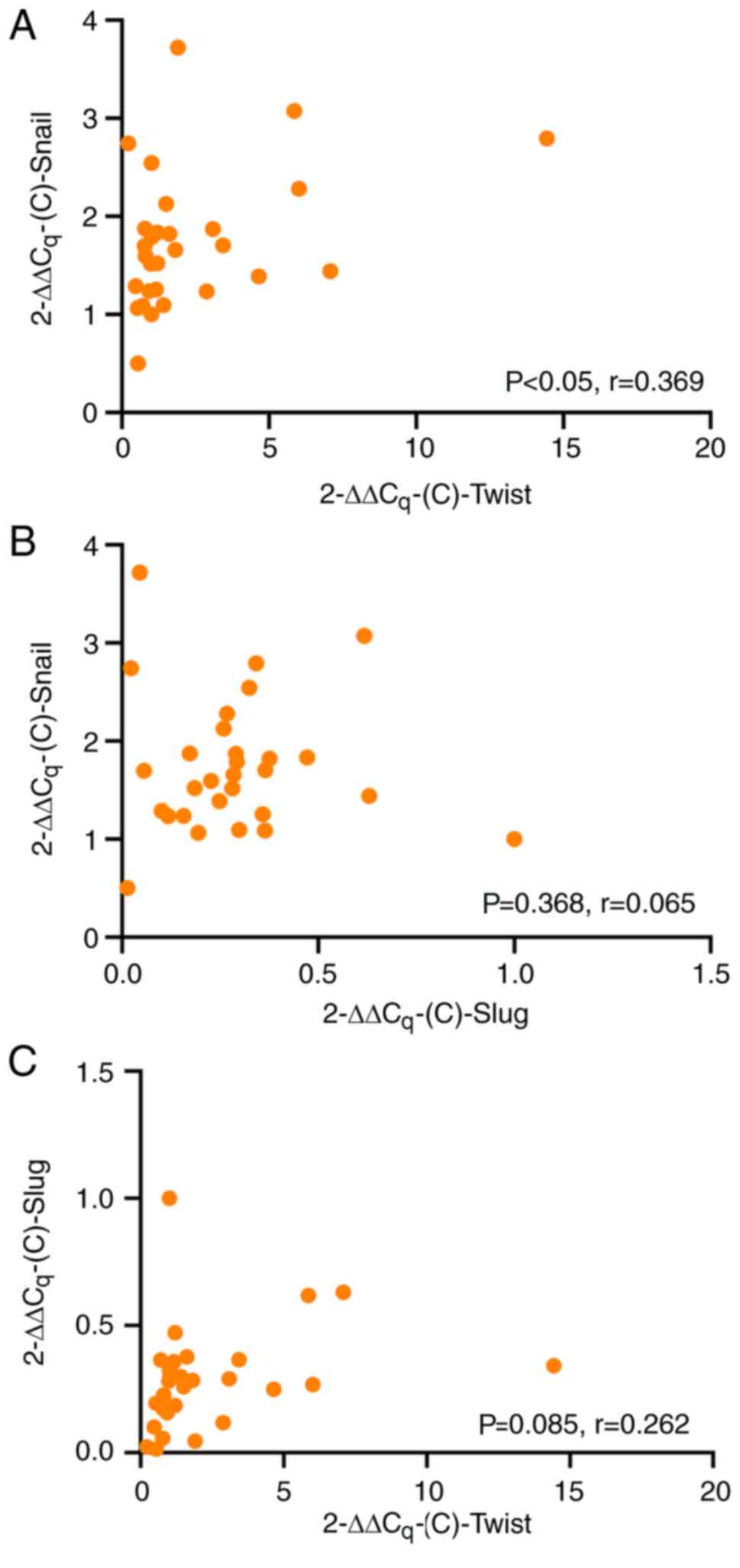
Pearson's correlation analysis revealed that there
was a positive correlation between the mRNA expression of Twist and
that of Snail in ESCC (r=0.369, P=0.024; Fig. 3A), indicating that when the mRNA
expression of Twist was high, the mRNA expression of Snail was also
high. However, no significant correlation was observed between Slug
and Snail (r=0.065, P=0.368; Fig.
3B), and Twist and Slug (r=0.262, P=0.085; Fig. 3C) expression.
Influence of Twist, Snail, Slug and
clinicopathological parameters on the prognosis of ESCC
The influence of clinicopathological parameters on
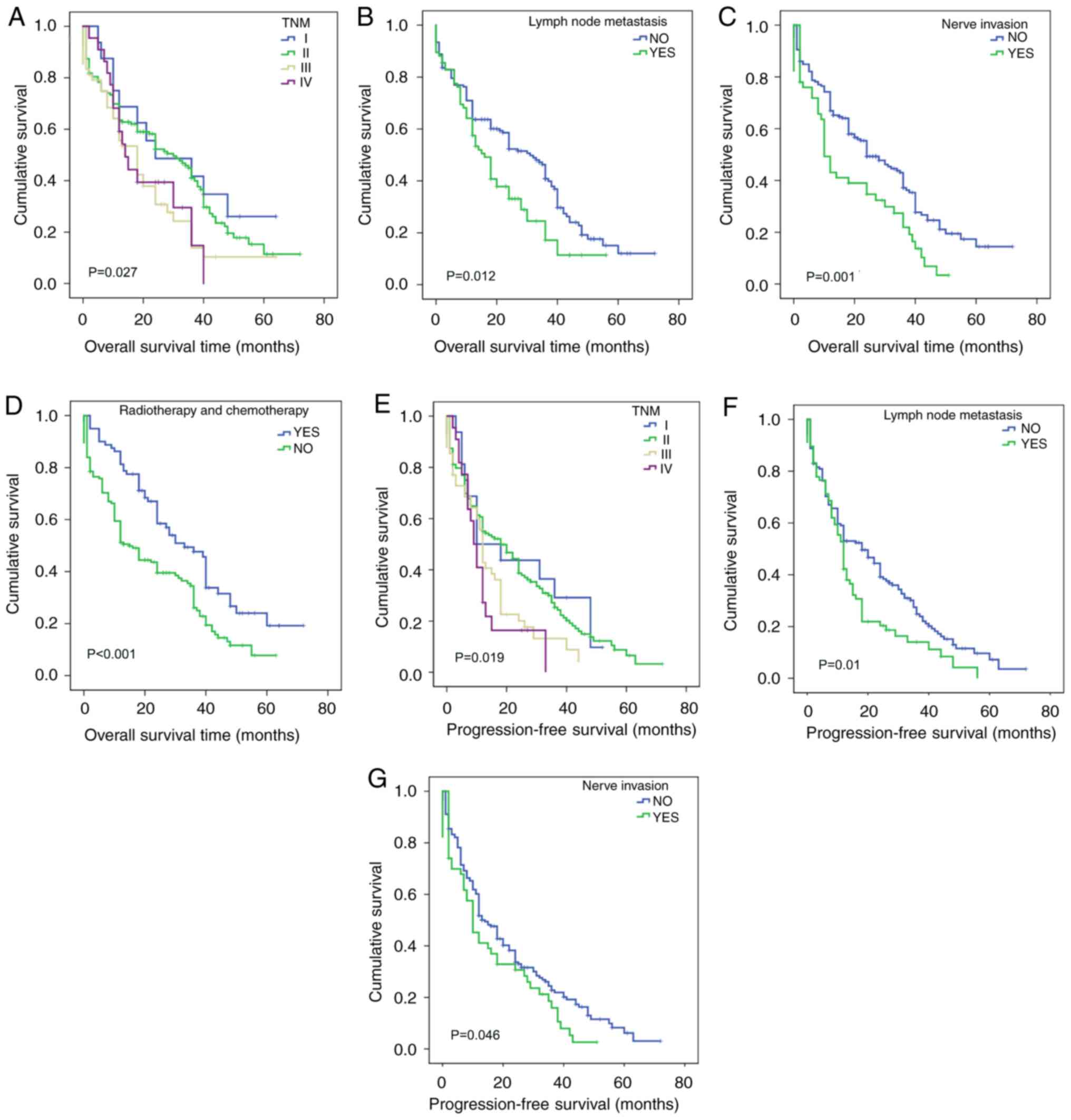
the prognosis of ESCC was analyzed (Table VII). Kaplan-Meier survival analysis
showed that TNM stage (P=0.027; Fig.
4A), lymph node metastasis (P=0.012; Fig. 4B), nerve invasion (P=0.001; Fig. 4C) and postoperative radiotherapy or
chemotherapy (P<0.001; Fig. 4D)
had an impact on the overall survival of patients with ESCC. Using
the Cox proportional hazards model (Table VIII), it was found that nerve
invasion (P=0.002), and postoperative radiotherapy or chemotherapy
(P<0.001) had an effect on the overall duration of survival;
therefore, nerve invasion and postoperative radiotherapy or
chemotherapy may be used as independent prognostic factors for
ESCC. However, lymph node metastasis (P=0.145) and TNM stage
(P=0.149) were not found to significantly affect the survival
status of patients with ESCC. The overall survival time of patients
with neurological invasion was shorter, and the risk of mortality
was 1.78 times higher in patients with neurological invasion than
in patients without [hazard ratio (HR)=1.78]. The overall survival
time of patients with ESCC who received postoperative radiotherapy
or chemotherapy was significantly prolonged (P<0.001), and the
death rate of patients who received radiotherapy or chemotherapy
was less that of patients who did not (HR=0.545).
| Table VII.Influence of clinicopathological
parameters on the prognosis of patients with esophageal squamous
cell carcinoma. |
Table VII.
Influence of clinicopathological
parameters on the prognosis of patients with esophageal squamous
cell carcinoma.
|
| OS | PFS |
|---|
|
|
|
|
|---|
| Clinicopathological
parameters | Mean survival
(months) | Median survival
(months) | Log rank
(Mantel-Cox) | P-value | Mean survival
(months) | Median survival
(months) | Log rank
(Mantel-Cox) | P-value |
|---|
| Age, years |
|
|
0.087 |
0.768 |
|
| 0.122 | 0.726 |
|
≤60 | 27.682 | 24 |
|
| 21.197 | 16 |
|
|
|
>60 | 27.615 | 22 |
|
| 20.364 | 12 |
|
|
| Sex |
|
|
1.495 |
0.222 |
|
| 0.627 | 0.429 |
|
Male | 26.485 | 24 |
|
| 20.224 | 13 |
|
|
|
Female | 29.780 | 30 |
|
| 22.422 | 12 |
|
|
| Ethnicity |
|
|
0.033 |
0.857 |
|
| 0.178 | 0.673 |
|
Han | 26.454 | 24 |
|
| 20.811 | 13 |
|
|
|
Kazakh | 28.189 | 24 |
|
| 20.187 | 12 |
|
|
| Tumor location |
|
|
1.429 |
0.489 |
|
| 1.127 | 0.569 |
|
Upper | 19.515 | 2 |
|
| 19.000 | 2 |
|
|
|
Middle | 27.575 | 24 |
|
| 21.739 | 12 |
|
|
|
Lower | 27.030 | 24 |
|
| 19.001 | 15 |
|
|
| Tumor size |
|
|
0.575 |
0.448 |
|
| 0.522 | 0.47 |
| <3
cm | 28.223 | 32 |
|
| 22.102 | 20 |
|
|
| ≥3
cm | 26.943 | 20 |
|
| 20.169 | 12 |
|
|
|
Differentiation |
|
|
4.314 |
0.116 |
|
| 1.678 | 0.432 |
|
Low | 23.756 | 18 |
|
| 19.082 | 16 |
|
|
|
Medium | 25.890 | 24 |
|
| 19.992 | 13 |
|
|
|
High | 34.360 | 24 |
|
| 24.594 | 11 |
|
|
| Depth of
invasion |
|
|
2.054 |
0.358 |
|
| 2.484 | 0.289 |
|
Mucosal | 37.833 | 48 |
|
| 34.000 | 48 |
|
|
|
Muscle | 28.697 | 24 |
|
| 20.375 | 12 |
|
|
|
Full | 25.635 | 22 |
|
| 20.182 | 12 |
|
|
| TNM stage |
|
|
9.200 |
0.027 |
|
| 10.006 | 0.019 |
| I | 35.266 | 36 |
|
| 23.899 | 10 |
|
|
| II | 29.520 | 28 |
|
| 23.049 | 18 |
|
|
|
III | 20.894 | 18 |
|
| 14.932 | 12 |
|
|
| IV | 19.756 | 13 |
|
| 12.427 | 9 |
|
|
| Lymph node
metastasis |
|
|
6.270 |
0.012 |
|
| 6.698 | 0.010 |
| No | 30.248 | 31 |
|
| 23.134 | 18 |
|
|
|
Yes | 22.128 | 15 |
|
| 15.776 | 12 |
|
|
| Vascular
invasion |
|
|
1.670 |
0.196 |
|
| 0.349 | 0.555 |
| No | 28.826 | 24 |
|
| 21.083 | 12 |
|
|
|
Yes | 22.719 | 22 |
|
| 18.200 | 12 |
|
|
| Nerve invasion |
|
| 10.255 |
0.001 |
|
| 3.990 | 0.046 |
| No | 30.223 | 24 |
|
| 22.089 | 14 |
|
|
|
Yes | 18.567 | 10 |
|
| 16.090 | 10 |
|
|
| Radiotherapy or
chemotherapy |
|
| 10.202 | <0.001 |
|
| 3.185 | 0.074 |
| No | 23.157 | 18 |
|
| 24.228 | 12 |
|
|
|
Yes | 35.152 | 28 |
|
| 18.907 | 18 |
|
|
| Table VIII.Cox analysis of the overall survival
of patients with esophageal squamous cell carcinoma. |
Table VIII.
Cox analysis of the overall survival
of patients with esophageal squamous cell carcinoma.
|
|
|
|
|
|
| HR 95% CI |
|---|
|
|
|
|
|
|
|
|
|---|
| Variables | B | SE | Wald | P-value | HR | Lower | Higher |
|---|
| Snail |
0.352 | 0.165 | 4.531 | 0.033 | 1.422 | 1.028 | 1.966 |
| Lymph node
metastasis | 0.33 | 0.226 | 2.128 | 0.145 | 1.391 | 0.893 | 2.168 |
| TNM stage |
0.208 | 0.144 | 2.077 | 0.149 | 1.231 | 0.928 | 1.634 |
| Nerve invasion |
0.577 | 0.182 | 10.036 | 0.002 | 1.78 | 1.246 | 2.543 |
| Radiotherapy or
chemotherapy | −0.607 | 0.174 | 12.207 | <0.001 | 0.545 | 0.388 | 0.766 |
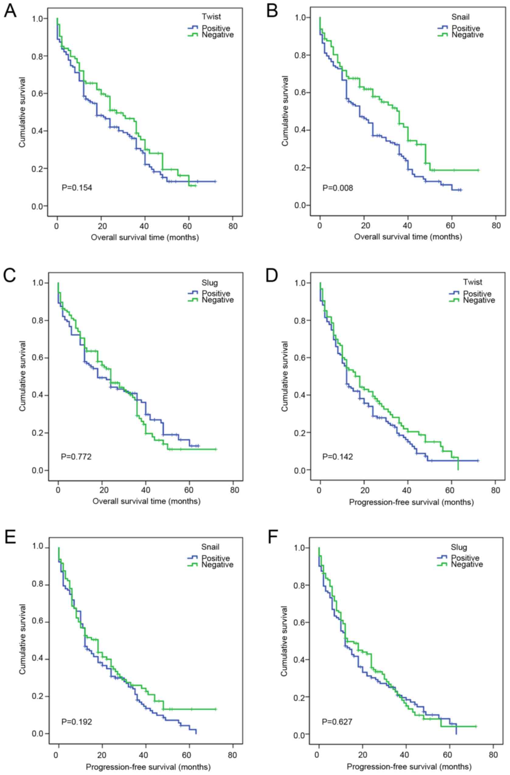
The effects of Twist, Snail and Slug expression on
the overall survival of patients with ESCC were also analyzed
(Table IX). The results did not
identify a statistically significant effect of Twist (P=0.154;
Fig. 5A) or Slug (P=0.772; Fig. 5C) on ESCC prognosis, but revealed a
significant effect of Snail on the overall survival of patients
with ESCC (P=0.008; Fig. 5B). Cox
multivariate model analysis revealed that Snail was an independent
factor (P=0.033) affecting the overall survival of patients with
ESCC (Table VIII). The prognosis
of patients who were Snail positive was poor, and their overall
survival duration was shorter than that of patients who were Snail
negative (HR=1.422).
| Table IX.Effects of Twist, Snail and Slug
expression on the prognosis of patients with esophageal squamous
cell carcinoma. |
Table IX.
Effects of Twist, Snail and Slug
expression on the prognosis of patients with esophageal squamous
cell carcinoma.
|
| OS | PFS |
|---|
|
|
|
|
|---|
| Protein | Mean survival
(months) | Median survival
(months) | Log rank
(Mantel-Cox) | P-value | Mean survival
(months) | Median survival
(months) | Log rank
(Mantel-Cox) | P-value |
|---|
| Twist |
|
| 2.033 | 0.154 |
|
| 2.158 | 0.142 |
|
Positive | 26.112 | 18 |
|
| 19.256 | 12 |
|
|
|
Negative | 29.408 | 27 |
|
| 23.012 | 18 |
|
|
| Snail |
|
| 7.076 | 0.008 |
|
| 2.158 | 0.192 |
|
Positive | 23.476 | 18 |
|
| 19.291 | 12 |
|
|
|
Negative | 33.169 | 36 |
|
| 23.772 | 18 |
|
|
| Slug |
|
| 0.126 | 0.772 |
|
| 0.236 | 0.627 |
|
Positive | 26.962 | 18 |
|
| 19.798 | 12 |
|
|
|
Negative | 27.399 | 24 |
|
| 21.364 | 13 |
|
|
Kaplan-Meier analysis showed that TNM staging
(P=0.019; Fig. 4E), lymph node
metastasis (P=0.01; Fig. 4F) and
nerve invasion (P=0.046; Fig. 4G)
had a significant effect on progression-free survival in ESCC
(Table VII), while no significant
association of Twist (P=0.142; Fig.
5D), Snail (P=0.192; Fig. 5E) or
Slug (P=0.627; Fig. 5F) with
progression-free survival in ESCC was observed (Table IX). Using Cox multivariate analysis
(Table X), it was found that nerve
invasion (P=0.05) was an independent factor affecting the
progression-free survival of patients with ESCC.
| Table X.Cox-analysis of progression-free
survival in patients with esophageal squamous cell carcinoma. |
Table X.
Cox-analysis of progression-free
survival in patients with esophageal squamous cell carcinoma.
|
|
|
|
|
|
| HR 95.0% CI |
|---|
|
|
|
|
|
|
|
|
|---|
| Variables | B | SE | Wald | P-value | HR | Lower | Upper |
|---|
| Lymph node
metastasis | 0.222 | 0.212 | 1.102 |
0.294 | 1.249 | 0.825 | 1.891 |
| TNM stage | 0.192 | 0.139 | 1.913 |
0.167 | 1.212 | 0.923 | 1.59 |
| Nerve invasion | 0.341 | 0.174 | 3.858 | 0.05 | 1.407 | 1.001 | 1.978 |
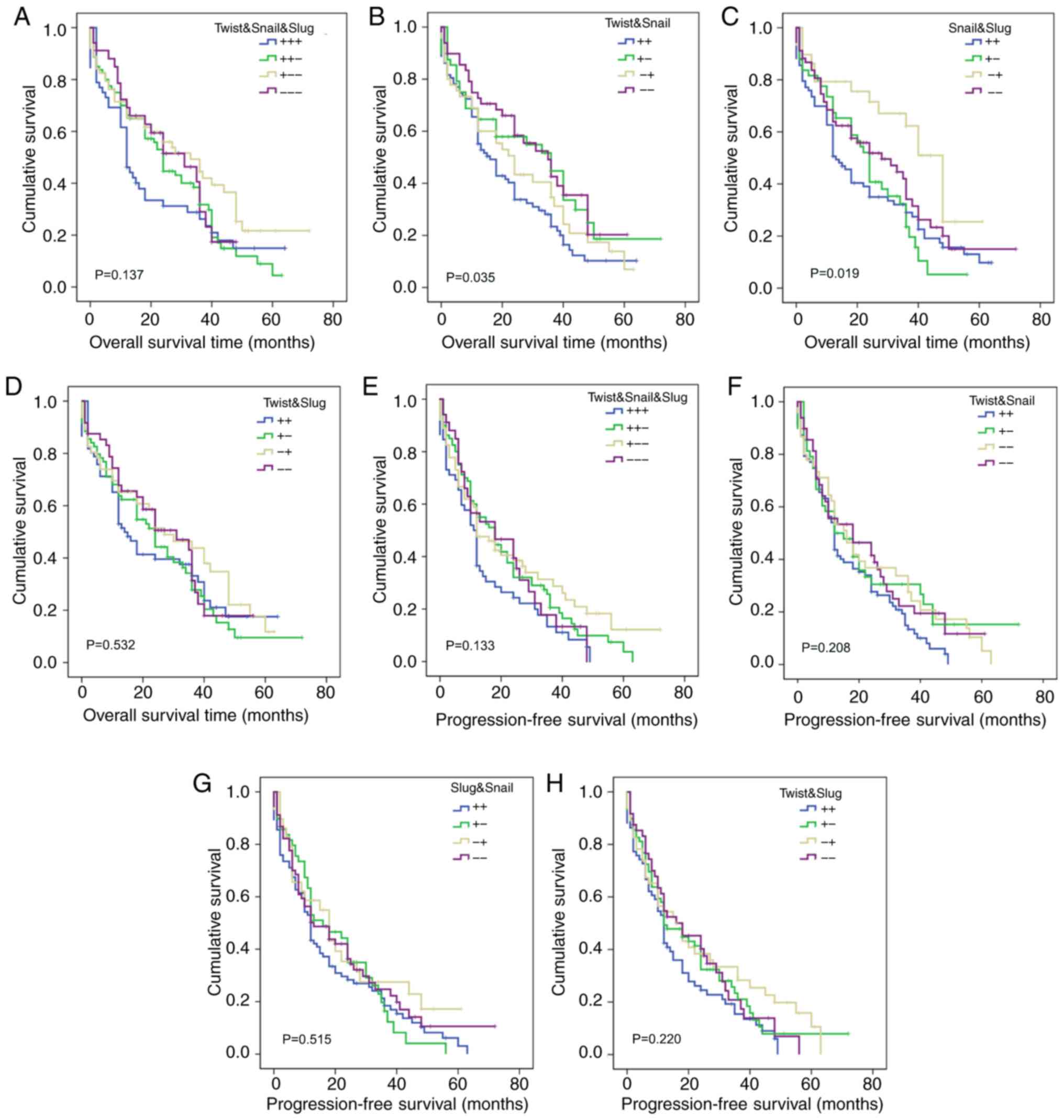
Effects of Twist, Snail and Slug
co-expression on the prognosis of ESCC
Kaplan-Meier analysis showed that the co-expression
of Twist and Snail (P=0.035; Fig.
6B), and Snail and Slug (P=0.019; Fig. 6C) had a significant effect on the
overall survival of patients with ESCC (Table XI), while the co-expression of
Twist, Snail and Slug (P=0.137; Fig.
6A), and Twist and Slug (P=0.532; Fig. 6D) had no effect on overall survival
(Table XI). The results also
indicate that the co-expression of Twist, Snail and Slug (P=0.133;
Fig. 6E), Twist and Snail (P=0.208;
Fig. 6F), Snail and Slug (P=0.515;
Fig. 6G) and Twist and Slug
(P=0.220; Fig. 6H) had no effect on
the progression-free survival of the 229 patients with ESCC
(Table XI). These results suggest
that the simultaneous inhibition of Twist and Snail protein
expression would be conducive to prolonging the overall survival of
patients with ESCC.
| Table XI.Effects of co-expression of Twist,
Snail and Slug on the prognosis of patients with esophageal
squamous cell carcinoma. |
Table XI.
Effects of co-expression of Twist,
Snail and Slug on the prognosis of patients with esophageal
squamous cell carcinoma.
|
|
| OS | PFS |
|---|
|
|
|
|
|
|---|
| Co-expression | No. | Mean survival
(months) | Median survival
(months) | Log rank
(Mantel-Cox) | P-value | Mean survival
(months) | Median survival
(months) | Log rank
(Mantel-Cox) | P-value |
|---|
|
Twist/Snail/Slug |
|
|
| 5.531 | 0.137 |
|
| 5.598 | 0.133 |
|
+/+/+ | 52 | 21.728 | 12 |
|
| 15.274 | 11 |
|
|
|
+/+/− | 80 | 25.799 | 24 |
|
| 21.582 | 18 |
|
|
|
+/−/− | 63 | 33.293 | 33 |
|
| 24.488 | 12 |
|
|
|
−/−/− | 34 | 26.352 | 31 |
|
| 20.262 | 18 |
|
|
| Twist/Snail |
|
|
| 8.593 | 0.035 |
|
| 4.551 | 0.208 |
|
+/+ | 87 | 22.113 | 16 |
|
| 17.073 | 12 |
|
|
|
+/− | 48 | 32.297 | 36 |
|
| 23.939 | 12 |
|
|
|
−/+ | 45 | 26.183 | 24 |
|
| 23.229 | 16 |
|
|
|
−/− | 49 | 32.263 | 35 |
|
| 22.549 | 18 |
|
|
| Snail/Slug |
|
|
| 9.917 | 0.019 |
|
| 2.287 | 0.515 |
|
+/+ | 83 | 23.001 | 13 |
|
| 18.444 | 12 |
|
|
|
+/− | 49 | 23.27 | 24 |
|
| 20.255 | 16 |
|
|
|
−/+ | 29 | 37.53 | 48 |
|
| 23.682 | 18 |
|
|
|
−/− | 68 | 29.704 | 28 |
|
| 22.654 | 13 |
|
|
| Twist/Slug |
|
|
| 2.198 | 0.532 |
|
| 4.412 | 0.22 |
|
+/+ | 66 | 24.941 | 14 |
|
| 16.368 | 12 |
|
|
|
+/− | 69 | 26.021 | 24 |
|
| 21.53 | 12 |
|
|
|
−/+ | 46 | 30.077 | 27 |
|
| 24.077 | 16 |
|
|
|
−/− | 48 | 27.544 | 31 |
|
| 21.207 | 18 |
|
|
Discussion
Surgical resection is the main treatment for early
esophageal cancer; however, the treatment of patients with
recurrence or progression is challenging. The identification of
highly specific molecular targets for the clinical treatment and
prognosis evaluation of patients with advanced esophageal cancer is
likely to have practical consequences. Therefore, the aim of the
present study was to investigate the expression of Twist, Slug and
Snail in ESCC and their prognostic significance.
Kazakhs have a high incidence of ESCC in Xinjiang.
Zheng et al (27) revealed
that ESCC is associated with Kazakhs' habits of smoking, drinking
alcohol and drinking hot milk tea. The present study revealed that
there were significant differences in age, tumor location and
vascular invasion between Han and Kazakh patients. The proportion
of Kazakh patients <60 years old was higher than that of Han
nationality, which indicates that Kazakh patients with esophageal
cancer are mainly young. The incidence of esophageal cancer in the
middle and lower parts of the esophagus in Kazakh patients was
higher than that in the upper part. It is suggested that changing
diet and lifestyle may decrease the incidence rate of ESCC.
Twist has been shown to be important indicator in
breast cancer, with a meta-analysis showing that Twist expression
is associated with increased tumor size, lymph node involvement,
high grade and human epidermal growth factor receptor 2 positivity
(28). In ESCC, it has been found
that the expression of Twist is significantly associated with lymph
node metastasis, and the overexpression of Twist can significantly
increase the invasion of ESCC cells and their expression of
vascular endothelial growth factor C (VEGF-C), while the knockdown
of Twist has the opposite effects, as confirmed by
immunohistochemical detection of the co-expression of Twist and
VEGF-C in ESCC (29).
In the present study, the positive expression rate
of Twist protein in ESCC was higher than that in normal esophageal
mucosa, and the RT-qPCR results confirmed that Twist was highly
expressed in ESCC at the transcriptional level. Among the 229 cases
of ESCC analyzed using immunohistochemistry, Twist was positive in
135 (59.0%), and negative in 94 cases (41.0%). The expression of
Twist in ESCC was found to be associated with the sex of the
patient. The positive Twist expression rate in male patients was
higher than that in female patients; if male patients were positive
for Twist expression, it was more likely to indicate ESCC.
Therefore, it is predicted that Twist may be a target gene for the
diagnosis of ESCC.
High expression levels of Slug are generally
considered to promote tumorigenesis and development. It has been
reported that Slug is associated with the invasion, migration and
cell regulation of tumor cells and, as an important transcription
factor during EMT, can directly inhibit E-cadherin transcription
and induce EMT, thereby promoting tumor progression (30). Furthermore, Slug has been shown to
promote lymph node metastasis in oropharyngeal squamous cell
carcinoma (31). Hasan et al
(32) studied the association
between the expression of Slug and the clinicopathological
parameters and prognosis of patients with ESCC, and found that Slug
expression increased during in the early stages of ESCC
development, and persisted as the disease progressed. Therefore, it
was suggested that Slug can be used as a diagnostic biomarker, as
well as a predictor of poor prognosis, to aid the early detection
of the possibility of postoperative esophageal cancer
recurrence.
In the present study, Slug expression was positive
in 112 (48.9%) and negative in 117/229 (51.1%) cases of ESCC. When
the association of Slug expression with clinicopathological
parameters was analyzed, it was found that the expression of Slug
differed according to the differentiation degree, TNM stage and
vascular invasion status of ESCC. The positive expression rate of
Slug in poorly differentiated ESCC was lower that than in highly
differentiated ESCC. Among the various TNM stages, the highest Slug
positive rate was detected in TNM stage I. The positive expression
rate of Slug in esophageal carcinoma without vascular invasion was
52.7%, which was significantly higher than that in esophageal
carcinoma with vascular invasion (32.6%). These results suggest
that Slug is a tumor suppressor gene in ESCC. In addition, RT-qPCR
showed that the mRNA expression of Slug in esophageal normal mucosa
was higher than that in ESCC (P<0.001). These results were
confirmed at the transcriptional level. In the study by Cui et
al (33), Slug was found to play
an anticancer role in the progression of cervical cancer. The
mechanism was suggested to be that Slug, by combining with the
E-box motif of the AKT1 gene promoter, trans-inhibits the
expression of AKT1, which upregulates p21/p27 and/or downregulates
the activity of the Wnt/β-catenin signaling pathway, thereby
inhibiting the proliferation of cervical cancer cells and tumor
formation. In the present study, no marked difference was found in
the expression of Slug between ESCC and the corresponding adjacent
tissues by immunohistochemistry, while the detection results of
Slug mRNA showed that its expression was higher in the adjacent
normal tissues than in ESCC. These results suggest that Slug may be
a tumor suppressor gene in ESCC. However, no significant
associations between Snail expression and the clinicopathological
parameters of patients with ESCC were detected herein.
The effects of Twist, Slug, Snail and
clinicopathological parameters on the prognosis of esophageal
cancer were also evaluated in the present study. Twist has been
demonstrated to play an important role in a variety of invasive
cancers, such as breast (17), lung
(18) and prostate cancer (19). In one study, Twist was shown to be
involved in nicotine-derived nitrosamine ketone-induced lung cancer
cell migration and invasion (18).
Lyu et al (19) demonstrated
that Twist is a prognostic marker of prostate cancer, and
identified potential downstream target and genes, which may be
useful for predicting the prognosis of Twist-mediated prostate
cancer. In addition, Sun and Liu (34) reported that the upregulation of Twist
activates the Wnt/β-catenin signaling pathway and promotes cervical
cancer. We hypothesize that Twist may also be of significance in
the development of ESCC. According to the results of the present
study, Twist is highly expressed in ESCC. However, Kaplan-Meier
survival analysis indicated that there was no significant
difference in the prognosis of ESCC between Twist-positive and
-negative cases, although a trend for poor overall survival in
Twist positive cases was shown in the survival curves.
In studies conducted by Salehi et al
(35) and Shenas et al
(36), when Snail was silenced, the
expression of vimentin was decreased, E-cadherin was induced and
EMT was inhibited, and Snail was indicated to play a crucial role
in the survival of bladder cancer. The high expression of Snail has
also been reported to indicate a poor prognosis in patients with
infiltrative breast lesions (37).
In the present study, the results of Kaplan-Meier survival analysis
showed that the overall survival for cases with positive Snail
expression was significantly worse than that for those with
negative expression, suggesting that the patients who were positive
for Snail expression had a poor prognosis and shorter survival
time. Multivariate Cox analysis indicated that Snail is an
independent prognostic factor for ESCC. This is consistent with the
results of several previous studies (35,36).
Kaplan-Meier survival analysis demonstrated that
patients with lymph node metastasis (P=0.012), a higher TNM stage
(P=0.027) and nerve invasion (P<0.001), or without postoperative
radiotherapy or chemotherapy (P<0.001), had a shorter overall
survival. Multivariate Cox analysis indicated that nerve invasion
and postoperative radiotherapy or chemotherapy are independent
prognostic factors for ESCC. When progression-free survival in ESCC
was analyzed, the results indicated that lower TNM stages were
associated with longer progression-free survival. Patients without
nerve invasion and lymph node metastasis also had a longer
progression-free survival. This suggests that postoperative
radiotherapy or chemotherapy can prolong the time taken for ESCC to
recur and metastasize, as well as the associated mortality.
The Twist/Snail axis has been demonstrated to be the
key to tropomyosin receptor kinase B-induced EMT, apoptosis
inhibition and metastasis. Twist, as the upstream gene of Snail,
affects the occurrence of EMT, and regulates and induces the
expression of E-cadherin in Snail, thus increasing the risk of
tumor metastasis (38). A study on
the expression of Snail and Twist revealed varying degrees of
interdependence; Snail and Twist were shown to cooperate in the
induction of zinc finger E-box-binding homeobox 1, while the
absence of Twist inhibited the upregulatory effect of TNF-β on
Snail, but Snail was essential for the rapid increase in Twist
protein and upregulation of Twist mRNA induced by TNF-β (39).
In the present study, a significant difference was
detected between the protein expression of Twist and that of Snail;
when Twist was highly expressed, Snail was also likely to be highly
expressed. A significant correlation between Twist and Snail was
also observed at the transcriptional level. We hypothesize that
Twist and Snail are co-expressed, and their co-expression suggests
a poor prognosis in ESCC patients. Although no significant
correlation was observed between Twist and Slug, a positive
correlation was observed for Slug and Snail proteins (r=0.326;
P<0.001). Considering that Slug is a member of the Snail family,
the functional structures of Slug and Snail have a certain
similarity. However, no significant correlation between Slug and
Snail was identified at the transcriptional level. We hypothesize
that the expression of Slug in ESCC may be inhibited at the
transcriptional level. However, as the sample size was too small,
more experiments are required to confirm this.
Twist, Slug and Snail are key transcription factors
in tumorigenesis and development. Twist and Snail have been shown
to affect the epithelial stromal transformation of odontogenic
epithelial tumors, with the expression of Twist being associated
with the aggressive behavior of the tumors, whose occurrence and
development may involve the Twist/Snail pathway, while Snail may
mediate interaction between the tumors and stroma (40). In a study of chronic obstructive
pulmonary disease (COPD) conducted by Mahmood et al
(41), it was found that the
transcription factor complex β-catenin-Snail1-Twist was upregulated
in smokers and patients with COPD, and translocated to the nucleus.
Furthermore, its expression was closely associated with EMT
activity and airway obstruction. In a study by Casas et al
(42), it was found that Slug was
necessary for Twist to induce EMT, as the knockdown of Slug
completely blocked the ability of Twist to inhibit E-cadherin
transcription. Experiments in mice demonstrated that Slug was
necessary for Twist to induce tumor cell invasion and metastasis.
Furthermore, in human breast tumors, the expression of Twist and
Slug was highly correlated. Thus, Twist and Slug were shown to work
together to promote EMT and tumor metastasis (42). Although no significant correlation
was observed between Twist and Slug in ESCC in the present study, a
positive correlation was found for Twist and Snail expression, and
Slug and Snail have a certain homology. These observations requires
further research.
In conclusion, the present study found that Twist
was highly expressed in ESCC and was associated with male sex. The
expression of Slug was found to be associated with the degree of
differentiation, TNM stage and vascular invasion in ESCC, and may
have an inhibitory effect on the development of ESCC. Snail was
found to be an independent prognostic factor in patients with ESCC,
and the expression of Twist was positively correlated with the
expression of Snail. Furthermore, the co-expression of Twist and
Snail had a significant effect on the overall survival of patients
with ESCC, suggesting that the inhibition of Twist and Snail
expression simultaneously may be conducive to prolonging the
overall survival of ESCC patients. These results provide a
theoretical basis for the identification of a new therapeutic
target for ESCC. The present study has certain limitations. It only
preliminarily confirmed the roles of Twist, Slug and Snail in ESCC
and their influence on prognosis. The mechanisms of the proteins
cannot be identified by the statistical analysis of the
immunohistochemistry and RT-qPCR results. In addition, the RT-qPCR
results showed that the mRNA expression level of Slug in ESCC was
lower than that in normal esophageal mucosa. However, the
expression of slug in ESCC was higher than that in normal
esophageal mucosa in the immunohistochemical experiment. In-depth
studies combining experiments with tissues, cells and animal models
are required to reveal the underlying mechanisms.
Acknowledgements
Not applicable.
Funding
The present study was supported by the National
Natural Science Foundation of China (grant no. 81860422) and the
graduate innovation and entrepreneurship start-up fund of Xinjiang
Medical University (grant no. CXCY2018024).
Availability of data and materials
The datasets used and/or analyzed during the current
study are available from the corresponding author on reasonable
request.
Authors' contributions
SX and YZ designed and performed the experiments,
analyzed the data and were the main contributors to the manuscript.
HB and HW performed the experiments and interpreted data. CL and WZ
were involved in the experiments and data collection. YM designed
the experimental program. All authors read and approved the final
manuscript.
Ethics approval and consent to
participate
The study was approved by the Medical Ethics
Committee of the First Affiliated Hospital of Xinjiang Medical
University (approval no. 20180223-08). Written informed consent was
obtained from all participants.
Patient consent for publication
Not applicable.
Competing interests
The authors declare that they have no competing
interests.
References
|
1
|
Bray F, Ferlay J, Soerjomataram I, Siegel
RL, Torre LA and Jema A: Global cancer statistics 2018: GLOBOCAN
estimates of incidence and mortality worldwide for 36 cancers in
185 countries. CA Cancer J Clin. 68:394–424. 2018. View Article : Google Scholar : PubMed/NCBI
|
|
2
|
Chen R, Zheng RS, Zhang SW, Zeng HM, Wang
SM, Sun KX, Gu XY, Wei WW and He J: Analysis of incidence and
mortality of esophageal cancer in China, 2015. Zhonghua Yu Fang Yi
Xue Za Zhi. 53:1094–1097. 2019.(In Chinese). PubMed/NCBI
|
|
3
|
Napier KJ, Scheerer M and Misra S:
Esophageal cancer: A review of epidemiology, pathogenesis, staging
workup and treatment modalities. World J Gastrointest Oncol.
6:112–120. 2014. View Article : Google Scholar : PubMed/NCBI
|
|
4
|
Zheng S, Vuitton L, Sheyhidin I, Vuitton
DA, Zhang Y and Lu X: Northwestern China: A place to learn more on
oesophageal cancer. Part two: Gene alterations and polymorphisms.
Eur J Gastroenterol Hepatol. 23:1087–1099. 2011. View Article : Google Scholar : PubMed/NCBI
|
|
5
|
Qing L, Liang M, Liu T, Vuitton L, Zheng
S, Gao X, Lu M, Li X, Sheyhidin I and Lu X: M2 isoform of pyruvate
kinase (PKM2) is upregulated in Kazakh's ESCC and promotes
proliferation and migration of ESCC cells. Tumour Biol.
37:2665–2672. 2016. View Article : Google Scholar : PubMed/NCBI
|
|
6
|
Pang L, Li Q, Wei C, Zou H, Li S, Cao W,
He J, Zhou Y, Ju X, Lan J, et al: TGF-β1/Smad signaling pathway
regulates epithelial-to-mesenchymal transition in esophageal
squamous cell carcinoma: In vitro and clinical analyses of cell
lines and nomadic Kazakh patients from northwest Xinjiang, China.
PLoS One. 9:e1123002014. View Article : Google Scholar : PubMed/NCBI
|
|
7
|
de la Iglesia JE, de la Calle MA, pérez
GC, Pérez RR and Delgado AA: Esophageal cancer: Anatomic,
particularities, staging, and, imaging techniques. Radiologia.
58:352–365. 2016.PubMed/NCBI
|
|
8
|
Liu B, Bo Y, Wang K, Liu Y, Tang X, Zhao
Y, Zhao E and Yuan L: Concurrent neoadjuvant chemoradiotherapy
could improve survival outcomes for patients with esophageal
cancer: A metaanalysis based on random clinical trials. Oncotarget.
21:20410–20417. 2017. View Article : Google Scholar
|
|
9
|
Huang Y, Wang H, Luo G, Zhang Y, Wang L
and Li K: A systematic review and network meta-analysis of
neoadjuvant therapy combined with surgery for patients with
resectable esophageal squamous cell carcinoma. Int J Surg.
38:41–47. 2017. View Article : Google Scholar : PubMed/NCBI
|
|
10
|
Hu Y, Li Z, Mi DM, Cao N, Zu SW, Wen ZZ,
Yu XL and Qu Y: Chemoradiation combined with regional hyperthermia
for advanced oesophageal cancer: A systematic review and
meta-analysis. J Clin Pharm Ther. 42:155–164. 2017. View Article : Google Scholar : PubMed/NCBI
|
|
11
|
Parvani JG and Schiemann WP: Sox4, EMT
programs, and the metastatic progression of breast cancers:
Mastering the masters of EMT. Breast Cancer Res. 15:R722013.
View Article : Google Scholar : PubMed/NCBI
|
|
12
|
Bruner HC and Derksen WB: Loss of
E-cadherin-dependent cell-cell adhesion and the development and
progression of cancer. Cold Spring Herb Perspect Biol.
1:102018.
|
|
13
|
Siar CH and Ng KH: Differential expression
of transcription factors snail, slug, SIP1, and twist in
ameloblastoma. J Oral Pathol Med. 43:45–52. 2014. View Article : Google Scholar : PubMed/NCBI
|
|
14
|
Qian Q, Young X, Tao H, Qin C and Xu J:
Normal and disease-related biological functions of Twist1 and
underlying molecular mechanisms. Cell Res. 22:90–106. 2012.
View Article : Google Scholar : PubMed/NCBI
|
|
15
|
Yu L, Mu Y, Sa N, Wang H and Xu W: Tumor
necrosis factor α induces epithelial-mesenchymal transition and
promotes metastasis via NF-κB signaling pathway-mediated twist
expression in hypopharyngeal cancer. Oncol Rep. 31:321–327. 2014.
View Article : Google Scholar : PubMed/NCBI
|
|
16
|
Yang J and Weinberg RA:
Epithelial-mesenchymal transition: At the crossroads of development
and tumor metastasis. Dev Cell. 14:818–829. 2008. View Article : Google Scholar : PubMed/NCBI
|
|
17
|
Cao J, Wang X, Dai T, Wu Y, Zhang M, Cao
R, Zhang R, Wang G, Jiang R, Zhou BP, et al: Twist promotes tumor
metastasis in basal-like breast cancer by transcriptionally
upregulating ROR1. Theranostics. 8:2739–2751. 2018. View Article : Google Scholar : PubMed/NCBI
|
|
18
|
Wang Y, Shi L, Li J, Wang H and Yang H:
Involvement of twist in NNK exposure-promoted lung cancer cell
migration and invasion. Toxicol In Vitro. 63:1047402019. View Article : Google Scholar : PubMed/NCBI
|
|
19
|
Lyu P, Zhang SD, Yuen HF, McCrudden CM,
Wen Q, Chan KW and Kwok HF: Identification of Twist-interacting
genes in prostate cancer. Sci China Life Sci. 60:386–396. 2017.
View Article : Google Scholar : PubMed/NCBI
|
|
20
|
Hajra KM, Chen DY and Fearon ER: The slug
zinc-finger protein represses E-cadherin in breast cancer. Cancer
Res. 62:1613–1618. 2002.PubMed/NCBI
|
|
21
|
de Souza Palma C, Grassi ML, Thomé CH,
Ferreira GA, Albuquerque D, Pinto MT, Melo FU, Kashima S, Covas DT,
Pitteri SJ and Faça VM: Proteomic analysis of epithelial to
mesenchymal transition (EMT) reveals cross-talk between snail and
HDAC1 proteins in breast cancer cells. Mol Cell Proteomics.
15:906–917. 2016. View Article : Google Scholar : PubMed/NCBI
|
|
22
|
Wang Y, Shi J, Chai K, Ying X and Zhou BP:
The role of snail in EMT and tumorigenesis. Curr Cancer Drug
Targets. 13:963–972. 2013. View Article : Google Scholar : PubMed/NCBI
|
|
23
|
Kaufhold S and Bonavida B: Central role of
snail1 in the regulation of EMT and resistance in cancer: A target
for therapeutic intervention. J Exp Clin Cancer Res. 33:622014.
View Article : Google Scholar : PubMed/NCBI
|
|
24
|
Phillips S and Kuperwasser C: SLUG:
Critical regulator of epithelial cell identity in breast
development and cancer. Cell Adh Migr. 8:578–587. 2014. View Article : Google Scholar : PubMed/NCBI
|
|
25
|
Alves CC, Carneiro F, Hoefler H and Becker
KF: Role of the epithelial-mesenchymal transition regulator Slug in
primary human cancers. Front Biosci (Landmark ED). 14:3035–3050.
2009. View Article : Google Scholar : PubMed/NCBI
|
|
26
|
Livak KJ and Schmittgen TD: Analysis of
relative gene expression data using real-time quantitative PCR and
the 2(-Delta Delta C(T)) method. Methods. 25:402–408. 2001.
View Article : Google Scholar : PubMed/NCBI
|
|
27
|
Zheng S, Vuitton L, Sheyhidin I, Vuitton
DA, Zhang Y and Lu X: Northwestern China: A place to learn more on
oesophageal cancer. Part one: Behavioural and environmental risk
factors. Eur J Gastroenterol Hepatol. 22:917–925. 2010. View Article : Google Scholar : PubMed/NCBI
|
|
28
|
Qiao W, Jia Z, Liu H, Liu Q, Zhang T, Guo
W, Li P, Deng M and Li S: Prognostic and clinicopathological value
of twist expression in breast cancer: A meta-analysis. PLoS One.
12:e01861912017. View Article : Google Scholar : PubMed/NCBI
|
|
29
|
Gong T, Xue Z, Tang S, Zheng X, Xu G, Gao
L, Zhao G, Hong L, Tang G, Zhang H, et al: Nuclear expression of
twist promotes lymphatic metastasis in esophageal squamous cell
carcinoma. Cancer Biol Ther. 13:606–613. 2012. View Article : Google Scholar : PubMed/NCBI
|
|
30
|
Shih JY and Yang PC: The EMT regulator
slug and lung carcinogenesis. Carcinogenesis. 32:1299–1304. 2011.
View Article : Google Scholar : PubMed/NCBI
|
|
31
|
Cho YA, Kim EK, Cho BC, Koh YW and Yoon
SO: Twist and snail/slug expression in oropharyngeal squamous cell
carcinoma in correlation with lymph node metastasis. Anticancer
Res. 39:6307–6316. 2019. View Article : Google Scholar : PubMed/NCBI
|
|
32
|
Hasan MR, Sharma R, Saraya A,
Chattopadhyay TK, DattaGupta S, Walfish PG, Chauhan SC and Ralhan
R: Slug is a predictor of poor prognosis in esophageal squamous
cell carcinoma patients. PLoS One. 8:e828462013. View Article : Google Scholar : PubMed/NCBI
|
|
33
|
Cui N, Yang WT and Zheng PS: Slug inhibits
the proliferation and tumor formation of human cervical cancer
cells by up-regulating the p21/p27 proteins and down-regulating the
activity of the wnt/β-catenin signaling pathway via the
trans-suppression akt1/p-akt1 expression. Oncotarget.
7:26152–26167. 2016. View Article : Google Scholar : PubMed/NCBI
|
|
34
|
Sun X and Liu Y: Activation of the
wnt/β-catenin signaling pathway may contribute to cervical cancer
pathogenesis via upregulation of twist. Oncol Lett. 14:4841–4844.
2017. View Article : Google Scholar : PubMed/NCBI
|
|
35
|
Salehi S, Mansoori B, Mohammadi A,
Davoudian S, Shenas SM, Shajari N, Majidi J and Baradaran B: An
analysis of suppressing migratory effect on human urinary bladder
cancer cell line by silencing of snail-1. Biomed Pharmacother.
96:545–550. 2017. View Article : Google Scholar : PubMed/NCBI
|
|
36
|
Shenas SM, Mansoori B, Mohammadi A, Salehi
S, Kaffash B, Talebi B, Babaloo Z, Shanehbandi D and Baradaran B:
SiRNA-Mediated silencing of snail-1 induces apoptosis and alters
micro RNA expression in human urinary bladder cancer cell line.
Artif Cells Nanomed Biotechnol. 45:969–974. 2017. View Article : Google Scholar : PubMed/NCBI
|
|
37
|
Chang HY, Tseng YK, Chen YC, Shu CW, Lin
MI, Liou HH, Fu TY, Lin YC, Ger LP, Yeh MH and Liu PF: High snail
expression predicts a poor prognosis in breast invasive ductal
carcinoma patients with HER2/EGFR-positive subtypes. Surg Oncol.
27:314–320. 2018. View Article : Google Scholar : PubMed/NCBI
|
|
38
|
Smit MA, Geiger TR, Song JY, Gitelman I
and Peeper DS: A twist-snail axis critical for TrkB-induced
epithelial-mesenchymal transition-like transformation, anoikis
resistance, and metastasis. Mol Cell Biol. 29:3722–3737. 2009.
View Article : Google Scholar : PubMed/NCBI
|
|
39
|
Dave N, Guaita-Esteruelas S, Gutarra S,
Frias A, Beltran M, Peiró S and de Herreros AG: Functional
cooperation between snail1 and twist in the regulation of ZEB1
expression during epithelial to mesenchymal transition. J Biol
Chem. 286:12024–12032. 2011. View Article : Google Scholar : PubMed/NCBI
|
|
40
|
Oh KY, Yoon HJ, Lee JI, Ahn SH and Hong
SD: Twist and snail expression in tumor and stromal cells of
epithelial odontogenic tumors. J Oral Pathol Med. 46:127–133. 2017.
View Article : Google Scholar : PubMed/NCBI
|
|
41
|
Mahmood MQ, Walters EH, Shukla SD, Weston
S, Muller HK, Ward C and Sohal SS: β-Catenin, twist and snail:
Transcriptional regulation of EMT in smokers and COPD, and relation
to airflow obstruction. Sci Rep. 7:108322017. View Article : Google Scholar : PubMed/NCBI
|
|
42
|
Casas E, Kim J, Bendesky A, Ohno-Machado
L, Wolfe CJ and Yang J: Snail2 is an essential mediator of
twist1-induced epithelial mesenchymal transition and metastasis.
Cancer Res. 71:245–254. 2011. View Article : Google Scholar : PubMed/NCBI
|