With the development of targeted therapies and
cellular immunotherapies, such as T cell-based, natural killer (NK)
cell-based and dendritic cell (DC)-based immunotherapies, the
therapeutic efficacy of cancer treatment has been greatly improved
(1). However, the overall remission
and survival rate of patients with certain tumors has not been
fundamentally addressed. In recent decades, oncolytic viruses (OVs)
have generated widespread interest, and have become a major focus
of interest for clinicians and scientists (2,3). These
viruses include adenovirus, measles virus, reovirus, herpes simplex
virus, Newcastle disease virus, vesicular stomatitis, vaccinia
virus and poliovirus (4,5).
Previous preclinical and clinical studies have
demonstrated that the intratumoral injection of OVs is effective,
although the efficacy toward disseminated and metastatic tumors
remains modest (6,7). Numerous factors can affect viral
efficiency in reaching tumor tissue, including viral destruction by
the immune system and viral absorption by tissues and organs
(8,9). Therefore, appropriate carrier vehicles
are required to deliver OVs to tumor sites in order to improve
therapeutic efficacy.
In recent years, mesenchymal stem cells (MSCs) have
become a promising cellular vehicle for anti-tumor drug delivery,
thanks to their inherent tumor tropism (10–13).
MSCs can specifically migrate to the tumor or inflammatory site. A
recent review has reported that MSCs can be modified by advanced
approaches to suppress tumor growth (14). Furthermore, MSCs exert
immunosuppressive functions, by inhibiting NK proliferation,
cytotoxicity and cytokine production (15), suppressing differentiation and
function of DC (16) and inducing
therefore the emergence of regulatory T cells. These features make
MSCs ideal candidates for OVs delivery. In the present review, an
overview of MSC loading of OVs for oncolytic virotherapy was
provided. We briefly introduced MSC characteristics for OV delivery
and summarized developments in the MSC oncolytic virotherapy
arena.
In the last decades, great progress has been made in
elucidating the molecular mechanisms of OV infection. OVs can
infect target cells using low-affinity binding to sialic acid
residues, from where they internalize via specific high-affinity
receptors (17,18). The expression of OV strain receptors
on the cell surface is a crucial factor in determining viral
infection (19). However,
accumulating evidence from preclinical and clinical studies has
indicated that growth conditions and genetic background of tumor
cells can affect cell sensitivity to OVs (19). For example, cathepsin B and cathepsin
L are critical for viral shelling, which is associated with the
sensitivity of tumor cells to oncolytic reoviruses; however, virus
shelling is also limited by low levels of cathepsin B and cathepsin
L in normal cells (20). In
addition, Ras mutations can increase cell sensitization to
reoviruses (21,22). Following OV infection, virus progeny
replicates highly in tumor cells, eventually lysing and killing
infected cells. Subsequently, tumor cell lysis releases infectious
viral progeny that spreads to surrounding tumor cells, causing more
tumor cells to undergo oncolysis. However, OV replication is often
limited in healthy cells, thus viral clearance is rapid with
minimal oncolysis (23).
With expanding OV research, virotherapy has
gradually changed from direct oncolysis to virus mediated
anti-tumor immunity (24,25). It has been demonstrated that the
immune system serves a crucial role in oncolytic virotherapy. On
the one hand, inherent and adaptive immunities control viral
infections, reducing or eliminating their oncolytic potential. On
the other hand, viruses can trigger anti-tumor immune responses
through a variety of mechanisms. Firstly, tumor-associated antigens
(TAAs) and neoantigens (TANs), which are released by tumor cells,
are captured by antigen-presenting cells and are ultimately
activated by tumor specific T cells in order to respond to tumor
antigens (26,27). Secondly, OVs can promote immunogenic
cell death by cell necrosis, immunogenic apoptosis and autophagic
cell death (27–30), subsequently releasing
danger-associated molecular patterns (DAMPs), including ATP and
high-mobility group box 1 protein (28,31,32). In
addition, virus-induced tumor cell death also leads to the release
of pathogen-associated molecular patterns (PAMPs), such as nucleic
acids, proteins and viral capsid components (33,34).
DAMPs and PAMPs are recognized by pattern recognition receptors
(PRRs) on innate immune cells, such as DC and NK cells, in turn
activating NF-κB signaling and releasing type I interferon (IFN),
proinflammatory cytokines and chemokines (35,36).
However, these molecules promote the recruitment and activation of
macrophages, NK, DC and tumor specific cytotoxic T lymphocytes to
the tumor microenvironment (TME), and help reverse the
immunosuppressive state of TME (32,35–38). In
addition, tumor cells infected with OVs express virus-specific
antigens on their surface, which facilitate their destruction by
anti-viral T cells (39). Therefore,
OVs can induce anti-tumor immune response, even if the virus does
not effectively replicate (40).
In 2015, the US Food and Drug Administration
approved Amgen's talimogene laherparepvec (T-VEC or
Imlygic®) for the treatment of melanoma (41), and in December of the same year,
T-VEC was approved by the European Medicines Agency for the
treatment of unresectable stage IIIB/C and stage IVM1a melanoma
(42). The T-VEC success has
significantly promoted OV research and clinical applications, and
aroused great interest in the academic and industry communities
(43,44). However, in most cases, the elicited
immune response limits the killing effects of OVs, the efficacy
remains modest, and the ultimate therapeutic efficacy of OVs as a
systemic administration reagent is limited (45–47).
There are four reasons that may explain this phenomenon: i)
Individuals carry anti-viral antibodies, such as anti-reovirus and
anti-measles virus antibodies. After systemic administration, OVs
are quickly cleared by pre-existing antibodies, which hinders OV
efficacy (48,49); ii) OVs are cleared by macrophages
located in the liver and spleen; iii) for solid tumors, OVs must
pass through the endothelial layer to reach target cells, therefore
physical barriers pose a significant challenges to viral
transmission; and iv) due to interactions between OVs and antigen
presenting cells, extensive anti-viral immunity, pre-existing
circulating antibodies and blood factors, such as coagulation
factors and complement proteins, OVs are easily cleared by the
host's immune system (50). Taken
together, these factors suggest that it may be difficult to
determine whether enough OV particles could reach the tumor site.
In the following sections of this review, current strategies for
OVs loading by MSCs for anti-tumor therapy will be discussed.
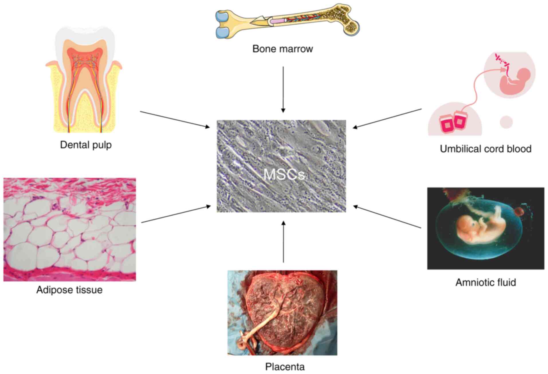
MSCs are adult stem cells derived from the mesoderm
that can be isolated from various tissues, including bone marrow,
adipose tissue, dental pulp, placenta, amniotic fluid, umbilical
cord, Wharton's jelly and umbilical cord blood (51,52)
(Fig. 1). Although MSCs derived from
these tissues contain diverse background genetic lineages, they can
exert intrinsic and extrinsic effects, and MSCs cultured in
vitro may share common features in agreement with the
International Society of Cell Therapy (ISCT) criteria established
in 2006 (53). Firstly, under in
vitro culture conditions, MSCs exhibit spindle-shaped or
fusiform morphology. Secondly, in vitro cultured MSCs
express CD73, CD90 and CD105 markers on their surface; however,
they express no monocyte markers, such as HLA-DR, CD14 or CD11b,
CD79α or CD19, and no hematopoietic markers, such as CD34 and CD45
(53). In addition, MSCs can
differentiate into osteoblasts, adipocytes and chondroblasts
following specific in vitro differentiation conditions
(53). Although MSCs have the
potential to express surface antigens and differentiate, other
characteristics of MSCs that would support anti-tumor therapeutic
interests are vital. In the following section, MSC functions,
including inherent tumor tropisms, as well as the immunosuppression
and paracrine characteristics of anti-tumor MSC carrying OVs will
therefore be discussed.
MSCs migrate to damaged tissue or inflammatory sites
and release simultaneous secretory cytokines (58,59). In
addition to tumor cells, the TME also contains immune cells,
fibroblasts, vascular endothelial cells, adipocytes and tumor
stromal cells, which secrete large numbers of cytokines, such as
vascular endothelial growth factor (VEGF), platelet derived growth
factor (PDGF), interleukin (IL)-8, IL-6, stromal cell-derived
factor-1 (SDF-1), basic fibroblast growth factor (bFGF),
granulocyte colony-stimulating factor (G-CSF),
granulocyte-macrophage colony-stimulating factor (GM-CSF), monocyte
chemoattractant protein-1 (MCP-1), hepatocyte growth factor (HGF),
tumor necrosis factor-α (TNF-α), transforming growth factor-β
(TGF-β), urokinase type plasminogen activator receptor, vascular
cell and intercellular cell adhesion molecules (VCAM, ICAM), C-X-C
motif chemokine ligand-12 (CXCL-12), C-C motif chemokine ligand-2
(CCL-2), C-C motif chemokine ligand-3 (CCL-3), C-C motif chemokine
receptor 4 (CCR4) and C-X-C motif chemokine receptor 4 (CXCR4)
(59–63).
MSC immunological characteristics serve crucial
roles in the therapeutic efficacy of MSCs loaded with OVs towards
tumors. Evidence indicates that MSCs amplified in vitro do
not express HLA-II or costimulatory molecules, such as CD40, CD80,
CD83, CD86 and CD154 (67).
Therefore, no additional immunosuppressants are required for
autologous or allogeneic MSC transplantation. In addition, MSCs
exert strong immunosuppressive functions. For example, MSCs produce
and release a variety of soluble cytokines, including IL-6, IL-10,
TGF-β, heme oxygenase-1, inducible nitric oxide synthase and
indoleamine-2-dioxygenase-3 (68),
which play major roles in immunosuppression. At present, MSCs are
used for immunomodulation, mostly for immune rejection and
autoimmune diseases, such as hematopoietic stem cell
transplantation, organ transplantation, rheumatoid arthritis and
systemic lupus erythematosus (69,70).
However, the underlying mechanisms of MSC immunosuppressive
function in vivo remain unclear.
In recent years, increasing evidence from
preclinical and clinical studies has indicated that MSCs exert
immunosuppressive functions by inhibiting the activity of certain
types of immune cell, including T, B lymphocytes and NKs, thereby
affecting monocytes, DC and macrophage function (71–74).
MSCs affect the activation, proliferation, maturation, cytokine
production and cytotoxic activity of innate and adaptive immune
cells (68). Indeed, MSCs can reduce
cytokine secretion from helper T cells, weaken the killing effects
of effector T lymphocytes (75),
hinder B lymphocyte differentiation and impede their ability to
secrete immunoglobulin (76,77), and inhibit INF-γ secretion by NK
cells and reduce their killing effects (78). In addition, MSCs prevent
CD14+ monocytes and CD34+ progenitor cells
from differentiating into mature DC cells (79). Importantly, MSCs promote the
emergence of regulatory immune subsets, including
CD8+CD28− T lymphocytes (80),
CD4+CD25+FOXP3+ T lymphocytes
(81), IL-10-producing B lymphocytes
(82) and IL-10-producing DCs
(83). Therefore, inhibiting immune
cell functions and promoting the emergence of regulatory immune
cell subsets, could serve positive roles in MSC immunosuppressive
functions. These functions are key MSC features in protecting OVs
from immune system clearance, and a guarantee to enhance OV spread
and increase viral persistence (84).
It has been reported that MSCs promote tumorigenesis
through various mechanisms, such as inhibition of local immune
responses (51), stimulation of
epithelial-mesenchymal transformation, inhibition of tumor cell
apoptosis and promotion of angiogenesis and tumor metastasis
(85). Previous studies have
demonstrated that MSCs, in contrast to their tumorigenic functions,
can inhibit tumor growth by inhibiting angiogenesis (86), inducing cell cycle arrest (14,87),
enhancing inflammatory infiltration (88) and inhibiting proliferation-associated
signaling pathways (14).
Although there is some controversy over whether MSCs
inhibit or promote tumor growth, emerging evidence indicates that
oncolytic adenovirus (OAD)-infected MSCs induce anti-tumor immune
responses and increase leukocyte infiltration into tumor lesions
(89). Similarly, Mahasa et
al (10) predicted the
therapeutic efficacy of MSCs loaded with OAD in a Hep3B cell tumor
model using an integrated mathematical-experimental model, and
demonstrated that MSCs loaded with OAD can promote tumor
therapeutic efficacy. In addition, a phase I clinical trial
(NCT01844661) of bone marrow-derived MSCs carrying Celyvir for the
treatment of metastatic or refractory tumors was completed and
reported that the combination of MSCs and Celyvir is safe (90). Following treatment with MSCs carrying
Celyvir, except for the increase in the amount of oncolytic virus
administered to patients, minimizing toxicities and avoiding direct
tumor injections, no grades 2–5 toxicities were reported (90). However, the safety and efficacy of
MSCs carrying Celyvir require further evaluation in a phase II
setting.
The majority of preclinical studies indicate
efficacy factors for MSCs as carriers for OV delivery (92–94). Du
et al (95) used MSCs as
cellular carriers for oncolytic herpes simplex virus (HSV) in order
to assess efficacy in immune-deficient and immune-competent mouse
melanoma metastasis models. The results demonstrated that
transplanted MSCs carrying HSV could migrate to the tumor site and
significantly prolong mouse survival. Furthermore, in
immune-competent mice, the combination of MSC-HSV and the
anti-programmed death ligand 1 (anti-PD-L1) immune checkpoint
inhibitor could increase CD8+ T lymphocyte infiltration,
leading to the production of IFN-γ and significant prolongation of
mouse survival.
For enveloped OVs, MSCs can deliver viruses to tumor
sites via hetero-cellular fusion. Ong et al (96) loaded bone marrow-derived MSCs with
oncolytic measles virus, and co-cultured them with human
hepatocellular carcinoma cells in vitro. The results
demonstrated that syncytia number increases when MSCs carries the
measles virus, which is not the case with non-enveloped virus.
Furthermore, in the presence of high titer anti-measles virus
antibodies, virus-infected MSCs significantly induce heterocellular
formation when compared with naked virus. In addition, MSCs
precisely deliver the measles virus to tumor lesions in a
patient-derived hepatocellular carcinoma model (96). These results were consistent with
Castleton et al (97) who
reported MSC delivery of the measles virus in a model for acute
lymphoblastic leukemia, suggesting that OV infected MSCs could
significantly prolong survival and improve anti-tumor efficacy when
compared with the naked virus.
In addition, genetic engineering improves MSC
delivery efficiency, enhances viral oncolytic activity and reduces
virotherapy side effects. Yoon et al (55) reported that the OAD infection
capability of MSCs is enhanced after modification of the fiber
domain of OADs, allowing the virus to replicate efficiently in
MSCs. These MSCs infected with OADs could effectively lyse
hepatocellular carcinoma cells in vitro. Importantly,
following MSC-OAD transplantation, MSCs home to the tumor site,
facilitating a high accumulation of virions at the site, and
ultimately leading to tumor growth inhibition. In another study,
Kaczorowski et al (66)
deleted the anti-apoptotic gene E1B19K from OAD and inserted the
cell death ligand TRAIL gene of OAD. After intravenous injection of
virally infected MSCs, adenovirus capsid protein is detected in
tumor xenografts established by cancer stem cell of pancreatic
ductal adenocarcinoma. Similarly, following viral MSC treatment,
the tumor size decreases significantly, the tumor cell
proliferation-associated Ki67 and CD24 expression decreases and the
tumor cell apoptosis-associated caspase-3 activity increases
(66). In addition, OADs
significantly increase virus release from MSCs following the
deletion of the anti-apoptotic virus gene E1B19K, or the
overexpression of the cell death ligand TRAIL, while MSC migration
ability remains unaffected (98).
These data suggest that genetic modification of OADs can induce
effective oncolysis, which may represent a promising strategy for
OVs in clinical applications. Similarly, MSCs as carriers for the
delivery of genetically modified OVs may be considered as a useful
method for improving oncolytic virotherapy efficacy (Table I)(99–106).
However, MSCs can also be modified by genetic modification or
preconditioned to modification in order to improve their inherent
properties, such as enhanced migration, adhesion and survival, and
reduced premature senescence (107). OV delivery and virotherapy efficacy
may therefore be improved.
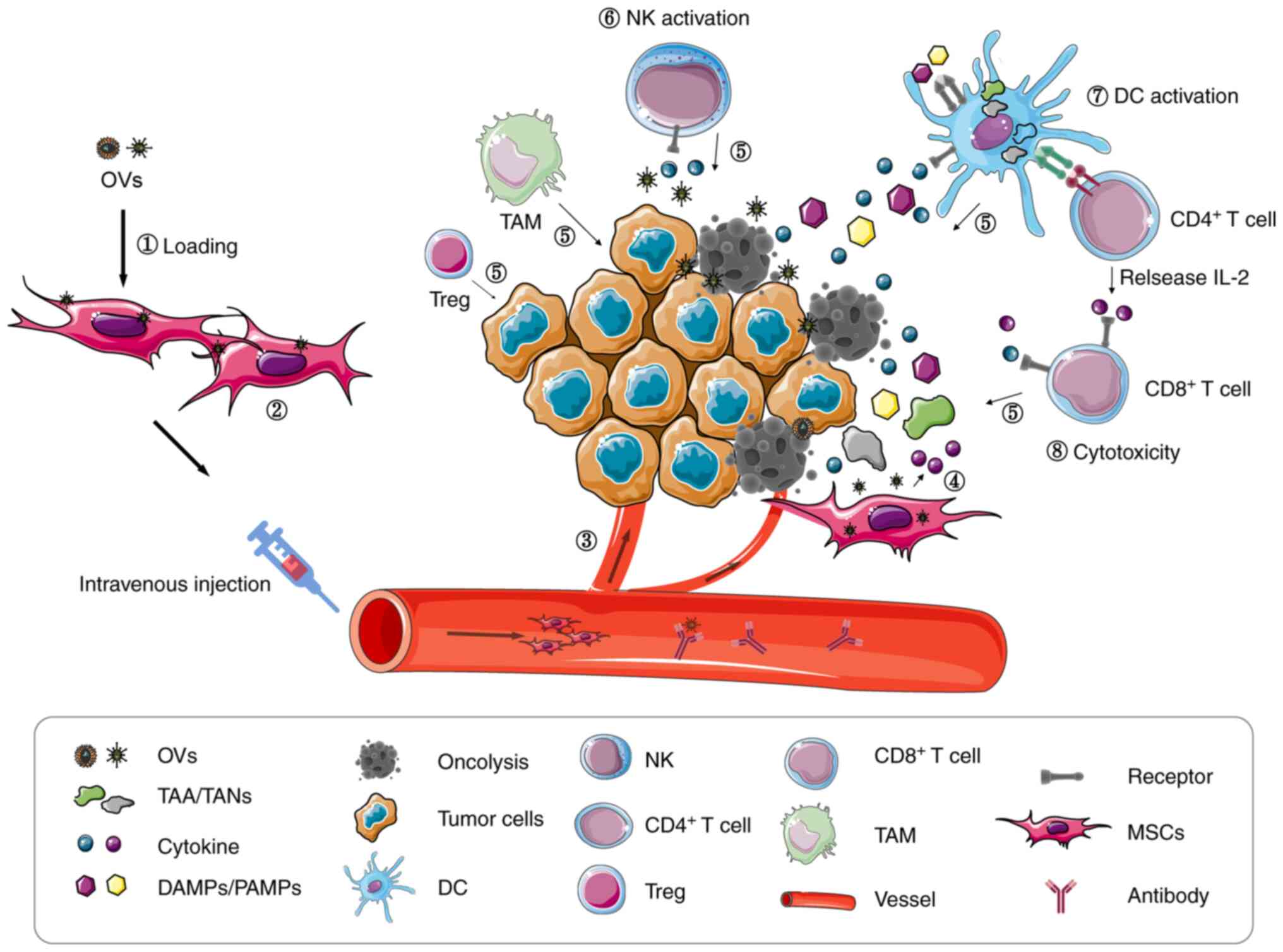
In summary, MSCs enhance the anti-tumor efficacy of
virotherapy through numerous factors. Firstly, MSCs provide a
replication location for OVs, facilitating the production of more
virus particles, which is beneficial for virotherapy. Secondly, the
tumor tropism and immunosuppression function of MSCs allow the
virus to accurately reach the tumor site and enhance the
transmission and persistence of the virus. Thirdly, oncolysis leads
to the release of ‘dangerous’ signals, such as TAAs/TANs and
DAMPs/PAMPs, activating local anti-tumor immune responses, and
converting the TME from an immunosuppressive to an
immunostimulatory environment (93,103).
However, cytokines released by MSCs recruit immune cells to the
TME, further enhancing the anti-tumor immune response. Therefore,
MSC carriers are considered as promising cellular vehicles for OV
delivery. Assuming the high quality of MSCs and appropriate
conditions of MSCs loading the virus, it is worth treating
malignant tumors with such therapy, which could lead to a restrain
of tumor growth progression in patients. However, further
investigation is required to evaluate the effects of MSC loading
viruses and explore the immune regulation mechanisms of MSCs on
anti-viral and anti-tumor immune responses.
In TME, cancer-associated fibroblasts, adipocytes,
Tregs, mesenchymal stromal cells and tumor-associated macrophages
release numerous cytokines, such as IL-10, which support immune
evasion and tumor growth (108). In
recent years, oncolytic virotherapy (OVT) has been demonstrated to
relieve the tumor immunosuppressive environments, and enhance
anti-tumor immune responses (109,110).
OVs stimulate anti-tumor immune responses which in turn, enhance
the efficacy of immune checkpoint inhibitors (ICIs) (111). For this reason, emerging evidence
from preclinical and clinical trials has indicated that combined
OVT and ICIs could improve the anti-tumor therapeutic efficacy
(112–114). In view of the contribution of MSCs
to the activation of immune responses in virotherapy, combined MSC
loading OVs with ICIs could be considered as a major therapeutic
area for future anti-tumor research.
Not applicable.
This study was supported by the National Natural
Science Foundation of China (grant no. 81871313), the Graduate
Student Innovation Program in Guizhou Province [grant no. Qian Jiao
He YJSCXJH (2020) 143], Key projects of Guizhou Provincial
Department of Science and Technology [grant no. Qian Ke He Zhi
Cheng (2020) 4Y192], the Guizhou Provincial Natural Science
Foundation [grant no. (2019)5663], the Program for Top Scientifc
and Technological Talents in Guizhou Province [grant no. KY
(2018)049], the Guizhou Province Science and Technology Talent
Platform Project [grant no. (2019)5406], the Non-profit Central
Research Institute Fund of Chinese Academy of Medical Sciences
(grant nos. 2018PT31048 and 2019PT310013) and the Special Grant for
Central Government Supporting Local Science and Technology
Development, Science and Technology Department of Guizhou Province
[grant no. (2019)4008].
Not applicable.
XW and ZH conceived the review. XW wrote the review.
ZH and XZ revised the review. XW, XZ and ZH proofread the
manuscript and revised the manuscript for intellectual content. All
authors read and approved the final version.
Not applicable.
Not applicable.
The authors declare that they have no competing
interests.
|
1
|
Hayes C: Cellular immunotherapies for
cancer. Ir J Med Sci. Jul 1–2020.(Epub ahead of print). doi:
10.1007/s11845-020-02264-w.
|
|
2
|
Alard E, Butnariu AB, Grillo M, Kirkham C,
Zinovkin DA, Newnham L, Macciochi J and Pranjol MZI: Advances in
anti-cancer immunotherapy: Car-T cell, checkpoint inhibitors,
dendritic cell vaccines, and oncolytic viruses, and emerging
cellular and molecular targets. Cancers (Basel). 12:18262020.
View Article : Google Scholar
|
|
3
|
Hemminki O, Dos Santos JM and Hemminki A:
Oncolytic viruses for cancer immunotherapy. J Hematol Oncol.
13:842020. View Article : Google Scholar : PubMed/NCBI
|
|
4
|
Romero D: Immunotherapy: Oncolytic viruses
prime antitumour immunity. Nat Rev Clin Oncol. 15:1352018.
View Article : Google Scholar : PubMed/NCBI
|
|
5
|
Engeland CE and Bell JC: Introduction to
oncolytic virotherapy. Methods Mol Biol. 2058:1–6. 2020. View Article : Google Scholar : PubMed/NCBI
|
|
6
|
Kolb EA, Sampson V, Stabley D, Walter A,
Sol-Church K, Cripe T, Hingorani P, Ahern CH, Weigel BJ, Zwiebel J
and Blaney SM: A phase I trial and viral clearance study of
reovirus (Reolysin) in children with relapsed or refractory
extra-cranial solid tumors: A children's oncology group phase I
consortium report. Pediatr Blood Cancer. 62:751–758. 2015.
View Article : Google Scholar : PubMed/NCBI
|
|
7
|
Hamid O, Ismail R and Puzanov I:
Intratumoral immunotherapy-update 2019. Oncologist. 25:e423–e438.
2020. View Article : Google Scholar : PubMed/NCBI
|
|
8
|
Roy DG, Bell JC and Bourgeois-Daigneault
MC: Magnetic targeting of oncolytic VSV-based therapies improves
infection of tumor cells in the presence of virus-specific
neutralizing antibodies in vitro. Biochem Biophys Res Commun.
526:641–646. 2020. View Article : Google Scholar : PubMed/NCBI
|
|
9
|
Schirrmacher V, van Gool S and Stuecker W:
Breaking therapy resistance: An update on oncolytic newcastle
disease virus for improvements of cancer therapy. Biomedicines.
7:662019. View Article : Google Scholar
|
|
10
|
Mahasa KJ, de Pillis L, Ouifki R, Eladdadi
A, Maini P, Yoon AR and Yun CO: Mesenchymal stem cells used as
carrier cells of oncolytic adenovirus results in enhanced oncolytic
virotherapy. Sci Rep. 10:4252020. View Article : Google Scholar : PubMed/NCBI
|
|
11
|
Hadrys A, Sochanik A, McFadden G and
Jazowiecka-Rakus J: Mesenchymal stem cells as carriers for systemic
delivery of oncolytic viruses. Eur J Pharmacol. 874:1729912020.
View Article : Google Scholar : PubMed/NCBI
|
|
12
|
Naseri Z, Oskuee RK, Forouzandeh-Moghadam
M and Jaafari MR: Delivery of LNA-antimiR-142-3p by mesenchymal
stem cells-derived exosomes to breast cancer stem cells reduces
tumorigenicity. Stem Cell Rev Rep. 16:541–556. 2020. View Article : Google Scholar : PubMed/NCBI
|
|
13
|
Altaner C and Altanerova U: Mesenchymal
stem cell exosome-mediated prodrug gene therapy for cancer. Methods
Mol Biol. 1895:75–85. 2019. View Article : Google Scholar : PubMed/NCBI
|
|
14
|
Kostadinova M and Mourdjeva M: Potential
of mesenchymal stem cells in anti-cancer therapies. Curr Stem Cell
Res Ther. 15:482–491. 2020. View Article : Google Scholar : PubMed/NCBI
|
|
15
|
Spaggiari GM, Capobianco A, Abdelrazik H,
Becchetti F, Mingari MC and Moretta L: Mesenchymal stem cells
inhibit natural killer-cell proliferation, cytotoxicity, and
cytokine production: Role of indoleamine 2,3-dioxygenase and
prostaglandin E2. Blood. 111:1327–1333. 2008. View Article : Google Scholar : PubMed/NCBI
|
|
16
|
Jiang XX, Zhang Y, Liu B, Zhang SX, Wu Y,
Yu XD and Mao N: Human mesenchymal stem cells inhibit
differentiation and function of monocyte-derived dendritic cells.
Blood. 105:4120–4126. 2005. View Article : Google Scholar : PubMed/NCBI
|
|
17
|
Lei J, Jacobus EJ, Taverner WK, Fisher KD,
Hemmi S, West K, Slater L, Lilley F, Brown A, Champion B, et al:
Expression of human CD46 and trans-complementation by murine
adenovirus 1 fails to allow productive infection by a group B
oncolytic adenovirus in murine cancer cells. J Immunother Cancer.
6:552018. View Article : Google Scholar : PubMed/NCBI
|
|
18
|
Koehler M, Aravamudhan P, Guzman-Cardozo
C, Dumitru AC, Yang J, Gargiulo S, Soumillion P, Dermody TS and
Alsteens D: Glycan-mediated enhancement of reovirus receptor
binding. Nat Commun. 10:44602019. View Article : Google Scholar : PubMed/NCBI
|
|
19
|
Phillips MB, Stuart JD, Rodriguez Stewart
RM, Berry JT, Mainou BA and Boehme KW: Current understanding of
reovirus oncolysis mechanisms. Oncolytic Virother. 7:53–63. 2018.
View Article : Google Scholar : PubMed/NCBI
|
|
20
|
Sakurai F, Inoue S, Kaminade T, Hotani T,
Katayama Y, Hosoyamada E, Terasawa Y, Tachibana M and Mizuguchi H:
Cationic liposome-mediated delivery of reovirus enhances the tumor
cell-killing efficiencies of reovirus in reovirus-resistant tumor
cells. Int J Pharm. 524:238–247. 2017. View Article : Google Scholar : PubMed/NCBI
|
|
21
|
Mahalingam D, Goel S, Aparo S, Patel Arora
S, Noronha N, Tran H, Chakrabarty R, Selvaggi G, Gutierrez A,
Coffey M, et al: A phase II study of pelareorep
(REOLYSIN(R)) in combination with gemcitabine for
patients with advanced pancreatic adenocarcinoma. Cancers (Basel).
10:1602018. View Article : Google Scholar
|
|
22
|
Jonker DJ, Tang PA, Kennecke H, Welch SA,
Cripps MC, Asmis T, Chalchal H, Tomiak A, Lim H, Ko YJ, et al: A
randomized phase II study of FOLFOX6/bevacizumab with or without
pelareorep in patients with metastatic colorectal cancer: IND.210,
a canadian cancer trials group trial. Clin Colorectal Cancer.
17:231–239 e7. 2018. View Article : Google Scholar : PubMed/NCBI
|
|
23
|
Davola ME and Mossman KL: Oncolytic
viruses: How ‘lytic’ must they be for therapeutic efficacy?
Oncoimmunology. 8:e15815282019. View Article : Google Scholar : PubMed/NCBI
|
|
24
|
Luo Y, Lin C, Zou Y, Ju F, Ren W, Lin Y,
Wang Y, Huang X, Liu H, Yu Z, et al: Tumor-targeting oncolytic
virus elicits potent immunotherapeutic vaccine responses to tumor
antigens. Oncoimmunology. 9:17261682020. View Article : Google Scholar : PubMed/NCBI
|
|
25
|
Pidelaserra-Marti G and Engeland CE:
Mechanisms of measles virus oncolytic immunotherapy. Cytokine
Growth Factor Rev. 56:28–38. 2020. View Article : Google Scholar : PubMed/NCBI
|
|
26
|
Pol JG, Bridle BW and Lichty BD: Detection
of tumor antigen-specific T-cell responses after oncolytic
vaccination. Methods Mol Biol. 2058:191–211. 2020. View Article : Google Scholar : PubMed/NCBI
|
|
27
|
Keshavarz M, Solaymani-Mohammadi F, Miri
SM and Ghaemi A: Oncolytic paramyxoviruses-induced autophagy; a
prudent weapon for cancer therapy. J Biomed Sci. 26:482019.
View Article : Google Scholar : PubMed/NCBI
|
|
28
|
Bommareddy PK, Zloza A, Rabkin SD and
Kaufman HL: Oncolytic virus immunotherapy induces immunogenic cell
death and overcomes STING deficiency in melanoma. Oncoimmunology.
8:15918752019. View Article : Google Scholar : PubMed/NCBI
|
|
29
|
Ma J, Ramachandran M, Jin C, Quijano-Rubio
C, Martikainen M, Yu D and Essand M: Characterization of
virus-mediated immunogenic cancer cell death and the consequences
for oncolytic virus-based immunotherapy of cancer. Cell Death Dis.
11:482020. View Article : Google Scholar : PubMed/NCBI
|
|
30
|
Wang X, Shao X, Gu L, Jiang K, Wang S,
Chen J, Fang J, Guo X, Yuan M, Shi J, et al: Targeting STAT3
enhances NDV-induced immunogenic cell death in prostate cancer
cells. J Cell Mol Med. 24:4286–4297. 2020. View Article : Google Scholar : PubMed/NCBI
|
|
31
|
Shao X, Wang X, Guo X, Jiang K, Ye T, Chen
J, Fang J, Gu L, Wang S, Zhang G, et al: STAT3 contributes to
oncolytic newcastle disease virus-induced immunogenic cell death in
melanoma cells. Front Oncol. 9:4362019. View Article : Google Scholar : PubMed/NCBI
|
|
32
|
Xu Q, Rangaswamy US, Wang W, Robbins SH,
Harper J, Jin H and Cheng X: Evaluation of newcastle disease virus
mediated dendritic cell activation and cross-priming tumor-specific
immune responses ex vivo. Int J Cancer. 146:531–541. 2020.
View Article : Google Scholar : PubMed/NCBI
|
|
33
|
Garg AD and Agostinis P: Cell death and
immunity in cancer: From danger signals to mimicry of pathogen
defense responses. Immunol Rev. 280:126–148. 2017. View Article : Google Scholar : PubMed/NCBI
|
|
34
|
Jiang H and Fueyo J: Healing after death:
Antitumor immunity induced by oncolytic adenoviral therapy.
Oncoimmunology. 3:e9478722014. View Article : Google Scholar : PubMed/NCBI
|
|
35
|
Kepp O, Senovilla L, Vitale I, Vacchelli
E, Adjemian S, Agostinis P, Apetoh L, Aranda F, Barnaba V, Bloy N,
et al: Consensus guidelines for the detection of immunogenic cell
death. Oncoimmunology. 3:e9556912014. View Article : Google Scholar : PubMed/NCBI
|
|
36
|
Garg AD, Galluzzi L, Apetoh L, Baert T,
Birge RB, Bravo-San Pedro JM, Breckpot K, Brough D, Chaurio R,
Cirone M, et al: Molecular and translational classifications of
DAMPs in immunogenic cell death. Front Immunol. 6:5882015.
View Article : Google Scholar : PubMed/NCBI
|
|
37
|
Das K, Urbiola C, Spiesschaert B, Mueller
P and Wollmann G: Analysis of immunological treatment effects of
virotherapy in tumor tissue. Methods Mol Biol. 2058:155–177. 2020.
View Article : Google Scholar : PubMed/NCBI
|
|
38
|
Reale A, Vitiello A, Conciatori V, Parolin
C, Calistri A and Palu G: Perspectives on immunotherapy via
oncolytic viruses. Infect Agent Cancer. 14:52019. View Article : Google Scholar : PubMed/NCBI
|
|
39
|
Sobol PT, Boudreau JE, Stephenson K, Wan
Y, Lichty BD and Mossman KL: Adaptive antiviral immunity is a
determinant of the therapeutic success of oncolytic virotherapy.
Mol Ther. 19:335–344. 2011. View Article : Google Scholar : PubMed/NCBI
|
|
40
|
Gujar S, Pol JG, Kim Y, Lee PW and Kroemer
G: Antitumor benefits of antiviral immunity: An underappreciated
aspect of oncolytic virotherapies. Trends Immunol. 39:209–221.
2018. View Article : Google Scholar : PubMed/NCBI
|
|
41
|
Ledford H: Cancer-fighting viruses win
approval. Nature. 526:622–623. 2015. View Article : Google Scholar : PubMed/NCBI
|
|
42
|
O'Donoghue C, Doepker MP and Zager JS:
Talimogene laherparepvec: Overview, combination therapy and current
practices. Melanoma Manag. 3:267–272. 2016. View Article : Google Scholar : PubMed/NCBI
|
|
43
|
Sunshine JC, Sosman J, Shetty A and Choi
JN: Successful treatment of in-transit metastatic melanoma in a
renal transplant patient with combination T-VEC/Imiquimod
immunotherapy. J Immunother. 43:149–152. 2020. View Article : Google Scholar : PubMed/NCBI
|
|
44
|
Masoud SJ, Hu JB, Beasley GM, Stewart JH
IV and Mosca PJ: Efficacy of talimogene laherparepvec (T-VEC)
therapy in patients with in-transit melanoma metastasis decreases
with increasing lesion size. Ann Surg Oncol. 26:4633–4641. 2019.
View Article : Google Scholar : PubMed/NCBI
|
|
45
|
Howard F and Muthana M: Designer
nanocarriers for navigating the systemic delivery of oncolytic
viruses. Nanomedicine (Lond). 15:93–110. 2020. View Article : Google Scholar : PubMed/NCBI
|
|
46
|
Phan M, Watson MF, Alain T and Diallo JS:
Oncolytic viruses on drugs: Achieving higher therapeutic efficacy.
ACS Infect Dis. 4:1448–1467. 2018. View Article : Google Scholar : PubMed/NCBI
|
|
47
|
Rosewell Shaw A and Suzuki M: Oncolytic
viruses partner with T-cell therapy for solid tumor treatment.
Front Immunol. 9:21032018. View Article : Google Scholar : PubMed/NCBI
|
|
48
|
Hwang CC, Igase M, Sakurai M, Haraguchi T,
Tani K, Itamoto K, Shimokawa T, Nakaichi M, Nemoto Y, Noguchi S, et
al: Oncolytic reovirus therapy: Pilot study in dogs with
spontaneously occurring tumours. Vet Comp Oncol. 16:229–238. 2018.
View Article : Google Scholar : PubMed/NCBI
|
|
49
|
Mok DZL and Chan KR: The effects of
pre-existing antibodies on live-attenuated viral vaccines. Viruses.
12:5202020. View Article : Google Scholar
|
|
50
|
Harrington K, Freeman DJ, Kelly B, Harper
J and Soria JC: Optimizing oncolytic virotherapy in cancer
treatment. Nat Rev Drug Discov. 18:689–706. 2019. View Article : Google Scholar : PubMed/NCBI
|
|
51
|
Naji A, Eitoku M, Favier B, Deschaseaux F,
Rouas-Freiss N and Suganuma N: Biological functions of mesenchymal
stem cells and clinical implications. Cell Mol Life Sci.
76:3323–3348. 2019. View Article : Google Scholar : PubMed/NCBI
|
|
52
|
Volarevic V, Markovic BS, Gazdic M,
Volarevic A, Jovicic N, Arsenijevic N, Armstrong L, Djonov V, Lako
M and Stojkovic M: Ethical and safety issues of stem cell-based
therapy. Int J Med Sci. 15:36–45. 2018. View Article : Google Scholar : PubMed/NCBI
|
|
53
|
Dominici M, Le Blanc K, Mueller I,
Slaper-Cortenbach I, Marini F, Krause D, Deans R, Keating A,
Prockop DJ and Horwitz E: Minimal criteria for defining multipotent
mesenchymal stromal cells. The international society for cellular
therapy position statement. Cytotherapy. 8:315–317. 2006.
View Article : Google Scholar : PubMed/NCBI
|
|
54
|
Salmasi Z, Hashemi M, Mahdipour E, Nourani
H, Abnous K and Ramezani M: Mesenchymal stem cells engineered by
modified polyethylenimine polymer for targeted cancer gene therapy,
in vitro and in vivo. Biotechnol Prog. 36:e30252020. View Article : Google Scholar : PubMed/NCBI
|
|
55
|
Yoon AR, Hong J, Li Y, Shin HC, Lee H, Kim
HS and Yun CO: Mesenchymal stem cell-mediated delivery of an
oncolytic adenovirus enhances antitumor efficacy in hepatocellular
carcinoma. Cancer Res. 79:4503–4514. 2019. View Article : Google Scholar : PubMed/NCBI
|
|
56
|
Vangala G, Imhoff FM, Squires CML, Cridge
AG and Baird SK: Mesenchymal stem cell homing towards cancer cells
is increased by enzyme activity of cathepsin D. Exp Cell Res.
383:1114942019. View Article : Google Scholar : PubMed/NCBI
|
|
57
|
Kwon S, Yoo KH, Sym SJ and Khang D:
Mesenchymal stem cell therapy assisted by nanotechnology: A
possible combinational treatment for brain tumor and central nerve
regeneration. Int J Nanomedicine. 14:5925–5942. 2019. View Article : Google Scholar : PubMed/NCBI
|
|
58
|
Thomas JG, Parker Kerrigan BC, Hossain A,
Gumin J, Shinojima N, Nwajei F, Ezhilarasan R, Love P, Sulman EP
and Lang FF: Ionizing radiation augments glioma tropism of
mesenchymal stem cells. J Neurosurg. 128:287–295. 2018. View Article : Google Scholar : PubMed/NCBI
|
|
59
|
Choi SA, Lee JY, Kwon SE, Wang KC, Phi JH,
Choi JW, Jin X, Lim JY, Kim H and Kim SK: Human adipose
tissue-derived mesenchymal stem cells target brain tumor-initiating
cells. PLoS One. 10:e01292922015. View Article : Google Scholar : PubMed/NCBI
|
|
60
|
Verdelli C, Vaira V and Corbetta S:
Parathyroid tumor microenvironment. Adv Exp Med Biol. 1226:37–50.
2020. View Article : Google Scholar : PubMed/NCBI
|
|
61
|
Karagiannis K, Proklou A, Tsitoura E,
Lasithiotaki I, Kalpadaki C, Moraitaki D, Sperelakis I, Kontakis G,
Antoniou KM and Tzanakis N: Impaired mRNA expression of the
migration related chemokine receptor CXCR4 in mesenchymal stem
cells of COPD patients. Int J Inflam. 2017:60894252017. View Article : Google Scholar : PubMed/NCBI
|
|
62
|
Armakolas A, Dimakakos A, Loukogiannaki C,
Armakolas N, Antonopoulos A, Florou C, Tsioli P, Papageorgiou E,
Alexandrou TP, Stathaki M, et al: IL-6 is associated to IGF-1Ec
upregulation and Ec peptide secretion, from prostate tumors. Mol
Med. 24:62018. View Article : Google Scholar : PubMed/NCBI
|
|
63
|
Lejmi E, Perriraz N, Clement S, Morel P,
Baertschiger R, Christofilopoulos P, Meier R, Bosco D, Buhler LH
and Gonelle-Gispert C: Inflammatory chemokines MIP-1δ and MIP-3α
are involved in the migration of multipotent mesenchymal stromal
cells induced by hepatoma cells. Stem Cells Dev. 24:1223–1235.
2015. View Article : Google Scholar : PubMed/NCBI
|
|
64
|
Pavon LF, Sibov TT, de Souza AV, da Cruz
EF, Malheiros SM, Cabral FR, de Souza JG, Boufleur P, de Oliveira
DM, de Toledo SR, et al: Tropism of mesenchymal stem cell toward
CD133+ stem cell of glioblastoma in vitro and promote
tumor proliferation in vivo. Stem Cell Res Ther. 9:3102018.
View Article : Google Scholar : PubMed/NCBI
|
|
65
|
Ramirez M, Garcia-Castro J, Melen GJ,
Gonzalez-Murillo A and Franco-Luzon L: Patient-derived mesenchymal
stem cells as delivery vehicles for oncolytic virotherapy: Novel
state-of-the-art technology. Oncolytic Virother. 4:149–155. 2015.
View Article : Google Scholar : PubMed/NCBI
|
|
66
|
Kaczorowski A, Hammer K, Liu L, Villhauer
S, Nwaeburu C, Fan P, Zhao Z, Gladkich J, Gross W, Nettelbeck DM
and Herr I: Delivery of improved oncolytic adenoviruses by
mesenchymal stromal cells for elimination of tumorigenic pancreatic
cancer cells. Oncotarget. 7:9046–9059. 2016. View Article : Google Scholar : PubMed/NCBI
|
|
67
|
Mehler VJ, Burns C and Moore ML: Concise
review: Exploring immunomodulatory features of mesenchymal stromal
cells in humanized mouse models. Stem Cells. 37:298–305. 2019.
View Article : Google Scholar : PubMed/NCBI
|
|
68
|
Gao F, Chiu SM, Motan DA, Zhang Z, Chen L,
Ji HL, Tse HF, Fu QL and Lian Q: Mesenchymal stem cells and
immunomodulation: Current status and future prospects. Cell Death
Dis. 7:e20622016. View Article : Google Scholar : PubMed/NCBI
|
|
69
|
Abbasi-Kangevari M, Ghamari SH,
Safaeinejad F, Bahrami S and Niknejad H: Potential therapeutic
features of human amniotic mesenchymal stem cells in multiple
sclerosis: Immunomodulation, inflammation suppression, angiogenesis
promotion, oxidative stress inhibition, neurogenesis induction,
MMPs regulation, and remyelination stimulation. Front Immunol.
10:2382019. View Article : Google Scholar : PubMed/NCBI
|
|
70
|
Ma ZJ, Wang YH, Li ZG, Wang Y, Li BY, Kang
HY and Wu XY: Immunosuppressive effect of exosomes from mesenchymal
stromal cells in defined medium on experimental colitis. Int J Stem
Cells. 12:440–448. 2019. View Article : Google Scholar : PubMed/NCBI
|
|
71
|
Carreras-Planella L, Monguio-Tortajada M,
Borras FE and Franquesa M: Immunomodulatory effect of MSC on B
cells is independent of secreted extracellular vesicles. Front
Immunol. 10:12882019. View Article : Google Scholar : PubMed/NCBI
|
|
72
|
Wilson A, Chee M, Butler P and Boyd AS:
Isolation and characterisation of human adipose-derived stem cells.
Methods Mol Biol. 1899:3–13. 2019. View Article : Google Scholar : PubMed/NCBI
|
|
73
|
Zhang F, Wang C, Wen X, Chen Y, Mao R, Cui
D, Li L, Liu J, Chen Y, Cheng J and Lu Y: Mesenchymal stem cells
alleviate rat diabetic nephropathy by suppressing CD103+
DCs-mediated CD8+ T cell responses. J Cell Mol Med.
24:5817–5831. 2020. View Article : Google Scholar : PubMed/NCBI
|
|
74
|
Haddad R and Saldanha-Araujo F: Mechanisms
of T-cell immunosuppression by mesenchymal stromal cells: What do
we know so far? Biomed Res Int. 2014:2168062014. View Article : Google Scholar : PubMed/NCBI
|
|
75
|
Rozenberg A, Rezk A, Boivin MN, Darlington
PJ, Nyirenda M, Li R, Jalili F, Winer R, Artsy EA, Uccelli A, et
al: Human mesenchymal stem cells impact Th17 and Th1 responses
through a prostaglandin E2 and myeloid-dependent mechanism. Stem
Cells Transl Med. 5:1506–1514. 2016. View Article : Google Scholar : PubMed/NCBI
|
|
76
|
Khare D, Or R, Resnick I, Barkatz C,
Almogi-Hazan O and Avni B: Mesenchymal stromal cell-derived
exosomes affect mRNA expression and function of B-lymphocytes.
Front Immunol. 9:30532018. View Article : Google Scholar : PubMed/NCBI
|
|
77
|
Corcione A, Benvenuto F, Ferretti E,
Giunti D, Cappiello V, Cazzanti F, Risso M, Gualandi F, Mancardi
GL, Pistoia V and Uccelli A: Human mesenchymal stem cells modulate
B-cell functions. Blood. 107:367–372. 2006. View Article : Google Scholar : PubMed/NCBI
|
|
78
|
Rezaei Kahmini F, Shahgaldi S and Moazzeni
SM: Mesenchymal stem cells alter the frequency and cytokine profile
of natural killer cells in abortion-prone mice. J Cell Physiol.
235:7214–7223. 2020. View Article : Google Scholar : PubMed/NCBI
|
|
79
|
Xu LL, Fu HX, Zhang JM, Feng FE, Wang QM,
Zhu XL, Xue J, Wang CC, Chen Q, Liu X, et al: Impaired function of
bone marrow mesenchymal stem cells from immune thrombocytopenia
patients in inducing regulatory dendritic cell differentiation
through the Notch-1/Jagged-1 signaling pathway. Stem Cells Dev.
26:1648–1661. 2017. View Article : Google Scholar : PubMed/NCBI
|
|
80
|
Liu Q, Zheng H, Chen X, Peng Y, Huang W,
Li X, Li G, Xia W, Sun Q and Xiang AP: Human mesenchymal stromal
cells enhance the immunomodulatory function of CD8(+)CD28(−)
regulatory T cells. Cell Mol Immunol. 12:708–718. 2015. View Article : Google Scholar : PubMed/NCBI
|
|
81
|
El Omar R, Xiong Y, Dostert G, Louis H,
Gentils M, Menu P, Stoltz JF, Velot E and Decot V: Immunomodulation
of endothelial differentiated mesenchymal stromal cells: Impact on
T and NK cells. Immunol Cell Biol. 94:342–356. 2016. View Article : Google Scholar : PubMed/NCBI
|
|
82
|
Cho KA, Lee JK, Kim YH, Park M, Woo SY and
Ryu KH: Mesenchymal stem cells ameliorate B-cell-mediated immune
responses and increase IL-10-expressing regulatory B cells in an
EBI3-dependent manner. Cell Mol Immunol. 14:895–908. 2017.
View Article : Google Scholar
|
|
83
|
Liu X, Qu X, Chen Y, Liao L, Cheng K, Shao
C, Zenke M, Keating A and Zhao RC: Mesenchymal stem/stromal cells
induce the generation of novel IL-10-dependent regulatory dendritic
cells by SOCS3 activation. J Immunol. 189:1182–1192. 2012.
View Article : Google Scholar : PubMed/NCBI
|
|
84
|
Ahmed AU, Rolle CE, Tyler MA, Han Y,
Sengupta S, Wainwright DA, Balyasnikova IV, Ulasov IV and Lesniak
MS: Bone marrow mesenchymal stem cells loaded with an oncolytic
adenovirus suppress the anti-adenoviral immune response in the
cotton rat model. Mol Ther. 18:1846–1856. 2010. View Article : Google Scholar : PubMed/NCBI
|
|
85
|
Atiya H, Frisbie L, Pressimone C and
Coffman L: Mesenchymal stem cells in the tumor microenvironment.
Adv Exp Med Biol. 1234:31–42. 2020. View Article : Google Scholar : PubMed/NCBI
|
|
86
|
Cai C, Hou L, Zhang J, Zhao D, Wang Z, Hu
H, He J, Guan W and Ma Y: The inhibitory effect of mesenchymal stem
cells with rAd-NK4 on liver cancer. Appl Biochem Biotechnol.
183:444–459. 2017. View Article : Google Scholar : PubMed/NCBI
|
|
87
|
Fathi E, Sanaat Z and Farahzadi R:
Mesenchymal stem cells in acute myeloid leukemia: A focus on
mechanisms involved and therapeutic concepts. Blood Res.
54:165–174. 2019. View Article : Google Scholar : PubMed/NCBI
|
|
88
|
El-Khadragy MF, Nabil HM, Hassan BN,
Tohamy AA, Waaer HF, Yehia HM, Alharbi AM and Moneim AEA: Bone
marrow cell therapy on 1,2-Dimethylhydrazine (DMH)-induced colon
cancer in rats. Cell Physiol Biochem. 45:1072–1083. 2018.
View Article : Google Scholar : PubMed/NCBI
|
|
89
|
Morales-Molina A, Gambera S, Cejalvo T,
Moreno R, Rodriguez-Milla MA, Perise-Barrios AJ and Garcia-Castro
J: Antitumor virotherapy using syngeneic or allogeneic mesenchymal
stem cell carriers induces systemic immune response and
intratumoral leukocyte infiltration in mice. Cancer Immunol
Immunother. 67:1589–1602. 2018. View Article : Google Scholar : PubMed/NCBI
|
|
90
|
Ruano D, Lopez-Martin JA, Moreno L,
Lassaletta A, Bautista F, Andion M, Hernandez C, Gonzalez-Murillo
A, Melen G, Alemany R, et al: First-in-human, first-in-child trial
of autologous MSCs carrying the oncolytic virus Icovir-5 in
patients with advanced tumors. Mol Ther. 28:1033–1042. 2020.
View Article : Google Scholar : PubMed/NCBI
|
|
91
|
Rincon E, Cejalvo T, Kanojia D, Alfranca
A, Rodriguez-Milla MA, Gil Hoyos RA, Han Y, Zhang L, Alemany R,
Lesniak MS and García-Castro J: Mesenchymal stem cell carriers
enhance antitumor efficacy of oncolytic adenoviruses in an
immunocompetent mouse model. Oncotarget. 8:45415–45431. 2017.
View Article : Google Scholar : PubMed/NCBI
|
|
92
|
Banijamali RS, Soleimanjahi H, Soudi S,
Karimi H, Abdoli A, Seyed Khorrami SM and Zandi K: Kinetics of
oncolytic reovirus T3D replication and growth pattern in
mesenchymal stem cells. Cell J. 22:283–292. 2020.PubMed/NCBI
|
|
93
|
Keshavarz M, Ebrahimzadeh MS, Miri SM,
Dianat-Moghadam H, Ghorbanhosseini SS, Mohebbi SR, Keyvani H and
Ghaemi A: Oncolytic newcastle disease virus delivered by
mesenchymal stem cells-engineered system enhances the therapeutic
effects altering tumor microenvironment. Virol J. 17:642020.
View Article : Google Scholar : PubMed/NCBI
|
|
94
|
Hai C, Jin YM, Jin WB, Han ZZ, Cui MN,
Piao XZ, Shen XH, Zhang SN and Sun HH: Application of mesenchymal
stem cells as a vehicle to deliver replication-competent adenovirus
for treating malignant glioma. Chin J Cancer. 31:233–240. 2012.
View Article : Google Scholar : PubMed/NCBI
|
|
95
|
Du W, Seah I, Bougazzoul O, Choi G, Meeth
K, Bosenberg MW, Wakimoto H, Fisher D and Shah K: Stem
cell-released oncolytic herpes simplex virus has therapeutic
efficacy in brain metastatic melanomas. Proc Natl Acad Sci USA.
114:E6157–E6165. 2017. View Article : Google Scholar : PubMed/NCBI
|
|
96
|
Ong HT, Federspiel MJ, Guo CM, Ooi LL,
Russell SJ, Peng KW and Hui KM: Systemically delivered measles
virus-infected mesenchymal stem cells can evade host immunity to
inhibit liver cancer growth. J Hepatol. 59:999–1006. 2013.
View Article : Google Scholar : PubMed/NCBI
|
|
97
|
Castleton A, Dey A, Beaton B, Patel B,
Aucher A, Davis DM and Fielding AK: Human mesenchymal stromal cells
deliver systemic oncolytic measles virus to treat acute
lymphoblastic leukemia in the presence of humoral immunity. Blood.
123:1327–1335. 2014. View Article : Google Scholar : PubMed/NCBI
|
|
98
|
Hammer K, Kazcorowski A, Liu L, Behr M,
Schemmer P, Herr I and Nettelbeck DM: Engineered adenoviruses
combine enhanced oncolysis with improved virus production by
mesenchymal stromal carrier cells. Int J Cancer. 137:978–990. 2015.
View Article : Google Scholar : PubMed/NCBI
|
|
99
|
Kazimirsky G, Jiang W, Slavin S, Ziv-Av A
and Brodie C: Mesenchymal stem cells enhance the oncolytic effect
of newcastle disease virus in glioma cells and glioma stem cells
via the secretion of TRAIL. Stem Cell Res Ther. 7:1492016.
View Article : Google Scholar : PubMed/NCBI
|
|
100
|
Melen GJ, Franco-Luzon L, Ruano D,
Gonzalez-Murillo A, Alfranca A, Casco F, Lassaletta A, Alonso M,
Madero L, Alemany R, et al: Influence of carrier cells on the
clinical outcome of children with neuroblastoma treated with high
dose of oncolytic adenovirus delivered in mesenchymal stem cells.
Cancer Lett. 371:161–170. 2016. View Article : Google Scholar : PubMed/NCBI
|
|
101
|
Leoni V, Gatta V, Palladini A, Nicoletti
G, Ranieri D, Dall'Ora M, Grosso V, Rossi M, Alviano F, Bonsi L, et
al: Systemic delivery of HER2-retargeted oncolytic-HSV by
mesenchymal stromal cells protects from lung and brain metastases.
Oncotarget. 6:34774–34787. 2015. View Article : Google Scholar : PubMed/NCBI
|
|
102
|
Hoyos V, Del Bufalo F, Yagyu S, Ando M,
Dotti G, Suzuki M, Bouchier-Hayes L, Alemany R and Brenner MK:
Mesenchymal stromal cells for linked delivery of oncolytic and
apoptotic adenoviruses to non-small-cell lung cancers. Mol Ther.
23:1497–1506. 2015. View Article : Google Scholar : PubMed/NCBI
|
|
103
|
Franco-Luzon L, Gonzalez-Murillo A,
Alcantara-Sanchez C, Garcia-Garcia L, Tabasi M, Huertas AL, Chesler
L and Ramirez M: Systemic oncolytic adenovirus delivered in
mesenchymal carrier cells modulate tumor infiltrating immune cells
and tumor microenvironment in mice with neuroblastoma. Oncotarget.
11:347–361. 2020. View Article : Google Scholar : PubMed/NCBI
|
|
104
|
Morales-Molina A, Rodriguez-Milla MA,
Gimenez-Sanchez A, Perise-Barrios AJ and Garcia-Castro J: Cellular
virotherapy increases tumor-infiltrating lymphocytes (TIL) and
decreases their PD-1+ subsets in mouse immunocompetent
models. Cancers (Basel). 12:19202020. View Article : Google Scholar
|
|
105
|
Mader EK, Maeyama Y, Lin Y, Butler GW,
Russell HM, Galanis E, Russell SJ, Dietz AB and Peng KW:
Mesenchymal stem cell carriers protect oncolytic measles viruses
from antibody neutralization in an orthotopic ovarian cancer
therapy model. Clin Cancer Res. 15:7246–7255. 2009. View Article : Google Scholar : PubMed/NCBI
|
|
106
|
Hakkarainen T, Sarkioja M, Lehenkari P,
Miettinen S, Ylikomi T, Suuronen R, Desmond RA, Kanerva A and
Hemminki A: Human mesenchymal stem cells lack tumor tropism but
enhance the antitumor activity of oncolytic adenoviruses in
orthotopic lung and breast tumors. Hum Gene Ther. 18:627–641. 2007.
View Article : Google Scholar : PubMed/NCBI
|
|
107
|
Ocansey DKW, Pei B, Yan Y, Qian H, Zhang
X, Xu W and Mao F: Improved therapeutics of modified mesenchymal
stem cells: An update. J Transl Med. 18:422020. View Article : Google Scholar : PubMed/NCBI
|
|
108
|
Najafi M, Goradel NH, Farhood B, Salehi E,
Solhjoo S, Toolee H, Kharazinejad E and Mortezaee K: Tumor
microenvironment: Interactions and therapy. J Cell Physiol.
234:5700–5721. 2019. View Article : Google Scholar : PubMed/NCBI
|
|
109
|
Oh CM, Chon HJ and Kim C: Combination
immunotherapy using oncolytic virus for the treatment of advanced
solid tumors. Int J Mol Sci. 21:77432020. View Article : Google Scholar
|
|
110
|
Sostoa J, Dutoit V and Migliorini D:
Oncolytic viruses as a platform for the treatment of malignant
brain tumors. Int J Mol Sci. 21:74492020. View Article : Google Scholar
|
|
111
|
Sivanandam V, LaRocca CJ, Chen NG, Fong Y
and Warner SG: Oncolytic viruses and immune checkpoint inhibition:
The best of both worlds. Mol Ther Oncolytics. 13:93–106. 2019.
View Article : Google Scholar : PubMed/NCBI
|
|
112
|
Heinio C, Havunen R, Santos J, de Lint K,
Cervera-Carrascon V, Kanerva A and Hemminki A: TNFα and IL2
encoding oncolytic adenovirus activates pathogen and
danger-associated immunological signaling. Cells. 9:7982020.
View Article : Google Scholar
|
|
113
|
Ribas A, Dummer R, Puzanov I, VanderWalde
A, Andtbacka RHI, Michielin O, Olszanski AJ, Malvehy J, Cebon J,
Fernandez E, et al: Oncolytic virotherapy promotes intratumoral T
cell infiltration and improves Anti-PD-1 immunotherapy. Cell.
174:1031–1032. 2018. View Article : Google Scholar : PubMed/NCBI
|
|
114
|
Sun L, Funchain P, Song JM, Rayman P,
Tannenbaum C, Ko J, McNamara M, Marcela Diaz-Montero C and Gastman
B: Talimogene laherparepvec combined with anti-PD-1 based
immunotherapy for unresectable stage III–IV melanoma: A case
series. J Immunother Cancer. 6:362018. View Article : Google Scholar : PubMed/NCBI
|