Introduction
Canonical transient receptor potential (TRPC)
channels are non-selective cation channels that are permeable to
Ca2+ and, thus, under both physiological and
pathological conditions facilitate Ca2+ entry into the
cell. In humans, there are seven TRPCs known which can be divided
into subgroups according to their degree of homology: C1/C4/C5,
C3/C6/C7 and the inactive pseudogene C2 (1,2). TRPC
channels exhibit baseline activity, which, however, can be
increased via stimulation of G-protein coupled receptors (GPCR) or
receptor tyrosine kinases. In this way, receptor agonist-dependent
activation of Gq-PLC signal transduction pathways leading to
IP3-mediated Ca2+ release from internal stores (SR; ER)
represents the main mechanism of generating intracellular
Ca2+ transients in non-excitable cells via potentially
any TRPC expressed. TRPCs are known to form heteromers in a large
variety of cell types, which adds another level of complexity to
the existing TRP network and complicates functional analyses.
Ca2+ transients activate a variety of cellular effector
processes, including proliferation, apoptosis, mitochondrial
function, metabolism, migration, invasion and angiogenesis
(1,3,4).
Furthermore, TRPC expression is altered in tumor tissue when
compared to non-malignant tissue [reviewed in ref. 1]. Against this background, it is not
surprising that TRPC channels have been linked to the development
and progression of cancer and represent promising targets for the
pharmacological treatment of different tumor entities (5–9).
Although the importance of calcium for the proliferation of
osteosarcoma cells is well established (10,11),
data on the expression and role of TRPC in osteosarcoma is scarce.
It could be demonstrated in various cell models that TRPCs are
involved in store operated calcium entry (SOCE) (12) and TRPC1 is one of the major
components involved here (13). In
line with the crucial role of Ca2+ for cytoskeletal
reorganization, migration, differentiation, survival and apoptosis,
and polarization, various TRPC members have been implicated in all
these processes. Pharmacological inhibition or knock-down of TRPC1
was shown to reduce cell polarization of U-2 OS osteosarcoma cell
line (14). It has been shown that
the TRP family, including subfamilies TRPC, TRPM, and TRPV is
highly expressed in human and murine osteosarcoma cell lines
(15). Human osteosarcoma cell lines
including U-2 OS were shown to share abundant expression of the
TRPC members TRPC1, TRPC3 and TRPC6 (15).
We have shown previously that the alternative axis
of the renin-angiotensin-system (RAS), ACE2/angiotensin-(1–7)/Mas, is
expressed and functional in U-2 OS and MNNG-HOS osteosarcoma cell
lines. The knock-down of Mas substantially increased the
proliferation of both cell lines (16). Whether calcium transients mediate
these effects on osteosarcoma proliferation is not clear. Mas has
been associated with G protein-mediated Ca2+-signaling
in Mas-transfected HEK293 cells (17), but other signaling pathways such as
MAPK and PI3K may well contribute to Mas-dependent alterations in
osteosarcoma cell proliferation or TRPC expression. In this study,
we provide a basic description of TRPC expression in both
osteosarcoma cell lines and, in addition, address the question if
the angiotensin-(1–7) receptor, Mas, is involved in the
regulation of TRPC expression in osteosarcoma and if this has
functional implications for cell migration. Furthermore, the
contribution of other GPCRs or receptor tyrosine kinases to the
regulation of TRCP expression is studied by applying the selective
pharmacological blockers of either PI3 kinase or MEK/Erk1/2
signaling, Ly294002 and PD98059.
Materials and methods
Cell culture
The osteosarcoma cell lines U-2 OS and MNNG-HOS were
purchased from the American Type Culture Collection (ATCC). U-2 OS
cells were maintained in McCoys 5A medium (PAA) and MNNG-HOS in MEM
with Earles Salt (Gibco; Thermo Fisher Scientific, Inc.), both
supplemented with 10% fetal calf serum, 2 mM L-glutamine, 1 mM
sodium pyruvate, 100 U/ml penicillin and 100 µg/ml streptomycin at
37°C in a 5% CO2 humidified atmosphere. Cells were free
from contamination with mycoplasma, as concluded from negative
results obtained in regularly performed testing by means of the PCR
Mycoplasma Testkit I/C (PromoCell).
Identification of cell lines was performed using the
PowerPlex® 21 System (Promega) and the software
GeneMapper® ID-X v1.4 (Applied Biosystems; Thermo Fisher
Scientific, Inc.), allowing automatic identification of the gender
marker amelogenin and 20 highly polymorphic chromosomal markers
(short tandem repeats) by fluorescent multiplex PCR. This marker
panel was used to control an eventual sample mix-up or
contamination during cell culture. Amelogenin and the eight STR
markers incl. the specific alleles [CSF1PO, D13S317, D16S539,
D5S818, D7S820, TH01, TPOX, vWA] are published for the cell lines
U-2 OS (ATCC® HTB-96™) and MNNG/HOS Cl #5 [R-1059-D]
(ATCC® CRL-1547™) in the American Type Culture
Collection (ATCC) database (www.ATCC.org).
The pattern of these eight markers observed in our cell lines
corresponded to the published alleles.
RNA preparation and reverse
transcription-quantitative PCR (RT-qPCR)
For experiments to investigate the influence of the
Mas receptor agonist, AVE0991 (1 µM; kindly provided by
Sanofi-Aventis) or D-Ala7-(Ang(1–7) (A779) (1 µM,
Sigma-Aldrich; Merck KGaA), on mRNA expression, the cells were
seeded at a density of 5×10⁵ cells/well of a 6-well
plate in a final volume of 3 ml for 24 h. The dosage is based on
previous studies in which 1 µM was identified to be the lowest
effective dose with which insulin secretion from isolated islets
can be maximally stimulated (18).
RNA was prepared using the innuPrep RNA Mini Kit (Analytik Jena)
according to the manufacturers instructions. cDNA synthesis and
RT-qPCR were performed in a CFX96 thermocycler (Bio-Rad
Laboratories) as described recently (19). Quantitative analysis was performed
using ΔΔCq-method (20) included in
the CFX Maestro RT-qPCR detection system software (Bio-Rad
Laboratories). Size and purity of the PCR amplificates was
determined by melt curve analysis. The amounts of mRNA were
normalized to α-tubulin mRNA. The relative expression levels of the
individual TRPCs were compared on the basis of their Cq values
using a calibration curve. Assuming 100% PCR efficiency, 3.3 cycles
lower Cq values correspond to a 10-fold higher number of mRNA
copies. Primers were designed with the aid of NCBIs Primer Designer
and were obtained from Invitrogen (Thermo Fisher Scientific, Inc.).
Primer-sequences [downstream (DS) and upstream (US)], size of
amplified DNA fragments in base pairs (bp), and the optimized
annealing temperatures are listed in Table I.
 | Table I.Primers used for reverse
transcription-quantitative PCR. |
Table I.
Primers used for reverse
transcription-quantitative PCR.
| Primer | Sequence (5–3) | Amplicon size,
bp | Annealing
temperature, °C |
|---|
| Mas-DS |
CAATGCCGACTGGTACTTG | 407 | 62 |
| Mas-US |
ACATCTCACTGGCAGGAAC |
|
|
| TRPC1-DS |
CTGACAACCGTAGTCCAAAAG | 177 | 60 |
| TRPC1-US |
TTCTTGCTGGCGTGCGACAAG |
|
|
| TRPC3-DS |
GTCATTCTTGAACTCCTTCTC | 492 | 60 |
| TRPC3-US |
AAGAAGGAGAACCTGGCGCG |
|
|
| TRPC4-DS |
CTGCTTATCAAGGAGTATAGGA | 126 | 56 |
| TRPC4-US |
GTCTATGTTGGAGATGCTCTAT |
|
|
| TRPC5-DS |
GTTGTAACTTGTTCTTCCTGTC | 210 | 56 |
| TRPC5-US |
CAGTGAGGTAGAATTAGGTGAA |
|
|
| TRPC6-DS |
CCATCGTAACATTATAGACTCC | 191 | 55 |
| TRPC6-US |
GTTCAATCTCTACTCCTACTAC |
|
|
| TRPC7-DS |
GAACTTCCATTCCACATCTG | 194 | 64 |
| TRPC7-US |
GGTCCATATTCGGCTTATCT |
|
|
| α-tubulin-DS |
CATTTCACCATCTGTTGGCTGGCTC | 528 | 58 |
| α-tubulin-US |
CACCCGTCTTCAGGGTTCTTGGTTT |
|
|
Cell migration assay
Cells were seeded at a density of 2×10³
per well into 96-well plates (Eppendorf). After the cells were
allowed to adhere, drugs (10 nM AVE0991, 1 µM
D-Ala7-(Ang(1–7), 3.5 µM Pyr 3 (Bio-Techne GmbH) and 35
µM AC1903 (Bio-Techne GmbH) or vehicle controls were added as
indicated. Cell migration was analyzed using live-cell imaging and
a high-content imaging device (Operetta CLS; PerkinElmer) every 20
min for at least 6 h at 37°C and 5% CO2. The culture
plates have a rim that can be filled with water to protect the
cells from evaporation during live-cell imaging. Image acquisition
was made using a 10× air (NA: 0.3) objective (Carl Zeiss). For each
well, four fields of view were acquired. Image analysis was done by
segmenting cells based on their digital phase-contrast signal
intensity, followed by removing border objects and the calculation
of kinetic properties based on signal overlaps between adjacent
time points. For each well, approximately 500 cells were included
in the analysis, which was done using Harmony 4.9 quantitative
image analysis software algorithms (PerkinElmer).
Statistical analysis
Mann-Whitney tests were applied in the case of n ≥4
when two groups were compared. When three or more groups were
compared, ANOVA was used prior to the use of Tukeys post hoc test.
Non-parametric data were illustrated as boxplots with medians,
quartiles, and an interquartile range (IQR) ± 1.5 × IQR with
outliers as indicated (Tukeys method). P-values <0.05 were
considered significant. All analyses were performed using GraphPad
Prism version 6.03 (Graphpad Software, Inc.).
Results
TRPC family members and Mas are
expressed in U-2 OS and MNNG-HOS cells
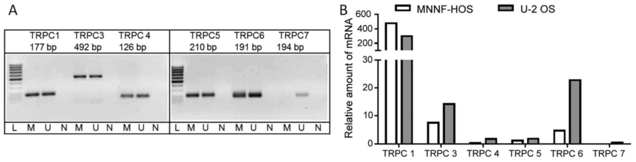
First, the expression of all human TRPC isoforms was
analyzed at the mRNA level in both U-2 OS and MNNG-HOS cells under
basic conditions by means of RT-qPCR. TRPCs 1, 3, 4, 5, 6 and 7
mRNAs could be detected in comparable amounts in both cell lines.
As illustrated in Fig. 1A, a single
amplicon of the expected size was obtained for any TRPC isoform as
confirmed by agarose gel electrophoresis as well as the melting
curve obtained as part of the PCR protocol (data not shown). The
level of expression of the individual TRPC isoforms is very
different, but the ‘TRPC profile’ shows extensive similarity
between the two cell lines (Fig.
1B). By far the highest mRNA levels could be detected for TRPC1
followed by TRPC6. TRPCs 3, 4 and 5 appeared to be moderately
expressed (Fig. 1B). RT-qPCR
analysis of TRPC7 provided Cq-values >38 in both cell lines and,
thus, this isoform is regarded as practically not expressed in
MNNG-HOS and U-2 OS cells.
Next, before studying the effects of the Mas
agonist, AVE0991 or of the antagonist,
D-Ala7-Ang-(1–7) (A779), on TRPC expression, mRNA
expression of the Ang-(1–7) receptor, Mas, was confirmed by RT-qPCR.
As previously observed (13) and
shown in Fig. 2A, Mas mRNA could be
detected in both U-2 OS and MNNG-HOS cells, with MNNG-HOS cells
showing stronger expression levels than U-2 OS. In MNNG-HOS cells,
the administration of AVE0991 led to a significant [3.35 (1.57;
7.16), p<0.001] increase in Mas-mRNA levels (Fig. 2B). In U-2 OS cells, there was no such
increase in Mas mRNA {1.4 [1.29 (0.39; 3.89)], p=0.92} (Fig. 2C). However, in response to the Mas
receptor antagonist, D-Ala7-Ang-(1–7), both
MNNG-HOS and U-2 OS showed an induction of Mas mRNA expression
[3.25 (0.83; 5.97), p<0.05 or 2.41 (1.05; 5.42), p=0.12]
(Fig. 2B and C).
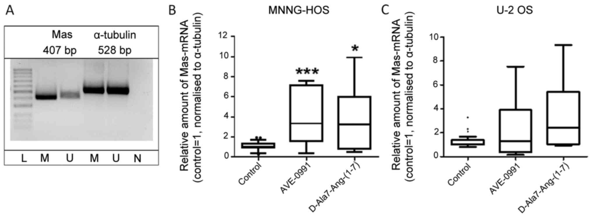 | Figure 2.Expression levels of Mas in MNNG-HOS
and U-2 OS osteosarcoma cells. (A) Reverse
transcription-quantitative PCR analysis revealed the expression
levels of Mas in MNNG-HOS and U-2 OS cell lines. The formation of a
unique amplicon of the correct size only was verified by agarose
gel electrophoresis and melting curve analysis. Mas-mRNA levels
were subject to regulation in response to Mas receptor agonist,
AVE0991, or antagonist, D-Ala7-Ang-(1–7), in (B)
MNNG-HOS osteosarcoma cells or (C) U-2 OS osteosarcoma cells (n=8;
*P<0.05, ***P<0.001 vs. control). bp, base pairs; L, 100
bp-ladder; M, MNNG-HOS; U, U-2 OS; N, negative control. |
Effects of AVE0991 or
D-Ala7-Ang-(1–7) on the mRNA expression of TRPC family
members
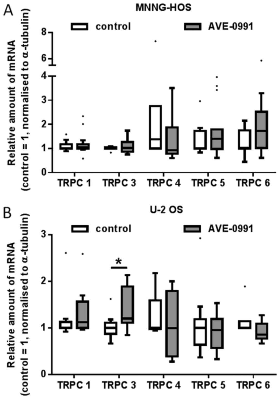
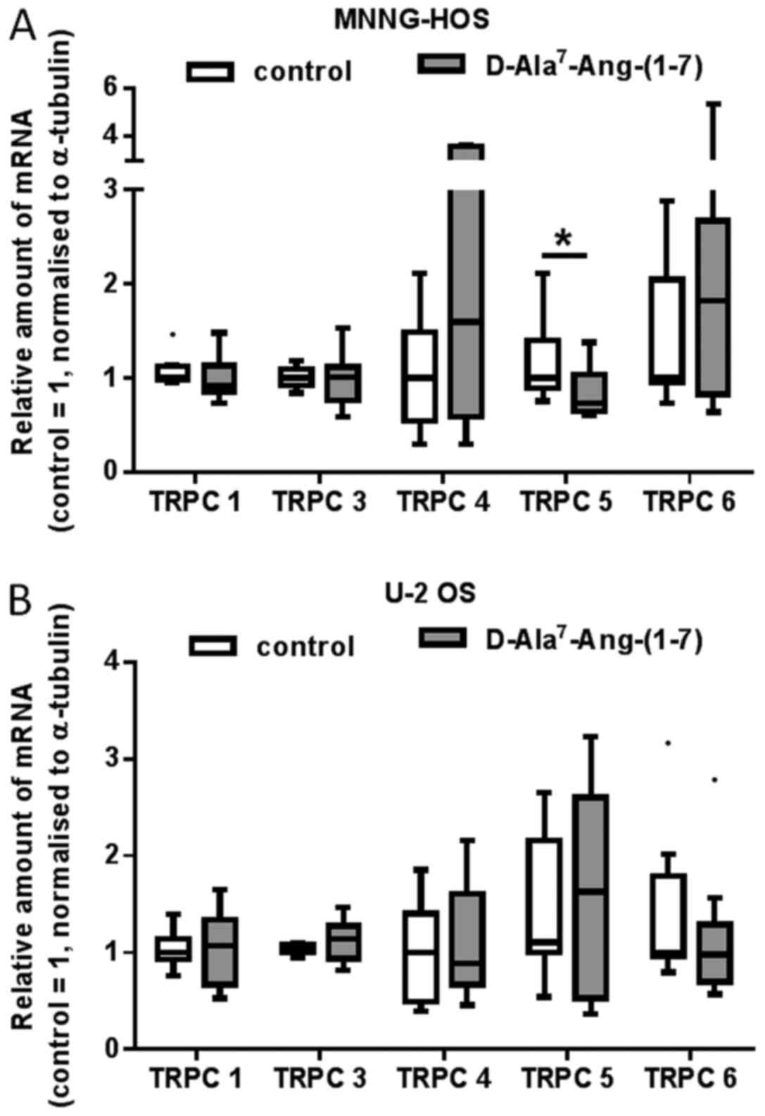
Next, the possible effects of a 24-h exposure of U-2
OS and MNNG-HOS cells to the Mas receptor agonist, AVE0991, or the
antagonist D-Ala7-Ang-(1–7) on TRPC
mRNA levels were investigated. In general, Mas receptor ligands
provoked no or only minor alterations in TRPC mRNA expression, as
summarized in Fig. 3 (AVE0991) and
Fig. 4
(D-Ala7-Ang-(1–7). In MNNG-HOS cells, AVE0991 led to a
non-significant elevation of TRPC5 mRNA levels by 70%, whereas in
U-2 OS cells, a small but significant increase in amounts of TRPC3
mRNA could be observed [1.2 (0.85; 2.13), p<0.05].
D-Ala7-Ang-(1–7) provoked
a small but significant decrease of TRCP5 mRNA amounts in MNNG-HOS
cells [0.73 (0.61; 1.38), p<0.05].
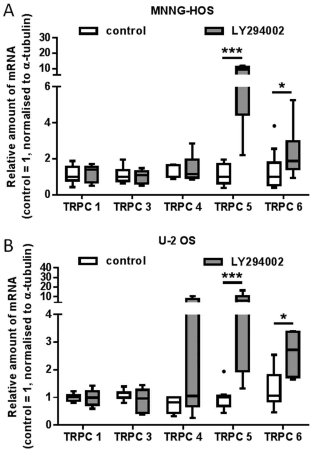
Effects of Ly294002 and PD98059 on the
mRNA expression of TRPC family members
The exposure of MNNG-HOS and U-2 OS osteosarcoma
cells to the selective pathway inhibitors Ly294002 or PD98059,
respectively, led to substantial alterations in the mRNA expression
levels of individual TRPC family members.
In detail, Ly294002 provoked a profound and highly
significant increase in TRPC5 mRNA amounts in both MNNG-HOS [9.6
(2.192; 11.68), p<0.0001] and U-2 OS cells [5.87 (1.32; 16.52)
p<0.001]. Slightly weaker were the stimulatory effects of
Ly294002 on the expression of TRPC6 in both cell lines; MNNG-HOS:
1.87[0.93; 5.27] p<0.05, U-2 OS: 2.72 [1.64; 3.38], p<0.05.
In contrast, the compound did not affect the mRNA levels of TRPC1,
3 and 4 (Fig. 5).
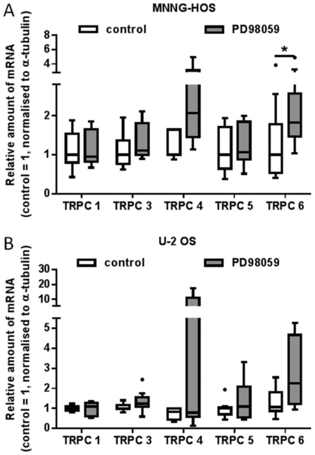
In MNNG-HOS cells, the administration of PD98059 led
to an about 2.07-fold increase in mRNA levels of TRPC4 2.07 [1.13;
4.95], p<0.01; and of TRPC6: 1.83 [1.04;4.90], p<0.05
(Fig. 6A). An increase of TRPC4 and
TRPC6 mRNA amounts in U-2 OS cells did not reach statistical
significance (Fig. 6B). A small but
significant increase in TRPC3 mRNA amounts could be observed in U-2
OS cells: 1.29 [1.02; 2.44], p<0.05, whereas in MNNG HOS cells,
this effect is only hinted at.
Effects of Ly294002 and PD98059 on the
Mas mRNA expression
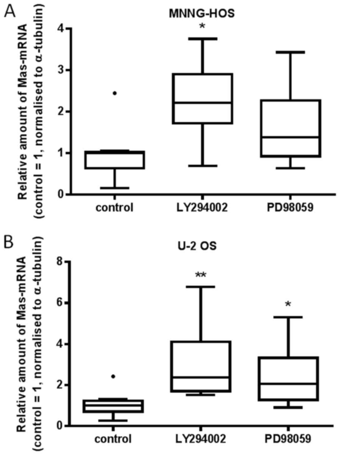
The administration of Ly294002 provoked a very
consistent 2.4-fold increase of Mas mRNA amounts in both MNNG-HOS
[2.212 (1.72; 2.89), p<0.05] and U-2 OS cells [2.38 (1.70;
4.11), p<0.001]. A similar increase could be observed in
response to PD98059. Whereas a 2.1-fold increase [2.1 (1.28; 3.33),
p<0.05] in Mas mRNA amounts could be observed in U-2 OS cells, a
1.4-fold [1.38 (0.93; 2.26), p=0.08] increase in MNNG-HOS cells
could be observed as a tendency only (Fig. 7).
Effects of AVE0991 or
D-Ala7-Ang-(1–7) and TRPC inhibitors AC1903 or Pyr3 on the
migration of MNNG-HOS and U-2 OS cells
The administration of Ang-(1–7) has been
previously shown to reduce cell migration of U-2 OS cells without
affecting that of MNNG-HOS cells in a Cultrex® 24 Well
Cell Migration Assay (16). In the
present assay, this could be observed here as a tendency only: U-2
OS cells showed slightly less migration upon exposure to the Mas
receptor agonist, AVE0991, and slightly increased migration when
exposed to the antagonist, D-Ala7-Ang-(1–7) (A779;
Fig. 8B). To assess the possible
contribution of Mas-dependent TRPC activity modulation to
alterations in cell migration, the inhibitors of TRPC5 or TRPC3,
AC1903 or Pyr3, respectively, were applied alone or in combination
with AVE0991 or A779.
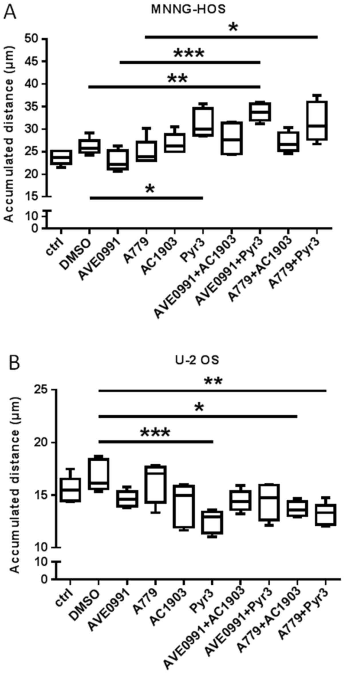 | Figure 8.Effects of AVE0991, A779, AC1903 and
Pyr3 on the migration of (A) MNNG-HOS and (B) U-2 OS osteosarcoma
cells. Cells were exposed to AVE0991 (1 µM),
D-Alaets-Ang-(1–7) (1 µM), AC1903 (35 µM), Pyr3 (3.5 µM), or
combinations thereof as indicated for 24 h. Subsequently, migration
was measured over 6 h (accumulated distance). Data are presented as
boxplots with medians, quartiles and an IQR ±1.5 × IQR with
outliers as indicated (Tukey method; n=5; *P<0.05, **P<0.01,
***P<0.001). ctrl, control; IQR, interquartile range. |
In MNNG-HOS cells, the TRPC5 inhibitor, AC1903, on
its own or in combination with A779 or AVE0991 did not affect cell
migration (Fig. 8A). However,
administration of the TRPC3 inhibitor, Pyr3, significantly
increased migration [Pyr3: 30.0 (28.6; 34.6) vs. DMSO: 25.8 (24.8;
27.48), p<0.05], an effect that could be further increased by
simultaneous administration of AVE0991 [Pyr3+AVE0991: 33.7 (31.9;
35.6), P=0.087 vs. Pyr3].
As shown in Fig. 8B,
AC1903, although not affecting U-2 OS migration per se, caused a
significant decrease of migration when applied together with A779.
The administration of Pyr3 significantly reduced the migration of
U-2 OS cells, an effect that could be prevented by the simultaneous
administration of AVE0991.
Discussion
TRPCs transform various stimuli into appropriate
cellular responses involving functions such as proliferation,
migration, and survival. These processes are of paramount
importance for normal growth, repair, and tissue homeostasis, and
therefore their dysregulation is a feature typically observed
during tumorigenesis and tumor progression. TRPCs have been
associated with various diseases and, in particular, with cancer.
With respect to the latter, altered expression, subcellular
localization, and gene mutations of TRPC subtypes have been
reported for an increasing number of tumor entities, including
glioma as well as lung, breast, prostate, gastric, and hepatic
cancer (reviewed in references 1,21-23). The abnormal regulation of
the intracellular calcium balance certainly plays a major role
here, but additional cellular processes, aside of enhancing
Ca2+ entry, have been identified (22). Reinforcing this phenomenon, TRPC
activity and/or expression could be modulated by inadequate signal
input from aberrant GPCR or growth factor receptor activation in
normal and (pre-)malignant cells. This has been observed for e.g.
TRPC6, the membrane expression and activity of which appeared to be
regulated by epidermal growth factor (EGF) receptor stimulation
(24). Mechanistically, a number of
different signal transduction pathways mediate TRPC signaling
including extracellular signal-regulated kinase Erk1/2,
calcium/calmodulin-dependent protein kinase IIα (CaMKIIα), and
calpain (21,25). Furthermore, TRPCs 3, 4 and 5 have all
been shown to contribute to cancer angiogenesis through modulating
VEGF pathway (26,27).
For cancer in general it is well established that
dysregulation of Ca2+ signaling promotes tumor
initiation, progression, metastasis, and angiogenesis (22). Little is known about the pathological
relevance of dysregulated TRPC expression or activity specifically
in osteosarcoma. It was previously shown that the knock-down of the
GPCR, Mas, decreased the proliferation of MNNG-HOS and U-2 OS
cells. In the present study, we examined the specific question of
whether and to what extent this effect can be attributed to a
Mas-dependent change in the TRPC expression. In addition, we
investigated whether signaling pathways typically activated by GPCR
or receptor tyrosine kinases are, in principle, able to influence
TRPC expression in osteosarcoma cells. Selective inhibitors of the
MEK/ERK1/2 and PI3 kinase signaling pathways, PD98059 and Ly294002,
were used for this purpose.
The results of the present study show that neither
the Mas receptor agonist, AVE0991 nor the antagonist,
D-Ala7-Ang-(1–7), caused substantial changes in TRPC
expression. The principle effectiveness of both compounds under the
conditions applied was reflected in the marked change in the
Mas-mRNA concentrations they provoked in both osteosarcoma cell
lines. From this, it can be concluded that Mas-dependent regulation
of osteosarcoma cell proliferation is not due to altered TRPC
expression. However, the TRPC inhibitors applied here were capable
of modulating effects of AVE0991 and A779 on osteosarcoma cell
migration, which strongly implies that Mas-receptor signaling is
associated with TRPC activity (rather than expression), and
migration in osteosarcoma cell lines. Supporting this view are
reports demonstrating: i) a cAMP-dependent increase in stimulated
TRPC6 cation currents in TRPC6-expressing HEK293 cells, which could
be diminished by LY294002 or PD 98059 (28); and ii) the increase of cAMP in
response to Mas receptor agonists such as Ang-(1–7) observed
in different cells (18,29).
The increase in Mas mRNA levels in response to the
antagonist very likely reflects a direct compensatory feedback
mechanism, the equanimous effect of the non-peptide agonist can be
explained as a similar response to functional desensitization
following rapid activation of Mas (17).
The results of our study further show that
inhibition of either PI3 kinase or MEK/Erk1/2 signal transduction
provokes substantial changes in the mRNA expression of distinct
TRPC subtypes. The administration of Ly294002 caused the strongest
changes in the amounts of TRPC5-mRNA, which were induced 9.6-fold
and 5.9-fold, respectively. In comparison, the induction of TRCP6
was less pronounced, whereas mRNA levels of TRPCs 1, 3 and 4
remained completely unaffected. The contribution of PI3 kinase
signaling to TRPC6 expression has been described by others
previously. However, in podocytes, vascular endothelial growth
factor (VEGF) appeared to increase TRPC6 mRNA and protein levels,
and this effect could be blocked by PI3 kinase inhibitors,
including Ly294002 (30). As a more
general mechanism, hormonal stimuli and growth factors via
activation of TRPC activity and Ca2+ influx contribute
to down-stream activation of MEK/Erk1/2 or PI3 kinase signaling
(21,31). This has been shown for e.g. TRPC6 in
hippocampal neurons and PC12 cells (32) and for TRPC3 in DT40 B-cells (33).
In the osteosarcoma cell lines MNNG-HOS and U-2 OS,
the administration of the MEK/Erk inhibitor, PD98059, increased
mRNA levels of TRPC6 whereas that of TRPC4 and TRPC3 appeared to be
affected only in one of the cell lines. Erk-dependent alterations
in TRPC subtype expression have been observed by others in
pulmonary arterial smooth muscle cells (34). Exposure of these cells to bone
morphogenetic protein (BMP) 4 increased TRPC1, TRPC4 and TRPC6
expression, and this effect was abolished by pharmacological
inhibition (PD98059) as well as by siRNA-mediated knock-down of
Erk1/2 (34).
Our data consistently show a stimulatory effect of
the pathway inhibitors on TRPC mRNA expression, which is
inconsistent with the observations of other groups described
before. It could be speculated, however, that different cell types
react differently. In any case, this even applies to osteosarcoma
cell lines from different species that differ in terms of their
TRPC expression profile (14).
Furthermore, each cell type has its special equipment with
G-protein-coupled receptors (GPCRs), which can then result in very
unique context- and stimulus-dependent activation patterns of
TRPCs. It could be speculated that it is this difference in GPCRs
that led to the different responses partially observed here for U-2
OS and MNNG HOS, a finding that reflects quite well the challenge
posed with treating patients with ‘individual’ osteosarcoma. In our
experiments, cells were exposed to the action of the inhibitors for
a relatively long time, namely 24 h. During this time, stimulatory
feedback mechanisms may already be at play.
In accordance with a previous report (16), it could be demonstrated that the Mas
receptor agonist AVE0991 negatively regulates U-2 OS cell
migration, but is significantly increased in response to the Mas
receptor antagonist A779.
In summary, the present study shows that
Mas-dependent alterations in osteosarcoma cell line proliferation
are not mediated by any changes in TRPC subtype gene expression.
However, available data suggest that there are Mas-mediated changes
in TRPC activity. TRPC activities have not been studied here, which
is a clear limitation of our study. Another limitation is the use
of only two cell lines, however, the previous observation that
human osteosarcoma exhibit similar TRPC expression profiles
(14) could be confirmed here. The
results of our study show in principle and consistent with the
literature, that: i) members of the TRPC family are abundantly
expressed in osteosarcoma cell lines; and ii) that the
cAMP-PI3K-PKB-MEK-ERK1/2 signaling pathway examined can regulate
the expression of TRPCs at the level of mRNA and may well
contribute to Mas-dependent changes of TRPC activity and related
functions including cell migration. TRPCs potential relevance for
diagnosis and, in particular, direct or signaling pathway-targeted
treatment of osteosarcoma needs to be elucidated in further
studies.
Acknowledgements
The authors would like to thank Mrs. Manja Möller
and Mrs. Ines Schultz (Institute of Medical Biochemistry and
Molecular Biology, University Medicine Greifswald, Greifswald,
Germany) for their technical assistance.
Funding
No funding was received.
Availability of data and materials
The datasets used and/or analyzed during the current
study are available from the corresponding author on reasonable
request.
Authors contributions
FL, SB, AK and CW performed the experiments. CW and
UL were responsible for confirming the authenticity of all the raw
data. FL, SB, AK, UL and CW conducted the data analyses. FL, CW and
UL wrote the manuscript. UL and CW designed the study and planned
experiments. All authors read and approved the final
manuscript.
Ethics approval and consent to
participate
Not applicable.
Patient consent for publication
Not applicable.
Competing interests
The authors declare that they have no competing
interests.
References
|
1
|
Gautier M, Dhennin-Duthille I, Ay AS,
Rybarczyk P, Korichneva I and Ouadid-Ahidouch H: New insights into
pharmacological tools to TR(i)P cancer up. Br J Pharmacol.
171:2582–2592. 2014. View Article : Google Scholar : PubMed/NCBI
|
|
2
|
Wu LJ, Sweet TB and Clapham DE:
International Union of Basic and Clinical Pharmacology. LXXVI.
Current progress in the mammalian TRP ion channel family. Pharmacol
Rev. 62:381–404. 2010. View Article : Google Scholar : PubMed/NCBI
|
|
3
|
Capiod T: The need for calcium channels in
cell proliferation. Recent Patents Anticancer Drug Discov. 8:4–17.
2013. View Article : Google Scholar
|
|
4
|
Hodeify R, Yu F, Courjaret R, Nader N, Dib
M, Sun L, Adap E, Hubrack S and Machaca K: Regulation and role of
store-operated Ca2+ entry in cellular proliferation.
Calcium entry channels in non-excitable cells. Kozak JA and Putney
JW Jr: CRC Press/Taylor & Francis; Boca Raton, FL: pp. 215–240.
2018
|
|
5
|
Bomben VC and Sontheimer HW: Inhibition of
transient receptor potential canonical channels impairs cytokinesis
in human malignant gliomas. Cell Prolif. 41:98–121. 2008.
View Article : Google Scholar : PubMed/NCBI
|
|
6
|
Cai R, Ding X, Zhou K, Shi Y, Ge R, Ren G,
Jin Y and Wang Y: Blockade of TRPC6 channels induced G2/M phase
arrest and suppressed growth in human gastric cancer cells. Int J
Cancer. 125:2281–2287. 2009. View Article : Google Scholar : PubMed/NCBI
|
|
7
|
Hwang JA, Hwang MK, Jang Y, Lee EJ, Kim
JE, Oh MH, Shin DJ, Lim S, Ji G, Oh U, et al:
20-O-β-d-glucopyranosyl-20(S)-protopanaxadiol, a metabolite of
ginseng, inhibits colon cancer growth by targeting TRPC
channel-mediated calcium influx. J Nutr Biochem. 24:1096–1104.
2013. View Article : Google Scholar : PubMed/NCBI
|
|
8
|
Jiang HN, Zeng B, Zhang Y, Daskoulidou N,
Fan H, Qu JM and Xu SZ: Involvement of TRPC channels in lung cancer
cell differentiation and the correlation analysis in human
non-small cell lung cancer. PLoS One. 8:e676372013. View Article : Google Scholar : PubMed/NCBI
|
|
9
|
Gaunt HJ, Vasudev NS and Beech DJ:
Transient receptor potential canonical 4 and 5 proteins as targets
in cancer therapeutics. Eur Biophys J. 45:611–620. 2016. View Article : Google Scholar : PubMed/NCBI
|
|
10
|
Huang Z, Fan G and Wang D: Downregulation
of calbindin 1, a calcium-binding protein, reduces the
proliferation of osteosarcoma cells. Oncol Lett. 13:3727–3733.
2017. View Article : Google Scholar : PubMed/NCBI
|
|
11
|
Wang Y, Yang Z, Meng Z, Cao H, Zhu G, Liu
T and Wang X: Knockdown of TRPM8 suppresses cancer malignancy and
enhances epirubicin-induced apoptosis in human osteosarcoma cells.
Int J Biol Sci. 10:90–102. 2013. View Article : Google Scholar : PubMed/NCBI
|
|
12
|
Berna-Erro A, Redondo PC and Rosado JA:
Store-operated Ca2+ entry. Adv Exp Med Biol.
740:349–382. 2012. View Article : Google Scholar : PubMed/NCBI
|
|
13
|
Clapham DE, Runnels LW and Strübing C: The
TRP ion channel family. Nat Rev Neurosci. 2:387–396. 2001.
View Article : Google Scholar : PubMed/NCBI
|
|
14
|
Huang YW, Chang SJ, Harn HI, Huang HT, Lin
HH, Shen MR, Tang MJ and Chiu WT: Mechanosensitive store-operated
calcium entry regulates the formation of cell polarity. J Cell
Physiol. 230:2086–2097. 2015. View Article : Google Scholar : PubMed/NCBI
|
|
15
|
Abed E, Labelle D, Martineau C, Loghin A
and Moreau R: Expression of transient receptor potential (TRP)
channels in human and murine osteoblast-like cells. Mol Membr Biol.
26:146–158. 2009. View Article : Google Scholar : PubMed/NCBI
|
|
16
|
Ender SA, Dallmer A, Lässig F, Lendeckel U
and Wolke C: Expression and function of the
ACE2/angiotensin(1–7)/Mas axis in osteosarcoma cell lines U-2 OS
and MNNG-HOS. Mol Med Rep. 10:804–810. 2014. View Article : Google Scholar : PubMed/NCBI
|
|
17
|
Tirupula KC, Desnoyer R, Speth RC and
Karnik SS: Atypical signaling and functional desensitization
response of MAS receptor to peptide ligands. PLoS One.
9:e1035202014. View Article : Google Scholar : PubMed/NCBI
|
|
18
|
Sahr A, Wolke C, Maczewsky J,
Krippeit-Drews P, Tetzner A, Drews G, Venz S, Gürtler S, van den
Brandt J, Berg S, et al: The angiotensin-(1–7)/Mas axis improves
pancreatic β-cell function in vitro and in vivo. Endocrinology.
157:4677–4690. 2016. View Article : Google Scholar : PubMed/NCBI
|
|
19
|
Chilukoti RK, Mostertz J, Bukowska A,
Aderkast C, Felix SB, Busch M, Völker U, Goette A, Wolke C, Homuth
G, et al: Effects of irbesartan on gene expression revealed by
transcriptome analysis of left atrial tissue in a porcine model of
acute rapid pacing in vivo. Int J Cardiol. 168:2100–2108. 2013.
View Article : Google Scholar : PubMed/NCBI
|
|
20
|
Livak KJ and Schmittgen TD: Analysis of
relative gene expression data using real-time quantitative PCR and
the 2(−ΔΔC(T)) Method. Methods. 25:402–408. 2001. View Article : Google Scholar : PubMed/NCBI
|
|
21
|
Chen J, Luan Y, Yu R, Zhang Z, Zhang J and
Wang W: Transient receptor potential (TRP) channels, promising
potential diagnostic and therapeutic tools for cancer. Biosci
Trends. 8:1–10. 2014. View
Article : Google Scholar : PubMed/NCBI
|
|
22
|
Bacsa B, Tiapko O, Stockner T and
Groschner K: Mechanisms and significance of Ca2+ entry
through TRPC channels. Curr Opin Physiol. 17:25–33. 2020.
View Article : Google Scholar : PubMed/NCBI
|
|
23
|
Chen X, Sooch G, Demaree IS, White FA and
Obukhov AG: Transient receptor potential canonical (TRPC) channels:
Then and now. Cells. 9:2020. View Article : Google Scholar
|
|
24
|
Odell AF, Scott JL and Van Helden DF:
Epidermal growth factor induces tyrosine phosphorylation, membrane
insertion, and activation of transient receptor potential channel
4. J Biol Chem. 280:37974–37987. 2005. View Article : Google Scholar : PubMed/NCBI
|
|
25
|
Xu J, Wang H, Hu Y, Zhang YS, Wen L, Yin
F, Wang Z, Zhang Y, Li S, Miao Y, et al: Inhibition of CaMKIIα
activity enhances antitumor effect of fullerene C60 nanocrystals by
suppression of autophagic degradation. Adv Sci (Weinh).
6:18012332019. View Article : Google Scholar : PubMed/NCBI
|
|
26
|
Asghar MY, Magnusson M, Kemppainen K,
Sukumaran P, Löf C, Pulli I, Kalhori V and Törnquist K: Transient
receptor potential canonical 1 (TRPC1) channels as Regulators of
sphingolipid and VEGF receptor expression: Impolications for
thyroid cancer cell migration and proliferation. J Biol Chem.
290:16116–16131. 2015. View Article : Google Scholar : PubMed/NCBI
|
|
27
|
Tao X, Zhao N, Jin H, Zhang Z, Liu Y, Wu
J, Bast RC Jr, Yu Y and Feng Y: FSH enhances the proliferation of
ovarian cancer cells by activating transient receptor potential
channel C3. Endocr Relat Cancer. 20:415–429. 2013. View Article : Google Scholar : PubMed/NCBI
|
|
28
|
Shen B, Kwan HY, Ma X, Wong CO, Du J,
Huang Y and Yao X: cAMP activates TRPC6 channels via the
phosphatidylinositol 3-kinase (PI3K)-protein kinase B
(PKB)-mitogen-activated protein kinase kinase (MEK)-ERK1/2
signaling pathway. J Biol Chem. 286:19439–19445. 2011. View Article : Google Scholar : PubMed/NCBI
|
|
29
|
Tetzner A, Gebolys K, Meinert C, Klein S,
Uhlich A, Trebicka J, Villacañas Ó and Walther T: G-protein-coupled
receptor MrgD is a receptor for angiotensin-(1–7) involving
adenylyl cyclase, cAMP, and phosphokinase A. Hypertension.
68:185–194. 2016. View Article : Google Scholar : PubMed/NCBI
|
|
30
|
Thilo F, Liu Y, Loddenkemper C, Schuelein
R, Schmidt A, Yan Z, Zhu Z, Zakrzewicz A, Gollasch M and Tepel M:
VEGF regulates TRPC6 channels in podocytes. Nephrol Dial
Transplant. 27:921–929. 2012. View Article : Google Scholar : PubMed/NCBI
|
|
31
|
Jardin I, Diez-Bello R, Lopez JJ, Redondo
PC, Salido GM, Smani T and Rosado JA: TRPC6 channels are required
for proliferation, migration and invasion of breast cancer cell
lines by modulation of Orai1 and Orai3 surface exposure. Cancers
(Basel). 10:3312018. View Article : Google Scholar
|
|
32
|
Heiser JH, Schuwald AM, Sillani G, Ye L,
Müller WE and Leuner K: TRPC6 channel-mediated neurite outgrowth in
PC12 cells and hippocampal neurons involves activation of
RAS/MEK/ERK, PI3K, and CAMKIV signaling. J Neurochem. 127:303–313.
2013. View Article : Google Scholar : PubMed/NCBI
|
|
33
|
Numaga-Tomita T, Nishida M, Putney JW Jr
and Mori Y: TRPC3 amplifies B-cell receptor-induced ERK signalling
via protein kinase D-dependent Rap1 activation. Biochem J.
473:201–210. 2016. View Article : Google Scholar : PubMed/NCBI
|
|
34
|
Li X, Lu W, Fu X, Zhang Y, Yang K, Zhong
N, Ran P and Wang J: BMP4 increases canonical transient receptor
potential protein expression by activating p38 MAPK and ERK1/2
signaling pathways in pulmonary arterial smooth muscle cells. Am J
Respir Cell Mol Biol. 49:212–220. 2013. View Article : Google Scholar : PubMed/NCBI
|