Introduction
Globally, gastric cancer (GC) is one of the most
common tumours of the alimentary system, ranking fifth in terms of
incidence and third in terms of mortality among all cancer types in
2018 (1). Chemotherapy is one of the
main treatments for GC, with oxaliplatin or cisplatin plus
fluoropyrimidine being used as the first-line treatment (2). Nevertheless, the duration of the
response to chemotherapy is limited, and the 5-year overall
survival (OS) rate is still low in advanced GC. Therefore, there is
an urgent requirement to find a novel and effective treatment for
patients with GC.
The tumour microenvironment (TME) is composed of a
variety of different components, including endothelial cells,
fibroblasts, inflammatory mediators, mesenchymal cells, immune
cells and stromal cells (3). The
function of stromal cells and immune cells in the TME is crucial.
However, our understanding of the TME is still far from sufficient.
ESTIMATE, an algorithm designed by Yoshihara et al (4), can be used to evaluate stromal cells
and immune cells according to the expression of specific genes.
Recently, the ESTIMATE algorithm has been used in the study of
various malignancies, such as acute myeloid leukaemia (AML)
(5), clear cell renal cell carcinoma
(ccRCC) (6) and glioma (7). However, to the best of our knowledge,
the ESTIMATE algorithm has rarely used in the study of GC.
In the present study, ESTIMATE was utilized to
perform TME evaluation to calculate stromal and immune scores. Hub
genes associated with the TME were identified using Cytoscape
software (version 3.7.1; http://cytoscape.org/). More importantly, the impact
of these hub genes on survival was evaluated and validated, and the
association between the expression level of RGS1 and the
clinicopathological characteristics of patients with GC was
analysed.
Materials and methods
The cancer genome atlas (TCGA) data
collection and processing
The gene transcriptome and clinical profiles of 343
patients with stomach adenocarcinoma from TCGA database (http://tcga-data.nci.nih.gov/tcga/) were
downloaded and processed (March 13, 2020). Immune, stromal and
ESTIMATE scores were calculated by the ESTIMATE algorithm to
predict the level of infiltrating immune cells, stromal cells and
the tumour purity in tumour tissue, respectively (4).
Survival and association analysis
Kaplan-Meier survival curves were plotted, and the
log-rank test was employed to compare the differences in OS for
patients with GC based on the immune, stromal and ESTIMATE scores.
Moreover, the association between score levels and multiple
subgroups of clinical variables, including grade, American Joint
Committee on Cancer (AJCC) stage and TNM stage (8), was also analysed.
Differentially expressed genes (DEGs),
heatmaps and clustering analysis
The patients with GC were divided into high and low
scoring groups according to the median immune, stromal and
ESITIMATE scores (1088.7, 92.0 and 1106.1, respectively). DEGs were
screened out with |log (FC)|>1 and false discovery rate
(FDR)<0.05. Moreover, clustering analysis was applied to
identify significant upregulated and downregulated gene sets
between the immune and stromal subgroups. Corresponding heatmaps
were plotted to illustrate the DEGs. Furthermore, the ‘VennDiagram’
package was utilized to identify and visualize the intersecting
genes of upregulated and downregulated gene sets from the
differential analysis of immune scores and stromal scores (9).
Functional enrichment analysis and
protein-protein interaction (PPI) network construction
Intersect genes were subjected to comprehensive Gene
Ontology (GO; http://geneontology.org/) analysis, including
biological processes (BPs), cellular components (CCs) and molecular
functions (MFs), and they were also subjected to Kyoto
Encyclopaedia of Genes and Genomes (KEGG; http://www.kegg.jp/) pathway analysis. At the same
time, the STRING database (https://string-db.org/) was used to construct the PPI
network with the highest confidence (0.900).
Batch survival analysis of hub
genes
The hub genes were identified from the PPI network
using cytoHubba (10) from Cytoscape
software (version 3.7.1; http://cytoscape.org/), and used for further survival
analysis. Kaplan-Meier curves were plotted and the log-rank test
was applied to evaluate the difference between the high and low
groups based on the gene expression level with regard to the 5-year
OS rate.
Collection of tissue samples and
clinicopathological characteristics
Between August 2016 and December 2018, a total of 50
tumour tissue specimens were collected from patients with primary
GC without metastasis who underwent curative gastrectomy in the
Department of General Surgery of Tianjin Medical University General
Hospital (Tianjin, China). All tissues were stored at −80°C. The
patients were followed up, and corresponding clinicopathological
characteristics, including age, sex, BMI, tumour location,
differentiation degree, T stage, N stage and AJCC stage, were
collected. The age range of all patients was 52–85 years, and 34
patients were male and 16 were females. All patients provided
written informed consent before tissue collection.
Reverse transcription quantitative PCR
(RT-qPCR) assay
Total RNA was extracted from tissue specimens with
an RNAprep Pure Tissue kit (Tiangen Biotech, Co., Ltd). After
quantification with a NanoDrop-2000 spectrophotometer (Thermo
Fisher Scientific, Inc.), a total of 500 ng RNA was reverse
transcribed into cDNA using FastQuant RT Super mix kit (Tiangen
Biotech, Co., Ltd.) using the following temperature protocol: 15
min at 42°C and 3 min at 95°C. The cDNA was stored at −20°C.
Subsequently, mRNA quantitation in tumour tissues was detected
using a RT-qPCR assay with SYBR-Green qPCR Master Mix (Bimake
Biotechnology) on a DNA Engine Opticon 2 (Bio-Rad Laboratories,
Inc.). The thermocycling conditions were as follows: 95°C for 30
sec, then 40 cycles of 95°C for 15 sec, 60°C for 30 sec and 72°C
for 30 sec, followed by dissociation at 95°C for 15 sec, 60°C for 1
min and 95°C for 15 sec. mRNA expression was quantified using the
2−∆∆Cq method (11) and
normalized to the internal reference gene (β-actin). The primers
are listed in Table I.
 | Table I.Summary of primer sequences used for
reverse transcription-quantitative PCR. |
Table I.
Summary of primer sequences used for
reverse transcription-quantitative PCR.
| Gene | Forward primer
(5′-3′) | Reverse primer
(5′-3′) |
|---|
| CXCR4 |
AATGACTTGTGGGTGGTTG |
AAGAAAGCCAGGATGAGGA |
| PTGFR |
GTTCCTGTCCACTCTGAAGG |
TCACCATGCTCTGTAGTTTGA |
| RGS1 |
CAGCCAAGAAGATTAAAGCAC |
TTGAGGAACCTGGGATAAGA |
| β-actin |
CTCCTCCACCTTTGACGCTG |
TCCTCTTGTGCTCTTGCTGG |
Statistical analysis
R software (version 3.6.0; http://www.r-project.org/) was applied for the
statistical analysis of TCGA data. SPSS (version 23.0; IBM, Corp.)
was applied for the statistical analysis of the data from the
Department of General Surgery of Tianjin Medical University General
Hospital. The χ2 test and Fisher's exact test were
applied for categorical variables, and Student's t-test was applied
for continuous variables. The Kruskal-Wallis test and Wilcoxon rank
sum test were used to compare ESTIMATE, immune and stromal scores
between 2 or more subgroups of clinical variables, respectively.
P<0.05 was considered to indicate a statistically significant
difference.
Results
Stromal scores and immune scores are
associated with the histological grade, AJCC stage, T stage and
prognosis of GC patients
A total of 343 patients with GC from the TCGA
database were included. The patients were divided into high and low
scoring groups based on the median stromal, immune and ESTIMATE
scores. There was no significant association between the ESTIMATE
score and OS (P=0.4073; Fig. 1A).
However, higher stromal scores were associated with worse outcomes
in patients with GC (P=0.0178; Fig.
1B). There was no significant association between immune scores
and OS (P=0.6624; Fig. 1C).
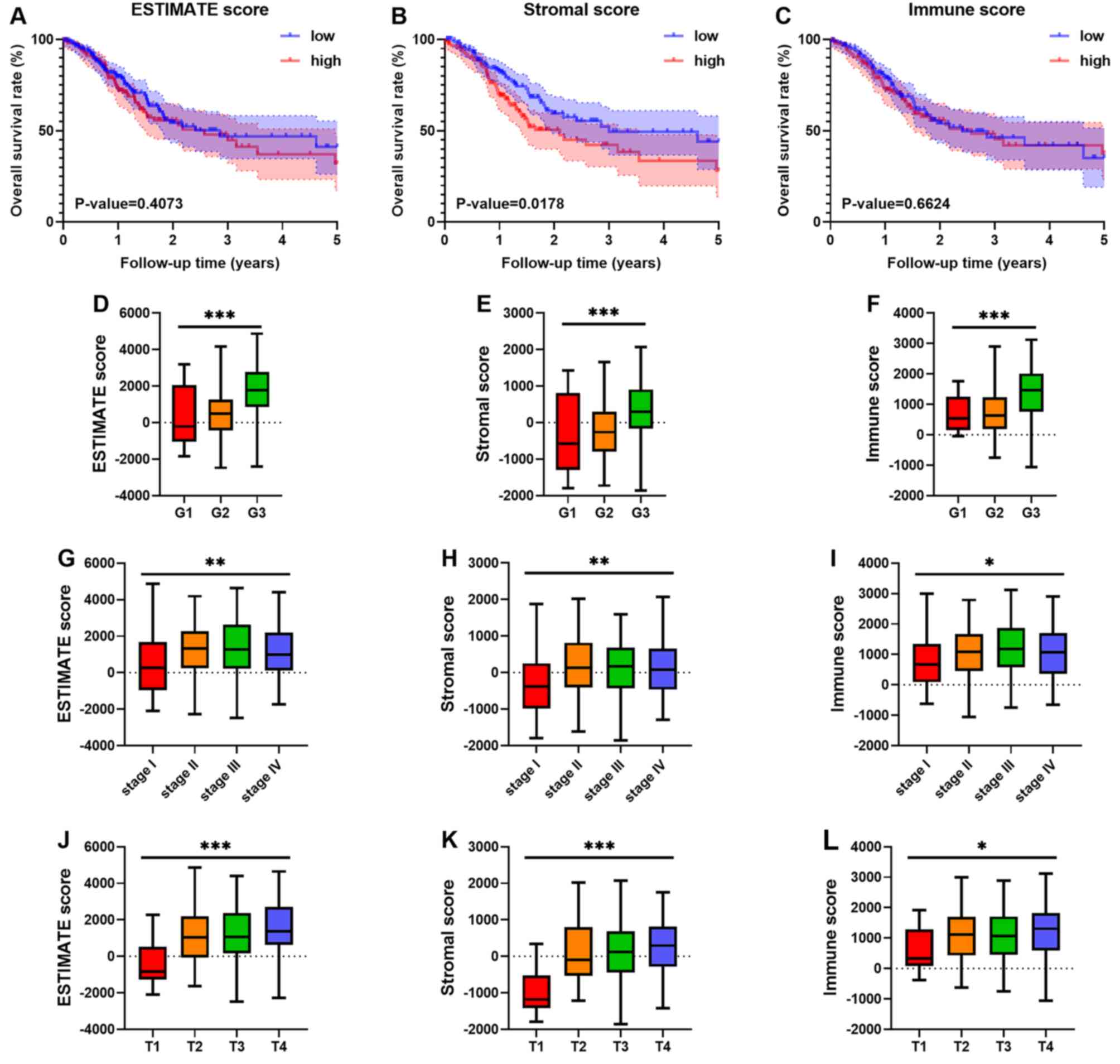 | Figure 1.Stromal scores are associated with the
survival outcome of GC, and immune scores and stromal scores are
associated with GC tumour grade, AJCC stage and T staging.
Kaplan-Meier survival curves suggested that (A) higher ESTIMATE
scores (P=0.4073) were not significantly related to survival
outcome, (B) stromal scores (P=0.0178) were significantly related
to worse prognosis and (C) immune scores (P=0.6624) were not
significantly related to survival outcome. (D-F) The distribution
of ESTIMATE scores, stromal scores and immune scores for
histological grade; (G-I) the distribution of ESTIMATE scores,
stromal scores and immune scores for AJCC stage; and (J-L) the
distribution of ESTIMATE scores, stromal scores and immune scores
in T staging. *P<0.05, **P<0.01 and ***P<0.001. AJCC,
American Joint Committee on Cancer; low, low score group; high,
high score group; G, histological grade; stage, AJCC stage; T, AJCC
T staging; GC, gastric cancer. |
In addition, the associations between scores and
clinical variables were also analysed. There were significant
differences in ESTIMATE, stromal and immune scores (Fig. 1D-F) among different histological
grades. Similar to those results observed for histological grade,
there were significant differences in ESTIMATE, stromal and immune
scores (Fig. 1G-I) for AJCC stage.
For TNM staging, significant differences in ESTIMATE, stromal and
immune scores (Fig. 1J-L) were found
only between the subgroups of T staging. For ESTIMATE scores, no
significant difference was observed in N and M staging (Fig. S1A and B). Similar results were also
observed in stromal scores (Fig. S1C
and D) and immune scores (Fig. S1E
and F).
DEGs of stromal scores and immune
scores for GC
The patients were divided into high and low groups
based on the median scores. The results of the clustering analysis
in the immune score group are shown in Fig. S2. The heatmap based on stromal
scores is shown in Fig. S3. A total
of 1,174 DEGs were identified based on immune scores, including 853
upregulated genes and 321 downregulated genes. Moreover,
differential gene analysis was conducted in the same way based on
the stromal score. In addition, 1,513 upregulated genes and 218
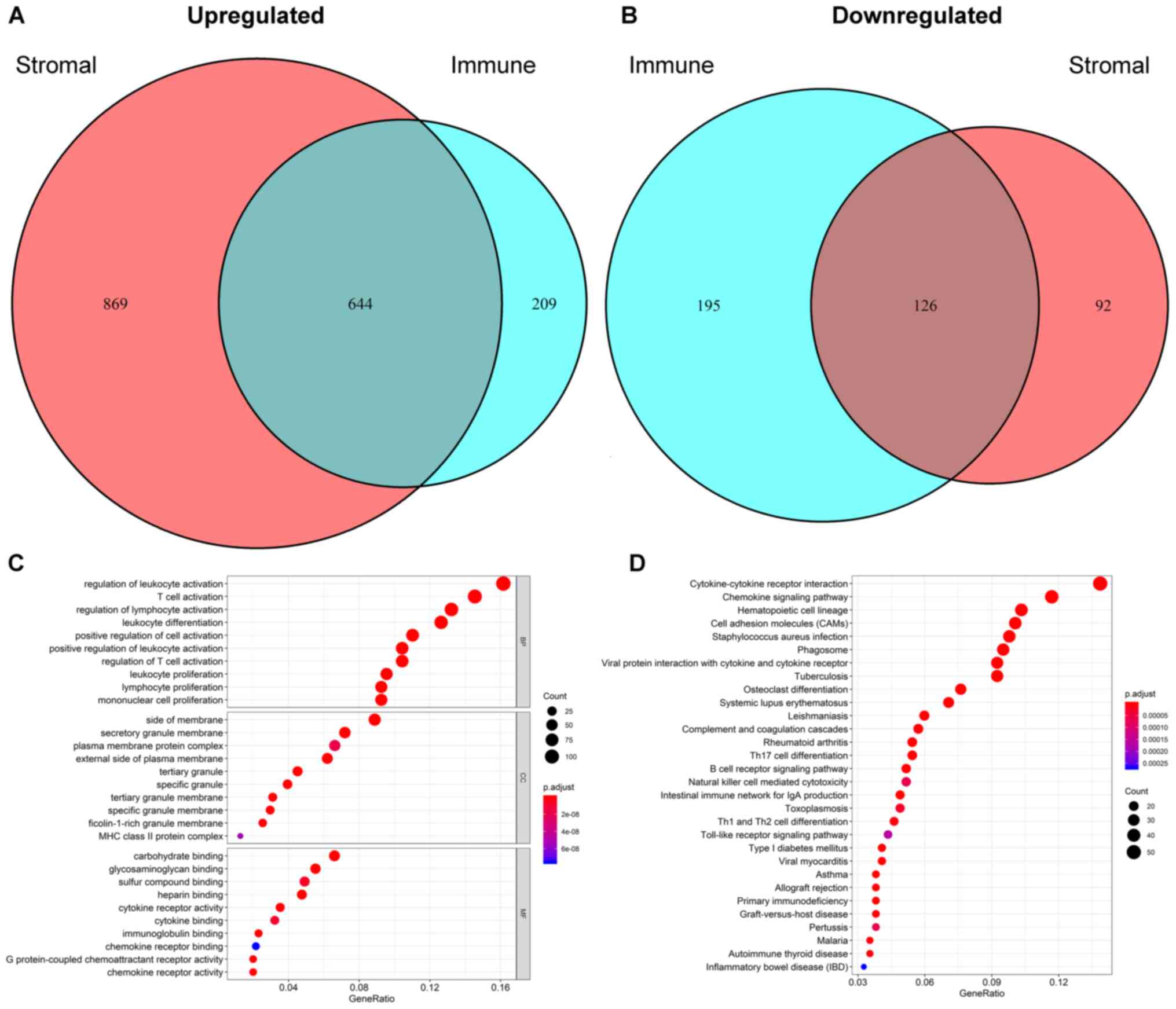
downregulated genes were identified (Fig. 2A and B). A Venn diagram was applied
to distinguish 770 intersect genes, consisting of 644 upregulated
and 126 downregulated intersect genes (Fig. 2A and B). The details of the DEGs of
immune and stromal scores are provided in Tables SI and SII, respectively.
Functional enrichment analysis and PPI
network
The functional enrichment analysis results indicated
that the intersecting genes were significantly associated with the
immune response. The top 10 GO terms in each category of BP, CC and
MF (Fig. 2C) were screened out.
‘Regulation of leukocyte activation’, ‘side of membrane’ and
‘carbohydrate binding’ were the top GO terms in the GO analysis.
For the KEGG pathway enrichment analysis, the top 30 pathways are
shown in Fig. 2D. Among them,
‘cytokine-cytokine receptor interaction’, ‘chemokine signalling
pathway’ and ‘haematopoietic cell lineage’ were the top KEGG
pathways, in which 770 intersecting genes might be involved.
Intersecting genes were used to construct the PPI network,
consisting of 294 nodes and 1,984 edges. As shown in Fig. 3A, a total of 71 TME-associated hub
genes were identified by 12 algorithms from cytoHubba.
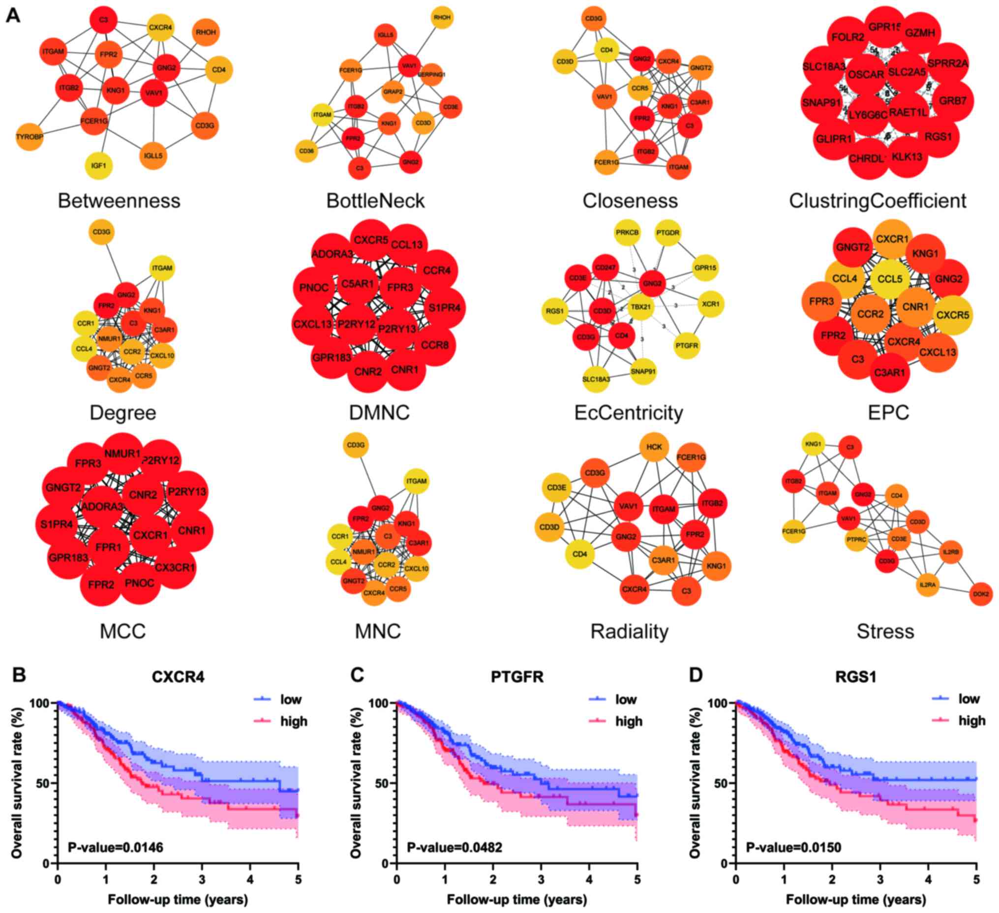 | Figure 3.Hub genes of algorithms from cytoHubba
and further batch survival analysis. (A) The PPI network data from
STRING were further analysed by Cytoscape, and hub gene
identification was performed by cytoHubba based on 12 algorithms.
Three hub genes, (B) CXCR4 (P=0.0146), (C) PTGFR (P=0.0482) and (D)
RGS1 (P=0.0150), were identified by batch survival analysis. The
colours of the nodes represent the ranks of the nodes, and the
darker the colour of the nodes, the higher the rank. Low, low
expression group; high, high expression group; PPI, protein-protein
interaction; DMNC, density of maximum neighbourhood component; MCC,
maximal clique centrality; MNC, maximum neighbourhood component;
EPC, edge percolated component. |
Batch processing of hub gene survival
analysis
To further elaborate the relationship between hub
genes and outcome, the 71 hub genes from 12 algorithms were used
for survival analysis. Among them, only three hub genes, CXCR4
(P=0.0146), PTGFR (P=0.0482) and RGS1 (P=0.0150), were identified
with significant differences in 5-year OS rate between the high and
low level groups (Fig. 3B-D).
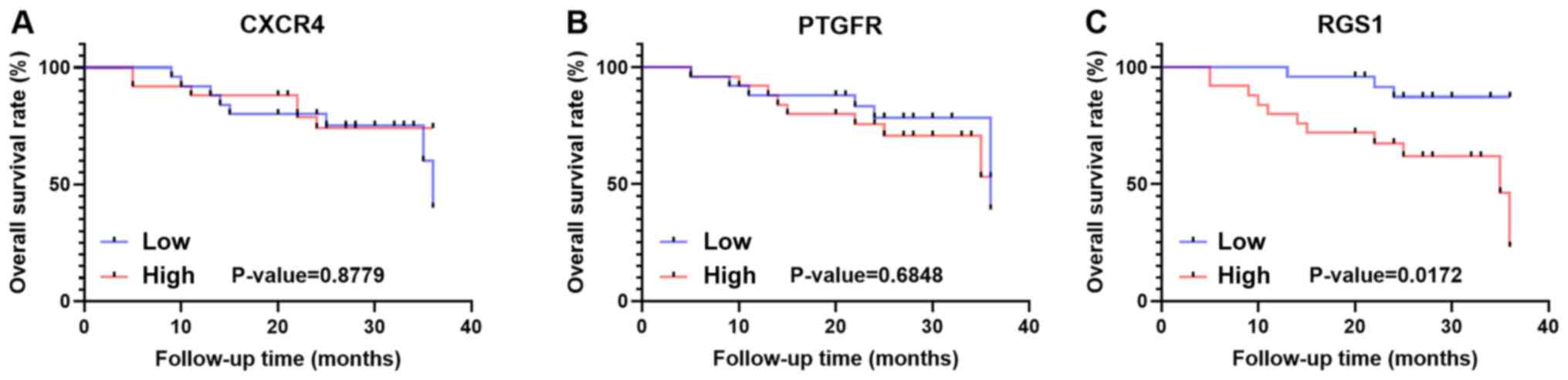
Validation of survival of CXCR4, PTGFR
and RGS1
To further verify the impact of the three hub genes
on prognosis in the study population, 50 patients who were
diagnosed with GC and underwent gastrectomy were included and
followed up. These patients were classified into a high and low
group based on the median 2−∆∆Cq value of each gene
(Fig. S4A-C; CXCR4, 0.8353; PTGFR,
1.0189; RGS1, 1.000). However, the results revealed that the high
and low CXCR4 and PTGFR groups exhibited no significant differences
in OS rate in the population (Fig. 4A
and B). However, the prognosis of the high RGS1 group
(P=0.0172) was significantly worse than that of the low RGS1 group
(Fig. 4C).
Clinicopathological characteristics of
the validation cohort
The clinicopathological characteristics of 50
patients were retrospectively collected. These patients were
divided into low and high level groups according to the median
2−∆∆Cq value of RGS1. The mean age of all patients was
68.1 years old, and 34 patients (68%) were male. The baseline
characteristics of the patients are shown in Table II. There was no significant
difference in age between the low and high level groups (67.6±7.6
vs. 68.7±9.0 years). In addition, there were also no significant
differences between the low and high level groups in terms of sex,
BMI index, tumour location, T stage, N stage or AJCC stage. With
regard to the degree of differentiation, however, the number of
poorly differentiated tumours in the low level group was
significantly less than that in the high level group (P<0.01;
Table II).
| Table II.Clinicopathological characteristics
of validation cohort based on RGS1 expression. |
Table II.
Clinicopathological characteristics
of validation cohort based on RGS1 expression.
| Characteristic | Low | High | P-value |
|---|
| Mean age ± SD,
years | 67.6±7.6 | 68.7±9.0 | 0.625 |
| Male, n (%) | 18 (72) | 16 (64) | 0.544 |
| Mean BMI ± SD,
kg/m2 | 22.71±3.42 | 22.16±2.23 | 0.509 |
| Location, n |
|
| 0.758 |
| Gastric
body | 8 | 7 |
|
|
Antrum | 17 | 18 |
|
| Degree of
differentiation, n |
|
| 0.005 |
| Poorly
differentiated | 4 | 14 |
|
|
Moderately differentiated | 15 | 5 |
|
| Highly
differentiated | 6 | 6 |
|
| T stage, n |
|
| 0.663 |
| 1 | 5 | 4 |
|
| 2 | 3 | 3 |
|
| 3 | 11 | 8 |
|
| 4 | 6 | 10 |
|
| N stage, n |
|
| 0.844 |
| 0 | 8 | 7 |
|
| 1 | 7 | 5 |
|
| 2 | 5 | 6 |
|
| 3 | 5 | 7 |
|
| AJCC stage, n |
|
| 0.693 |
| I | 6 | 5 |
|
| II | 9 | 7 |
|
|
III | 10 | 13 |
|
Discussion
GC is a global health threat with high incidence and
mortality. Recently, immunotherapy has been used to treat various
solid tumours, playing an anti-tumour role by inhibiting immune
checkpoints, such as programmed cell death protein 1 and programmed
cell death ligand 1, to enhance the activity of T cells. For
example, nivolumab, an emerging immune checkpoint inhibitor, has
shown promising therapeutic prospects in phase III clinical trials
of metastatic GC (12). Thus, the
TME may play an indispensable role in the treatment of tumours. Due
to the development of next-generation sequencing technology, we
have a better understanding of the molecular biological level of
tumours. To the best of our knowledge, TME-related genes have been
identified in other malignancies, such as AML (5) and ccRCC (6), but in GC, they are limited. Therefore,
the present study aimed to identify novel genetic targets for GC in
the TME.
Immune, stromal and ESTIMATE scores were calculated
in the present study based on the ESTIMATE algorithm. The results
showed that higher stromal scores were significantly associated
with worse outcomes in GC patients. In addition, higher scores were
significantly related to higher tumour grades, advanced AJCC stage
and higher AJCC T-stage with regard to immune, stromal and ESTIMATE
scores. Thus, these findings suggested that stromal cells may play
a more important role in the survival and prognosis of GC than
immune cells. However, not only stromal cells but also immune cells
may be involved in the development of GC. According to the results
of GO analysis, the present study also provided some evidence for
the biological basis of immunotherapy. In addition, functional
enrichment analysis was performed and a PPI network was
constructed. ‘Regulation of leukocyte activation’, ‘T-cell
activation’ and ‘regulation of lymphocytes’ were the top 3 BP terms
of GO analysis. The results revealed that the regulation of
different immune cells might be involved in the progression of GC.
Potential KEGG pathways, such as ‘cytokine-cytokine receptor
interaction’, ‘chemokine signalling pathway’ and ‘haematopoietic
cell lineage’, were identified. Moreover, a total of 71 hub genes
of the PPI network were identified. Subsequently, higher expression
of CXCR4 (P=0.0146), PTGFR (P=0.0482) and RGS1 (P=0.0150) was found
to be significantly related to worse outcome.
CXCR4 is a member of the C-X-C chemokine receptor
family. The binding of CXCL12, also known as stromal cell-derived
factor-1, to CXCR4 triggers a series of downstream signalling
pathways that result in several aspects of tumour progression,
including angiogenesis, metastasis and survival (13,14).
CXCR4 is highly expressed in >20 different types of cancer, such
as breast (15), colorectal
(16) and pancreatic ductal
(17) cancer. A meta-analysis by
Jiang et al (18) revealed
that high CXCR4 expression is related to a worse prognosis in GC
and that CXCR4 might be a prognostic biomarker in gastrointestinal
cancer. In addition, Xiang et al (19) also found that CXCR4 is upregulated in
GC tissues and is associated with more advanced tumour stage and
poor outcomes. This study also reported that CXCR4 and CXCR2 can
activate each other to promote the metastasis of GC. Together,
these findings suggest that CXCR4 is closely related to the
prognosis of GC. However, in the population from the Department of
General Surgery of Tianjin Medical University General Hospital,
there was no significant difference in survival rate between the
low and high level groups. Although the effect of CXCR4 on patient
survival was not verified, we hypothesize this may be due the small
sample size that was included.
The PTGFR protein belongs to the G-protein coupled
receptor family, which is the receptor of prostaglandin F2-α.
Romanuik et al (20) found
that PTGFR might have a positive effect on the proliferation of
castration-recurrent prostate cancer based on transcriptome data.
Akiyama et al (21) found
that the expression of PTGFR is upregulated in human tumour
endothelial cells and that PTGFR is expressed in human tumour blood
vessels in vivo based on immunostaining. The results
indicated that PTGFR may be applied for antiangiogenic therapy for
RCC. However, in colorectal cancer, PTGFR is expressed at low
levels in tumours but is hypermethylated in >40% of tumours
(22). Accordingly, PTGFR may serve
as a biomarker associated with GC, and the epigenetic modification
of PTGFR may play a crucial role in the progression of GC. However,
similarly to the results for CXCR4, there was no significant
difference in OS of the validation population according to
PTGFR.
RGS1, known as regulator of G-protein signalling 1,
is a member of the regulator of the G-protein family that can
activate G-protein signalling (23).
RGS1 is highly expressed in a variety of immune cells, such as B
lymphocytes (24,25), T lymphocytes (26), natural killer cells (27) and dendritic cells (28). The present results showed that high
expression of RGS1 was related to a low degree of differentiation
and worse outcomes. In metastatic head and neck squamous cell
carcinoma (HNSCC), RGS1 expression is significantly upregulated,
and knockdown of RGS1 was shown to inhibit the anchoring growth of
HNSCC, which is not conducive to cell transformation (29). A previous study has shown that RGS1
expression is negatively correlated with the migration ability of
regulatory T (Treg) cells (26).
Moreover, the expression of RGS1 in Treg cells in peripheral blood
from patients with metastatic castration-resistant prostate cancer
was significantly higher than that of healthy donors. The
overexpression of RGS1 may inhibit the migration of Treg cells and
lead to Treg accumulation (30). The
immune escape TME formed by the accumulation of Treg cells is
conducive to the distant metastasis of prostate cancer. Therefore,
we hypothesize that RGS1 may inhibit the migration of Treg cells in
the TME, leading to their accumulation, which is conducive to the
immune escape and distant metastasis of GC cells. The results of
the present study have shown that RGS1 can also inhibit the
differentiation of GC and increase the degree of malignancy,
leading to the poor prognosis of GC patients.
In summary, the present study identified three
TME-related hub genes from the PPI network according to the
ESTIMATE algorithm. In addition, the effect of RGS1, one of the
three hub genes, on survival was further verified in patients from
the Department of General Surgery of Tianjin Medical University
General Hospital, and RGS1 might play an important role in the
differentiation of GC cells. However, there were also some limits
to the present study. Firstly, the sample size of GC specimens from
the Department of General Surgery of Tianjin Medical University
General Hospital was relatively small, which may cause some bias to
the results. Furthermore, only the gene expression level of RGS1
was verified, and the influence of RGS1 protein expression level on
the prognosis of patients with GC was not explored. In the future
further expansion of the sample size of GC specimens and
revalidation of the relationship between the expression of CXCR4,
PTGFR and RGS1 and the prognosis of patients with GC will be
performed. Further mechanism studies will also be conducted to gain
an in-depth understanding of the functions of RGS1 in the TME for
GC.
In conclusion, the present study analysed
transcriptome data from TCGA public database based on
bioinformatics algorithms and identified genes associated with
immune cells and stromal cells in the TME of GC. In addition, the
validity of the three hub genes for predicting survival was
verified. The present study lays a foundation for future research
on the mechanism of the TME in GC.
Supplementary Material
Supporting Data
Acknowledgements
Not applicable.
Funding
Funding information is not applicable.
Availability of data and materials
The datasets used and/or analysed during the current
study are available from the corresponding author upon reasonable
request.
Authors' contributions
ShiL and HY performed the data processing and data
analysis, and these authors contributed equally to the manuscript.
ShuL collected and interpreted the patient data regarding gastric
cancer. DW and ZZ participated in the study design. WF led the
design of the study and is the corresponding author. All authors
have read and approved the manuscript, and confirm the authenticity
of all the raw data.
Ethics approval and consent to
participate
This study was approved by the Ethical Committee of
Tianjin Medical University General Hospital (Tianjin, China) and
written informed consent was obtained from all patients.
Patient consent for publication
Consent for publication was obtained from all
patients.
Competing interests
The authors declare that they have no competing
interests.
References
|
1
|
Bray F, Ferlay J, Soerjomataram I, Siegel
RL, Torre LA and Jemal A: Global cancer statistics 2018: GLOBOCAN
estimates of incidence and mortality worldwide for 36 cancers in
185 countries. CA Cancer J Clin. 68:394–424. 2018. View Article : Google Scholar : PubMed/NCBI
|
|
2
|
Smyth EC, Verheij M, Allum W, Cunningham
D, Cervantes A and Arnold D; ESMO Guidelines Committee, : Gastric
cancer: ESMO Clinical Practice Guidelines for diagnosis, treatment
and follow-up. Ann Oncol. 27:v38–v49. 2016. View Article : Google Scholar : PubMed/NCBI
|
|
3
|
Wu T and Dai Y: Tumor microenvironment and
therapeutic response. Cancer Lett. 387:61–68. 2017. View Article : Google Scholar : PubMed/NCBI
|
|
4
|
Yoshihara K, Shahmoradgoli M, Martinez E,
Vegesna R, Kim H, Torres-Garcia W, Treviño V, Shen H, Laird PW,
Levine DA, et al: Inferring tumour purity and stromal and immune
cell admixture from expression data. Nat Commun. 4:26122013.
View Article : Google Scholar : PubMed/NCBI
|
|
5
|
Ni J, Wu Y, Qi F, Li X, Yu S, Liu S, Feng
J and Zheng Y: Screening the cancer genome atlas database for genes
of prognostic value in acute myeloid Leukemia. Front Oncol.
9:15092019. View Article : Google Scholar : PubMed/NCBI
|
|
6
|
Luo J, Xie Y, Zheng Y, Wang C, Qi F, Hu J
and Xu Y: Comprehensive insights on pivotal prognostic signature
involved in clear cell renal cell carcinoma microenvironment using
the ESTIMATE algorithm. Cancer Med. 9:4310–4323. 2020. View Article : Google Scholar : PubMed/NCBI
|
|
7
|
Ni J, Liu S, Qi F, Li X, Yu S, Feng J and
Zheng Y: Screening TCGA database for prognostic genes in lower
grade glioma microenvironment. Ann Transl Med. 8:2092020.
View Article : Google Scholar : PubMed/NCBI
|
|
8
|
Washington K: 7th edition of the AJCC
cancer staging manual: Stomach. Ann Surg Oncol. 17:3077–3079. 2010.
View Article : Google Scholar : PubMed/NCBI
|
|
9
|
Chen H and Boutros PC: VennDiagram: A
package for the generation of highly-customizable Venn and Euler
diagrams in R. BMC Bioinformatics. 12:352011. View Article : Google Scholar : PubMed/NCBI
|
|
10
|
Chin CH, Chen SH, Wu HH, Ho CW, Ko MT and
Lin CY: cytoHubba: Identifying hub objects and sub-networks from
complex interactome. BMC Syst Biol. 8 (Suppl 4):S112014. View Article : Google Scholar : PubMed/NCBI
|
|
11
|
Livak KJ and Schmittgen TD: Analysis of
relative gene expression data using real-time quantitative PCR and
the 2(-Delta Delta C(T)) method. Methods. 25:402–408. 2001.
View Article : Google Scholar : PubMed/NCBI
|
|
12
|
Kang YK, Boku N, Satoh T, Ryu MH, Chao Y,
Kato K, Chung HC, Chen JS, Muro K, Kang WK, et al: Nivolumab in
patients with advanced gastric or gastro-oesophageal junction
cancer refractory to, or intolerant of, at least two previous
chemotherapy regimens (ONO-4538-12, ATTRACTION-2): A randomised,
double-blind, placebo-controlled, phase 3 trial. Lancet.
390:2461–2471. 2017. View Article : Google Scholar : PubMed/NCBI
|
|
13
|
Chatterjee S, Behnam Azad B and Nimmagadda
S: The intricate role of CXCR4 in cancer. Adv Cancer Res.
124:31–82. 2014. View Article : Google Scholar : PubMed/NCBI
|
|
14
|
Teicher BA and Fricker SP: CXCL12
(SDF-1)/CXCR4 pathway in cancer. Clin Cancer Res. 16:2927–2931.
2010. View Article : Google Scholar : PubMed/NCBI
|
|
15
|
Xu C, Zhao H, Chen H and Yao Q: CXCR4 in
breast cancer: Oncogenic role and therapeutic targeting. Drug Des
Devel Ther. 9:4953–4964. 2015.PubMed/NCBI
|
|
16
|
Xu C, Zheng L, Li D, Chen G, Gu J, Chen J
and Yao Q: CXCR4 overexpression is correlated with poor prognosis
in colorectal cancer. Life Sci. 208:333–340. 2018. View Article : Google Scholar : PubMed/NCBI
|
|
17
|
Ding Y and Du Y: Clinicopathological
significance and prognostic role of chemokine receptor CXCR4
expression in pancreatic ductal adenocarcinoma, a meta-analysis and
literature review. Int J Surg. 65:32–38. 2019. View Article : Google Scholar : PubMed/NCBI
|
|
18
|
Jiang Q, Sun Y and Liu X: CXCR4 as a
prognostic biomarker in gastrointestinal cancer: A meta-analysis.
Biomarkers. 24:510–516. 2019. View Article : Google Scholar : PubMed/NCBI
|
|
19
|
Xiang Z, Zhou ZJ, Xia GK, Zhang XH, Wei
ZW, Zhu JT, Yu J, Chen W, He Y, Schwarz RE, et al: A positive
crosstalk between CXCR4 and CXCR2 promotes gastric cancer
metastasis. Oncogene. 36:5122–5133. 2017. View Article : Google Scholar : PubMed/NCBI
|
|
20
|
Romanuik TL, Wang G, Morozova O, Delaney
A, Marra MA and Sadar MD: LNCaP Atlas: Gene expression associated
with in vivo progression to castration-recurrent prostate cancer.
BMC Med Genomics. 3:432010. View Article : Google Scholar : PubMed/NCBI
|
|
21
|
Akiyama K, Ohga N, Maishi N, Hida Y,
Kitayama K, Kawamoto T, Osawa T, Suzuki Y, Shinohara N, Nonomura K,
et al: The F-prostaglandin receptor is a novel marker for tumor
endothelial cells in renal cell carcinoma. Pathol Int. 63:37–44.
2013. View Article : Google Scholar : PubMed/NCBI
|
|
22
|
Cebola I, Custodio J, Muñoz M,
Díez-Villanueva A, Paré L, Prieto P, Aussó S, Coll-Mulet L, Boscá
L, Moreno V and Peinado MA: Epigenetics override pro-inflammatory
PTGS transcriptomic signature towards selective hyperactivation of
PGE2 in colorectal cancer. Clin Epigenetics. 7:742015. View Article : Google Scholar : PubMed/NCBI
|
|
23
|
Xie Z, Chan EC and Druey KM: R4 regulator
of G protein signaling (RGS) proteins in inflammation and immunity.
AAPS J. 18:294–304. 2016. View Article : Google Scholar : PubMed/NCBI
|
|
24
|
Moratz C, Kang VH, Druey KM, Shi CS,
Scheschonka A, Murphy PM, Kozasa T and Kehrl JH: Regulator of G
protein signaling 1 (RGS1) markedly impairs Gi alpha signaling
responses of B lymphocytes. J Immunol. 164:1829–1838. 2000.
View Article : Google Scholar : PubMed/NCBI
|
|
25
|
Moratz C, Hayman JR, Gu H and Kehrl JH:
Abnormal B-cell responses to chemokines, disturbed plasma cell
localization, and distorted immune tissue architecture in
Rgs1-/-mice. Mol Cell Biol. 24:5767–5775. 2004. View Article : Google Scholar : PubMed/NCBI
|
|
26
|
Agenes F, Bosco N, Mascarell L, Fritah S
and Ceredig R: Differential expression of regulator of G-protein
signalling transcripts and in vivo migration of CD4+ naive and
regulatory T cells. Immunology. 115:179–188. 2005. View Article : Google Scholar : PubMed/NCBI
|
|
27
|
Kveberg L, Ryan JC, Rolstad B and
Inngjerdingen M: Expression of regulator of G protein signalling
proteins in natural killer cells, and their modulation by Ly49A and
Ly49D. Immunology. 115:358–365. 2005. View Article : Google Scholar : PubMed/NCBI
|
|
28
|
Shi GX, Harrison K, Han SB, Moratz C and
Kehrl JH: Toll-like receptor signaling alters the expression of
regulator of G protein signaling proteins in dendritic cells:
Implications for G protein-coupled receptor signaling. J Immunol.
172:5175–5184. 2004. View Article : Google Scholar : PubMed/NCBI
|
|
29
|
Liu CJ, Liu TY, Kuo LT, Cheng HW, Chu TH,
Chang KW and Lin SC: Differential gene expression signature between
primary and metastatic head and neck squamous cell carcinoma. J
Pathol. 214:489–497. 2008. View Article : Google Scholar : PubMed/NCBI
|
|
30
|
Huen NY, Pang AL, Tucker JA, Lee TL,
Vergati M, Jochems C, Intrivici C, Cereda V, Chan WY, Rennert OM,
et al: Up-regulation of proliferative and migratory genes in
regulatory T cells from patients with metastatic
castration-resistant prostate cancer. Int J Cancer. 133:373–382.
2013. View Article : Google Scholar : PubMed/NCBI
|