Introduction
Colorectal cancer (CRC) is one of the most common
types of cancer worldwide, with up to 50% of patients developing
either synchronous or metachronous liver metastases (1). Approximately 50% of the metastasized
CRCs have mutations in KRAS and NRAS (2). Patients with distant metastases can be
treated with combination chemotherapy, such as folinic acid, 5-FU
and oxaliplatin (FOLFOX) or folinic acid, 5-FU and irinotecan
(FOLFIRI). Treatment of metastatic CRC (mCRC) is influenced by a
variety of variables like tumor side, microsatellite status and
genetic alterations (3). In
addition, depending on the tumor's mutational status, epidermal
growth factor receptor (EGFR) or vascular endothelial growth factor
(VEGF) inhibitors can reduce disease progression (4, 5).
Bevacizumab represents an anti-angiogenic recombinant humanized
monoclonal antibody against VEGF-A (6). It induces vascular regression leading
to intratumoral hypoxia (7). Its
usage is approved for mCRC patients as first and second line
therapy (8). Moreover, an improved
outcome in mCRC patients has been demonstrated (9).
The EGFR family comprises different receptor
isoforms, including EGFR/ErbB1, ErbB2/Her2, ErbB3/Her3, and
ErbB4/Her4 (10). EGFR ligands, such
as EGF, heregulin, amphiregulin, and TGF-α, bind to EGFR and
activate downstream signaling pathways, including the
phosphatidylinositol 3-kinase/AKT (PI3K/AKT), RAS/RAF
mitogen-activated protein kinase, extracellular signal regulated
kinase, and Janus kinase/signal transducers and activators of
transcription (JAK/STAT) pathways (11,12).
Over-activation of EGFR and its family members promote cell
proliferation, transformation and metastasis, and inhibit
apoptosis; therefore, anti-EGFR monoclonal antibodies like
cetuximab or panitumumab have been used to reduce the metastatic
potential of (4,12,13).
However, only patients whose tumors express wild-type KRAS benefit
from anti-EGFR therapy (14); tumors
with de novo mutations in exons 2, 3 and 4 of the KRAS gene acquire
resistance to cetuximab (15–17). The
BOND-2 study showed that the objective therapy response rate is 11%
for cetuximab alone and 22% for cetuximab combined with other
chemotherapy regimens (18,19).
Reliable identification of therapeutic responders
and non-responders is of utmost importance for treatment success.
Until now, high response rates to cetuximab have only been
described in patients with chemorefractory mCRC with overexpression
of epiregulin and amphiregulin in primary colorectal cancer tissue
(20). Biomarkers that can predict
therapeutic success and response would help us to identify which
patients with mCRC would benefit from anti-EGFR treatment. In this
study, we examined whether differences in EGFR and EGFR ligand
protein expression in colorectal cancer liver metastases correlate
with the response to cetuximab therapy.
Materials and methods
Patient characteristics and data
collection
Twenty-eight patients who underwent liver resection
for metastasized colorectal cancer between 2005 and 2009 were
included in this study. Tissues collected from liver metastases
during liver resection were stored at −80°C for further use. The
study was approved by the Ethics Committee of the University of
Heidelberg (S-168/2008) and written informed consent was obtained
from each patient. Inclusion criteria were: age >18 years, liver
metastases due to colorectal cancer, postoperative cetuximab
treatment, and computed tomography (CT) or magnetic resonance
imaging (MRI) scans prior to and after cetuximab treatment. All
patients underwent liver resection and received cetuximab
additionally to a combination chemotherapy regime afterwards. None
of the patients received cetuximab as an induction therapy before
surgery. Patient information, including diagnosis, age, sex, tumor
location, primary TNM classification, tumor grading, resection
margin status, KRAS mutational status, chemotherapy, CT/MRI based
response according to RECIST criteria, and overall survival (time
from diagnosis to death or last follow-up) was obtained for all
patients. Every patient received a CT or MRI scan <4 weeks
before and 6–8 weeks after cetuximab therapy. All patients who were
included in the study had target lesions. We used the liver
metastases as target lesions to evaluate the efficacy of cetuximab.
According to RECIST criteria two liver metastases or one - if only
a single liver metastasis existed - were measured and the sum of
the largest diameters was used as reference value. Single
metastasis had a diameter of ≥10 mm. For the evaluation of the
effect of cetuximab therapy metastases were measured 6–8 weeks
after cetuximab therapy. Therefore, again the sum of the largest
diameter was calculated. If a metastasis was not detectable anymore
or to small it was calculated with 5 mm generalized. If metastases
were confluent and therefore not delimitable anymore, the vector of
the largest diameter was taken for calculation. Compared with the
reference measurement before cetuximab treatment the following
RECIST criteria were used and patients were divided into three
groups: partial response (PR): sum of the largest diameters ≥30%
less than the reference; progressive disease (PD): sum of the
largest diameter ≥20% larger than the reference, increase of the
sum of ≥5 mm and/or new lesions; stable disease (SD): neither
response nor progressive disease criteria fulfilled (21). RECIST criteria were measured by
spiral CT or MRI with maximum 5-mm sections in our Department of
Radiology. RECIST criteria were assessed using the Picture
Archiving and Communication System (GE Healthcare). Two CT scans
were evaluated for each patient, one before and one after cetuximab
therapy. Patients underwent the same type of scan (either CT or
MRI) before and after cetuximab therapy for better comparison.
Histopathological assessment
Four-micron sections were cut from formalin-fixed
paraffin-embedded tissue samples. Sections were stained with
hematoxylin-eosin to evaluate sample quality before further use.
Tumor tissue samples were evaluated by a board-certified
pathologist from the Institute of Pathology, University of
Heidelberg, Heidelberg, Germany.
Tissue preparation
Cells were pulverized using the Covaris CryoPrep™
system (KBiosciences) and tissue was lysed using a cell lysis
buffer kit (Merrimack Pharmaceuticals, Inc.) according to the
manufacturer's protocol. Total protein concentration was measured
using the Pierce® BCA Protein Assay Kit (Thermo Fisher
Scientific Inc.) and Infinite 200® PRO Reader (Tecan
Group Ltd.). Data were analyzed using Magellan™ Data Analysis
Software (Tecan Group Ltd.). For each sample, 0.5 µg/µl total
protein was used in the assays.
Luminex® based multiplex assay and
protein enzyme-linked immunosorbent assay (ELISA). Tissue lysates
were processed using Procarta™ Transcription Factor Assay Kits
(Panomics/Affymetrix Inc.) according to the manufacturer's
protocol. The Procarta™ Transcription Factor Assay Kit was used to
analyze EGFR/ErB1, ErbB2, ErbB3, and heregulin expression on a
Luminex® 200™ reader (Luminex® Corporation).
Amphiregulin (R&D Systems) protein concentration was measured
by ELISA using a Tecan Infinite M200 plate reader (Tecan GmbH).
Statistical analysis
Statistical analyses were conducted with Excel 2010
(Microsoft Corporation) and SPSS version 22 (SPSS, IBM
Corporation). The Kruskal-Wallis test was used to determine
differences in EGFR and EGFR ligand expression between patients
with progressive disease, partial response, or stable disease.
Expression data are presented as medians with interquartile ranges.
Pearsons chi-square test was performed for evaluation of RESCIST
criteria vs. KRAS status. Survival analysis was performed with
Kaplan-Meier method. Statistical differences between subgroups were
analysed using log-rank-test. A P-value <0.05 indicated
statistical significance.
Results
Patient characteristics
Twenty-eight patients were included in this study.
Each patient underwent liver resection to remove colorectal cancer
metastases. Mean age at the time of operation was 52 years. All
patients underwent tumor board evaluation for interdisciplinary
discussion of further treatment. Twenty patients revealed
synchronous liver metastases, 8 patients metachronous liver
metastases. Two patients received left hemihepatectomy, 6 patients
right hemihepatectomy, 18 patients partial liver resection and one
patient received a liver transplantation in the context of another
study. Eight patients (28.6%) received cetuximab in combination
with FOLFOX, 15 (53.6%) in combination with FOLFIRI, and 5 (17.8%)
in combination with other drugs. No patient received cetuximab
therapy alone. Mean duration of chemotherapy was 7.9 months. Based
on RECIST criteria, 12 patients showed a partial response, nine
showed stable disease, and seven showed progressive disease after
treatment. All patients exhibited KRAS wild-type in primary cancer.
Comparison of KRAS status (mutated vs. wild-type) in liver
metastases vs. RECIST criteria (PD vs. SD vs. PR) revealed no
significant difference (P=0.260). Patient characteristics are shown
in detail in Table I.
 | Table I.Clinicopathological characteristics
in patients with metastatic colorectal cancer (n=28). |
Table I.
Clinicopathological characteristics
in patients with metastatic colorectal cancer (n=28).
| Patient
characteristics | Value |
|---|
| Mean age ± SD,
years | 52.3 ±10.6 |
| Sex, n (%) |
|
|
Female | 7
(25.0) |
|
Male | 21 (75.0) |
| Location of tumor,
n (%) |
|
|
Colon | 16 (57.1) |
|
Rectum | 12 (42.9) |
| Primary T
classification, n (%) |
|
|
pT2 | 1
(3.6) |
|
pT3 | 21 (75.0) |
|
pT4 | 6
(21.4) |
| Primary N
classification, n (%) |
|
|
pN0 | 1
(3.6) |
|
pN1 | 7
(25.0) |
|
pN2 | 20 (71.4) |
| Primary M
classification, n (%) |
|
|
pM0 | 4
(14.3) |
|
pM1 | 24 (85.7) |
| Primary R status, n
(%) |
|
| R0 | 24 (85.7) |
| R1 | 3
(10.7) |
| R2 | 1
(3.6) |
| Primary
histopathological grade, n (%) |
|
| G2 | 15 (53.6) |
| G3 | 12 (42.8) |
| NA | 1
(3.6) |
| Chemotherapy
regimen, n (%) |
|
|
FOLFIRI/cetuximab | 15 (53.6) |
|
FOLFOX/cetuximab | 8
(28.6) |
|
Cetuximab combined with other
regimens | 5
(17.8) |
| KRAS mutation of
liver metastases, n (%) |
|
|
Mutated | 9
(32.1) |
|
Wild-type | 19 (67.9) |
| Response after
cetuximab therapy, n (%) |
|
| Partial
response | 12 (42.9) |
| Stable
disease | 9
(32.1) |
|
Progressive disease | 7
(25.0) |
Protein expression analysis
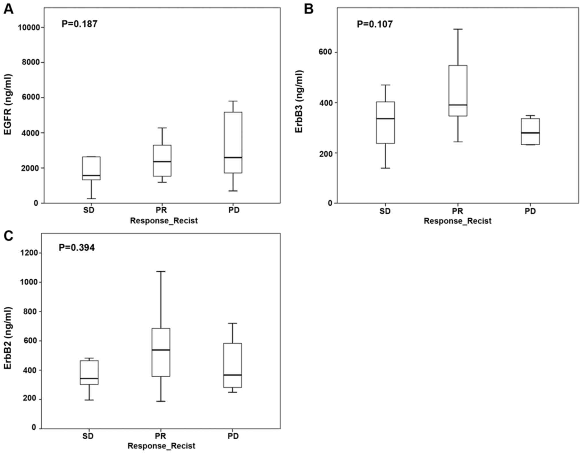
Expression of EGFR protein in tumor samples did not
significantly correlate with treatment response to anti-EGFR
therapy according to RECIST criteria. The median EGFR/ErbB1 protein
level was 2,278.8 ng/ml (IQR 1,387.3–4,127.1 ng/ml) and was not
different in groups of patients with partial response, stable
disease, or progressive disease. Similarly, tumor protein levels of
ErbB2 (median 348.8 ng/ml, IQR 244.1–453.6 ng/ml) and ErbB3 (median
426.5 ng/ml, IQR 292.3–642.4 ng/ml) did not correlate with
treatment response (Fig. 1A-C).
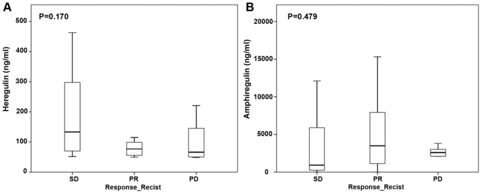
Expression of EGFR ligands heregulin and
amphiregulin did not correlate with treatment response to anti-EGF
therapy. The median heregulin protein level was 76.9 ng/ml (IQR
55–196 ng/ml) and the median amphiregulin protein level was 2,900.2
ng/ml (IQR 664.8–5,884.6 ng/ml). EGFR ligand protein expression did
not differ between patients with partial response, stable disease,
or progressive disease (Fig. 2A and
B).
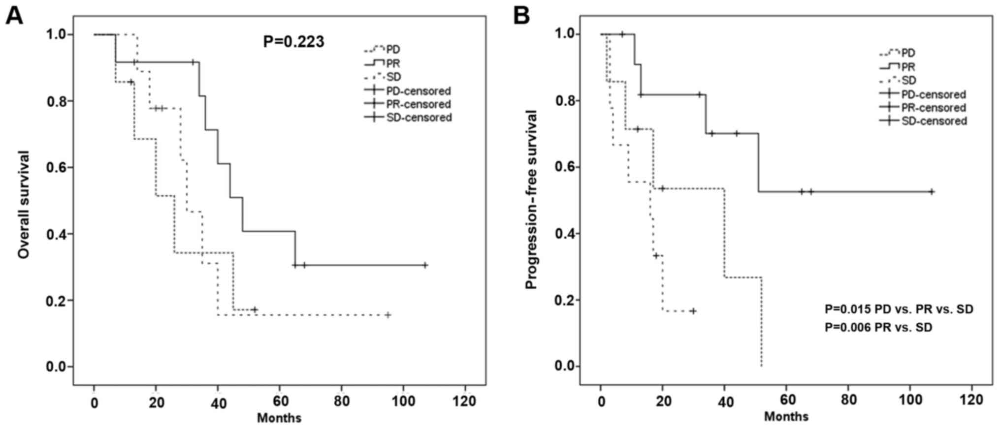
Survival analysis
Median overall survival of all patients was 40
months. Overall survival showed no significant difference between
the three RECIST groups: median OAS PR group: 48 months [95% CI,
36–60 months]; median OAS SD group: 30 months [95% CI 22- 38
months]; median OAS PD group: 26 months [95% CI, 11.5–41 months]
(P=0.223).
Median progression free survival of all patients was
34 months. Progression free survival showed a significant
difference between the three RECIST groups: median PFS PR group: 35
months [95% CI, 21–59 months]; median PFS SD group: 16 months [95%
CI, 0–36 months]; median PFS PD group: 17 months [95% CI, 5–38
months] (P=0.015). Therefore, we evaluated the differences in
progression free survival between PR vs SD, PR vs PD and SD vs PD.
There was a significant difference in progression free survival
between the PR and the SD group (P=0.006), for PR vs PD
respectively SD vs PD there was no significant difference in
progression free survival (P=0.77 respectively P=0.268) (Fig. 3).
Discussion
In this study, we determined protein levels of
different EGFRs and EGFR ligands in tumor samples taken from
patients with mCRC. All patients were treated with a combination
chemotherapy regimen plus cetuximab. Patients were grouped into
three groups based on their treatment response (determined by
RECIST criteria): partial response, stable disease, and progressive
disease. We observed no correlation between protein expression of
EGFR and EGFR ligands in tumor tissues and treatment response.
The EGF signaling pathway comprises 13 ligands.
Interactions between the four known EGFRs and their ligands are
remarkably complex. These interactions have been analyzed using
different techniques, including immunohistochemistry, real-time
PCR, fluorescence in situ hybridization (FISH), and ELISA,
which differ in reliability and sensitivity. Consequently, reported
EGFR and EGFR ligand expression rates vary a lot in colorectal
cancer specimens in the published literature. Differences between
RNA and protein expression due to posttranscriptional alterations
should also be considered (22,23).
Approximately 60–85% of primary colorectal cancer
tissues overexpress EGFR (24,25). We
expected ErbB1 to be overexpressed in responding patients, but we
observed no significant associations between ErbB1 expression and
treatment response. This finding is in line with the results of
Italiano et al (26). Others
have described varying associations between EGFR expression and
response to treatment and/or survival (27–29).
Several studies have examined EGFR and EGFR ligand
expression and have described associations between these expression
levels and response to cetuximab (20,23,30–51).
However, for the reasons mentioned above, these results are
conflicting. The mRNA expression reported in some studies may not
reflect the expression of corresponding proteins that would compete
with cetuximab for EGFR binding in tumor cells.
Although anti-EGFR therapy has proven to be
beneficial in KRAS wild-type advanced colorectal cancer (52), results are contradictory and
underlying mechanisms are not fully understood. Some patients with
selected KRAS mutations have responded to anti-EGFR therapy
(53). Possible reasons could be the
heterogeneity of KRAS mutations in different primary tumors and
metastases (22). Furthermore, other
factors may influence the therapeutic response, such as the
percentage of viable tumor cells and the tumor microenvironment,
which plays a pivotal role in tumor progression (54). The FOCUS-4 study has shown that
combined inhibition of EGFR, ErbB2/Her2, and ErbB3/Her3 does not
improve the outcome in patients with advanced colorectal cancer
expressing wild-type KRAS, NRAS, B rat fibrosarcoma, and PIK3CA, so
the study was closed after the first interim analysis (55).
Some limitations of the study have to be named.
First limitation is the small sample size of patients included into
the study. Second the retrospective study design. Third there might
be a bias because of the higher frequency of male patients in the
study, which was a random side effect because study patients were
selected due to their received chemotherapy regime containing
cetuximab regardless their sex.
Because of some unclear aspects further studies on
this topic are necessary investigating RNA as well as protein level
of EGF receptors and EGFR ligands to assess posttranscriptional
differences and to evaluate the varying associations between EGFR
expression and response to treatment and/or survival. Furthermore,
it is needed to examine why selected KRAS mutations still respond
to anti-EGFR therapy and to assess the influence and heterogeneity
of the expressional differences in the viable tumor cells and the
tumor microenvironment in order to improve the personalized
treatment of patients with anti-EGF agents.
Acknowledgements
Not applicable.
Funding
FK was funded by the Olympia-Morata Postdoctoral
Fellowship of the Medical Faculty of the University of Heidelberg
(Heidelberg, Germany).
Availability of data and materials
The datasets used and/or analyzed during the current
study are available from the corresponding author on reasonable
request.
Authors' contributions
FK, MS, FB, MH, AM and YK contributed to the study
design. FK, MS, EK, OG, MH and YK collected the data. FK, MS, OG,
MH and YK assessed and confirmed the authenticity of all the raw
data. FK, MS, EK, OG, FB, MH, AM and YK contributed to data
analysis. FK, MS and YK drafted the manuscript, and MS, EK, OG, FB,
MH, AM and YK reviewed the manuscript. All authors read and
approved the final manuscript.
Ethics approval and consent to
participate
The study was conducted ethically in accordance with
the Declaration of Helsinki and the appropriate guidelines for
human studies. The study was approved by the Ethics Committee of
the University of Heidelberg (Heidelberg, Germany; approval no.
S-168/2008) and written informed consent was obtained from each
patient.
Patient consent for publication
Not applicable.
Competing interests
The authors declare that they have no competing
interests.
References
|
1
|
Soerjomataram I, Lortet-Tieulent J, Parkin
DM, Ferlay J, Mathers C, Forman D and Bray F: Global burden of
cancer in 2008: A systematic analysis of disability-adjusted
life-years in 12 world regions. Lancet. 380:1840–1850. 2012.
View Article : Google Scholar : PubMed/NCBI
|
|
2
|
Douillard JY, Oliner KS, Siena S,
Tabernero J, Burkes R, Barugel M, Humblet Y, Bodoky G, Cunningham
D, Jassem J, et al: Panitumumab-FOLFOX4 treatment and RAS mutations
in colorectal cancer. N Engl J Med. 369:1023–1034. 2013. View Article : Google Scholar : PubMed/NCBI
|
|
3
|
Mody K, Baldeo C and Bekaii-Saab T:
Antiangiogenic Therapy in Colorectal Cancer. Cancer J. 24:165–170.
2018. View Article : Google Scholar : PubMed/NCBI
|
|
4
|
Van Cutsem E, Köhne CH, Hitre E, Zaluski
J, Chang Chien CR, Makhson A, D'Haens G, Pintér T, Lim R, Bodoky G,
et al: Cetuximab and chemotherapy as initial treatment for
metastatic colorectal cancer. N Engl J Med. 360:1408–1417. 2009.
View Article : Google Scholar : PubMed/NCBI
|
|
5
|
Misale S, Di Nicolantonio F,
Sartore-Bianchi A, Siena S and Bardelli A: Resistance to anti-EGFR
therapy in colorectal cancer: From heterogeneity to convergent
evolution. Cancer Discov. 4:1269–1280. 2014. View Article : Google Scholar : PubMed/NCBI
|
|
6
|
Ferrara N, Hillan KJ and Novotny W:
Bevacizumab (Avastin), a humanized anti-VEGF monoclonal antibody
for cancer therapy. Biochem Biophys Res Commun. 333:328–335. 2005.
View Article : Google Scholar : PubMed/NCBI
|
|
7
|
Itatani Y, Kawada K, Yamamoto T and Sakai
Y: Resistance to Anti-Angiogenic Therapy in Cancer-Alterations to
Anti-VEGF Pathway. Int J Mol Sci. 19:12322018. View Article : Google Scholar : PubMed/NCBI
|
|
8
|
McCormack PL and Keam SJ: Bevacizumab: A
review of its use in metastatic colorectal cancer. Drugs.
68:487–506. 2008. View Article : Google Scholar : PubMed/NCBI
|
|
9
|
Van Cutsem E, Cervantes A, Nordlinger B,
Arnold D and group EGW; ESMO Guidelines Working Group, : Metastatic
colorectal cancer: ESMO Clinical Practice Guidelines for diagnosis,
treatment and follow-up. Ann Oncol. 25 (Suppl 3):iii1–iii9. 2014.
View Article : Google Scholar
|
|
10
|
Yarden Y: The EGFR family and its ligands
in human cancer. signalling mechanisms and therapeutic
opportunities. Eur J Cancer. 37 (Suppl 4):S3–S8. 2001. View Article : Google Scholar
|
|
11
|
Olayioye MA, Neve RM, Lane HA and Hynes
NE: The ErbB signaling network: Receptor heterodimerization in
development and cancer. EMBO J. 19:3159–3167. 2000. View Article : Google Scholar : PubMed/NCBI
|
|
12
|
Yamaoka T, Ohba M and Ohmori T:
Molecular-Targeted Therapies for Epidermal Growth Factor Receptor
and Its Resistance Mechanisms. Int J Mol Sci. 18:24202017.
View Article : Google Scholar : PubMed/NCBI
|
|
13
|
Petrelli F, Borgonovo K, Cabiddu M,
Ghilardi M and Barni S: Cetuximab and panitumumab in KRAS wild-type
colorectal cancer: A meta-analysis. Int J Colorectal Dis.
26:823–833. 2011. View Article : Google Scholar : PubMed/NCBI
|
|
14
|
Rizzo S, Bronte G, Fanale D, Corsini L,
Silvestris N, Santini D, Gulotta G, Bazan V, Gebbia N, Fulfaro F,
et al: Prognostic vs predictive molecular biomarkers in colorectal
cancer: Is KRAS and BRAF wild type status required for anti-EGFR
therapy? Cancer Treat Rev. 36 (Suppl 3):S56–S61. 2010. View Article : Google Scholar
|
|
15
|
Karapetis CS, Khambata-Ford S, Jonker DJ,
O'Callaghan CJ, Tu D, Tebbutt NC, Simes RJ, Chalchal H, Shapiro JD,
Robitaille S, et al: K-ras mutations and benefit from cetuximab in
advanced colorectal cancer. N Engl J Med. 359:1757–1765. 2008.
View Article : Google Scholar : PubMed/NCBI
|
|
16
|
Misale S, Yaeger R, Hobor S, Scala E,
Janakiraman M, Liska D, Valtorta E, Schiavo R, Buscarino M,
Siravegna G, et al: Emergence of KRAS mutations and acquired
resistance to anti-EGFR therapy in colorectal cancer. Nature.
486:532–536. 2012. View Article : Google Scholar : PubMed/NCBI
|
|
17
|
Van Cutsem E, Lenz HJ, Köhne CH, Heinemann
V, Tejpar S, Melezínek I, Beier F, Stroh C, Rougier P, van Krieken
JH, et al: Fluorouracil, leucovorin, and irinotecan plus cetuximab
treatment and RAS mutations in colorectal cancer. J Clin Oncol.
33:692–700. 2015. View Article : Google Scholar : PubMed/NCBI
|
|
18
|
Jones C, Taylor MA and McWilliams B: The
role of cetuximab as first-line treatment of colorectal liver
metastases. HPB (Oxford). 15:11–17. 2013. View Article : Google Scholar : PubMed/NCBI
|
|
19
|
Saltz LB, Lenz HJ, Kindler HL, Hochster
HS, Wadler S, Hoff PM, Kemeny NE, Hollywood EM, Gonen M, Quinones
M, et al: Randomized phase II trial of cetuximab, bevacizumab, and
irinotecan compared with cetuximab and bevacizumab alone in
irinotecan-refractory colorectal cancer: The BOND-2 study. J Clin
Oncol. 25:4557–4561. 2007. View Article : Google Scholar : PubMed/NCBI
|
|
20
|
Jacobs B, De Roock W, Piessevaux H, Van
Oirbeek R, Biesmans B, De Schutter J, Fieuws S, Vandesompele J,
Peeters M, Van Laethem JL, et al: Amphiregulin and epiregulin mRNA
expression in primary tumors predicts outcome in metastatic
colorectal cancer treated with cetuximab. J Clin Oncol.
27:5068–5074. 2009. View Article : Google Scholar : PubMed/NCBI
|
|
21
|
Eisenhauer EA, Therasse P, Bogaerts J,
Schwartz LH, Sargent D, Ford R, Dancey J, Arbuck S, Gwyther S,
Mooney M, et al: New response evaluation criteria in solid tumours:
Revised RECIST guideline (version 1.1). Eur J Cancer. 45:228–247.
2009. View Article : Google Scholar : PubMed/NCBI
|
|
22
|
Roskoski R Jr: The ErbB/HER family of
protein-tyrosine kinases and cancer. Pharmacol Res. 79:34–74. 2014.
View Article : Google Scholar : PubMed/NCBI
|
|
23
|
Valtorta E, Martino C, Sartore-Bianchi A,
Penaullt-Llorca F, Viale G, Risio M, Rugge M, Grigioni W,
Bencardino K, Lonardi S, et al: Assessment of a HER2 scoring system
for colorectal cancer: results from a validation study. Mod Pathol.
28:1481–1491. 2015. View Article : Google Scholar : PubMed/NCBI
|
|
24
|
Cunningham D, Humblet Y, Siena S, Khayat
D, Bleiberg H, Santoro A, Bets D, Mueser M, Harstrick A, Verslype
C, et al: Cetuximab monotherapy and cetuximab plus irinotecan in
irinotecan-refractory metastatic colorectal cancer. N Engl J Med.
351:337–345. 2004. View Article : Google Scholar : PubMed/NCBI
|
|
25
|
Porebska I, Harlozińska A and Bojarowski
T: Expression of the tyrosine kinase activity growth factor
receptors (EGFR, ERB B2, ERB B3) in colorectal adenocarcinomas and
adenomas. Tumour Biol. 21:105–115. 2000. View Article : Google Scholar : PubMed/NCBI
|
|
26
|
Italiano A, Follana P, Caroli FX, Badetti
JL, Benchimol D, Garnier G, Gugenheim J, Haudebourg J, Keslair F,
Lesbats G, et al: Cetuximab shows activity in colorectal cancer
patients with tumors for which FISH analysis does not detect an
increase in EGFR gene copy number. Ann Surg Oncol. 15:649–654.
2008. View Article : Google Scholar : PubMed/NCBI
|
|
27
|
Uhlyarik A, Piurko V, Papai Z, Raso E,
Lahm E, Kiss E, Sikter M, Vachaja J, Kenessey I and Timar J: EGFR
Protein Expression in KRAS Wild-Type Metastatic Colorectal Cancer
Is Another Negative Predictive Factor of the Cetuximab Therapy.
Cancers (Basel). 12:6142020. View Article : Google Scholar : PubMed/NCBI
|
|
28
|
Laurent-Puig P, Cayre A, Manceau G, Buc E,
Bachet JB, Lecomte T, Rougier P, Lievre A, Landi B, Boige V, et al:
Analysis of PTEN, BRAF, and EGFR status in determining benefit from
cetuximab therapy in wild-type KRAS metastatic colon cancer. J Clin
Oncol. 27:5924–5930. 2009. View Article : Google Scholar : PubMed/NCBI
|
|
29
|
Personeni N, Fieuws S, Piessevaux H, De
Hertogh G, De Schutter J, Biesmans B, De Roock W, Capoen A,
Debiec-Rychter M, Van Laethem JL, et al: Clinical usefulness of
EGFR gene copy number as a predictive marker in colorectal cancer
patients treated with cetuximab: A fluorescent in situ
hybridization study. Clin Cancer Res. 14:5869–5876. 2008.
View Article : Google Scholar : PubMed/NCBI
|
|
30
|
Nagaoka T, Kitaura K, Miyata Y, Kumagai K,
Kaneda G, Kanazawa H, Suzuki S, Hamada Y and Suzuki R:
Downregulation of epidermal growth factor receptor family receptors
and ligands in a mutant K-ras group of patients with colorectal
cancer. Mol Med Rep. 13:3514–3520. 2016. View Article : Google Scholar : PubMed/NCBI
|
|
31
|
Park DI, Kang MS, Oh SJ, Kim HJ, Cho YK,
Sohn CI, Jeon WK, Kim BI, Han WK, Kim H, et al: HER-2/neu
overexpression is an independent prognostic factor in colorectal
cancer. Int J Colorectal Dis. 22:491–497. 2007. View Article : Google Scholar : PubMed/NCBI
|
|
32
|
Conradi LC, Styczen H, Sprenger T, Wolff
HA, Rödel C, Nietert M, Homayounfar K, Gaedcke J, Kitz J,
Talaulicar R, et al: Frequency of HER-2 positivity in rectal cancer
and prognosis. Am J Surg Pathol. 37:522–531. 2013. View Article : Google Scholar : PubMed/NCBI
|
|
33
|
Gharib E, Salmanipour R, Nazemalhosseini
Mojarad E, Yaghoob Taleghani M, Sarlak S, Malekzade-Moghani M,
Nasrabadi PN, Meiary MA, Asadzadeh Aghdaei H and Zali MR:
HER2+ mCRC patients with exon 20 R784G substitution
mutation do not respond to the cetuximab therapy. J Cell Physiol.
234:13137–13144. 2019. View Article : Google Scholar : PubMed/NCBI
|
|
34
|
Aurisicchio L, Marra E, Roscilli G,
Mancini R and Ciliberto G: The promise of anti-ErbB3 monoclonals as
new cancer therapeutics. Oncotarget. 3:744–758. 2012. View Article : Google Scholar : PubMed/NCBI
|
|
35
|
Beji A, Horst D, Engel J, Kirchner T and
Ullrich A: Toward the prognostic significance and therapeutic
potential of HER3 receptor tyrosine kinase in human colon cancer.
Clin Cancer Res. 18:956–968. 2012. View Article : Google Scholar : PubMed/NCBI
|
|
36
|
Scartozzi M, Giampieri R, Maccaroni E,
Mandolesi A, Giustini L, Silva R, Zaniboni A, Biscotti T, Biagetti
S, Galizia E, et al: Analysis of HER-3, insulin growth factor-1,
nuclear factor-κB and epidermal growth factor receptor gene copy
number in the prediction of clinical outcome for K-RAS wild-type
colorectal cancer patients receiving irinotecan-cetuximab. Ann
Oncol. 23:1706–1712. 2012. View Article : Google Scholar : PubMed/NCBI
|
|
37
|
Lédel F, Hallström M, Ragnhammar P,
Öhrling K and Edler D: HER3 expression in patients with primary
colorectal cancer and corresponding lymph node metastases related
to clinical outcome. Eur J Cancer. 50:656–662. 2014. View Article : Google Scholar : PubMed/NCBI
|
|
38
|
Cushman SM, Jiang C, Hatch AJ, Shterev I,
Sibley AB, Niedzwiecki D, Venook AP, Owzar K, Hurwitz HI and Nixon
AB: Gene expression markers of efficacy and resistance to cetuximab
treatment in metastatic colorectal cancer: results from CALGB 80203
(Alliance). Clin Cancer Res. 21:1078–1086. 2015. View Article : Google Scholar : PubMed/NCBI
|
|
39
|
Stahler A, Heinemann V, Neumann J, Crispin
A, Schalhorn A, Stintzing S, Giessen-Jung C, Fischer von
Weikersthal L, Vehling-Kaiser U, Stauch M, et al: Prevalence and
influence on outcome of HER2/neu, HER3 and NRG1 expression in
patients with metastatic colorectal cancer. Anticancer Drugs.
28:717–722. 2017. View Article : Google Scholar : PubMed/NCBI
|
|
40
|
Busser B, Sancey L, Brambilla E, Coll JL
and Hurbin A: The multiple roles of amphiregulin in human cancer.
Biochim Biophys Acta. 1816:119–131. 2011.PubMed/NCBI
|
|
41
|
Zvibel I, Brill S, Halpern Z and Papa M:
Amphiregulin and hepatocyte-derived extracellular matrix regulate
proliferation and autocrine growth factor expression in colon
cancer cell lines of varying liver-colonizing capability. J Cell
Biochem. 76:332–340. 1999. View Article : Google Scholar : PubMed/NCBI
|
|
42
|
Jing C, Jin YH, You Z, Qiong Q and Jun Z:
Prognostic value of amphiregulin and epiregulin mRNA expression in
metastatic colorectal cancer patients. Oncotarget. 7:55890–55899.
2016. View Article : Google Scholar : PubMed/NCBI
|
|
43
|
Lo Nigro C, Ricci V, Vivenza D, Granetto
C, Fabozzi T, Miraglio E and Merlano MC: Prognostic and predictive
biomarkers in metastatic colorectal cancer anti-EGFR therapy. World
J Gastroenterol. 22:6944–6954. 2016. View Article : Google Scholar : PubMed/NCBI
|
|
44
|
Nishimura T, Andoh A, Inatomi O, Shioya M,
Yagi Y, Tsujikawa T and Fujiyama Y: Amphiregulin and epiregulin
expression in neoplastic and inflammatory lesions in the colon.
Oncol Rep. 19:105–110. 2008.PubMed/NCBI
|
|
45
|
Yamada M, Ichikawa Y, Yamagishi S,
Momiyama N, Ota M, Fujii S, Tanaka K, Togo S, Ohki S and Shimada H:
Amphiregulin is a promising prognostic marker for liver metastases
of colorectal cancer. Clin Cancer Res. 14:2351–2356. 2008.
View Article : Google Scholar : PubMed/NCBI
|
|
46
|
Seligmann JF, Elliott F, Richman SD,
Jacobs B, Hemmings G, Brown S, Barrett JH, Tejpar S, Quirke P and
Seymour MT: Combined Epiregulin and Amphiregulin Expression Levels
as a Predictive Biomarker for Panitumumab Therapy Benefit or Lack
of Benefit in Patients With RAS Wild-Type Advanced Colorectal
Cancer. JAMA Oncol. 2:633–642. 2016. View Article : Google Scholar : PubMed/NCBI
|
|
47
|
Yoshida M, Shimura T, Sato M, Ebi M,
Nakazawa T, Takeyama H and Joh T: A novel predictive strategy by
immunohistochemical analysis of four EGFR ligands in metastatic
colorectal cancer treated with anti-EGFR antibodies. J Cancer Res
Clin Oncol. 139:367–378. 2013. View Article : Google Scholar : PubMed/NCBI
|
|
48
|
Sebio A, Páez D, Salazar J,
Berenguer-Llergo A, Paré-Brunet L, Lasa A, Del Río E, Tobeña M,
Martín-Richard M, Baiget M, et al: Intergenic polymorphisms in the
amphiregulin gene region as biomarkers in metastatic colorectal
cancer patients treated with anti-EGFR plus irinotecan.
Pharmacogenomics J. 14:256–262. 2014. View Article : Google Scholar : PubMed/NCBI
|
|
49
|
Breuleux M: Role of heregulin in human
cancer. Cell Mol Life Sci. 64:2358–2377. 2007. View Article : Google Scholar : PubMed/NCBI
|
|
50
|
Khurana A, Gonzalez-Guerrico A and Lupu R:
Heregulin in breast cancer: Old story, new paradigm. Curr Pharm
Des. 20:4874–4878. 2014. View Article : Google Scholar : PubMed/NCBI
|
|
51
|
Yonesaka K, Zejnullahu K, Okamoto I, Satoh
T, Cappuzzo F, Souglakos J, Ercan D, Rogers A, Roncalli M, Takeda
M, et al: Activation of ERBB2 signaling causes resistance to the
EGFR-directed therapeutic antibody cetuximab. Sci Transl Med.
3:99ra862011. View Article : Google Scholar : PubMed/NCBI
|
|
52
|
Tsilimigras DI, Ntanasis-Stathopoulos I,
Bagante F, Moris D, Cloyd J, Spartalis E and Pawlik TM: Clinical
significance and prognostic relevance of KRAS, BRAF, PI3K and TP53
genetic mutation analysis for resectable and unresectable
colorectal liver metastases: A systematic review of the current
evidence. Surg Oncol. 27:280–288. 2018. View Article : Google Scholar : PubMed/NCBI
|
|
53
|
De Roock W, Jonker DJ, Di Nicolantonio F,
Sartore-Bianchi A, Tu D, Siena S, Lamba S, Arena S, Frattini M,
Piessevaux H, et al: Association of KRAS p.G13D mutation with
outcome in patients with chemotherapy-refractory metastatic
colorectal cancer treated with cetuximab. JAMA. 304:1812–1820.
2010. View Article : Google Scholar : PubMed/NCBI
|
|
54
|
Pietras K and Ostman A: Hallmarks of
cancer: Interactions with the tumor stroma. Exp Cell Res.
316:1324–1331. 2010. View Article : Google Scholar : PubMed/NCBI
|
|
55
|
Adams R, Brown E, Brown L, Butler R, Falk
S, Fisher D, Kaplan R, Quirke P, Richman S, Samuel L, et al FOCUS4
Trial Investigators, : Inhibition of EGFR, HER2, and HER3
signalling in patients with colorectal cancer wild-type for BRAF,
PIK3CA, KRAS, and NRAS (FOCUS4-D): A phase 2–3 randomised trial.
Lancet Gastroenterol Hepatol. 3:162–171. 2018. View Article : Google Scholar : PubMed/NCBI
|