Introduction
Endometrial cancer (EC) is one of the three main
malignant tumors of the female reproductive system (1). Despite recent discoveries in the
field of cancer therapy, no significant improvement has been made
in the treatment of EC or its early detection. Most patients with
EC exhibit tumor progression during their visits to their medical
practitioners. ECs are usually spread locally or have metastasized
(2–4). It has been shown that the degree of
malignancy of EC is very high and is characterized by potent
invasive and metastatic activities (5). Invasion and metastasis are the main
obstacles to the long-term survival and quality of life of patients
with EC (5,6). These two processes are also
considered as crucial biological behaviors of ECs, and may
therefore be used as research directions for the treatment of
Ec.
Ubiquitination is intricately involved in
tumorigenesis and cancer development, including invasion and
metastasis (7,8). The E3 ubiquitination ligand is
particularly important in tumorigenesis and cancer development
(9–12). E3 ubiquitin ligases are a class of
enzymes that are involved in the regulation of the conversion and
activity of several target proteins. Significant differences are
often noted in the structure and expression of several E3 ubiquitin
ligases in tumor tissues compared with those of the corresponding
normal tissues. In addition, these E3 ubiquitin ligases can have a
significant impact on the regulation of malignant behavior, such as
the regulation of epithelial-to-mesenchymal transition (13,14).
Increasing evidence has strongly suggested that the abnormal
regulation of E3 ligases is related to the occurrence of cancer
(12–14). Proteins with E3 ubiquitin ligases
may be considered as potential cancer drug targets and prognostic
biomarkers.
As a member of the Ligand of NUMB protein-X family,
PDZ Domain Containing Ring Finger 3 (PDZRN3) contains the RING-type
ubiquitin E3 ligase. The PDZRN3 gene is located in the 3p13
chromosomal region and is ~242,568 bases in length. The molecular
mass of the corresponding protein is estimated to be 119,596 Da and
the protein is found in human and mouse tissues (15). Previous studies have indicated that
PDZRN3 can induce the differentiation of C2C12 mouse mesenchymal
progenitor cells into myotubes or osteoblasts. This indicates that
PDZRN3 exerts a certain effect on cell differentiation (16), which is also an extremely important
biological function of tumor cells. Furthermore, the E3-ubiquitin
ligase has been shown to affect the ubiquitination function of
PDZRN3. The latter can promote the modification of the
ubiquitination process of related molecules via the E3 ubiquitin
ligase domain. This phenomenon is mediated by the decrease in
Muscle-Specific Kinase expression via PDZRN3-induced ubiquitination
(17,18). PDZRN3 interacts with the E3
ubiquitin ligase domain. Cell differentiation provides a necessary
theoretical basis for PDZRN3 in tumor invasion and metastasis,
including EC.
The aim of the present study was to investigate the
effects of PDZRN3 on EC cell proliferation, migration and
invasiveness. Reverse transcription-quantitative PCR (RT-qPCR) was
used to detect the expression levels of PDZRN3 in EC cells, and the
role of PDZRN3 in EC progression was determined using western
blotting, and MTT, colony formation, Transwell, subcutaneous tumor
formation and pulmonary metastasis assays. A multi-pathway reporter
array and western blotting were performed to investigate the
potential biological mechanisms of PDZRN3 in EC.
Materials and methods
Cell culture and transfection
HEC-1-B and KLE cell lines were purchased from
Tongpai (Shanghai) Biotechnology Co., Ltd. HEC-1-B cells were
cultured in high glucose medium containing 10% FBS, 100 U/ml
penicillin and 100 mg/ml streptomycin (all Hyclone; Cytiva). KLE
cells were cultured in F12 medium (Hyclone; Cytiva) containing 10%
FBS, 100 U/ml penicillin and 100 mg/ml streptomycin. All cells were
placed at 37°C in a humidified incubator containing 5%
CO2.
The designed plasmid (en-PDZRN3) was transfected
into cells using Lipofectamine™ 3000 (Invitrogen; Thermo Fisher
Scientific, Inc). The plasmids were purchased from Wegene
Biosciences Co., Ltd. The number of cells transduced with
lentivirus was ~2×10⁵/well. The next day, the original
medium was replaced with 2 ml fresh medium with 1 µl/ml
Lipofectamine 3000 and adding 20 µl virus suspension by incubating
at 37°C for 24 h. After 24 h, the medium in the well was removed
and 2 ml of fresh medium (5 µg/ml puromycin) was added for 48 h.
The cells were amplified by changing the fresh medium, and the
transfection efficiency was checked using RT-qPCR and western
blotting. At 24 h before lentivirus transfection, the adherent
cells were treated with 1×10⁵/well in a 24-well
plate.
RT-qPCR
RNA was extracted from HEC-1-B/HEC-1-B-PDZRN3 and
KLE/KLE-PDZRN3 cells using TRIzol® (Invitrogen; Thermo
Fisher Scientific, Inc.) and reverse transcribed into cDNA using a
reverse transcription kit (Roche Applied Science) according to the
manufacturer's instructions, followed by amplification using a qPCR
kit (Roche Applied Science). The following thermocycling conditions
were used for qPCR: 95°C for 10 min; followed by 40 cycles of 95°C
for 30 sec, 60°C for 30 sec and 72°C for 30 sec, and final
extension at 72°C for 2 min. The expression level of PDZRN3 was
normalized to endogenous control GAPDH and expressed using the
2−ΔΔCq method (19).
Each assay was repeated three times. The sequences of the primers
were as follows: GAPDH forward, 5′-CACCGCAAATGCTTCTAGGC-3′ and
reverse, 5′-GATCTCCACACACCTGCACT-3′; and PDZRN3 forward,
5′-ATTATTGAGGTCAACGGCAG-3′ and reverse,
5′-AGGGCCATGATATGTTCAAAG-3′.
Western blotting
Cells were lysed using RIPA lysis buffer (New Cell
& Molecular Biotech Co., Ltd.). Following estimation of the
protein concentration using a BCA kit (Beyotime Institute of
Biotechnology), proteins (40 µg) were separated by 12% SDS-PAGE and
transferred onto a PVDF membrane (MilliporeSigma). Membranes were
incubated with primary antibodies against PDZRN3 (1:2,000; cat. no.
Ag24455; ProteinTech Group, Inc.), β-catenin (1:2,000; cat. no.
17565-1-AP; ProteinTech Group, Inc.) and tubulin (1:2,000; cat. no.
11224-1-AP; ProteinTech Group, Inc.) at 4°C overnight. Membranes
were washed by 1X PBST solution and incubated with the
corresponding secondary antibodies (HRP-conjugated Affinipure goat
anti-rabbit IgG (H+L); 1:5,000; cat. no. SA00001-2; ProteinTech
Group, Inc.). Enhanced chemiluminescence reagent (ebiogen, Inc.)
was used to detect the signal on the membrane. The data were
analyzed via densitometry and normalized to expression of the
internal control (tubulin) by ImageJ 1.8. (National Institutes of
Health).
MTT assay
Cells (1×10⁴) in the
HEC-1-B/HEC-1-B-PDZRN3 and KLE/KLE-PDZRN3 groups were incubated
into 96-well plates and cultured for 7 days. A total of 20 µl MTT
(5 mg/ml) was added into the culture medium and incubated for 4 h.
Subsequently, 200 µl DMSO was added following removal of the medium
to dissolve the MTT formazan. The absorbance was read at 490 nm on
a microplate reader.
Colony formation assay
Colony formation assay was performed to evaluate the
proliferative ability of EC cells. A total of 500 EC cells derived
from the HEC-1-B/HEC-1-B-PDZRN3 and KLE/KLE-PDZRN3 groups were
seeded into six-well plates and stained at room temperature for 10
min with 0.1% crystal violet following 14 days of incubation. The
number of colonies (cell clusters with a diameter >1 mm) was
counted manually using a microscope.
Invasion assay
A total of 5×10⁵ cells were resuspended
in 200 µl serum-free medium and subsequently seeded to the upper
chamber of a Transwell (Corning, Inc.) insert precoated with 1
µg/µl Matrigel (BD Biosciences). Complete medium was added to the
lower chamber to stimulate cell invasion. Following 48 h of cell
culture, the cells that did not cross the membrane were wiped with
a cotton swab, while the cells adhering to the lower surface of the
membrane were stained with 0.1% crystal violet solution at room
temperature for 10 min. The number of invading cells was counted in
five randomly selected fields using a light microscope
(magnification, ×200; Olympus Corporation). All experiments were
performed three times.
Migration assay
A total of 5×10⁵ cells were resuspended
in 200 µl serum-free medium and subsequently seeded to the upper
chamber of a Transwell insert without Matrigel. All other
procedures were performed as described for the invasion assay.
Subcutaneous tumor formation
assay
A total of 40 BALB/c-nude female mice aged 6-8 weeks
were purchased from the Slk Jing Da Laboratory Animal Co., Ltd. and
the experiments were performed in a SPF-level sterile laboratory at
the Animal Testing Center of Tongji University. All experimental
procedures were approved by the Animal Ethics Committee of Shanghai
Yangpu District Shidong Hospital. All mice were ~20 g in weight
when purchased. Mice were housed with free access to food and water
in a temperature- and light-regulated pathogen-free room
(temperature, 24±1°C; humidity, 60±5%; 12-h light/dark cycle). In
the present study, the maximum length and diameter of the tumor was
<15 mm and the volume and weight of the tumor did not exceed 20%
of body weight. The tumor load of each mouse was no more than 2
tumor lesions. Direct cervical dislocation was used for euthanasia,
which provides a rapid, painless and stress-free death. All mice
were judged dead following no response to stimulation, no chest
fluctuation, no heartbeat and significant decrease in body
temperature. After death, all mice were treated with humane care
and pollution-free treatment in the animal experiment center. In
the animal center, the animal keeper checks once a day to ensure
the health of mice. There were 5 nude mice in the
HEC-1-B/HEC-1-B-PDZRN3 and KLE/KLE-PDZRN3 groups. The
HEC-1-B/HEC-1-B-PDZRN3 cells and KLE/KLE-PDZRN3 cells
(5×10⁶) for each group were injected subcutaneously into
nude mice (7–8 weeks, female, BALB/c) to assess their tumorigenic
capacity. The tumor samples were collected following 4 weeks of
incubation and after euthanasia. Tumor volume was calculated as
follows: Tumor volume (mm3)=(AxB2)/2, where A
corresponds to length and B to width. The in vivo
experiments lasted for 4 weeks, during which the animals suffered
no pain or showed no sign of distress, such as rapid weight loss
(over 20%), loss of appetite, weakness, organ and tissue infection
or tumor overweight.
Pulmonary metastasis assay
The concentration of the HEC-1-B/HEC-1-B-PDZRN3
cells and KLE/KLE-PDZRN3 cells was adjusted to 5×10⁷
cells/ml using a cell counter. Each nude mouse was injected with
100 µl cell suspension twice a week. Following 4 weeks of
incubation, the nude mice were sacrificed by cervical dislocation.
The lungs of the nude mice were completely removed and subsequently
immersed in 10% formalin solution. The lungs of the nude mice were
embedded in paraffin and sectioned into ~5 µm thick slices.
Following hematoxylin and eosin staining, the metastasis of the
lung tumors was observed by microscopy and the number of metastatic
colonies was counted.
Hematoxylin and eosin (H&E)
staining and immunohistochemical (IHC) staining
For H&E staining, the slides were incubated at
62°C for 1 h. The slices were dewaxed in xylene solution and
rehydrated with graded alcohol (100, 95, 75 and 50%) and distilled
water. The slides were stained with hematoxylin solution at room
temperature for 5 min, then immersed in 1% acid ethanol at room
temperature for 5 min, prior to washing with distilled water. The
slides were stained with eosin solution for 3 min, then washed with
graded alcohol (50, 75, 95 and 100%) for dehydration. The slides
were then dried with xylene solution at room temperature. The
H&E images were captured at ×100 magnification using a
microscope camera system (Olympus Corporation).
For IHC staining, the slides were dewaxed in xylene
solution and rehydrated in graded alcohol (100, 95, 75 and 50%) and
distilled water, then treated with 3% methanol hydrogen peroxide at
room temperature for 30 min. After washing with PBS three times,
they were heated in a microwave oven with 10% citrate buffer for 10
min twice and cooled to room temperature, then 10% BSA was added
for 30 min. The slices were incubated with anti-Ki-67 antibody
(1:2,000; cat. no. 27309-1-AP; ProteinTech Group, Inc.) overnight
at 4°C. The next day, after washing with PBS, the sections were
incubated with HRP-conjugated secondary antibody at room
temperature for 1 h. DAB solution was used for staining for 5 min
and hematoxylin was used for re-staining the nucleus. The slides
were then washed with graded alcohol (50, 75, 95 and 100%) for
dehydration, then dried with xylene solution at room temperature.
The images were captured at ×400 magnification using a microscope
camera system (Olympus Corporation).
Multi-pathway reporter array
A Signal Finder 10-Pathway Reporter Array (SA
Biosciences) was used for the signaling pathway assessment using
the reverse transfection technique. The relative firefly luciferase
activity of the HEC-1-B/HEC-1-B-PDZRN3 and KLE /KLE-PDZRN3 groups
was calculated using a dual luciferase reporter and analysis system
(Promega Corporation) and normalized to that of Renilla
luciferase.
Statistical analysis
All data are expressed as the mean ± standard
deviation. SPSS 18.0 (SPSS, Inc.) and GraphPad Prism 8 (GraphPad
Software, Inc.) were used for data analysis. Statistical
comparisons of the data were performed using unpaired t-test
between PDZRN3-treated cell groups/cancer tissues and their control
cell groups. Associations between PDZRN3 expression and
clinicopathological characteristics were analyzed using Pearson's
χ2 test. One-way (or two-way ANOVA) was used for
multiple comparisons of different cell groups followed by
Bonferroni's correction, Benjamini-Hochberg method or Dunnett's
test. All experiments were performed at least three times.
P<0.05 was considered to indicate a statistically significant
difference.
Results
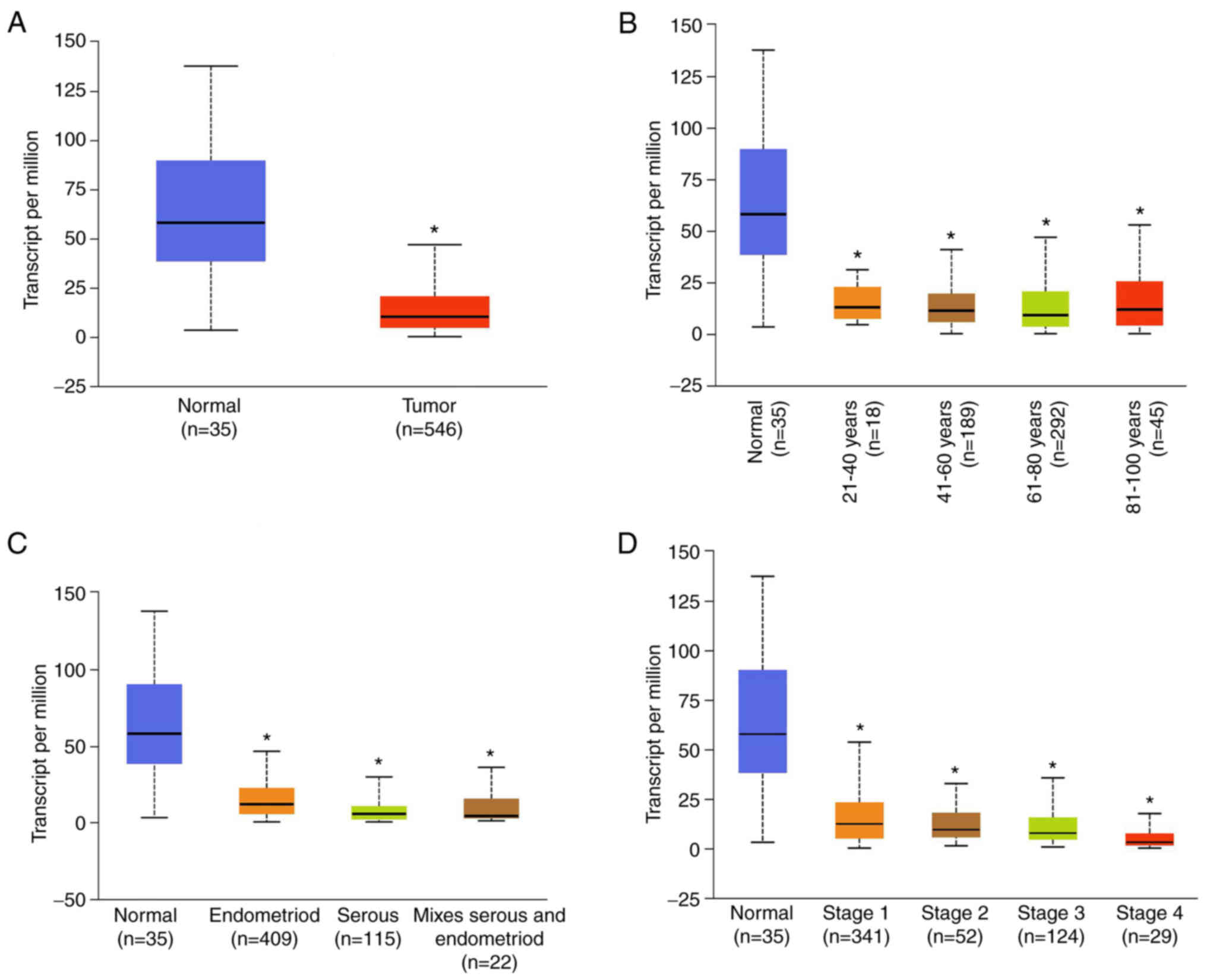
PDZRN3 expression is significantly
lower in EC tissues
The expression level of PDZRN3 in EC was examined
using the public database The Cancer Genome Atlas (TCGA). The
results demonstrated that PDZRN3 expression was significantly lower
in EC tissues compared with normal tissues (P<0.001; Fig. 1A). In addition, PDZRN3 expression
was associated with the age of the patients, the tumor grade and
the tumor subtype (Fig. 1).
Different age groups were selected from the TCGA database as
follows: 21-40, 41-60, 61-80 and 81-100 years. The expression level
of PDZRN3 in patients with EC was significantly lower than those in
normal endometrial tissues (P<0.001; Fig. 1B). Furthermore, the expression
level of PDZRN3 in EC of different pathological classification
types (endometrioid, serous or mixed) was significantly lower than
those in normal endometrial tissues (P<0.001; Fig. 1C). In addition, the clinical stages
of EC were also identified as follows: Stage 1, stage 2, stage 3
and stage 4. The data further demonstrated that the expression
level of PDZRN3 in EC tissues was significantly decreased
(P<0.001; Fig. 1D) compared
with normal group. These results suggested that lower PDZRN3
expression in ECs may therefore be correlated with EC progression
and accelerate invasion and metastasis of EC.
PDZRN3 promotes EC cell
proliferation
To evaluate the function of PDZRN3 in EC, PDZRN3 was
overexpressed using a lentiviral vector. Ectopic PDZRN3 was
constitutively expressed in HEC-1-B cells (HEC-1-B-PDZRN3) and KLE
cells (KLE-PDZRN3). The expression levels of PDZRN3 were assessed
by RT-qPCR and western blotting (Fig.
S1). Furthermore, HEC-1-B-PDZRN3 and KLE-PDZRN3 cells exhibited
lower absorbance compared with HEC-1-B- and KLE-control cells as
determined by the MTT assay, which indicated a decreased
proliferative rate (Fig. 2A). In
addition, the high PDZRN3 expression cell groups HEC-1-B-PDZRN3 and
KLE-PDZRN3 formed a lower number of colonies according to results
from the colony formation assay (Fig.
2B) compared with the control-PDZRN3 cell groups HEC-1-B- and
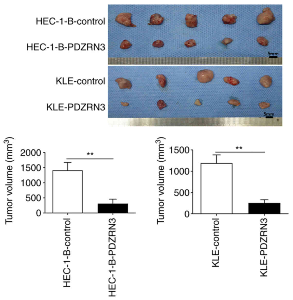
KLE-control cells. To verify these findings in vivo, subcutaneous
xenograft tumor models were established. Following 4 weeks of
incubation, the HEC-1-B-PDZRN3 and KLE-PDZRN3 cell-derived tumors
at the subcutaneous implantation sites were significantly smaller
and grew more slowly than those from the HEC-1-B- and KLE-control
groups (Fig. 3). In addition,
Ki-67 staining for these tumors confirmed that high PDZRN3
expression groups HEC-1-B-PDZRN3 and KLE-PDZRN3 had fewer Ki-67
positive cells (Fig. S2) compared
with the control-PDZRN3 groups HEC-1-B- and KLE-control cells.
Taken together, these results suggested that decreased PDZRN3
expression could promote EC cell proliferation.
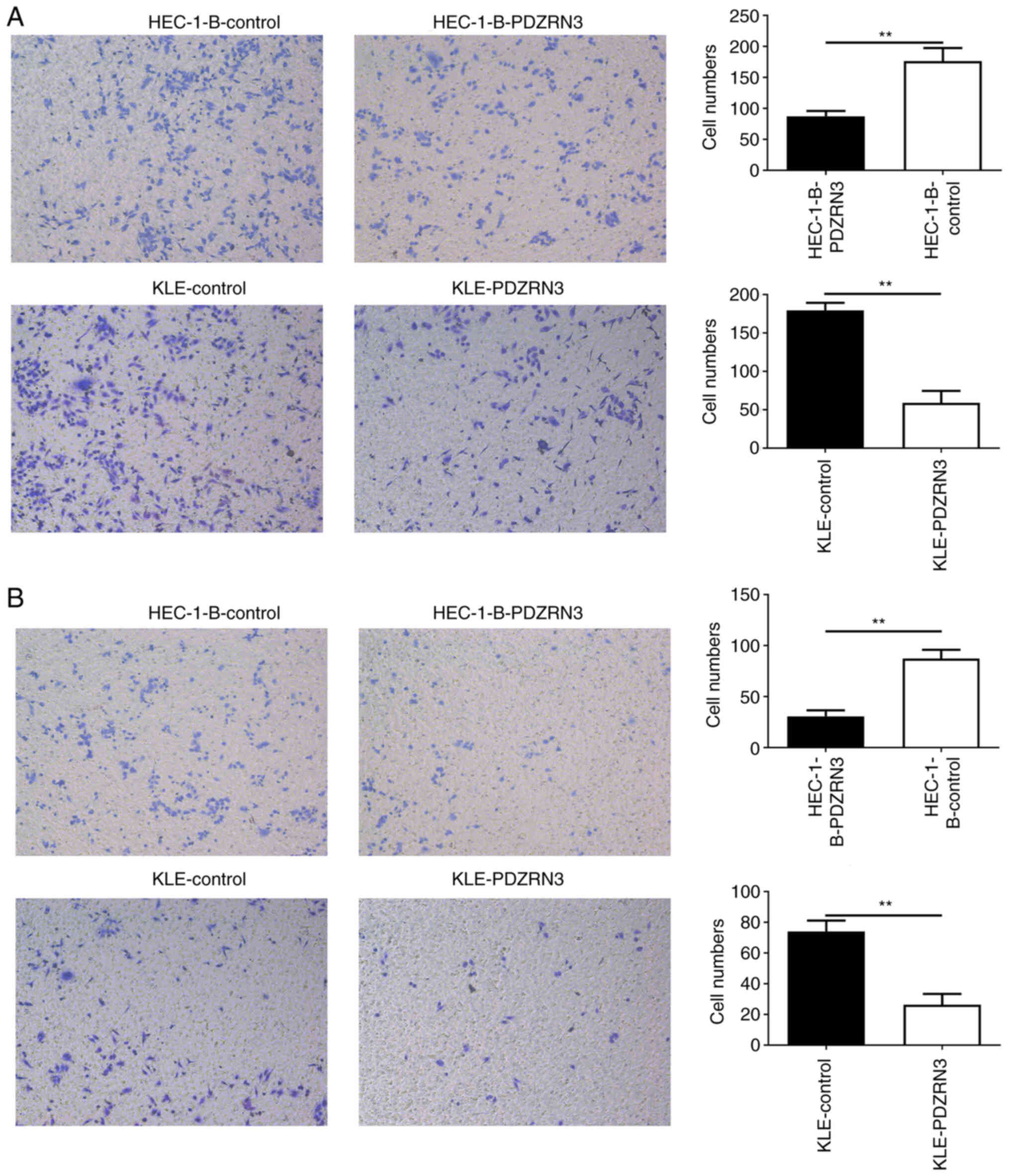
Lower PDZRN3 promotes EC cell
migration and invasion
Transwell invasion and migration assays were used to
investigate the invasive and migratory abilities of the cells. The
results indicated that HEC-1-B-PDZRN3 cells exhibited significantly
reduced migratory ability compared with the HEC-1-B-control cells.
Similarly, KLE- PDZRN3 cells exhibited significantly lower
migratory ability compared with the KLE-control cells (Fig. 4A). The results from the invasion
assays indicated that HEC-1-B-PDZRN3 cells exhibited a
significantly lower number of invading cells compared with the
HEC-1-B-control cells. In addition, KLE-PDZRN3 cells exhibited a
significantly lower number of invading cells compared with the
KLE-control cells (Fig. 4B).
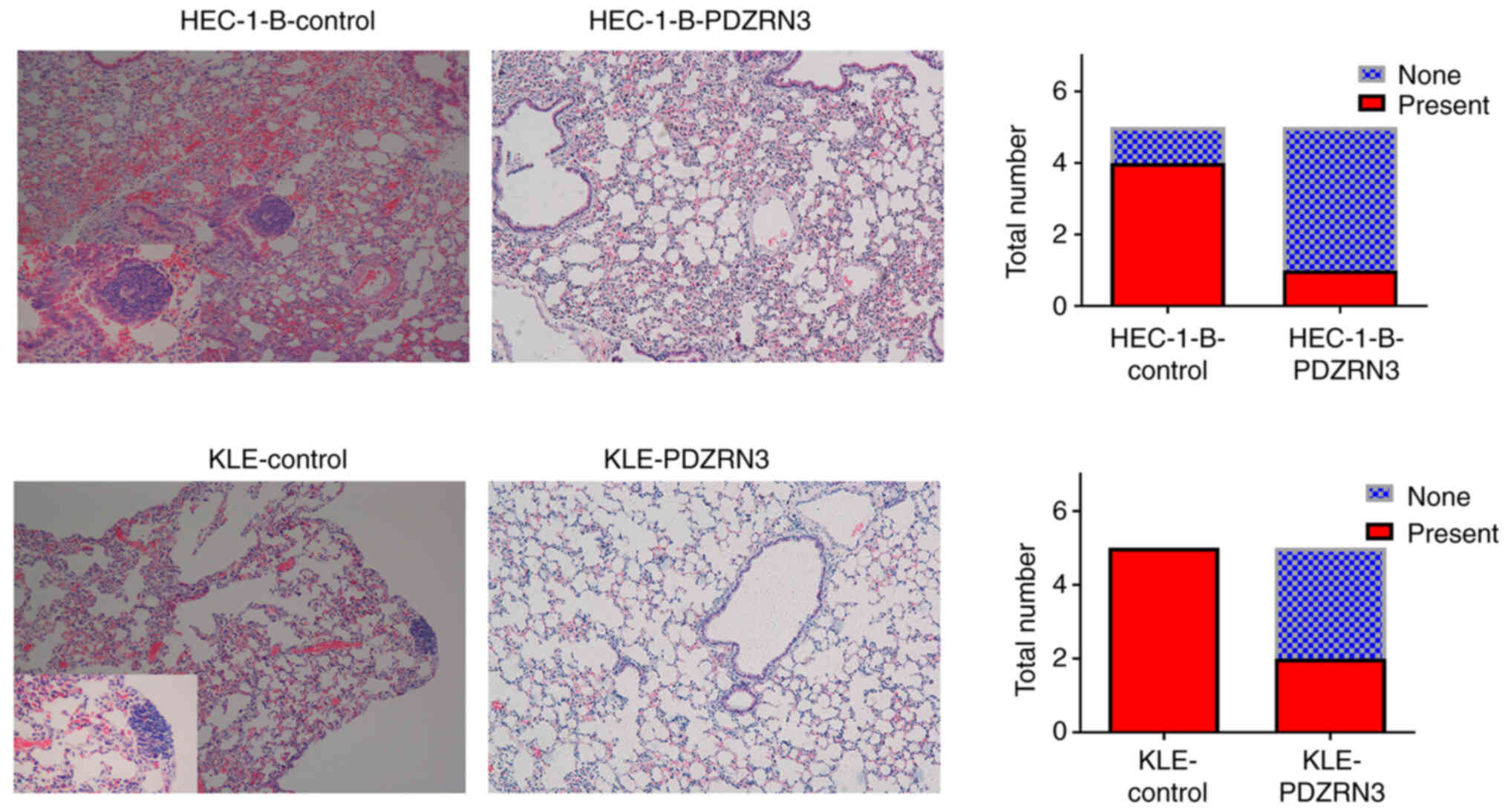
Furthermore, a pulmonary metastasis nude mouse model was
established and the invasive activity was assessed by
immunohistochemical analysis. The results indicated that the number
of lung metastases in tumors of mice injected with cells
overexpressing PDZRN3 (HEC-1-B-PDZRN3 and KLE-PDZRN3) was
significantly lower than that in the control-PDZRN3 cell group
(HEC-1-B- and KLE-control cells; Fig.
5). Taken together, these results indicated that lower
expression of PDZRN3 may promote the metastasis of EC cells via
increased proliferation, migration and invasion of EC cells.
PDZRN3 modulates EC progression via
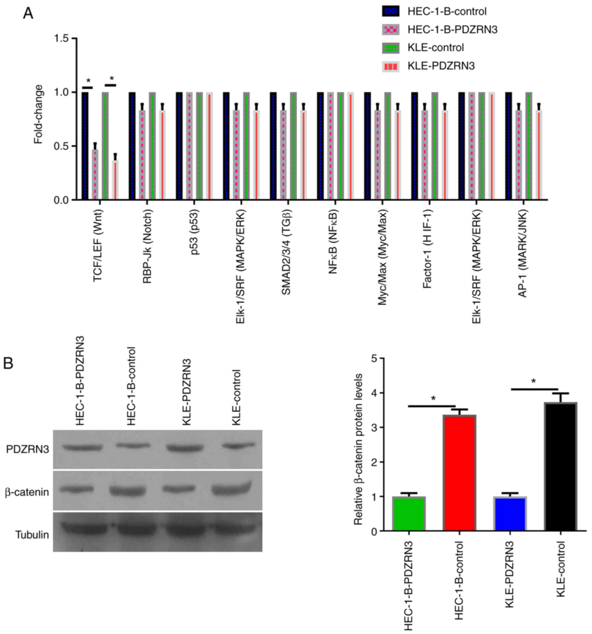
the canonical Wnt signaling pathway
PDZRN3 expression is associated with
canonical/non-canonical Wnt signaling in non-tumor cell
differentiation microenvironment (20). To systemically screen the potential
signaling molecules that could interact with PDZRN3, a Signal
Finder Cancer 10-Pathway Reporter Array was assessed by RT-qPCR
analysis. The results demonstrated that PDZRN3 significantly
attenuated the activity of Wnt signaling in the high PDZRN3
expression groups HEC-1-B-PDZRN3 and KLE-PDZRN3, whereas it
increased Wnt signaling activity in the control-PDZRN3 groups
HEC-1-B-control and KLE-control cells (Fig. 6A). Furthermore, β-catenin
expression level was assessed in these cell groups and the data
demonstrated that ectopic expression of PDZRN3 in HEC-1-B-PDZRN3
and KLE-PDZRN3 groups was accompanied with decreased β-catenin
accumulation (Fig. 6B). Conversely
to these observations, decreased PDZRN3 expression and increased
β-catenin accumulation were observed in HEC-1-B- and KLE-control
groups (Fig. 6B). These results
indicated that PDZRN3 could modulate the canonical Wnt signaling
pathway.
Discussion
In clinical, it is found that EC has high malignancy
and low survival rate. Tumor tissues exhibit uncontrolled
proliferation compared with normal tissues (21). Tumor invasion and metastasis are
often a direct cause of poor prognosis and high mortality rates in
patients with tumors, including patients with EC (22,23).
In addition, it is recognized that ubiquitination system serves a
crucial role in tumor genesis and development, including invasion
and metastasis. E3 ubiquitination ligands have been reported to
have important clinical significance in the regulation of cell
motility and of tumor invasion and metastasis (24,25).
Molecules with such structures and functions may therefore play a
crucial role in tumor invasion and metastasis and may have
potential value in clinical research. Based on this evidence, the
assessment of the structure and function of PDZRN3 may be of
considerable interest.
Previous studies reported that PDZRN3 and its E3
ubiquitin ligase domain serve an important role in ubiquitination
function and in promoting cell differentiation (26,27),
which confirms the hypothesis that it may be involved in promoting
tumor progression. Following analysis of the public database TCGA,
significant differences in PDZRN3 expression were identified in EC
tissues. PDZRN3 expression was associated with the age of the
patients, tumor grade and tumor subtype. In addition, PDZRN3
expression was associated with certain EC subtypes, suggesting that
PDZRN3 may be used to distinguish the clinical subtypes of EC. In
summary, this study demonstrated that PDZRN3 may have
ubiquitination function, and by checking the public database TCGA,
we found that PDZRN3 was significantly lower in EC compared with
normal tissues. The present study was incomplete since it lacked
clinical case data and because it failed to collect comprehensive
data relevant to the mechanism of action of PDZRN3.
The present study primarily focused on assessing the
invasive, metastatic and proliferative functions of EC with regard
to PDZRN3 expression. The results derived from the in vivo
and in vitro experiments indicated that low expression of
PDZRN3 could affect these characteristics of EC cells. One
limitation of the present study is that the impact of PDZRN3 on
other tumor cell characteristics, such as apoptosis and immune
escape, was not examined. Furthermore, the effect of PDZRN3 on
other biological behaviors could not be comprehensively evaluated.
In addition, it has been reported that PDZRN3 can promote the
differentiation of mesenchymal progenitor cells into myotubes in
2C12 mice, which indicates that PDZRN3 could promote the
transformation from poorly differentiated to highly differentiated
cells (16,28). Tumor cells are mainly poorly
differentiated cells. Whether the expression of PDZRN3 could induce
the differentiation of tumor cells is also worthy of investigation.
In addition, due to the limitation of research funds, the role of
PDZRN3 ubiquitination in tumors was not examined, and should thus
be investigated in future studies.
From preliminary screening, it was found that PDZRN3
was related to the activation of the canonical Wnt signaling
pathway. It is well established that the Wnt signaling pathway is
closely related to specific tumor biological characteristics
(29,30), such as proliferation or invasion,
and the PDZRN3-induced biological behavior is likely to require the
activation of the canonical Wnt signaling pathway (16). Additional investigation is required
to confirm the important role of the canonical Wnt signaling
pathway in EC to reveal its downstream target and determine whether
this target is ubiquitinated by the E3 ubiquitinated ligand. The
specific downstream targets and related functional verification of
the canonical Wnt signaling pathway were not investigated in the
present study.
In summary, the present study indicated that PDZRN3
inhibited the invasion, metastasis and proliferation of EC cells.
By using preliminary signaling pathway screening, PDZRN3 was
identified as a potential target responsible for the biological
behavior of EC via activation of the Wnt signaling pathway. PDZRN3
expression may therefore provide important clinical guidance for
predicting the survival of patients with EC and for the development
of targeted therapeutics for EC.
Supplementary Material
Supporting Data
Acknowledgements
Not applicable.
Funding
Funding: No funding was received.
Availability of data and materials
The datasets used and/or analyzed during the current
study are available from the corresponding author on reasonable
request.
Authors' contributions
YL designed the project and funded all the
experiments. QL completed all the experiments. JZ and SY were
involved in drafting the manuscript, revising it critically for
important intellectual content and assisted with the experiments.
YL, QL, JZ and SY confirm the authenticity of all the raw data. All
authors read and approved the final manuscript.
Ethics approval and consent to
participate
Animal experimental procedures were approved by the
Animal Ethics Committee of Shanghai Yangpu District Shidong
Hospital.
Patient consent for publication
Not applicable.
Competing interests
The authors declare that they have no competing
interests.
References
|
1
|
Bray F, Ferlay J, Soerjomataram I, Siegel
RL, Torre LA and Jemal A: Global cancer statistics 2018: GLOBOCAN
estimates of incidence and mortality worldwide for 36 cancers in
185 countries. CA Cancer J Clin. 68:394–424. 2018. View Article : Google Scholar : PubMed/NCBI
|
|
2
|
Washington CR, Haggerty A, Ronner W, Neff
PM and Ko EM: Knowledge of endometrial cancer risk factors in a
general gynecologic population. Gynecol Oncol. 158:137–142. 2020.
View Article : Google Scholar : PubMed/NCBI
|
|
3
|
Byrne FL, Martin AR, Kosasih M, Caruana BT
and Farrell R: The role of hyperglycemia in endometrial cancer
pathogenesis. Cancers (Basel). 12:11912020. View Article : Google Scholar : PubMed/NCBI
|
|
4
|
Nagao S: IV. Endometrial Cancer. Gan To
Kagaku Ryoho. 47:247–251. 2020.(In Japanese). PubMed/NCBI
|
|
5
|
Lamothe S and Ramia de Cap M: Survival
analysis and treatment effects in patients with endometrial cancer
and POLE mutations. Cancer. 127:4306–4307. 2021. View Article : Google Scholar : PubMed/NCBI
|
|
6
|
Luna C, Balcacer P, Castillo P, Huang M
and Alessandrino F: Endometrial cancer from early to advanced-stage
disease: An update for radiologists. Abdom Radiol (NY).
46:5325–5336. 2021. View Article : Google Scholar : PubMed/NCBI
|
|
7
|
Deng L, Meng T, Chen L, Wei W and Wang P:
The role of ubiquitination in tumorigenesis and targeted drug
discovery. Signal Transduct Target Ther. 5:112020. View Article : Google Scholar : PubMed/NCBI
|
|
8
|
Pérez-Benavente B, Nasresfahani AF and
Farràs R: Ubiquitin-regulated cell proliferation and cancer. Adv
Exp Med Biol. 1233:3–28. 2020. View Article : Google Scholar : PubMed/NCBI
|
|
9
|
Faktor J, Pjechová M, Hernychová L and
Vojtěšek B: Protein ubiquitination research in oncology. Klin
Onkol. 32 (Suppl 3):S56–S64. 2019. View Article : Google Scholar : PubMed/NCBI
|
|
10
|
Fan Q, Wang Q, Cai R, Yuan H and Xu M: The
ubiquitin system: orchestrating cellular signals in non-small-cell
lung cancer. Cell Mol Biol Lett. 25:12020. View Article : Google Scholar : PubMed/NCBI
|
|
11
|
Jiang Q, Li F, Cheng Z, Kong Y and Chen C:
The role of E3 ubiquitin ligase HECTD3 in cancer and beyond. Cell
Mol Life Sci. 77:1483–1495. 2020. View Article : Google Scholar : PubMed/NCBI
|
|
12
|
Liu X, Zurlo G and Zhang Q: The roles of
Cullin-2 E3 ubiquitin ligase complex in cancer. Adv Exp Med Biol.
1217:173–186. 2020. View Article : Google Scholar : PubMed/NCBI
|
|
13
|
Venuto S and Merla G: E3 ubiquitin ligase
TRIM proteins, cell cycle and mitosis. Cells. 8:5102019. View Article : Google Scholar : PubMed/NCBI
|
|
14
|
Wang ZW, Hu X, Ye M, Lin M, Chu M and Shen
X: NEDD4 E3 ligase: Functions and mechanism in human cancer. Semin
Cancer Biol. 67:92–101. 2020. View Article : Google Scholar : PubMed/NCBI
|
|
15
|
Katoh M and Katoh M: Identification and
characterization of PDZRN3 and PDZRN4 genes in silico. Int J Mol
Med. 13:607–613. 2004.PubMed/NCBI
|
|
16
|
Ko JA, Kimura Y, Matsuura K, Yamamoto H,
Gondo T and Inui M: PDZRN3 (LNX3, SEMCAP3) is required for the
differentiation of C2C12 myoblasts into myotubes. J Cell Sci.
119:5106–5113. 2006. View Article : Google Scholar : PubMed/NCBI
|
|
17
|
Marracci S, Vangelisti A, Raffa V,
Andreazzoli M and Dente L: pdzrn3 is required for pronephros
morphogenesis in Xenopus laevis. Int J Dev Biol. 60:57–63. 2016.
View Article : Google Scholar : PubMed/NCBI
|
|
18
|
Marunaka K, Furukawa C, Fujii N, Kimura T,
Furuta T, Matsunaga T, Endo S, Hasegawa H, Anzai N, Yamazaki Y, et
al: The RING finger- and PDZ domain-containing protein PDZRN3
controls localization of the Mg2+ regulator claudin-16
in renal tube epithelial cells. J Biol Chem. 292:13034–13044. 2017.
View Article : Google Scholar : PubMed/NCBI
|
|
19
|
Livak KJ and Schmittgen TD: Analysis of
relative gene expression data using real-time quantitative PCR and
the 2(−Delta Delta C(T)) method. Methods. 25:402–408. 2001.
View Article : Google Scholar : PubMed/NCBI
|
|
20
|
Sewduth RN, Jaspard-Vinassa B, Peghaire C,
Guillabert A, Franzl N, Larrieu-Lahargue F, Moreau C, Fruttiger M,
Dufourcq P, Couffinhal T and Duplàa C: The ubiquitin ligase PDZRN3
is required for vascular morphogenesis through Wnt/planar cell
polarity signaling. Nat Commun. 5:48322014. View Article : Google Scholar : PubMed/NCBI
|
|
21
|
Hanahan D and Weinberg RA: Hallmarks of
cancer: The next generation. Cell. 144:646–674. 2011. View Article : Google Scholar : PubMed/NCBI
|
|
22
|
Nikolaou S and Machesky LM: The stressful
tumour environment drives plasticity of cell migration programmes,
contributing to metastasis. J Pathol. 250:612–623. 2020. View Article : Google Scholar : PubMed/NCBI
|
|
23
|
Jamil A and Kasi A: Lung Metastasis.
StatPearls (Internet). StatPearls Publishing; Treasure Island, FL:
2021
|
|
24
|
Lu C, Ning G, Si P, Zhang C, Liu W, Ge W,
Cui K, Zhang R and Ge S: E3 ubiquitin ligase HECW1 promotes the
metastasis of non-small cell lung cancer cells through mediating
the ubiquitination of Smad4. Biochem Cell Biol. 99:675–681. 2021.
View Article : Google Scholar : PubMed/NCBI
|
|
25
|
Shen J, Yu Z and Li N: The E3 ubiquitin
ligase RNF146 promotes colorectal cancer by activating the
Wnt/β-catenin pathway via ubiquitination of Axin1. Biochem Biophys
Res Commun. 503:991–997. 2018. View Article : Google Scholar : PubMed/NCBI
|
|
26
|
Honda T and Inui M: PDZRN3 regulates
differentiation of myoblasts into myotubes through transcriptional
and posttranslational control of Id2. J Cell Physiol.
234:2963–2972. 2019. View Article : Google Scholar : PubMed/NCBI
|
|
27
|
Honda T and Inui M: PDZRN3 protects
against apoptosis in myoblasts by maintaining cyclin A2 expression.
Sci Rep. 10:11402020. View Article : Google Scholar : PubMed/NCBI
|
|
28
|
Sewduth RN, Kovacic H, Jaspard-Vinassa B,
Jecko V, Wavasseur T, Fritsch N, Pernot M, Jeaningros S, Roux E,
Dufourcq P, et al: PDZRN3 destabilizes endothelial cell-cell
junctions through a PKCζ-containing polarity complex to increase
vascular permeability. Sci Signal. 10:eaag32092017. View Article : Google Scholar : PubMed/NCBI
|
|
29
|
Chatterjee A, Paul S, Bisht B,
Bhattacharya S, Sivasubramaniam S and Paul MK: Advances in
targeting the WNT/β-catenin signaling pathway in cancer. Drug
Discov Today. Jul 10–2021.(Epub ahead of print).
|
|
30
|
Fatima I, Barman S, Rai R, Thiel KWW and
Chandra V: Targeting Wnt signaling in endometrial cancer. Cancers
(Basel). 13:23512021. View Article : Google Scholar : PubMed/NCBI
|