Introduction
Acute myeloid leukemia (AML) is a malignancy of the
hematopoietic stem cells characterized by the uncontrolled growth
of immature myeloid cells in the bone marrow, which interferes with
normal hematopoietic function (1,2).
Currently, it is the most common acute leukemia among adults, with
an occurrence of more than 20,000 cases every year in the US
(3). Furthermore, its prognosis is
relative unfavorable among all types of leukemia, including the
high probability of relapse and low survival rate (2,4–6).
Considering that AML is still a malignancy with unsatisfied
outcomes dependent on various factors (2), the exploration of novel biomarker for
predicting the treatment response of induction therapy and
indicating long-term prognosis in AML patients is crucial.
MicroRNAs (miRNAs) participate in various biological
processes, including hematopoietic differentiation, proliferation
and leukemogenesis (7). Among
them, it has been indicated that miR-181b is able to regulate drug
sensitivity in AML through targeting high mobility group protein
(HMGB1) and myeloid cell leukemia-1 (Mcl-1) (8). Moreover, it also has been illustrated
that miR-181b is abnormally expressed in AML compared to the normal
populations, as well as it correlates with the treatment response
of AML (8–12). Based on the above-mentioned
information, we hypothesized that miR-181b-5p expression could be a
potential prognostic marker in AML patients who undergo induction
therapy. However, such information is obscure.
Therefore, the aim of this study was to explore the
correlation of miR-181b-5p with the National Comprehensive Cancer
Network (NCCN) risk classification, treatment response and
long-term prognosis of AML.
Patients and methods
Subjects
Between January 2016 and December 2019, following
approval by the Ethics Committee of Tongji Hospital, School of
Medicine, Tongji University (Shanghai, China), 84 de novo
AML patients and 30 healthy donors were consecutively recruited in
this study. All eligible patients were confirmed as AML rather than
acute promyelocytic leukemia, with an age above 18 years, and had
no history of other malignancies. All health donors were enrolled
after they agreed to donate bone marrow, and the necessary
examinations were carried out for them to confirm the eligibility.
Pregnant or breast-feeding subjects were excluded from the study.
All subjects provided written informed consent.
Clinical data and sample
collection
After recording the clinical features of the AML
patients, collection of bone marrow sample was performed before
they started the induction therapy. Bone marrow samples of 30
healthy donors (age range, 42–65 years; male-to-female ratio, 3:2)
were collected during donation. Immediately after sample
collection, human bone marrow monocyte separation solution (Beijing
Biolabo Technology Co., Ltd.) was used for separation of the bone
marrow mononuclear cells (BMMCs), followed by quantitative analysis
of miR-181b-5p using reverse transcription-quantitative polymerase
chain reaction (RT-qPCR) assay.
RT-qPCR assay
The RT-qPCR procedures were performed as described
in a previous study (11), and the
following kits were used: TRIzol™ Reagent (Thermo Fisher
Scientific, Inc.) for extraction of total RNA; RT-PCR Quick Master
Mix (Toyobo) for reverse transcription; SYBR® Premix
DimerEraser™ (Takara Bio, Inc.) for qPCR. The expression of
miR-181b-5p was normalized to the U6 gene, and the relative
expression of miR-181b-5p was calculated by the 2−ΔΔCq
method (13). The primer sequences
for miR-181b-5p were as follows (14): Forward, 5′-GCGGATCATTCATTGCTGTCG-3′
and reverse, 5′-ATCTGGTGGCTCTCGGAGTAA-3′. For U6, the forward
sequence was 5′-CGCTTCGGCAGCACATATACTA-3′ and the reverse sequence
was 5′-ATGGAACGCTTCACGAATTTGC-3′.
The expression of miR-181b-5p was classified
according to 4 quantiles in survival analyses: quantile 1,
miR-181b-5p expression in the interval of 0–25% of total AML
patients; quantile 2, miR-181b-5p expression in the interval of
26–50% of total AML patients; quantile 3, miR-181b-5p expression in
the interval of 51–75% of total AML patients; quantile 4,
miR-181b-5p expression in the interval of 76–100% of total AML
patients. In particular, miR-181b-5p expression in the interval of
0–25% of total AML patients was defined as miR-181b-5p
insufficiency.
Response data and survival data
collection
All patients received standard induction therapy
with 3 days of an anthracycline (e.g., daunorubicin, at least 60
mg/m2, idarubicin, 10–12 mg/m2, or
anthracenedione mitoxantrone, 10–12 mg/m2) and 7 days of
cytarabine (100–200 mg/m2 cont. i.v.). Complete
remission (CR) patients after induction therapy were recorded for
the study analysis. Follow-up was conducted every 3 months for the
first 2 years, and then surveillance continued every 6 months for
the following 2–3 years. The final follow-up date for study was
December 31, 2020. Event-free survival (EFS) and overall survival
(OS) were calculated based on the recorded date of defined events
in the AML guideline (15).
Statistical analysis
Data analysis and figure plotting were performed
using SPSS 20.0 (IBM Corp.) and GraphPad Prism 6.01 (GraphPad
Software Inc.). Distribution characteristics of miR-181b-5p in the
different subjects were displayed using a Box plot. Comparison of
the expression difference of miR-181b-5p among the different
subjects was determined by Kruskal-Wallis test or Mann-Whitney U
test. Correlation analysis between miR-181b-5p expression and NCCN
risk classification was determined by Spearman rank correlation
test. The receiver-operating characteristic (ROC) curve and area
under the curve (AUC) were used for estimating profiles of
miR-181b-5p in distinguishing different subjects. Survival data
were described using the Kaplan-Meier method. The multiple
comparisons of survival data were examined by log-rank test and
corrected by Benjamini-Hochberg (B-H) method. Cox proportional
hazards regression with forward stepwise method was applied for
analysis of the prognostic factors. P-value <0.05 was considered
indicative of a statistical significant difference.
Results
Baseline characteristics
For the 84 AML patients, the mean age was 58.4±12.8
years. There were 42 (50.0%) patients >60 years and 42 (50.0%)
patients ≤60 years. Moreover, there were 51 (60.7%) males in these
AML patients. As for FAB classification, there were 5 (6.0%)
patients with M1, 28 (33.3%) patients with M2, 22 (26.2%) patients
with M4 and 29 (34.5%) patients with M5. In terms of cytogenetic
abnormities, there were 41 (48.8%) patients with a normal
karyotype, 9 (10.7%) patients with complex karyotype and 4 (4.8%)
patients with a monosomal karyotype. Regarding genetic mutations,
there were 25 (29.8%) patients with NPM1 mutation, 20
(23.8%) patients with FLT3-ITD mutation, 10 (11.9%) patients
with WT1 mutation and 7 (8.3%) patients with CEBPA
mutation. Furthermore, according to risk classification, there were
15 (17.9%) patients with better-risk, 44 (52.4%) patients with
intermediate-risk and 25 (29.8%) patients with poor-risk. The
detailed characteristics of the AML patients are shown in Table I.
 | Table I.Characteristics of the patients with
AML (n=84). |
Table I.
Characteristics of the patients with
AML (n=84).
| Item | Value |
|---|
| Age, years | 58.4±12.8 |
|
>60 | 42 (50.0) |
|
≤60 | 42 (50.0) |
| Male sex | 51 (60.7) |
| FAB
classification |
|
| M1 | 5 (6.0) |
| M2 | 28 (33.3) |
| M4 | 22 (26.2) |
| M5 | 29 (34.5) |
| Cytogenetic
abnormities |
|
| Normal
karyotype | 41 (48.8) |
| Complex
karyotype | 9 (10.7) |
|
Inv(16) or t(16;16) | 5 (6.0) |
|
Monosomal karyotype | 4 (4.8) |
| +8 | 4 (4.8) |
|
t(9;11) | 4 (4.8) |
| -7 or
7q- | 3 (3.6) |
| -5 or
5q- | 1 (1.2) |
|
inv(3), t(3;3) | 1 (1.2) |
|
t(6;9) | 1 (1.2) |
|
t(8;21) | 1 (1.2) |
| Others
non-defined | 14 (16.7) |
| Genetic
mutations |
|
|
NPM1 mutation | 25 (29.8) |
|
FLT3-ITD mutation | 20 (23.8) |
|
WT1 mutation | 10 (11.9) |
|
CEBPA mutation | 7 (8.3) |
| WBCs, 1/l |
|
|
>10×109 | 57 (67.9) |
|
≤10×109 | 27 (32.1) |
| BM blasts, % |
|
|
>75 | 40 (47.6) |
|
≤75 | 44 (52.4) |
| Risk
classification |
|
| High
risk | 15 (17.9) |
|
Intermediate risk | 44 (52.4) |
| Low
risk | 25 (29.8) |
Comparison of miR-181b-5p expression
between AML patients and healthy donors
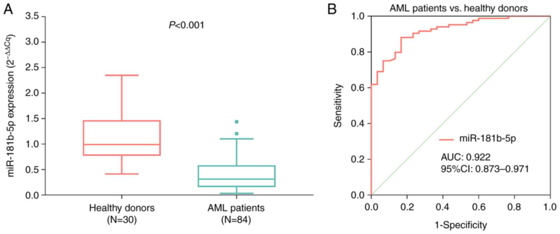
miR-181b-5p expression was reduced in the AML
patients [median value, 0.312 (0.170-0.571)] compared to the
healthy donors [median value, 0.990 (0.784-1.455)]
(P<0.001) (Fig. 1A).
Meanwhile, the ROC curve showed that miR-181b-5p had excellent
potential in discriminating AML patients from healthy donors with
AUC of 0.922 [95% confidence interval (CI): 0.873-0.971]. In
addition, miR-181b-5p expression was 0.735 at the best cut-off
point (the point with maximum value of the sum of sensitivity and
specificity); the sensitivity and specificity were 0.881 and 0.833
at the best cut-off point, respectively (Fig. 1B).
Comparison of miR-181b-5p expression
among patients with diverse characteristics
miR-181b-5p expression in patients stratified based
on various features is compared in Fig. 2. miR-181b-5p expression was
decreased in the patients with FLT3-ITD mutation compared to
those without FLT3-ITD mutation (P=0.032) (Fig. 2F). Moreover, miR-181b-5p expression
was attenuated in patients with WT1 mutation compared to
those without WT1 mutation (P=0.017) (Fig. 2G). In addition, miR-181b-5p
expression was highest in patients with better-risk classification,
followed by patients with intermediate-risk classification, and
lowest in patients with poor-risk classification (P=0.036)
(Fig. 2I). However, no difference
in miR-181b-5p expression was found among patients with different
FAB classification (M1, M2, M4 or M5) (P=0.578) (Fig. 2A). Furthermore, no difference was
found in miR-181b-5p expression in patients with or without normal
karyotype, complex karyotype, monosomal karyotype, NMP1
mutation or CEBPA mutation (all P>0.05) (Fig. 2B-E and H).
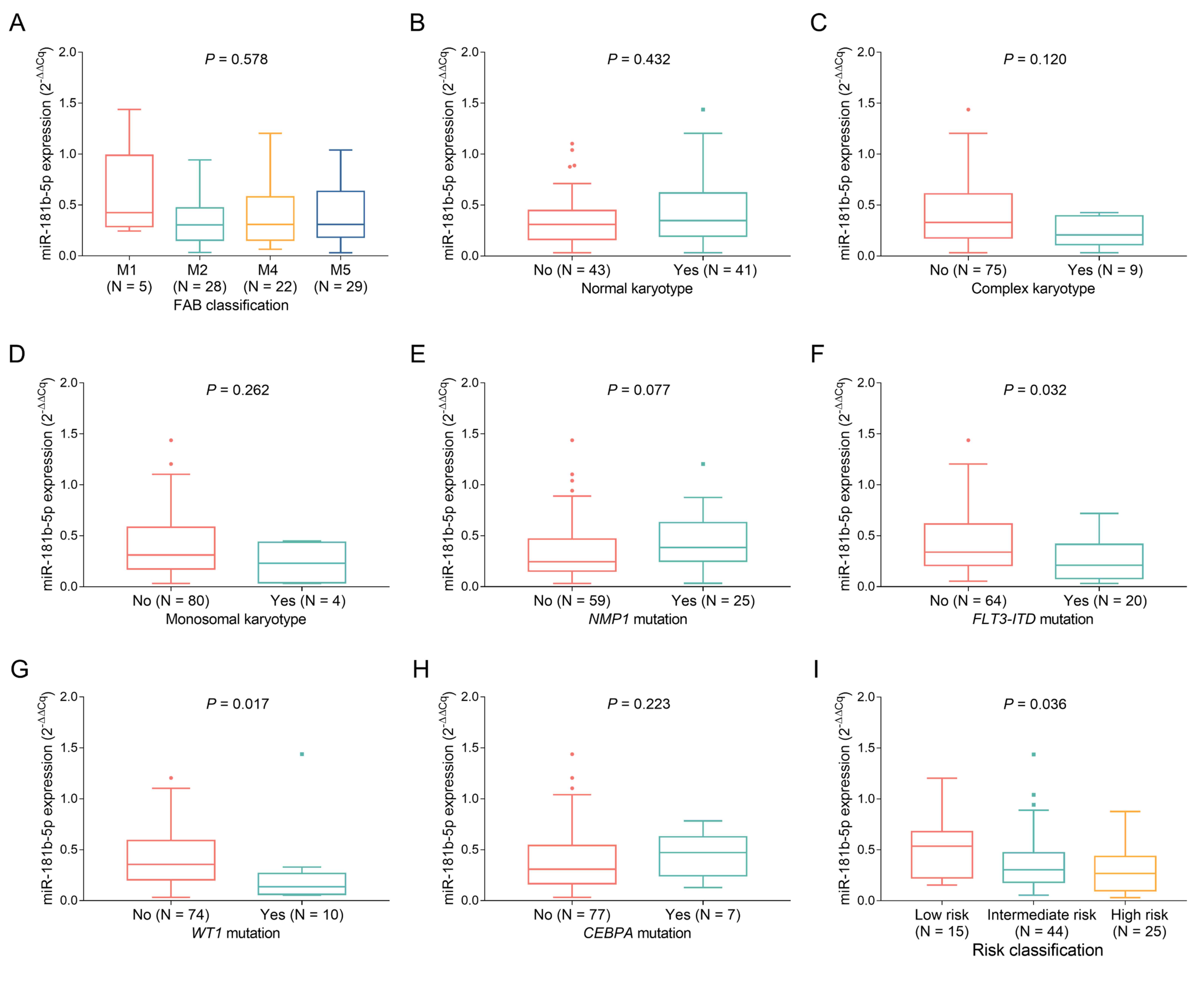 | Figure 2.miR-181b-5p in AML patients with
distinct clinical features. Association of miR-181b-5p with (A) FAB
classification, (B) normal karyotype, (C) complex karyotype, (D)
monosomal karyotype, (E) NMP1 mutation, (F) FLT3-ITD
mutation, (G) WT1 mutation, (H) CEBPA mutation and
(I) National Comprehensive Cancer Network (NCCN) risk
classification in AML patients. miR-181b-5p, microRNA-181b-5p; AML,
acute myeloid leukemia; FAB, France-American-Britain; NMP1,
nucleophosmin 1; FLT3-ITD, Fms-like tyrosine kinase
3-internal tandem duplication; WT1, Wilms' tumor 1;
CEBPA, CCAAT/enhancer binding protein α. |
Comparison of miR-181b-5p expression
between CR patients and non-CR patients
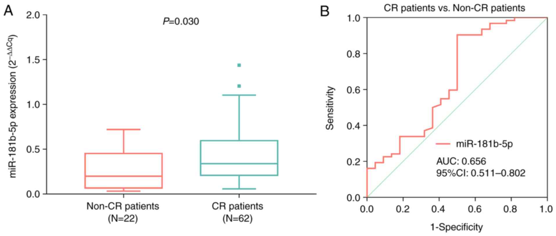
miR-181b-5p expression was increased in CR patients
[median value: 0.339 (0.209-0.595)] compared to non-CR patients
[median value: 0.199 (0.068-0.452)] (P=0.030) (Fig. 3A). Meanwhile, the ROC curve
illustrated that miR-181b-5p had certain ability in discriminating
CR patients from non-CR patients with AUC of 0.656 (95% CI:
0.511-0.802). In addition, miR-181b-5p expression was 0.142 at the
best cut-off point; the sensitivity and specificity were 0.903 and
0.500 at the best cut-off point, respectively (Fig. 3B).
Association of miR-181b-5p expression
with accumulating EFS
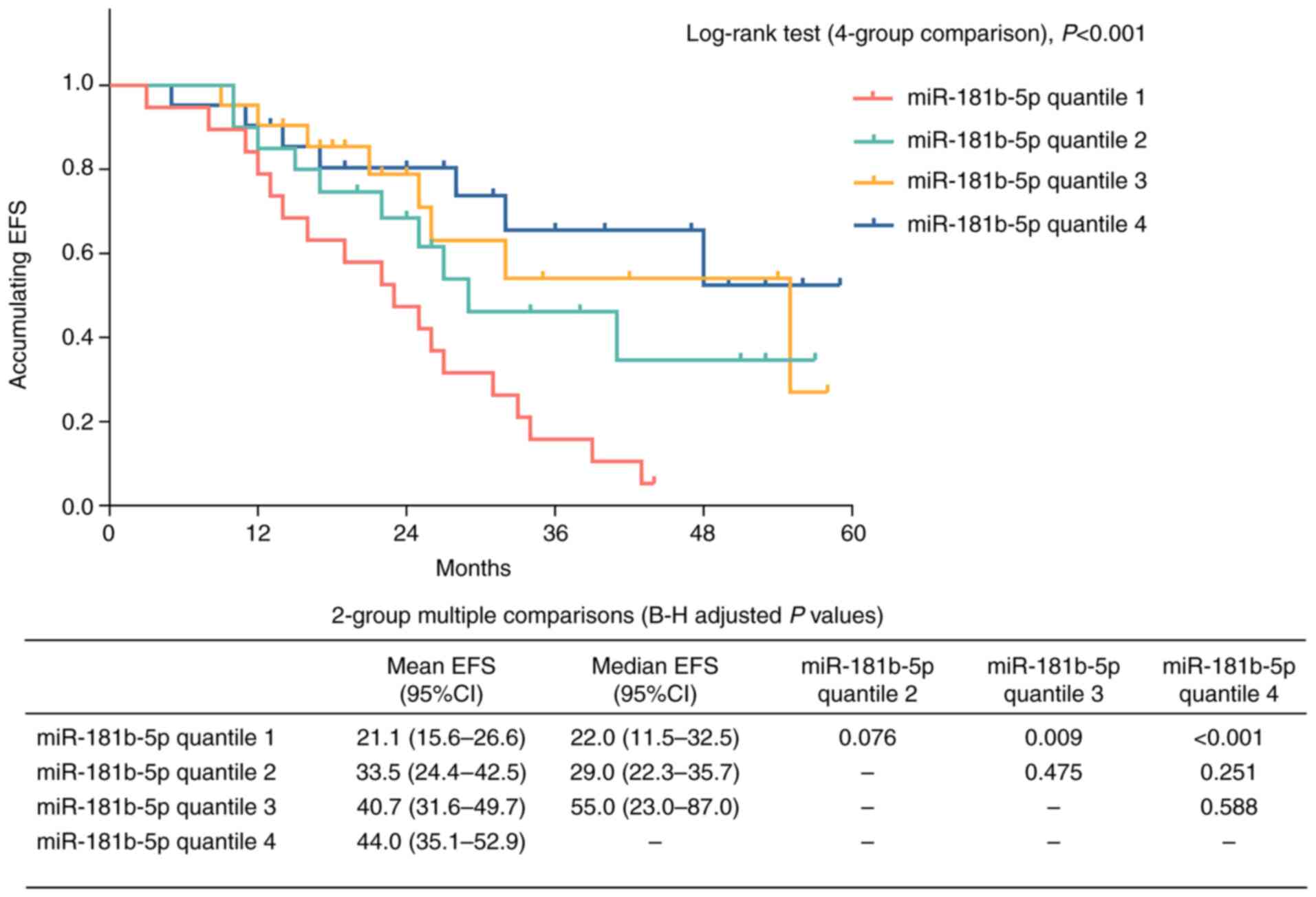
Higher miR-181b-5p expression was correlated with
increased accumulating EFS (P<0.001). Meanwhile, adjusted
multiple comparisons showed that accumulating EFS was attenuated in
patients with AML with miR-181b-5p quantile 1 compared to those
with miR-181b-5p quantile 3 (P=0.009) and miR-181b-5p quantile 4
(P<0.001). However, no difference in accumulating EFS was found
in patients with AML with miR-181b-5p quantile 1 vs. miR-181b-5p
quantile 2 (P=0.076), miR-181b-5p quantile 2 vs. miR-181b-5p
quantile 3 (P=0.475), miR-181b-5p quantile 2 vs. miR-181b-5p
quantile 4 (P=0.251) or miR-181b-5p quantile 3 vs. miR-181b-5p
quantile 4 (P=0.588) (Fig. 4). In
addition, forward stepwise multivariate Cox regression analysis
showed that higher miR-181b-5p expression (HR: 0.698, P=0.012) was
independently associated with better EFS (Table II). These above-mentioned data
imply that miR-181b-5p insufficiency is correlated with worse
EFS.
| Table II.Cox proportional hazards regression
analysis for event-free survival. |
Table II.
Cox proportional hazards regression
analysis for event-free survival.
| A, Univariate Cox
regression analysis |
|---|
|
|---|
| Item | P-value | HR (95% CI) |
|---|
| Higher miR-181b-5p
expression | <0.001 | 0.589
(0.445-0.780) |
| Age >60
years | 0.778 | 0.920
(0.515-1.642) |
| Male sex | 0.034 | 1.977
(1.051-3.718) |
| FAB
classification |
|
|
| M1 | Reference |
|
| M2 | 0.577 | 1.521
(0.349-6.631) |
| M4 | 0.639 | 0.694
(0.151-3.197) |
| M5 | 0.929 | 0.935
(0.215-4.066) |
| Cytogenetic
abnormities |
|
|
| Normal
karyotype | 0.258 | 0.709
(0.390-1.287) |
| Complex
karyotype | 0.181 | 1.739
(0.773-3.913) |
|
Monosomal karyotype | 0.268 | 1.947
(0.599-6.328) |
| Genetic
mutations |
|
|
|
NPM1 mutation | 0.683 | 0.868
(0.439-1.715) |
|
FLT3-ITD mutation | 0.005 | 2.414
(1.312-4.443) |
|
WT1 mutation | 0.066 | 2.066
(0.953-4.476) |
|
CEBPA mutation | 0.417 | 0.614
(0.189-1.994) |
| WBCs
>10×109/l | 0.013 | 2.450
(1.209-4.968) |
| BM blasts
>75% | 0.337 | 1.329
(0.743-2.377) |
| Poor risk
classification | <0.001 | 2.432
(1.538-3.846) |
|
| B, Forward
stepwise multivariate Cox regression analysis |
|
| Item | P-value | HR (95%
CI) |
|
| Higher miR-181b-5p
expression | 0.012 | 0.698
(0.528-0.924) |
| Male sex | 0.012 | 2.353
(1.211-4.571) |
| Poor risk
classification | <0.001 | 2.476
(1.503-4.079) |
Association of miR-181b-5p expression
with accumulating OS
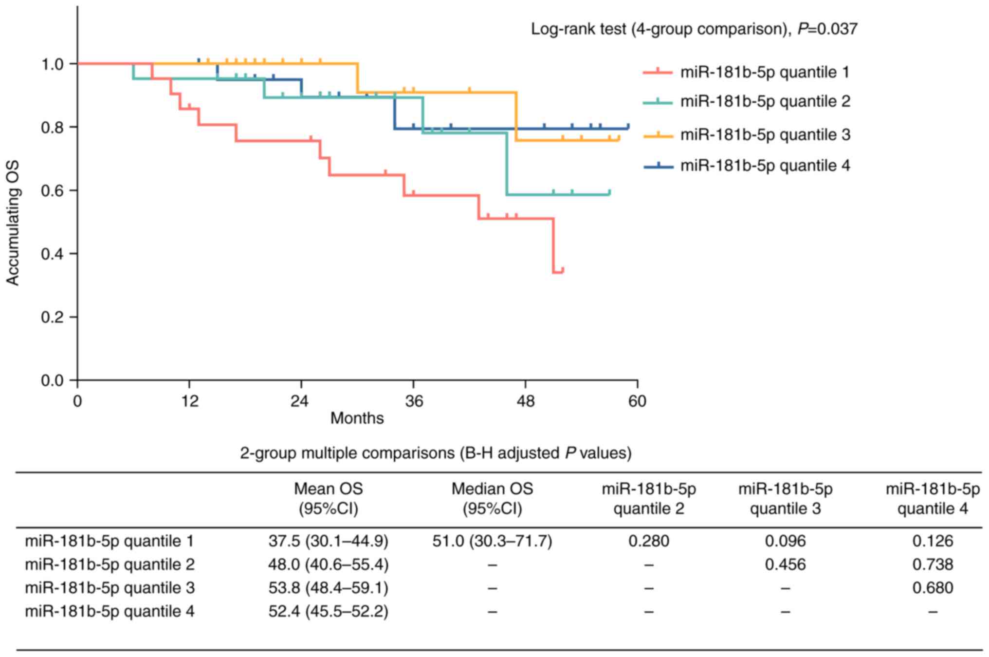
Higher miR-181b-5p expression was found to be
associated with enhanced accumulating OS (P=0.037). Furthermore,
adjusted multiple comparisons showed that no difference in
accumulating OS was found in patients with AML with miR-181b-5p
quantile 1 vs. miR-181b-5p quantile 2 (P=0.280), miR-181b-5p
quantile 1 vs. miR-181b-5p quantile 3 (P=0.096), miR-181b-5p
quantile 1 vs. miR-181b-5p quantile 4 (P=0.126), miR-181b-5p
quantile 2 vs. miR-181b-5p quantile 3 (P=0.456), miR-181b-5p
quantile 2 vs. miR-181b-5p quantile 4 (P=0.738), or miR-181b-5p
quantile 3 vs. miR-181b-5p quantile 4 (P=0.680) (Fig. 5). In addition, forward stepwise
multivariate Cox regression analysis illustrated that higher
miR-181b-5p expression was not independently associated with
accumulating OS (Table III).
| Table III.Cox proportional hazards regression
analysis for overall survival. |
Table III.
Cox proportional hazards regression
analysis for overall survival.
| A, Univariate Cox
regression analysis |
|---|
|
|---|
| Item | P-value | HR (95% CI) |
|---|
| Higher miR-181b-5p
expression | 0.017 | 0.583
(0.374-0.908) |
| Age >60
years | 0.311 | 0.624
(0.251-1.555) |
| Male sex | 0.017 | 4.490
(1.301-15.490) |
| FAB
classification |
|
|
| M1 | Reference |
|
| M2 | 0.630 | 1.678
(0.205-13.748) |
| M4 | 0.921 | 1.112
(0.136-9.107) |
| M5 | 0.509 | 0.477
(0.053-4.298) |
| Cytogenetic
abnormities |
|
|
| Normal
karyotype | 0.979 | 0.988
(0.398-2.452) |
| Complex
karyotype | 0.452 | 1.607
(0.467-5.531) |
|
Monosomal karyotype | 0.102 | 3.477
(0.782-15.459) |
| Genetic
mutations |
|
|
|
NPM1 mutation | 0.832 | 0.895
(0.322-2.491) |
|
FLT3-ITD mutation | 0.081 | 2.301
(0.902-5.871) |
|
WT1 mutation | 0.185 | 2.125
(0.698-6.468) |
|
CEBPA mutation | 0.442 | 0.452
(0.059-3.431) |
| WBCs
>10×109/l | 0.694 | 1.215
(0.461-3.204) |
| BM blasts
>75% | 0.436 | 1.441
(0.575-3.611) |
| Poor risk
classification | <0.001 | 4.947
(2.050-11.941) |
|
| B, Forward
stepwise multivariate Cox regression analysis |
|
| Item | P-value | HR (95%
CI) |
|
| Male sex | 0.004 | 6.877
(1.881-25.141) |
| Poor risk
classification | <0.001 | 7.401
(2.836-19.311) |
Discussion
In the present study, it was found that: i)
miR-181b-5p had excellent potential in discriminating AML patients
from non-AML populations; ii) miR-181b-5p insufficiency was
correlated with FLT3-ITD mutation, WT1 mutation and
poor NCCN risk classification; iii) miR-181b-5p insufficiency was
associated with treatment response failure and unfavorable
long-term prognosis of AML. Previous research indicated that
miR-181a-5p is a prognostic marker for AML (10). To date, only one study has proposed
a correlation of miR-181b with treatment response (11), while the correlation of miR-181b-5p
with survival in AML remains obscured. To the best of our
knowledge, this is the first study to explore the clinical role of
miR-181b-5p as a biomarker of both treatment response and survival
in AML.
Regarding miR-181b expression in AML patients, it
has been demonstrated that miR-181b is abnormally expressed in AML
patients compared to healthy populations (11,16).
In addition, it also has been illustrated that miR-181b expression
is decreased in relapsed/refractory AML patients (17). In this study, we discovered that
miR-181b-5p expression was attenuated in AML patients. A possible
reason might be that miR-181b-5p could regulate several
leukemogenic signaling pathways, including Wnt, protein kinase B
and Notch 1 pathways, which are correlated with the pathogenesis of
AML (18–23). Therefore, its expression was
attenuated in the AML patients.
In terms of the correlation between miR-181b
expression and AML clinical features, it has been found that
miR-181b expression is correlated with genetic mutations in AML,
such as DNA methyltransferase 3 α (DNMT3a), tet
methylcytosine dioxygenase 2 (TET2) and isocitrate
dehydrogenase 1/2 (IDH1/2) (17). In the present study, it was
demonstrated that miR-181b-5p expression was correlated with the
FLT3-ITD and WT1 mutation, respectively. This finding
was partially consistent with a previous study (17). In addition, insufficient expression
of miR-181b-5p was correlated with poor NCCN risk classification of
AML. A possible reason might be that FLT3-ITD and WTI
mutations are two important factors involved in NCCN risk
classification of AML (24–27).
Furthermore, miR-181b-5p expression is associated with the
FLT3-ITD mutation or WT1 mutation (as mentioned
above). Therefore, miR-181b-5p insufficiency is correlated with
poor NCCN risk classification of AML.
As for the association between miR-181b expression
and prognosis of AML, it was demonstrated that reduced miR-181b
expression is correlated with a lower complete remission (CR) rate
in AML patients (17). Another
study also illustrated that miR-181b expression is attenuated in
AML patients with unfavorable overall survival (OS) (9). In the present study, we discovered
that insufficient expression of miR-181b-5p was correlated with
lower CR, unfavorable event-free survival (EFS) and OS. Possible
explanations may be that: i) decreased expression of miR-181b-5p
reduces drug sensitivity via promotion of HMGB1 and Mcl-1
expression (8). Furthermore,
miR-181b-5p insufficiency was correlated with poor NCCN risk
classification of AML (mentioned above), which could result in the
treatment response failure of AML (28). Therefore, miR-181b-5p insufficiency
is associated with poor treatment response. ii) Reduced expression
of miR-181b-5p might have the capability of inhibiting apoptosis,
as well as accelerating the proliferation of AML cells, which may
indirectly lead to a worse long-term prognosis of AML patients
(9,17,29).
Thus, insufficient expression of miR-181b-5p is correlated with the
unfavorable survival profile of AML.
In this study, there were several limitations: i)
the sample size was not large enough and might have led to reduced
strong statistical power in the analyses; ii) the follow-up period
was not long enough, thus the association between miR-181b-5p
expression and long-term EFS or OS of AML could be investigated in
the future; iii) more comprehensive and in-depth understanding of
the underlying mechanisms of miR-181b-5p in AML need to be
investigated in the future, which may facilitate the development of
miR-181b-5p-based treatments; iv) the correlation of miR-181b-5p
with other genetic mutations in AML could be explored in the
future, such as DNMT3a, TET2 and IDH1/2; and v) the
correlation of miR-181b-5p with extramedullary diseases could be
explored in further study.
In conclusion, miR-181b-5p insufficiency was found
to be associated with high disease risk, poor induction therapy
response and unfavorable survival of AML, indicating that
miR-181b-5p may serve as a potential biomarker in AML and
consequently improve the management of AML.
Acknowledgements
Not applicable.
Funding
Funding: Not applicable.
Availability of data and materials
The datasets used and/or analyzed during the current
study are available from the corresponding author on reasonable
request.
Authors' contributions
AL and WZ were responsible for the conception of the
present study. HL acquired the clinical data. YDi and YDo analyzed
and interpreted the data. XL was responsible for statistical
analysis. XW and BX made substantial contributions to analysis and
interpretation of data, drafted the work and revised it critically
for important intellectual content. All authors have read and
approved the final manuscript. AL, WZ and HL confirm the
authenticity of all the raw data. All authors read and approved the
manuscript and agree to be accountable for all aspects of the
research in ensuring that the accuracy or integrity of any part of
the work are appropriately investigated and resolved.
Ethics approval and consent to
participate
This study was approved by the Ethics Committee of
Tongji Hospital, School of Medicine, Tongji University (Shanghai,
China) with approval number 279 on 15th April 2019. All subjects
provided written informed consent.
Patient consent for publication
Not applicable.
Competing interests
The authors declare that they have no competing
interests.
Glossary
Abbreviations
Abbreviations:
|
AML
|
acute myeloid leukemia
|
|
miRNAs
|
microRNAs
|
|
HMGB1
|
high mobility group protein
|
|
Mcl-1
|
myeloid cell leukemia-1
|
|
BMMCs
|
bone marrow mononuclear cells
|
|
RT-qPCR
|
reverse transcription-quantitative
polymerase chain reaction
|
|
CR
|
complete remission
|
|
EFS
|
event-free survival
|
|
OS
|
overall survival
|
|
ROC
|
receiver operating characteristic
|
|
AUC
|
area under the curve
|
|
CI
|
confidence interval
|
References
|
1
|
Aquino VM: Acute myelogenous leukemia.
Curr Probl Pediatr Adolesc Health Care. 32:50–58. 2002. View Article : Google Scholar : PubMed/NCBI
|
|
2
|
Shipley JL and Butera JN: Acute
myelogenous leukemia. Exp Hematol. 37:649–658. 2009. View Article : Google Scholar
|
|
3
|
De Kouchkovsky I and Abdul-Hay M: ‘Acute
myeloid leukemia: A comprehensive review and 2016 update’. Blood
Cancer J. 6:e4412016. View Article : Google Scholar : PubMed/NCBI
|
|
4
|
Malfuson JV, Konopacki J, Thepenier C,
Eddou H, Foissaud V and de Revel T: Fractionated doses of
gemtuzumab ozogamicin combined with 3 + 7 induction chemotherapy as
salvage treatment for young patients with acute myeloid leukemia in
first relapse. Ann Hematol. 91:1871–1877. 2012. View Article : Google Scholar
|
|
5
|
Mika T, Ladigan S, Schork K, Turewicz M,
Eisenacher M, Schmiegel W, Schroers R and Baraniskin A:
Monocytes–neutrophils-ratio as predictive marker for failure of
first induction therapy in AML. Blood Cells Mol Dis. 77:103–108.
2019. View Article : Google Scholar : PubMed/NCBI
|
|
6
|
Papaemmanuil E, Gerstung M, Bullinger L,
Gaidzik VI, Paschka P, Roberts ND, Potter NE, Heuser M, Thol F,
Bolli N, et al: Genomic classification and prognosis in acute
myeloid leukemia. N Engl J Med. 374:2209–2221. 2016. View Article : Google Scholar : PubMed/NCBI
|
|
7
|
Yendamuri S and Calin GA: The role of
microRNA in human leukemia: A review. Leukemia. 23:1257–1263. 2009.
View Article : Google Scholar
|
|
8
|
Lu F, Zhang J, Ji M, Li P, Du Y, Wang H,
Zang S, Ma D, Sun X and Ji C: miR-181b increases drug sensitivity
in acute myeloid leukemia via targeting HMGB1 and Mcl-1. Int J
Oncol. 45:383–392. 2014. View Article : Google Scholar
|
|
9
|
Li Z, Huang H, Li Y, Jiang X, Chen P,
Arnovitz S, Radmacher MD, Maharry K, Elkahloun A, Yang X, et al:
Up-regulation of a HOXA-PBX3 homeobox-gene signature following
down-regulation of miR-181 is associated with adverse prognosis in
patients with cytogenetically abnormal AML. Blood. 119:2314–2324.
2012. View Article : Google Scholar : PubMed/NCBI
|
|
10
|
Seipel K, Messerli C, Wiedemann G, Bacher
U and Pabst T: MN1, FOXP1 and hsa-miR-181a-5p as prognostic markers
in acute myeloid leukemia patients treated with intensive induction
chemotherapy and autologous stem cell transplantation. Leuk Res.
89:1062962020. View Article : Google Scholar : PubMed/NCBI
|
|
11
|
Saadi MI, Arandi N, Yaghobi R, Azarpira N,
Geramizadeh B and Ramzi M: Aberrant expression of the
miR-181b/miR-222 after hematopoietic stem cell transplantation in
patients with acute myeloid leukemia. Indian J Hematol Blood
Transfus. 35:446–450. 2019. View Article : Google Scholar : PubMed/NCBI
|
|
12
|
Xiang L, Li M, Liu Y, Cen J, Chen Z, Zhen
X, Xie X, Cao X and Gu W: The clinical characteristics and
prognostic significance of MN1 gene and MN1-associated microRNA
expression in adult patients with de novo acute myeloid leukemia.
Ann Hematol. 92:1063–1069. 2013. View Article : Google Scholar
|
|
13
|
Livak KJ and Schmittgen TD: Analysis of
relative gene expression data using real-time quantitative PCR and
the 2(−Delta Delta C(T)) method. Methods. 25:402–408. 2001.
View Article : Google Scholar : PubMed/NCBI
|
|
14
|
Liu B, Guo Z and Gao W: miR-181b-5p
promotes proliferation and inhibits apoptosis of hypertrophic scar
fibroblasts through regulating the MEK/ERK/p21 pathway. Exp Ther
Med. 17:1537–1544. 2019.PubMed/NCBI
|
|
15
|
Dohner H, Estey EH, Amadori S, Appelbaum
FR, Büchner T, Burnett AK, Dombret H, Fenaux P, Grimwade D, Larson
RA, et al: Diagnosis and management of acute myeloid leukemia in
adults: Recommendations from an international expert panel, on
behalf of the European LeukemiaNet. Blood. 115:453–474. 2010.
View Article : Google Scholar : PubMed/NCBI
|
|
16
|
Su R, Lin HS, Zhang XH, Yin XL, Ning HM,
Liu B, Zhai PF, Gong JN, Shen C, Song L, et al: MiR-181 family:
Regulators of myeloid differentiation and acute myeloid leukemia as
well as potential therapeutic targets. Oncogene. 34:3226–3239.
2015. View Article : Google Scholar : PubMed/NCBI
|
|
17
|
Weng H, Lal K, Yang FF and Chen J: The
pathological role and prognostic impact of miR-181 in acute myeloid
leukemia. Cancer Genet. 208:225–229. 2015. View Article : Google Scholar
|
|
18
|
Soares-Lima SC, Pombo-de-Oliveira MS and
Carneiro FRG: The multiple ways Wnt signaling contributes to acute
leukemia pathogenesis. J Leukoc Biol. 108:1081–1099. 2020.
View Article : Google Scholar
|
|
19
|
Ruan Z, Lu L, Zhang L and Dong M: Bone
marrow stromal cells-derived microRNA-181-containing extracellular
vesicles inhibit ovarian cancer cell chemoresistance by
downregulating MEST via the Wnt/beta-catenin signaling pathway.
Cancer Gene Ther. 28:785–798. 2021. View Article : Google Scholar : PubMed/NCBI
|
|
20
|
Strotbek M, Schmid S, Sanchez-Gonzalez I,
Boerries M, Busch H and Olayioye MA: miR-181 elevates Akt signaling
by co-targeting PHLPP2 and INPP4B phosphatases in luminal breast
cancer. Int J Cancer. 140:2310–2320. 2017. View Article : Google Scholar : PubMed/NCBI
|
|
21
|
Fragoso R, Mao T, Wang S, Schaffert S,
Gong X, Yue S, Luong R, Min H, Yashiro-Ohtani Y, Davis M, et al:
Modulating the strength and threshold of NOTCH oncogenic signals by
mir-181a-1/b-1. PLoS Genet. 8:e10028552012. View Article : Google Scholar
|
|
22
|
Nepstad I, Hatfield KJ, Gronningsaeter IS
and Reikvam H: The PI3K-Akt-mTOR signaling pathway in human acute
myeloid leukemia (AML) cells. Int J Mol Sci. 21:29072020.
View Article : Google Scholar
|
|
23
|
Liu Q, Li W, Zhou Y, Jian J, Han S, Liu C,
Li W, Zhu X, Ma D, Ji M and Ji C: PRKD2 promotes progression and
chemoresistance of AML via regulating notch1 pathway. Onco Targets
Ther. 12:10931–10941. 2019. View Article : Google Scholar : PubMed/NCBI
|
|
24
|
Abu-Duhier FM, Goodeve AC, Wilson GA, Gari
MA, Peake IR, Rees DC, Vandenberghe EA, Winship PR and Reilly JT:
FLT3 internal tandem duplication mutations in adult acute myeloid
leukaemia define a high-risk group. Br J Haematol. 111:190–195.
2000. View Article : Google Scholar
|
|
25
|
Niktoreh N, Walter C, Zimmermann M, von
Neuhoff C, von Neuhoff N, Rasche M, Waack K, Creutzig U, Hanenberg
H and Reinhardt D: Mutated WT1, FLT3-ITD, and NUP98-NSD1 fusion in
various combinations define a poor prognostic group in pediatric
acute myeloid leukemia. J Oncol. 2019:16091282019. View Article : Google Scholar
|
|
26
|
Larson RA: Micro-RNAs and copy number
changes: New levels of gene regulation in acute myeloid leukemia.
Chem Biol Interact. 184:21–25. 2010. View Article : Google Scholar
|
|
27
|
Marcucci G, Radmacher MD, Maharry K,
Mrózek K, Ruppert AS, Paschka P, Vukosavljevic T, Whitman SP,
Baldus CD, Langer C, et al: MicroRNA expression in cytogenetically
normal acute myeloid leukemia. N Engl J Med. 358:1919–1928. 2008.
View Article : Google Scholar : PubMed/NCBI
|
|
28
|
Pollyea DA, Bixby D, Perl A, Bhatt VR,
Altman JK, Appelbaum FR, de Lima M, Fathi AT, Foran JM, Gojo I, et
al: NCCN guidelines insights: Acute myeloid leukemia, version
2.2021. J Natl Compr Canc Netw. 19:16–27. 2021. View Article : Google Scholar : PubMed/NCBI
|
|
29
|
Chen CZ, Li L, Lodish HF and Bartel DP:
MicroRNAs modulate hematopoietic lineage differentiation. Science.
303:83–86. 2004. View Article : Google Scholar : PubMed/NCBI
|