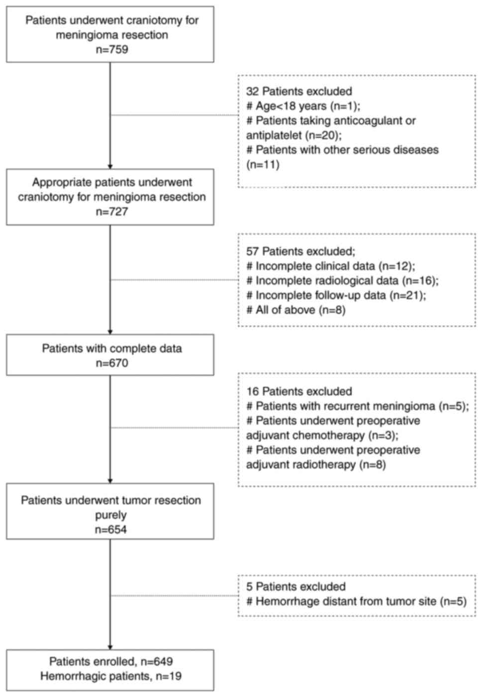
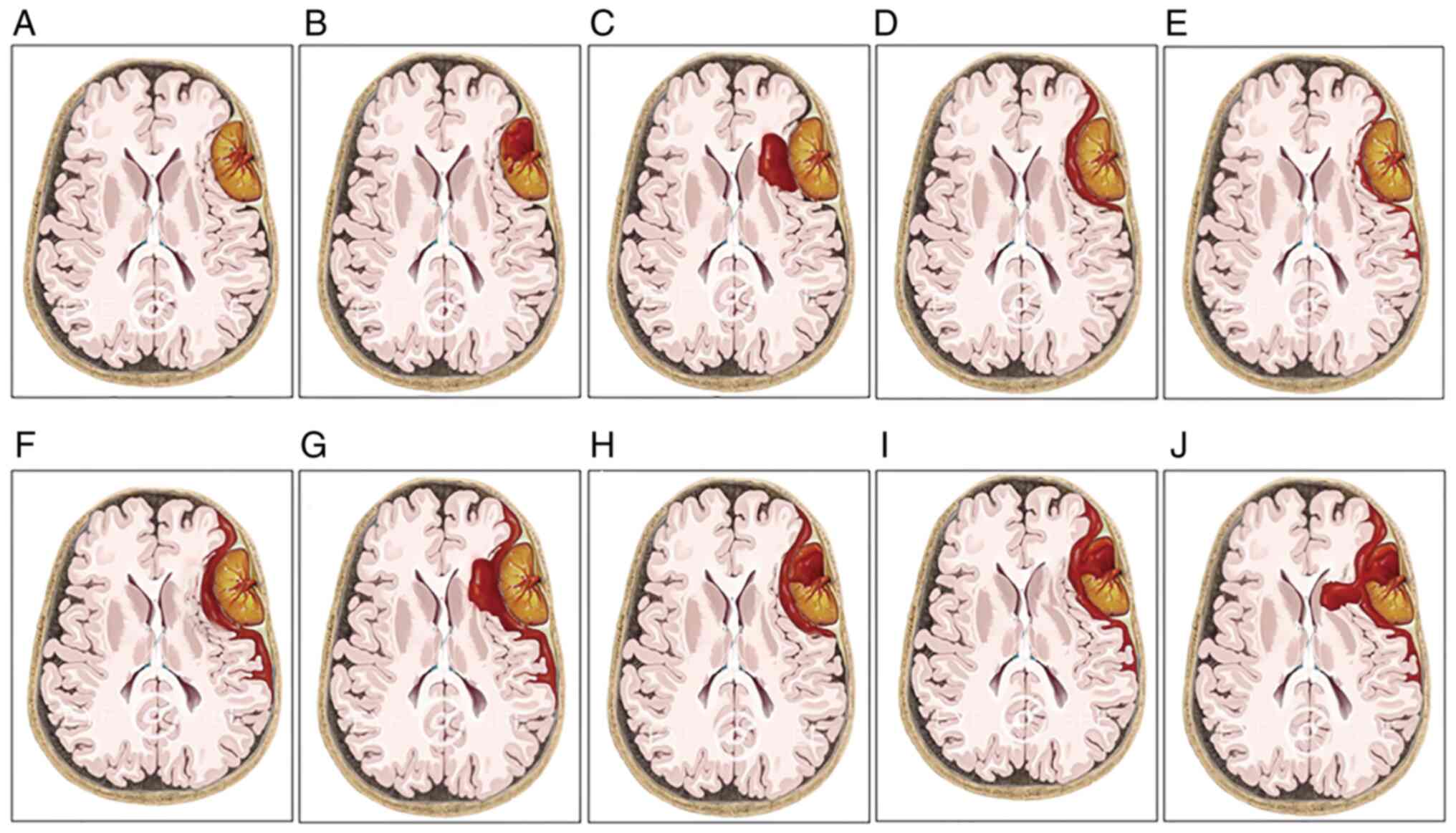
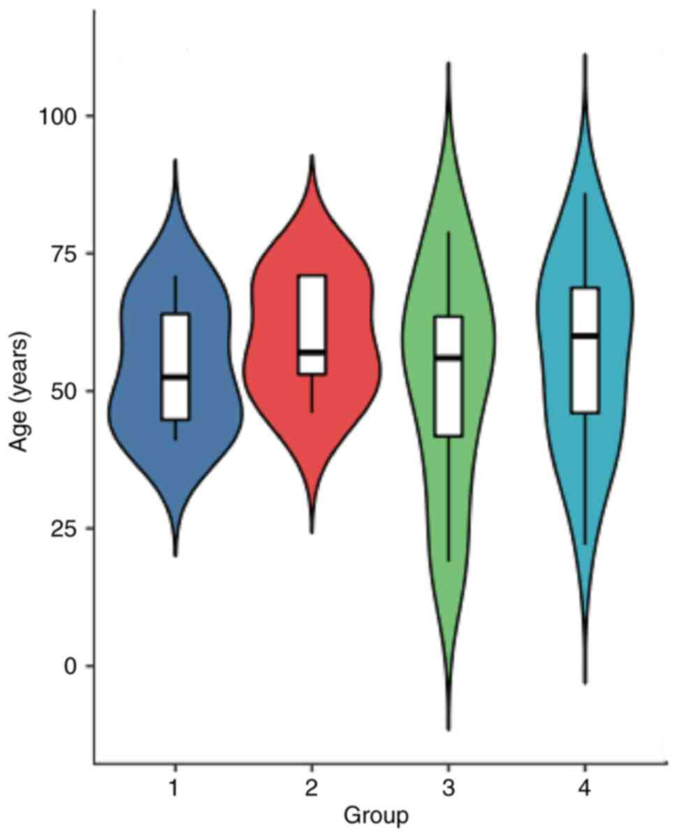
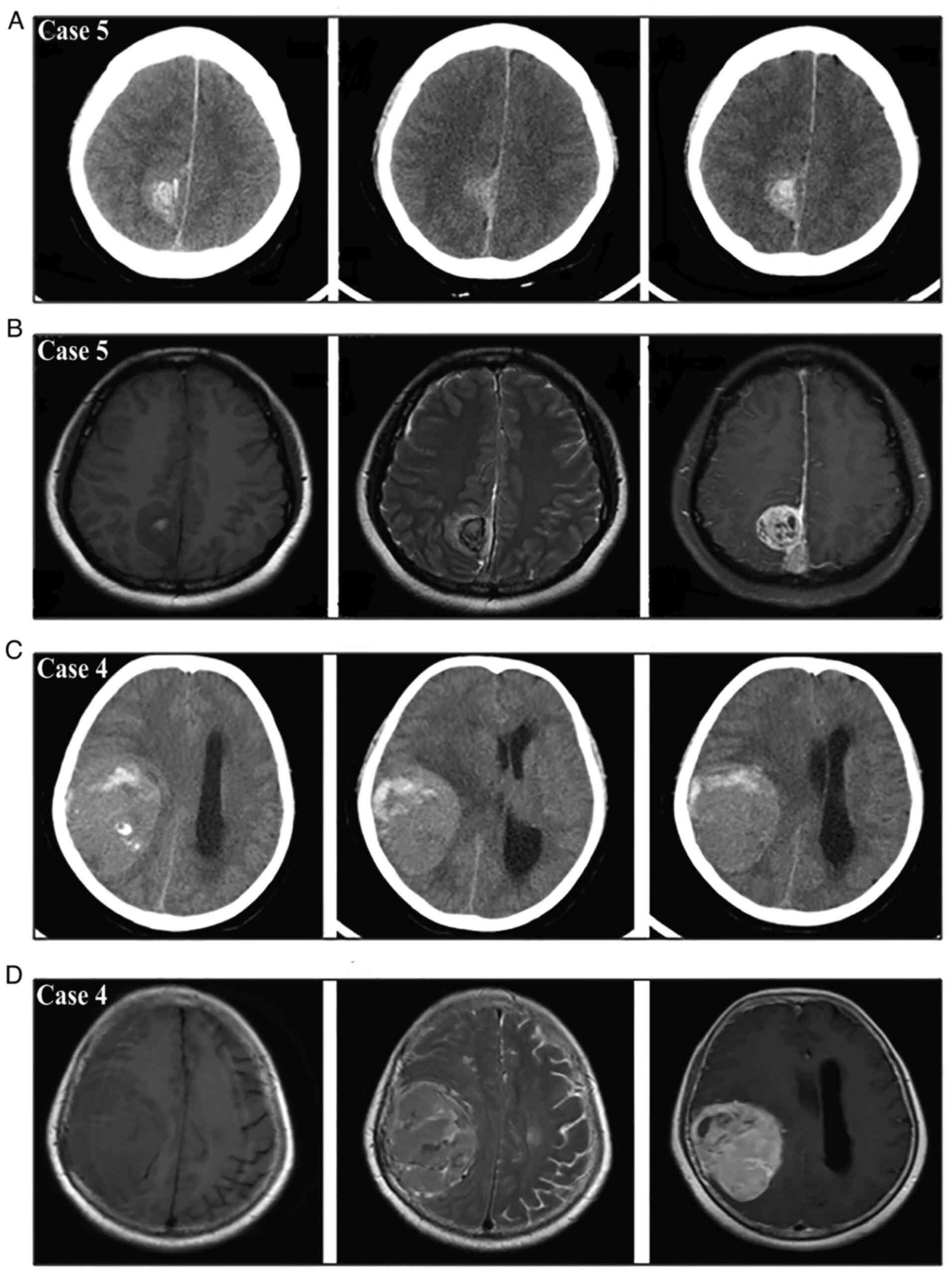
|
1
|
Goldbrunner R, Minniti G, Preusser M,
Jenkinson M, Sallabanda K, Houdart E, Deimling A, Stavrinou P,
Lefranc F, Lund-Johansen M, et al: EANO guidelines for the
diagnosis and treatment of meningiomas. Lancet Oncol. 17:e383–e912.
2016. View Article : Google Scholar : PubMed/NCBI
|
|
2
|
Boŝnjak R, Derham C, Popović M and Ravnik
J: Spontaneous intracranial meningioma bleeding:
Clinicopathological features and outcome. J Neurosurg. 103:473–484.
2005. View Article : Google Scholar : PubMed/NCBI
|
|
3
|
Pereira BJ, de Almeida AN, Paiva WS, de
Aguiar PH, Teixeira MJ and Marie SK: Assessment of hemorrhagic
onset on meningiomas: Systematic review. Clin Neurol Neurosurg.
199:1061752020. View Article : Google Scholar : PubMed/NCBI
|
|
4
|
Kouyialis AT, Stranjalis G, Analyti R,
Boviatsis EJ, Korfias S and Sakas DE: Peritumouralhaematoma and
meningioma: A common tumour with an uncommon presentation. J Clin
Neurosci. 11:906–909. 2004. View Article : Google Scholar : PubMed/NCBI
|
|
5
|
Cheng MH and Lin JW: Intracranial
meningioma with intratumoral hemorrhage. J Formos Med Assoc.
96:116–120. 1997.PubMed/NCBI
|
|
6
|
Huang RB, Chen LJ, Su SY, Wu XJ, Zheng YG,
Wang HP and Liu Y: Misdiagnosis and delay of diagnosis in
hemorrhagic meningioma: A case series and review of the literature.
World Neurosurg. 155:e836–e846. 2021. View Article : Google Scholar : PubMed/NCBI
|
|
7
|
Aloraidi A, Abbas M, Fallatah B,
Alkhaibary A, Ahmed ME and Alassiri AH: Meningioma presenting with
spontaneous subdural hematoma: A report of two cases and literature
review. World Neurosurg. 127:150–154. 2019. View Article : Google Scholar : PubMed/NCBI
|
|
8
|
Pressman E, Penn D and Patel NJ:
Intracranial hemorrhage from meningioma: 2 novel risk factors.
World Neurosurg. 135:217–221. 2020. View Article : Google Scholar : PubMed/NCBI
|
|
9
|
Hambra DV, Danilo de P, Alessandro R, Sara
M and Juan GR: Meningioma associated with acute subdural hematoma:
A review of the literature. Surg Neurol Int. 5 (Suppl
12):S469–S471. 2014. View Article : Google Scholar : PubMed/NCBI
|
|
10
|
Han S, Yang Y, Yang Z, Liu N, Qi X, Yan C
and Yu C: Continuous progression of hemorrhage of sphenoid ridge
meningioma causing cerebral hernia: A case report and literature
review. Oncol Lett. 20:785–793. 2020. View Article : Google Scholar : PubMed/NCBI
|
|
11
|
Wang HC, Wang BD, Chen MS, Li SW, Chen H,
Xu W and Zhang JM: An underlying pathological mechanism of
meningiomas with intratumoral hemorrhage: Undifferentiated
microvessels. World Neurosurg. 94:319–327. 2016. View Article : Google Scholar : PubMed/NCBI
|
|
12
|
Krisht KM, Altay T and Couldwell WT:
Myxoid meningioma: A rare metaplastic meningioma variant in a
patient presenting with intratumoral hemorrhage: Case report. J
Neurosurg. 116:861–865. 2012. View Article : Google Scholar : PubMed/NCBI
|
|
13
|
Bitzer M, Topka H, Morgalla M, Friese S,
Wöckel L and Voigt K: Tumor-related venous obstruction and
development of peritumoral brain edema in meningiomas.
Neurosurgery. 42:724–728. 1998. View Article : Google Scholar : PubMed/NCBI
|
|
14
|
Schrader B, Barth H, Lang EW, Buhl R, Hugo
HH, Biederer J and Mehdorn HM: Spontaneous intracranial haematomas
caused by neoplasms. Acta Neurochir (Wien). 142:979–985. 2000.
View Article : Google Scholar : PubMed/NCBI
|
|
15
|
Kim DG, Park CK, Paek SH, Choe GY, Gwak
HS, Yoo H and Jung HW: Meningioma manifesting intracerebral
haemorrhage: A possible mechanism of haemorrhage. Acta Neurochir
(Wien). 142:165–168. 2000. View Article : Google Scholar : PubMed/NCBI
|
|
16
|
Niiro M, Ishimaru K, Hirano H, Yunoue S
and Kuratsu J: Clinico-pathological study of meningiomas with
haemorrhagic onset. Acta Neurochir (Wien). 145:767–772. 2003.
View Article : Google Scholar : PubMed/NCBI
|
|
17
|
Jr SS, Shuangshoti S and Vajragupt L:
Meningiomas associated with hemorrhage: A report of two cases with
a review of the literature. Neuropathology. 19:150–160. 1999.
View Article : Google Scholar
|
|
18
|
Lin RH and Shen CC: Meningioma with purely
intratumoral hemorrhage mimicked intracerebral hemorrhage: Case
report and literature review. J Med Sci. 36:1582016. View Article : Google Scholar
|
|
19
|
Yamaguchi N, Kawase T, Sagoh M, Ohira T,
Shiga H and Toya S: Prediction of consistency of meningiomas with
preoperative magnetic resonance imaging. Surg Neurol. 48:579–583.
1997. View Article : Google Scholar : PubMed/NCBI
|
|
20
|
Kim JH, Gwak HS, Hong EK, Bang CW, Lee SH
and Yoo H: A case of benign meningioma presented with subdural
hemorrhage. Brain Tumor Res Treat. 3:30–33. 2015. View Article : Google Scholar : PubMed/NCBI
|
|
21
|
Sasagawa Y, Tachibana O, Iida T and Iizuka
H: Oncocytic meningioma presenting with intratumoral hemorrhage. J
Clin Neurosci. 20:1622–1624. 2013. View Article : Google Scholar : PubMed/NCBI
|
|
22
|
Dang DD, Mugge L, Awan O, Dang J and
Shenai M: Spontaneous or traumatic intratumoral hemorrhage? A rare
presentation of parafalcine meningioma. Cureus.
12:e114862020.PubMed/NCBI
|
|
23
|
Agazzi S, Burkhardt K and Rilliet B: Acute
haemorrhagic presentation of an intracranial meningioma. J Clin
Neurosci. 6:242–245. 1999. View Article : Google Scholar : PubMed/NCBI
|
|
24
|
Motegi H, Kobayashi H, Terasaka S, Ishii
N, Ito M, Shimbo D and Houkin K: Hemorrhagic onset of rhabdoid
meningioma after initiating treatment for infertility. Brain Tumor
Patholo. 29:240–244. 2012. View Article : Google Scholar : PubMed/NCBI
|
|
25
|
Lefranc F, Nagy N, Dewitte O, Balériaux D
and Brotchi J: Intracranial meningiomas revealed by non-traumatic
subdural haematomas: A series of four cases. Acta Neurochir (Wien).
143:977–983. 2001. View Article : Google Scholar : PubMed/NCBI
|
|
26
|
Mangubat EZ and Byrne RW: Major
Intratumoral hemorrhage of a petroclival atypical meningioma: Case
report and review of literature. Skull Base. 20:469–474. 2010.
View Article : Google Scholar : PubMed/NCBI
|


