Introduction
Hepatocellular carcinoma (HCC) is one of the most
common malignancy with the highest morbidity and mortality in
numerous countries across the world (1). A number of treatments are used to
treat HCC; however, the treatment effects are often limited,
especially for advanced stage carcinomas (1,2).
Therefore, it is urgent to further explore the carcinogenic
targeting molecules of HCC to enhance prognosis and individualized
treatments.
Syntaxin 6 (STX6) is a sensitive factor in the
soluble n-ethylmaleimide receptor protein and has been reported to
serve a role in Parkinson's disease (3,4). It
has been reported to be expressed in brain, lung and kidney
(5). Furthermore, STX6 is a
vesicle transporter, which serves a vital role in intracellular
protein transport and membrane structure changes (6). STX6 has been reported to be involved
in tumorigenesis in multiple malignant tumors, including esophageal
cancer (7), osteosarcoma (8) and renal cell carcinoma (9). Notably, a recent study reported that
STX6 is a key factor in macrophages during the immune response in
lipopolysaccharide-activated cells (10).
Macrophages are a major component of the
inflammatory infiltrate in tumor (11,12).
The high levels of tumor-associated macrophages (TAMs) are
associated with poor prognosis in a range of tumors, including
breast, gastric and colorectal carcinoma, and HCC (13–17).
The macrophages are classified as M1 phenotype or M2 phenotype
(18). It is now widely accepted
that the M2 phenotype supports tumor growth (13–15,17,19).
CD163 is widely reported as a scavenger receptor and is a highly
specific marker of M2 macrophages (20,21).
CD163 has been reported to be an anti-inflammatory molecule as it
is mainly expressed by M2 macrophages at sites of inflammation
(22). Interestingly, one study
reported that STX6 is associated with increased cytokine secretion
in activated macrophages (23).
However, the mechanisms of STX6 immune infiltration in HCC remain
unclear.
The present study analyzed the association between
STX6 expression and clinical characteristics and prognosis of
patients with HCC. Furthermore, the functions of STX6 in HCC and
potential immune infiltration-related molecular mechanisms were
assessed.
Materials and methods
Tissue sections
The tumor tissues and para-carcinoma tissue sections
were collected between January 2014 and December 2016 and stored in
the Human Resources Specimen Bank of the First Affiliated Hospital
of Nanchang University (Nanchang, China). Samples were obtained
from the Human Genetic Resources Center and Department of Pathology
of The First Affiliated Hospital of Nanchang University (Nanchang,
China) and examined independently by two pathologists. The patients
included 76 males and 14 females between 27 and 81 years old
(median 53). None of the patients had previously received other
tumor surgery, chemotherapy, radiation therapy, or any other
anticancer therapy. As this was a retrospective study, the
requirement for informed consent was waived by the ethics
committee. The present study was approved by the Clinical Medical
Research Ethics Committee of the First Affiliated Hospital of
Nanchang University (approval no. 202112020; Nanchang, China).
Immunohistochemistry (IHC)
To examine STX6 and CD163 expression in tumor
tissues, paired paraffin-embedded tumor slices were obtained from
the specimen bank. The sections were then incubated with anti-STX6
(1:100; cat. no. ab140607; Abcam) and anti-CD163 (1:500, ab182422;
Abcam) antibodies overnight at 4°C as previously reported (24). For statistical analysis, the
percentage coverage of the protein was scored manually as follows:
i) 1 (0–25%); ii) 2 (26–50%); iii) 3 (51–75%); and iv) 4 (76–100%)
(24). The intensity of positive
staining was also scored as follows: i) 0 (negative); ii) 1 (weak);
iii) 2 (moderate); and iv) 3 (strong). The final scores were
calculated by multiplying the aforementioned scores. The final
scores of the percentage and staining scores were defined as the
overall IHC scores (0–12). Scores <6 were considered to be low
expression (STX6-Low) and scores ≥6 were considered to be high
expression (STX6-High).
Gene expression profiling interactive
analysis (GEPIA2) database analysis
GEPIA2 (http://gepia2.cancer-pku.cn/#index) is an interactive
online platform, which contains information from >9,000 tumor
samples from The Cancer Genome Atlas (TCGA) (https://portal.gdc.cancer.gov/) and The
Genotype-Tissue Expression (GTEx) (https://xenabrowser.net/datapages/) databases, and
information from >8,000 control samples. The correlation between
STX6 and CD163 was determined by Pearson correlation coefficient
analysis in GEPIA2 (25). The
iCluster 1 and iCluster 3 datasets (Cutoff-High-75% and
Cutoff-Low-25%) were used to analyze the association between
prognosis and STX6 expression in patients with HCC (25).
Kaplan-Meier plotter (KM plotter)
survival analysis of STX6
Kaplan-Meier plotter (http://kmplot.com/analysis/) can perform survival
analysis on >54,000 genes (mRNA, microRNA and protein) in 21
types of tumors (including breast, ovarian, lung, gastric and liver
cancer). The data mainly come from the Gene Expression Omnibus and
TCGA databases (26). The results
were assessed based on the log rank P-value and hazard ratio (HR)
with 95% confidence intervals. The RNAseq ID: 10228 (STX6) and
Cutoff value used in analysis: 1046 were used. A follow-up
threshold of 60 months was used to analyze the association between
prognosis and STX6 expression in patients with HCC based on the log
rank P-value.
Tumor immune estimation resource
(TIMER) database
The TIMER database (https://cistrome.shinyapps.io/timer/) (27) is mainly divided into seven
sections: Gene, Survival, Mutation, somatic copy number
amplifications (sCNA), differential expression (Diffexp),
Correlation and Estimation. Among them, the Gene and Correlation
were used in this study. To facilitate the study of tumor immunity
and genomic data, the TIMER database applies a deconvolution method
(28) to infer the abundance of
tumor immune-infiltrating cells (TIICs) from gene expression
profiles, reanalyzes the gene expression data of 10,897 samples of
32 cancer types from TCGA and estimates the abundance of 6 TIIC
subgroups [B cells, CD4+ T cells, CD8+ T
cells, macrophages, neutrophils and dendritic cells (DCs)].
Deconvolution methods define the problem as mathematical equations
that model the gene expression of a tissue sample as the weighted
sum of the expression profiles from the cells in the population mix
(29). These methods are further
detailed in previous studies (27,30).
The statistical method used by the TIMER database in the present
study was the Spearman correlation coefficient analysis. P<0.05
was considered to be statistically significant.
University of California Santa Cruz
(UCSC) Xena
RNAseq data were extracted from the UCSC Xena portal
(https://xenabrowser.net/datapages/)
(31), including data from TCGA
Liver HCC (LIHC; n=371) and GTEx for corresponding normal tissue
(n=160). These data were used to assess the diagnostic effect of
STX6 and α-fetoprotein (AFP) in liver cancer using a receiver
operating characteristic (ROC) curve as previously reported
(32).
Statistical analysis
All bioinformatics analyses were performed using the
corresponding database websites. Other statistical analyses of data
were performed using GraphPad Prism 6 (GraphPad Software, Inc.) and
SPSS 18 (SPSS, Inc.). Data are presented as the median. The
Wilcoxon signed-rank test was used to compare the difference
between tumor tissues and normal tissues. A Mann-Whitney U test was
used to compare two distinct groups of patients (STX6-Low vs.
STX6-High). The association of clinicopathological factors with
STX6 expression was analyzed using the χ2 test. A
log-rank test was used to compare the survival curves by
Kaplan-Meier. The correlation between the IHC score of STX6 and the
IHC score of CD163 was analyzed using Pearson correlation
coefficient analysis. P<0.05 was considered to indicate a
statistically significant difference.
Results
STX6 is highly expressed in HCC
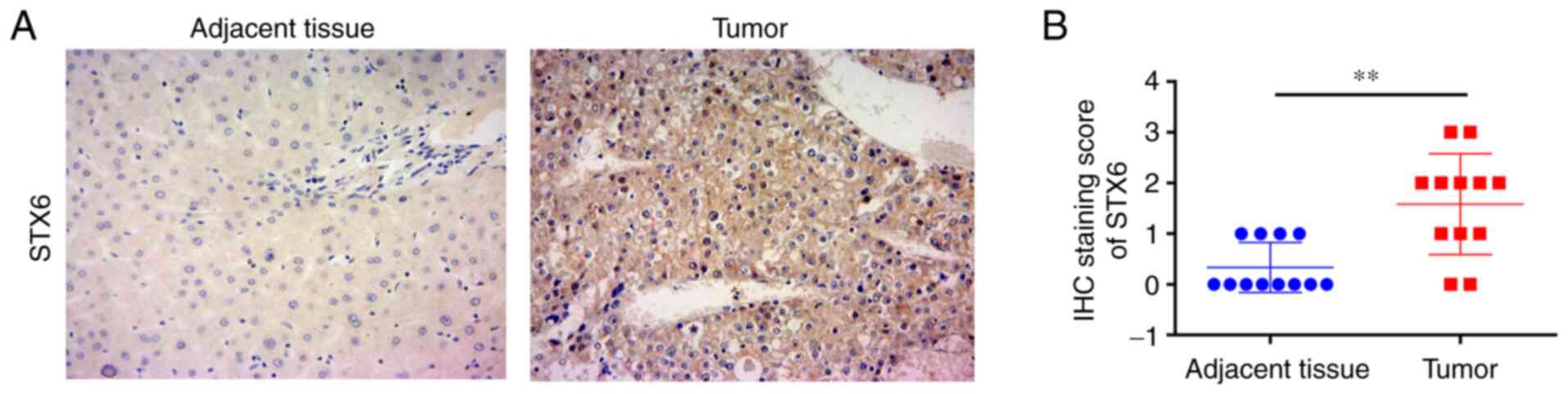
IHC was used to assess the protein expression levels
of STX6 in patients with HCC (Fig.
1A). The results demonstrated that the IHC scores of 12 pairs
of HCC samples stained for STX6 were significantly higher than
those of paired adjacent tissues (Fig.
1B). These results demonstrated that STX6 protein was highly
expressed in HCC tissues.
Association analysis between STX6
expression and clinical features of patients with HCC
The relationship between STX6 protein expression and
the clinicopathological characteristics of 90 patients with HCC was
evaluated. STX6 protein expression was significantly associated
with HCC tumor size (P=0.003), Edmondson grade (P=0.020) and AFP
level (P=0.019) (Table I).
 | Table I.Association between STX6 expression
and clinical characteristics of patients with hepatocellular
carcinoma. |
Table I.
Association between STX6 expression
and clinical characteristics of patients with hepatocellular
carcinoma.
|
|
| STX6 expression
(IHC score) |
|
|
|
|---|
|
|
|
|
|
|
|
|---|
| Variables | No. (%) | Low (<6), n | High (≥6), n | Df | χ2 | P-value |
|---|
| Sex |
|
|
| 1 | 0.017 | 0.897 |
|
Male | 76 (84.4) | 42 | 34 |
|
|
|
|
Female | 14 (15.6) | 8 | 6 |
|
|
|
| Age, years |
|
|
| 1 | 0.057 | 0.810 |
|
<55 | 37 (41.1) | 20 | 17 |
|
|
|
|
≥55 | 53 (58.9) | 30 | 23 |
|
|
|
| Size, cm |
|
|
| 1 | 9.085 | 0.003 |
|
<5 | 56 (62.2) | 38 | 18 |
|
|
|
| ≥5 | 34 (37.8) | 12 | 22 |
|
|
|
| Diolame
complete |
|
|
| 1 | 0.188 |
|
|
Yes | 54 | 31 | 23 |
|
| 0.665 |
| No | 36 | 19 | 17 |
|
|
|
| Number of
tumors |
|
|
| 1 | 1.125 | 0.289 |
|
Single | 72 (80) | 38 | 34 |
|
|
|
|
Multiple | 18 (20) | 12 | 6 |
|
|
|
| TNM staging |
|
|
| 1 | 1.798 | 0.180 |
|
I+II | 77 (85.6) | 45 | 32 |
|
|
|
|
III+IV | 13 (14.4) | 5 | 8 |
|
|
|
| Microvascular
invasion |
|
|
| 1 | 1.309 | 0.253 |
| No | 64 (71.1) | 38 | 26 |
|
|
|
|
Yes | 26 (28.9) | 12 | 14 |
|
|
|
| Edmondson
grade |
|
|
| 1 | 5.399 | 0.020 |
|
I+II | 67 (74.4) | 42 | 25 |
|
|
|
|
III | 23 (25.6) | 8 | 15 |
|
|
|
| Cirrhosis |
|
|
| 1 | 0.243 | 0.622 |
|
Negative | 16 (17.8) | 8 | 8 |
|
|
|
|
Positive | 74 (82.2) | 42 | 32 |
|
|
|
| HBV |
|
|
| 1 | 0.800 | 0.371 |
|
Absent | 25 (27.8) | 12 | 13 |
|
|
|
|
Present | 65 (72.2) | 38 | 27 |
|
|
|
| ALT, U/l |
|
|
| 1 | 0.458 | 0.499 |
|
<45 | 55 (61.1) | 29 | 26 |
|
|
|
|
≥45 | 35 (38.9) | 21 | 14 |
|
|
|
| AFP, ng/ml |
|
|
| 1 | 5.478 | 0.019 |
|
<400 | 69 (76.7) | 43 | 26 |
|
|
|
|
≥400 | 21 (23.3) | 7 | 14 |
|
|
|
Association between STX6 expression
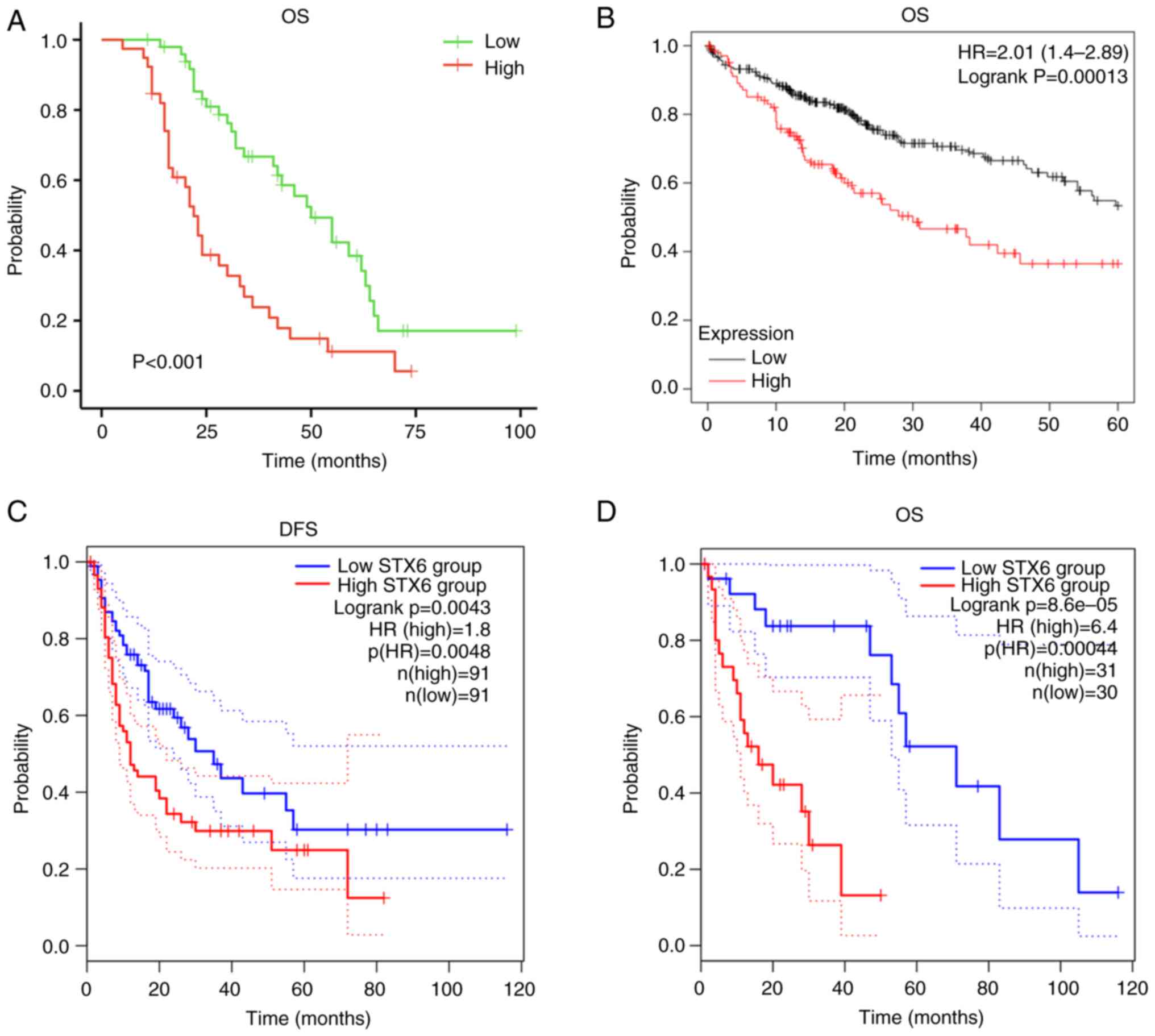
and patient survival in HCC
Survival analysis demonstrated that patients with
high STX6 expression had worse prognosis (Fig. 2A). Furthermore, analysis using KM
plotter (Fig. 2B) demonstrated
that high expression levels of STX6 were associated with worse
overall survival (OS; P=0.00013; HR, 2.01; 95% CI, 1.4-2.89) in
patients with HCC. Additionally, in the GEPIA2 database analysis,
high STX6 expression was associated with worse disease-free
survival [P=0.0043; HR(high), 1.8; p(HR), 0.0048] and OS
[P=0.000086; HR, 6.4; p(HR)=0.00044] in patients with HCC (Fig. 2C and D). These data demonstrated
that dysregulated expression of STX6 affected the clinical outcomes
of patients with HCC.
Analysis of immune infiltration
STX6 expression was significantly positively
correlated with infiltration by B cells (r=0.389;
P=6.93×10⁻¹⁴), CD4+ T cells (r=0.541;
P=1.50×10⁻²⁷), macrophages (r=0.535;
P=1.17×10⁻²⁶), neutrophils (r=0.457;
P=3.11×10⁻¹⁹) and DCs (r=0.416;
P=1.15×10⁻¹⁵), and significantly positively associated
with CD8+ T cell infiltration (r=0.247;
P=3.77×10⁻⁶; Fig. 3A),
which demonstrated that STX6 serves a crucial role in the immune
infiltration of HCC. STX6 expression was also significantly
positively associated with macrophage TAMs: CCL2 (r=0.271;
P=1.28×10⁻⁷), CD68 (r=0.298; P=5.38×10⁻⁹),
IL10 (r=0.31; P=1.11×10⁻⁹), MS4A4A (r=0.202;
P=9.06×10⁻⁵), MSR1 (r=0.371; P=1.62×10⁻¹³)
and VSIG4 (r=0.222; P=1.65×10⁻⁵) were also analyzed
(Fig. 3B). The results
demonstrated that STX6 mRNA expression was significantly correlated
with TAMs [CC motif chemokine ligand 2 (CCL2), CD68, IL10, V-set
and immunoglobulin domain containing 4, macrophage scavenger
receptors-type 1 and membrane spanning 4-domains A4A], DCs,
CD4+ T cells, CD163, etc. (Table SI). To further assess the
association of STX6 with CD163, STX6 and CD163 expression was
analyzed using the GEPIA2 and TIMER databases. These results
demonstrated a small association between STX6 and CD163 (cor=0.173
in Fig. 3C and R=0.18 in Fig. 3D). The results of the present study
demonstrated that STX6 expression was positively associated with B
and T-cell receptor signaling pathways during HCC pathogenesis,
indicating that STX6 was related to the immune response. These
findings suggested that STX6 expression may be associated with the
infiltration of immune cells in HCC.
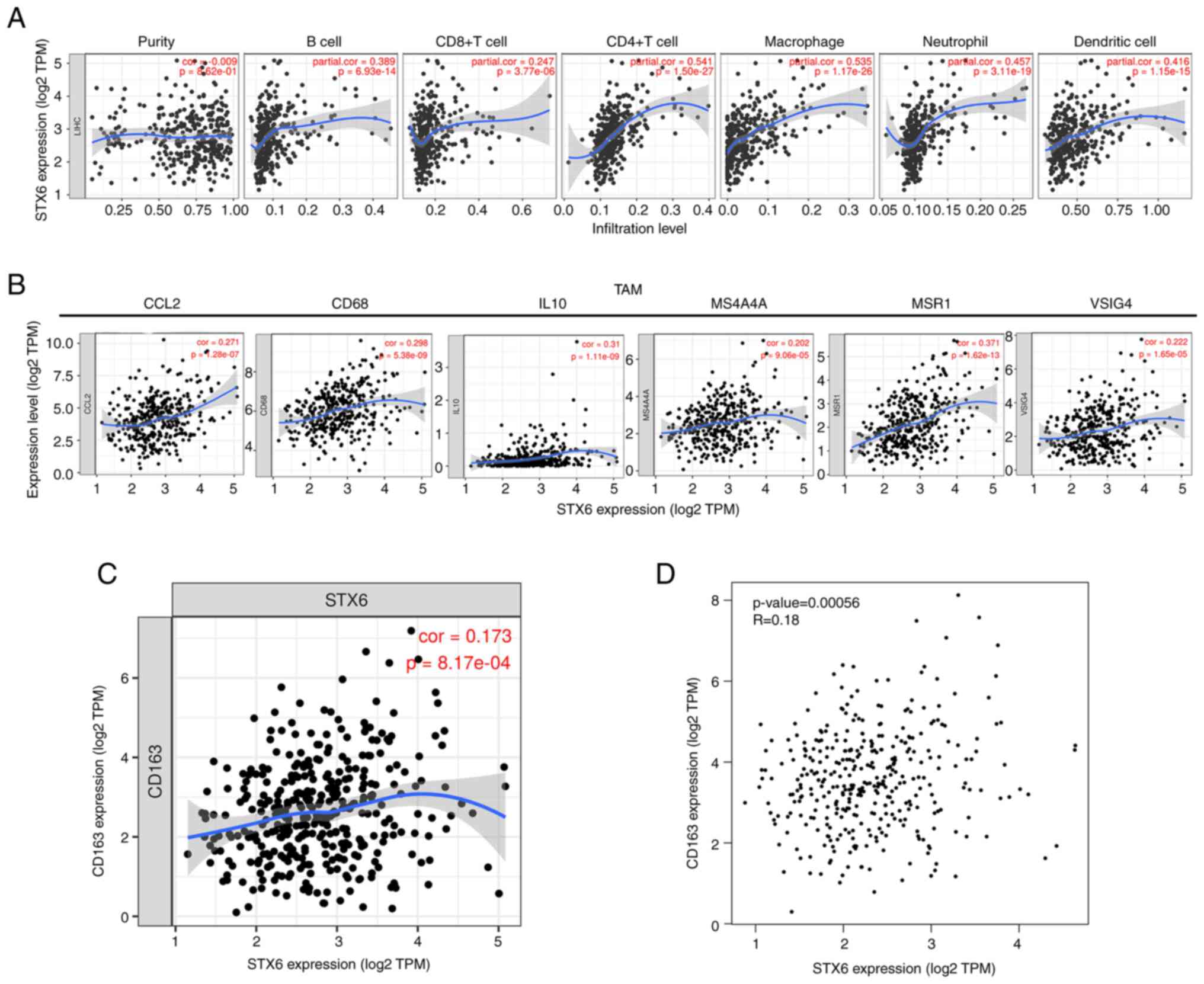 | Figure 3.Relationship between STX6 expression
and immune cell infiltration levels in hepatocellular carcinoma.
(A) Correlation of STX6 expression with tumor-infiltrating immune
cells in LIHC (n=371). Scatter plots presenting the correlations
between STX6 expression and marker molecules, including (B) TAMs
(CCL2, CD68, IL10, VSIG4, MSR1, MS4A4A), and (C and D) CD163. The
blue curve and gray area in the figure represent the general trend
direction. LIHC, liver hepatocellular carcinoma; STX6, syntaxin 6;
CCL2, CC motif chemokine ligand 2; MS4A4A, membrane spanning
4-domains A4A; MSR1, macrophage scavenger receptor 1; TAMs,
tumor-associated macrophages; TPM, transcripts per million; VSIG4,
V-set and immunoglobulin domain containing 4. |
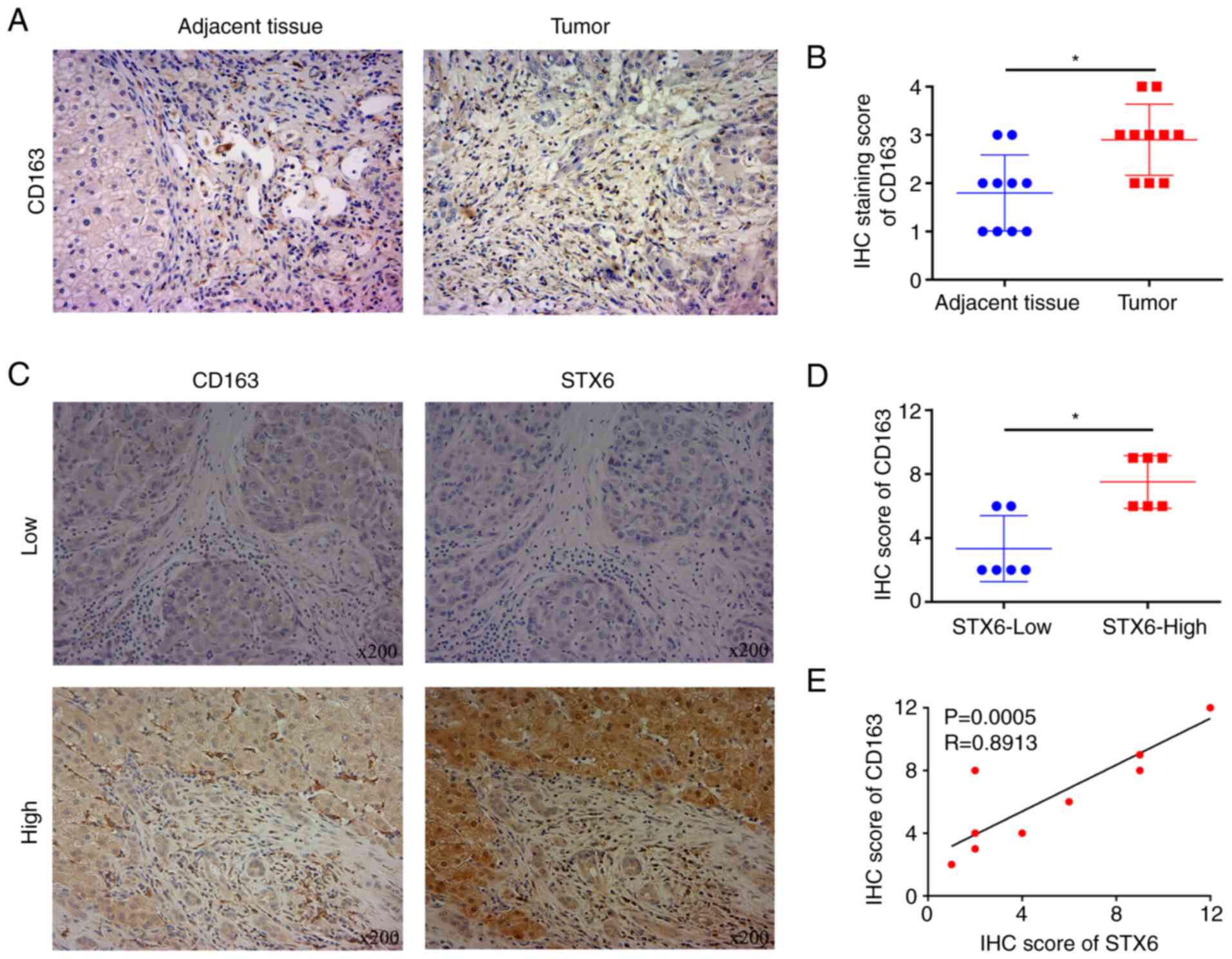
STX6 is positively associated with
CD163 according to IHC
STX6 and CD163 expression in HCC and adjacent
noncancerous tissues was assessed using IHC (Figs. 1A and 4A). The results demonstrated that the
protein expression levels of STX6 and CD163 in cancer tissues were
significantly increased compared with those in adjacent tissues
(Figs. 1B and 4B). To further examine the association
between STX6 and CD163, IHC was performed (Fig. 4C). The results demonstrated that
CD163 levels were significantly positively associated with the
levels of STX6 in paired paraffin-embedded tumor slices (Fig. 4D and E).
Diagnostic values of STX6 and AFP as
assessed using ROC curve analyses
The ROC curve analyses of two markers (STX6 and AFP)
in HCC and normal tissues are presented (Fig. 5). ROC curve analysis indicated that
the area under the curve (AUC) values for STX6 and AFP were 0.942
(CI, 0.916-0.967) and 0.720 (CI, 0.668-0.773), respectively, using
TCGA data (tumor, n=374; normal, n=50). AUC of STX6 (sensitivity,
0.940; specificity, 0.850) was higher than that of AFP
(sensitivity, 0.940; specificity, 0.516) in TCGA (Table II). Furthermore, the ROC curve
analysis demonstrated that the AUC values of STX6 and AFP were
0.844 and 0.746, respectively, according to the combined TCGA
(tumor, n=371) and GTEx (normal, n=160) datasets. The sensitivity
of the ROC curve for STX6 (0.827) was higher than that for AFP
(0.563). The specificity of the ROC curve of STX6 (0.762) was lower
than that of AFP (0.869). Compared with the aforementioned
datasets, STX6 had a higher AUC value (0.947) and specificity
(0.917) in the our HCC data of the present study. The sensitivity
of ROC curve of STX6 is 0.856 in the present HCC data. These
results demonstrated that STX6 may be a diagnostic marker for
HCC.
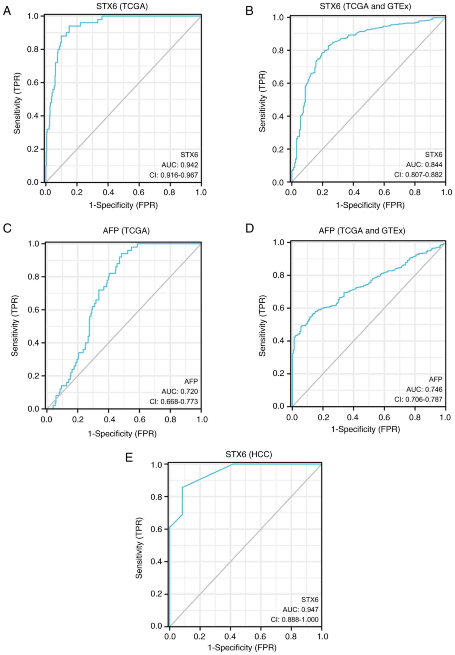 | Figure 5.Diagnostic values of STX6 and AFP
indicated by ROC curve analyses based on the TCGA and GTEx
datasets. (A) Diagnostic value of STX6 assessed using ROC curve
analysis of data from TCGA (tumor, n=374; normal, n=50). (B)
Diagnostic value of STX6 assessed using ROC curve analysis of data
from TCGA (tumor, n=371) and GTEx (normal, n=160) databases. (C)
Diagnostic value of AFP assessed using ROC curve analysis of data
from TCGA (tumor, n=374; normal, n=50). (D) Diagnostic value of AFP
assessed using ROC curve analysis of data from TCGA (tumor, n=371)
and GTEx (normal, n=160) databases. (E) Diagnostic value of STX6
assessed using ROC curve analysis of data from the present study
(tumor, n=90; normal, n=12). AFP, α-fetoprotein; AUC, area under
the curve; FPR, false positive rate; GTEx, Genotype-Tissue
Expression; HCC, hepatocellular carcinoma; ROC, receiver operating
characteristic; STX6, syntaxin 6; TCGA, The Cancer Genome Atlas;
TPR, true positive rate. |
| Table II.Diagnostic values of STX6 and AFP in
patients with liver hepatocarcinoma according to receiver operating
characteristic curve analyses. |
Table II.
Diagnostic values of STX6 and AFP in
patients with liver hepatocarcinoma according to receiver operating
characteristic curve analyses.
| Index | AUC | 95% CI | Cut-off | Sensitivity | Specificity | Youden index | Data |
|---|
| AFP | 0.720 | 0.668-0.773 | 2.613 | 0.940 | 0.516 | 1.456 | TCGA |
| STX6 | 0.942 | 0.916-0.967 | 2.334 | 0.940 | 0.850 | 1.790 | TCGA |
| AFP | 0.746 | 0.706-0.787 | 3.341 | 0.563 | 0.869 | 1.432 | TCGA + GTEx |
| STX6 | 0.844 | 0.807-0.882 | 1.702 | 0.827 | 0.762 | 1.590 | TCGA + GTEx |
| STX6 | 0.947 | 0.888-1.000 | 1.500 | 0.856 | 0.917 | 0.772 | HCC |
Discussion
The progression of liver HCC is rapid and numerous
patients present with advanced HCC (33,34).
Regulation of immune infiltration is increasingly recognized as
being important in tumor development (35). Finding breakthroughs in
immunotherapy has become the focus of current research.
Furthermore, an effective early detection method is still lacking
in the current treatment of HCC. AFP has been recognized as a tumor
marker for HCC but it has poor sensitivity and specificity
(36,37). Exploring novel therapeutic and
diagnostic markers is still the top priority of scientific research
in this field.
In the present study, the role of the STX6 gene in
the development of HCC was analyzed using IHC. STX6 protein
expression in HCC was analyzed and the results demonstrated that
STX6 expression was upregulated in HCC tissues compared with normal
tissues. The association between STX6 and clinical characteristics
of patients was further considered. These results demonstrated that
high protein expression levels of STX6 were significantly
associated with tumor size, Edmondson grade and the AFP level in
patients with HCC. Furthermore, these results demonstrated that
patients with high STX6 expression had a worse prognosis as
demonstrated by analysis of the survival data in the KM plotter and
GEPIA2 databases, which are based on the results of transcriptome
sequencing data analysis. Furthermore, patients with high STX6
protein expression also had a worse prognosis as demonstrated by
the survival data of patients with HCC in the present study. A
previous study reported that STX6 could be a prognostic biomarker
for patients with renal cell carcinoma based on TCGA transcriptome
sequencing data (9). This is
supported by the present study which combined analysis of two
different datasets, which demonstrated that STX6 could promote the
process of HCC malignancy.
The present study demonstrated that STX6 was
associated with infiltration of immune cells based on analysis
using the TIMER database. The results demonstrated that high STX6
mRNA expression in HCC was positively associated with high immune
infiltration. STX6 expression was significantly positively
correlated with the levels of immune infiltration, including B
cell, CD4+ T cell, DC, macrophage and neutrophil
infiltration, in HCC, and significantly positively associated with
CD8+ T cells. Furthermore, the gene markers of M2
macrophages (CD163 and CD115) and TAMs (CCL2 and IL10) were
significantly positively correlated with STX6 expression.
Association between STX6 and CD163 was assessed using IHC and the
results confirmed the aforementioned findings. It could be
concluded that the M2 macrophage CD163 expression was also enhanced
by increased levels of STX6. It has previously been reported that
high rates of infiltration of M2 macrophages into the tumor stroma
could inhibit T cell proliferation and downregulate antitumor
immune responses (38,39). STX6 upregulation could be one of
the routes that link immunosuppression and the development of HCC.
Overall, these results demonstrated the potential regulatory role
of STX6 in immune inflammatory responses in HCC.
AFP has a diagnostic value as a marker for liver
cancer in clinical settings (40–42).
Abnormal AFP levels in adult plasma have been reported to be a
marker of the pathological condition of HCC (43). In the present study, the results
demonstrated that the AUC of STX6 was significantly higher than the
AUC of AFP based on TCGA data (0.942 vs. 0.720) and the sensitivity
of STX6 was also higher than that of AFP based on combined TCGA and
GTEx data (0.827 vs. 0.563). Nevertheless, the specificity of the
AUC curve of STX6 was lower than the AUC of AFP based on combined
TCGA (tumor, n=371) and GTEx (normal, n=160) data (0.762 vs.
0.869). This could have been due to the high STX6 expression in
multiple other tumor types (7,8,44),
which reduce its diagnostic specificity in HCC. However, compared
with the aforementioned datasets, STX6 demonstrated a higher AUC
value (0.947) and specificity (0.917) in data from the present
study. Collectively, these results demonstrated that STX6 has the
potential to be a powerful diagnostic maker in HCC.
The present study demonstrated that STX6 protein
expression was significantly associated with HCC tumor size,
Edmondson grade, AFP level and prognosis of patients with HCC.
Furthermore, STX6 may be involved in the immune-inflammatory
response of HCC and may become a novel potential diagnostic marker
for patients with HCC. However, there were some limitations in the
present study. For the assessment of the association between STX6
and CD163, the absence of double-staining IHC or immunofluorescence
staining was a limitation of the present study. Furthermore, the
present study was only an initial early-stage experiment and future
work is required which should include experiments on larger numbers
of clinical samples and more in-depth research on the molecular
mechanism of STX6 in HCC. Further studies should analyze fresh
tissues and HCC cell lines and explore the effects of STX6 on the
phenotype of HCC cells.
In summary, the present study demonstrated that STX6
expression was associated with the clinical characteristics and
prognosis of patients with HCC. Furthermore, STX6 may associate
with CD163 to participate in the modulation of inflammatory
responses in HCC. Compared with AFP, STX6 may become a valuable
novel tumor marker for the diagnosis of patients with HCC and
combination of STX6 with AFP may have higher diagnostic value. The
present study also provided further insight into the molecular
mechanism of STX6 in HCC.
Supplementary Material
Supporting Data
Acknowledgements
Not applicable.
Funding
The present study was supported by the National Natural Science
Foundation of China (grant no. 82103165), The Key R & D General
Project of Jiangxi Science and Technology Department (grant no.
20203BBGL73187), the Natural Science Foundation of Jiangxi Province
(grant no. 20212BAB216036), The Education Department of Jiangxi
Province Science and Technology Research Projects (grant no.
GJJ160246) and the Young Talent Cultivation Project of the First
Affiliated Hospital of Nanchang University (grant no.
YFYPY202007).
Availability of data and materials
The datasets generated and/or analyzed during the
current study are available in the GEPIA2 repository, https://gepia2.cancer-pku.cn/#index; National
Cancer Institute GDC Data Portal repository, https://portal.gdc.cancer.gov/; the Kaplan-Meier
Plotter repository, https://kmplot.com/analysis/; TIMER repository,
https://cistrome.shinyapps.io/timer/;
TCGA repository, https://portal.gdc.cancer.gov/; and UCSC Xena
repository [TCGA cohort, GDC TCGA Liver Cancer (LIHC),
TCGA-LIHC.htseq_fpkm.tsv; GTEX cohort, GTEX, gtex_RSEM_gene_tpm],
https://xenabrowser.net/datapages/.
The other datasets used and/or analyzed during the current study
are available from the corresponding author on reasonable
request.
Authors' contributions
YZ, LL, ZF and YT analyzed the data and prepared the
manuscript. YL and JX designed the study. YL and JX confirm the
authenticity of all the raw data. All authors have read and
approved the final manuscript.
Ethics approval and consent to
participate
The paired paraffin-embedded tumor slices were
obtained from Human Genetic Resources Center of the First
Affiliated Hospital of Nanchang University (Nanchang, China). The
project was approved by the Clinical Medical Research Ethics
Committee of the First Affiliated Hospital of Nanchang University
(approval no. 202112020; Nanchang, China). As this was a
retrospective study, the requirement for informed consent was
waived by the ethics committee.
Patient consent for publication
Not applicable.
Competing interests
The authors declare that they have no competing
interests.
References
|
1
|
Anwanwan D, Singh SK, Singh S, Saikam V
and Singh R: Challenges in liver cancer and possible treatment
approaches. Biochim Biophys Acta Rev Cancer. 1873:1883142020.
View Article : Google Scholar : PubMed/NCBI
|
|
2
|
Orcutt ST and Anaya DA: Liver resection
and surgical strategies for management of primary liver cancer.
Cancer Control. 25:10732748177446212018. View Article : Google Scholar : PubMed/NCBI
|
|
3
|
Höglinger GU, Melhem NM, Dickson DW,
Sleiman PM, Wang LS, Klei L, Rademakers R, de Silva R, Litvan I,
Riley DE, et al: Identification of common variants influencing risk
of the tauopathy progressive supranuclear palsy. Nat Genet.
43:699–705. 2011. View
Article : Google Scholar : PubMed/NCBI
|
|
4
|
Bock JB, Matern HT, Peden AA and Scheller
RH: A genomic perspective on membrane compartment organization.
Nature. 409:839–841. 2001. View
Article : Google Scholar : PubMed/NCBI
|
|
5
|
Bock JB, Lin RC and Scheller RH: A new
syntaxin family member implicated in targeting of intracellular
transport vesicles. J Biol Chem. 271:17961–17965. 1996. View Article : Google Scholar : PubMed/NCBI
|
|
6
|
Wendler F and Tooze S: Syntaxin 6: The
promiscuous behaviour of a SNARE protein. Traffic. 2:606–611. 2001.
View Article : Google Scholar : PubMed/NCBI
|
|
7
|
Du J, Liu X, Wu Y, Zhu J and Tang Y:
Essential role of STX6 in esophageal squamous cell carcinoma growth
and migration. Biochem Biophys Res Commun. 472:60–67. 2016.
View Article : Google Scholar : PubMed/NCBI
|
|
8
|
Zhang PR, Ren J, Wan JS, Sun R and Li Y:
Circular RNA hsa_circ_0002052 promotes osteosarcoma via modulating
miR-382/STX6 axis. Hum Cell. 33:810–818. 2020. View Article : Google Scholar : PubMed/NCBI
|
|
9
|
Peak TC, Su Y, Chapple AG, Chyr J and Deep
G: Syntaxin 6: A novel predictive and prognostic biomarker in
papillary renal cell carcinoma. Sci Rep. 9:31462019. View Article : Google Scholar : PubMed/NCBI
|
|
10
|
West ZE, Aitcheson SM, Semmler ABT and
Murray RZ: The trans-SNARE complex VAMP4/Stx6/Stx7/Vti1b is a key
regulator of Golgi to late endosome MT1-MMP transport in
macrophages. Traffic. 22:368–376. 2021. View Article : Google Scholar : PubMed/NCBI
|
|
11
|
Bian Z, Shi L, Kidder K, Zen K,
Garnett-Benson C and Liu Y: Intratumoral SIRPα-deficient
macrophages activate tumor antigen-specific cytotoxic T cells under
radiotherapy. Nat Commun. 12:32292021. View Article : Google Scholar : PubMed/NCBI
|
|
12
|
Mantovani A, Sozzani S, Locati M, Allavena
P and Sica A: Macrophage polarization: Tumor-associated macrophages
as a paradigm for polarized M2 mononuclear phagocytes. Trends
Immunol. 23:549–555. 2002. View Article : Google Scholar : PubMed/NCBI
|
|
13
|
Yamaguchi T, Fushida S, Yamamoto Y,
Tsukada T, Kinoshita J, Oyama K, Miyashita T, Tajima H, Ninomiya I,
Munesue S, et al: Tumor-associated macrophages of the M2 phenotype
contribute to progression in gastric cancer with peritoneal
dissemination. Gastric Cancer. 19:1052–1065. 2016. View Article : Google Scholar : PubMed/NCBI
|
|
14
|
Munir MT, Kay MK, Kang MH, Rahman MM,
Al-Harrasi A, Choudhury M, Moustaid-Moussa N, Hussain F and Rahman
SM: Tumor-associated macrophages as multifaceted regulators of
breast tumor growth. Int J Mol Sci. 22:65262021. View Article : Google Scholar : PubMed/NCBI
|
|
15
|
Cersosimo F, Lonardi S, Bernardini G,
Telfer B, Mandelli GE, Santucci A, Vermi W and Giurisato E:
Tumor-associated macrophages in osteosarcoma: From mechanisms to
therapy. Int J Mol Sci. 21:52072020. View Article : Google Scholar : PubMed/NCBI
|
|
16
|
Wei C, Yang C, Wang S, Shi D, Zhang C, Lin
X, Liu Q, Dou R and Xiong B: Crosstalk between cancer cells and
tumor associated macrophages is required for mesenchymal
circulating tumor cell-mediated colorectal cancer metastasis. Mol
Cancer. 18:642019. View Article : Google Scholar : PubMed/NCBI
|
|
17
|
Arvanitakis K, Koletsa T, Mitroulis I and
Germanidis G: Tumor-associated macrophages in hepatocellular
carcinoma pathogenesis, prognosis and therapy. Cancers (Basel).
14:2262022. View Article : Google Scholar : PubMed/NCBI
|
|
18
|
Shapouri-Moghaddam A, Mohammadian S,
Vazini H, Taghadosi M, Esmaeili SA, Mardani F, Seifi B, Mohammadi
A, Afshari JT and Sahebkar A: Macrophage plasticity, polarization,
and function in health and disease. J Cell Physiol. 233:6425–6440.
2018. View Article : Google Scholar : PubMed/NCBI
|
|
19
|
Mantovani A, Marchesi F, Malesci A, Laghi
L and Allavena P: Tumour-associated macrophages as treatment
targets in oncology. Nat Rev Clin Oncol. 14:399–416. 2017.
View Article : Google Scholar : PubMed/NCBI
|
|
20
|
Kawamura K, Komohara Y, Takaishi K,
Katabuchi H and Takeya M: Detection of M2 macrophages and
colony-stimulating factor 1 expression in serous and mucinous
ovarian epithelial tumors. Pathol Int. 59:300–305. 2009. View Article : Google Scholar : PubMed/NCBI
|
|
21
|
Bao D, Zhao J, Zhou X, Yang Q, Chen Y, Zhu
J, Yuan P, Yang J, Qin T, Wan S and Xing J: Mitochondrial
fission-induced mtDNA stress promotes tumor-associated macrophage
infiltration and HCC progression. Oncogene. 38:5007–5020. 2019.
View Article : Google Scholar : PubMed/NCBI
|
|
22
|
Bover LC, Cardó-Vila M, Kuniyasu A, Sun J,
Rangel R, Takeya M, Aggarwal BB, Arap W and Pasqualini R: A
previously unrecognized protein-protein interaction between TWEAK
and CD163: Potential biological implications. J Immunol.
178:8183–8194. 2007. View Article : Google Scholar : PubMed/NCBI
|
|
23
|
Murray RZ, Wylie FG, Khromykh T, Hume DA
and Stow JL: Syntaxin 6 and Vti1b form a novel SNARE complex, which
is up-regulated in activated macrophages to facilitate exocytosis
of tumor necrosis factor-alpha. J Biol Chem. 280:10478–10483. 2005.
View Article : Google Scholar : PubMed/NCBI
|
|
24
|
Xiong J, Feng Z, Li Z, Zhong T, Yang Z, Tu
Y, Xiao T, Jie Z and Cao Y: Overexpression of TWA1 predicts poor
prognosis in patients with gastric cancer. Pathol Res Pract.
215:1525942019. View Article : Google Scholar : PubMed/NCBI
|
|
25
|
Tang Z, Li C, Kang B, Gao G, Li C and
Zhang Z: GEPIA: A web server for cancer and normal gene expression
profiling and interactive analyses. Nucleic Acids Res. 45((W1)):
W98–W102. 2017. View Article : Google Scholar : PubMed/NCBI
|
|
26
|
Lánczky A, Nagy Á, Bottai G, Munkácsy G,
Szabó A, Santarpia L and Győrffy B: miRpower: A web-tool to
validate survival-associated miRNAs utilizing expression data from
2178 breast cancer patients. Breast Cancer Res Treat. 160:439–446.
2016. View Article : Google Scholar : PubMed/NCBI
|
|
27
|
Li T, Fan J, Wang B, Traugh N, Chen Q, Liu
JS, Li B and Liu XS: TIMER: A web server for comprehensive analysis
of tumor-infiltrating immune cells. Cancer Res. 77:e108–e110. 2017.
View Article : Google Scholar : PubMed/NCBI
|
|
28
|
Becht E, Giraldo NA, Lacroix L, Buttard B,
Elarouci N, Petitprez F, Selves J, Laurent-Puig P, Sautès-Fridman
C, Fridman WH and de Reyniès A: Erratum to: Estimating the
population abundance of tissue-infiltrating immune and stromal cell
populations using gene expression. Genome Biol. 17:2492016.
View Article : Google Scholar : PubMed/NCBI
|
|
29
|
Racle J, de Jonge K, Baumgaertner P,
Speiser DE and Gfeller D: Simultaneous enumeration of cancer and
immune cell types from bulk tumor gene expression data. Elife.
6:e264762017. View Article : Google Scholar : PubMed/NCBI
|
|
30
|
Li T, Fu J, Zeng Z, Cohen D, Li J, Chen Q,
Li B and Liu XS: TIMER2.0 for analysis of tumor-infiltrating immune
cells. Nucleic Acids Res. 48((W1)): W509–W514. 2020. View Article : Google Scholar : PubMed/NCBI
|
|
31
|
Goldman MJ, Craft B, Hastie M, Repečka K,
McDade F, Kamath A, Banerjee A, Luo Y, Rogers D, Brooks AN, et al:
Visualizing and interpreting cancer genomics data via the Xena
platform. Nat Biotechnol. 38:675–678. 2020. View Article : Google Scholar : PubMed/NCBI
|
|
32
|
Vivian J, Rao AA, Nothaft FA, Ketchum C,
Armstrong J, Novak A, Pfeil J, Narkizian J, Deran AD,
Musselman-Brown A, et al: Toil enables reproducible, open source,
big biomedical data analyses. Nat Biotechnol. 35:314–316. 2017.
View Article : Google Scholar : PubMed/NCBI
|
|
33
|
Hartke J, Johnson M and Ghabril M: The
diagnosis and treatment of hepatocellular carcinoma. Semin Diagn
Pathol. 34:153–159. 2017. View Article : Google Scholar : PubMed/NCBI
|
|
34
|
Kulik L and El-Serag HB: Epidemiology and
management of hepatocellular carcinoma. Gastroenterology.
156:477–491.e1. 2019. View Article : Google Scholar : PubMed/NCBI
|
|
35
|
Pan Y, Yu Y, Wang X and Zhang T:
Tumor-associated macrophages in tumor immunity. Front Immunol.
11:5830842020. View Article : Google Scholar : PubMed/NCBI
|
|
36
|
Debruyne EN and Delanghe JR: Diagnosing
and monitoring hepatocellular carcinoma with alpha-fetoprotein: New
aspects and applications. Clin Chim Acta. 395:19–26. 2008.
View Article : Google Scholar : PubMed/NCBI
|
|
37
|
Lu Z, Zuo B, Jing R, Gao X, Rao Q, Liu Z,
Qi H, Guo H and Yin H: Dendritic cell-derived exosomes elicit tumor
regression in autochthonous hepatocellular carcinoma mouse models.
J Hepatol. 67:739–748. 2017. View Article : Google Scholar : PubMed/NCBI
|
|
38
|
Lee WC, Reuben A, Hu X, McGranahan N, Chen
R, Jalali A, Negrao MV, Hubert SM, Tang C, Wu CC, et al: Multiomics
profiling of primary lung cancers and distant metastases reveals
immunosuppression as a common characteristic of tumor cells with
metastatic plasticity. Genome Biol. 21:2712020. View Article : Google Scholar : PubMed/NCBI
|
|
39
|
Ho DW, Tsui YM, Chan LK, Sze KM, Zhang X,
Cheu JW, Chiu YT, Lee JM, Chan AC, Cheung ET, et al: Single-cell
RNA sequencing shows the immunosuppressive landscape and tumor
heterogeneity of HBV-associated hepatocellular carcinoma. Nat
Commun. 12:36842021. View Article : Google Scholar : PubMed/NCBI
|
|
40
|
Galle PR, Foerster F, Kudo M, Chan SL,
Llovet JM, Qin S, Schelman WR, Chintharlapalli S, Abada PB, Sherman
M and Zhu AX: Biology and significance of alpha-fetoprotein in
hepatocellular carcinoma. Liver Int. 39:2214–2229. 2019. View Article : Google Scholar : PubMed/NCBI
|
|
41
|
Luo P, Wu S, Yu Y, Ming X, Li S, Zuo X and
Tu J: Current status and perspective biomarkers in AFP negative
HCC: Towards screening for and diagnosing hepatocellular carcinoma
at an earlier stage. Pathol Oncol Res. 26:599–603. 2020. View Article : Google Scholar : PubMed/NCBI
|
|
42
|
Kim KI, Chung HK, Park JH, Lee YJ and Kang
JH: Alpha-fetoprotein-targeted reporter gene expression imaging in
hepatocellular carcinoma. World J Gastroenterol. 22:6127–6134.
2016. View Article : Google Scholar : PubMed/NCBI
|
|
43
|
Chen T, Dai X, Dai J, Ding C, Zhang Z, Lin
Z, Hu J, Lu M, Wang Z, Qi Y, et al: AFP promotes HCC progression by
suppressing the HuR-mediated Fas/FADD apoptotic pathway. Cell Death
Dis. 11:8222020. View Article : Google Scholar : PubMed/NCBI
|
|
44
|
Shi Y, Ye Z, Lu G, Yang N, Zhang J, Wang
L, Cui J, Del Oozo MA, Wu Y, Xia D and Shen HM:
Cholesterol-enriched membrane micro-domaindeficiency induces
doxorubicin resistancevia promoting autophagy in breast cancer. Mol
Ther Oncolytics. 23:311–329. 2021. View Article : Google Scholar : PubMed/NCBI
|