Introduction
Neuroendocrine tumors (NETs) are a spectrum of
malignancies that originate from the neuroendocrine Kulchitsky
cells located in the bronchial epithelium (1). These tumors have distinct patterns of
behavior, ranging from indolent to highly aggressive patterns, and
include large cell neuroendocrine carcinoma and small cell lung
cancer (SCLC). Among the NETs, pulmonary carcinoid tumors (PCTs)
are a rare subgroup of tumors with an estimated age-adjusted
incidence of 1.35 cases per 100,000 individuals in the USA
(2), accounting for 20–25% of all
NETs. The World Health Organization classifies PCTs into two
subtypes depending on the microscopic findings of mitosis and
necrosis (3). Typical carcinoid
(TC) has fewer than 2 mitoses per 2 mm2 and a lack of
necrosis, while atypical carcinoid (AC) has 2–10 mitoses per 2
mm2 and/or foci of necrosis.
As surgical resection remains the mainstay of
treatment for PCT, multiple retrospective studies can be found in
the literature reporting the outcomes of these patients, as well as
therapeutic and prognostic factors that are implicated with disease
outcomes (1,3). However, PCTs have patterns of
biological behavior that are different in comparison to other types
of non-SCLC (NSCLC), and due to this, the surgical approach, in
general, can be more aggressive for PCT than for other types of
NSCLC. For example, surgery for concomitant N2 disease and
resection for patients with distant metastatic disease can be
considered more often in PCT than in NSCLC. Additionally, the
long-term prognosis of patients with PCT, as well as the effect of
adjuvant therapies, under these circumstances is not entirely clear
and needs to be better evaluated. The pattern of recurrence (local,
regional and distant) for PCT is also different, considering the
latency of these tumors. Hence, the relationship between these
patterns and survival outcomes should be further analyzed through
long-term follow-up.
There has been a lack of clinical data in these
subgroups of patients, thus making identification of prognostic
factors and treatment recommendations challenging, since there have
been a number of inconsistent findings regarding the prognostic
factors associated with recurrence and survival in patients with
resected PCT, especially in those cases that are considered as
advanced disease. Due to these challenges, a retrospective study
was conducted of the Health Science Research (HSR) Study Data
Management System (Mayo Clinic, AZ, USA) to characterize the
prognostic factors associated with recurrence, survival in resected
PCT tumors and the effect of adjuvant therapies.
Patients and methods
Patients and protocols
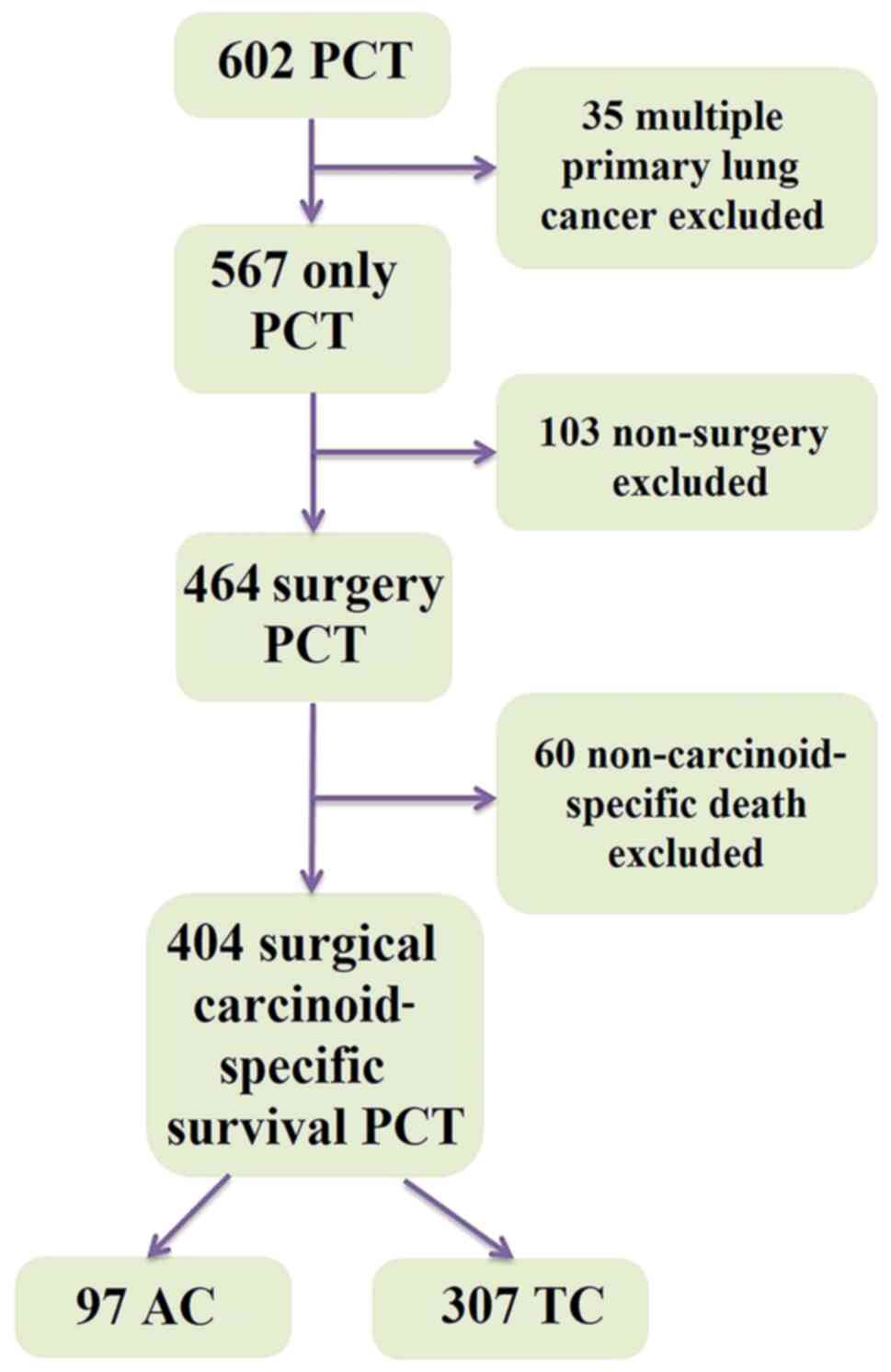
Patients with resected PCT were analyzed. All
patients underwent video-assisted thoracoscopic surgery or
thoracotomy with thoracic lymphadenectomy. Data for patients
histologically diagnosed with PCT between January 1, 1997, and
December 31, 2016, were retrieved from the lung cancer cohort,
where all primary lung cancer patients has been enrolled and
followed prospectively (4).
Patients with multiple primary lung cancer, non-surgical treatment
and non-carcinoid specific death were excluded. Eventually, 404
patients were included in the study. The selection of patients with
PCT is shown in Fig. 1.
The clinicopathological characteristics of the
patients were collected, including age, sex, BMI,
Tumor-Node-Metastasis (TNM) stage, tumor size, tumor lobe location,
smoking status, family history of cancer, surgical type and
comorbidity. Patients were restaged according to the eighth edition
of the TNM staging system of the American Joint Committee on Cancer
(5). Classification of smoking was
based on the Adult Tobacco Use Information in National Health
Interview Survey (6). Adequate
mediastinal lymphadenectomy consisted of node resection and mapping
(American Thoracic Society map) of at least three N2 groups
(7). For the purposes of the
present study, if the number of assessed N2 groups was <3, then
this was defined as inadequate mediastinal lymphadenectomy. The
diagnosis of recurrence was made with a combination of CT images,
and/or biopsy of the new suspected site of disease. The survival
data of each patient was collected through electronic medical
notes, registration database, next-of-kin reports, death
certificates, obituary documents filed in the patients' medical
records, The Mayo Clinic Institutional Tumor Registry and the
Social Security Death Index website (https://socialsecuritydeathindex-search.com).
Statistical analysis
The nominal categorical variables were analyzed by
the χ2 test. The continuous variables were reported as
the median and interquartile range, and were evaluated by the
unpaired independent sample t-test. For the carcinoid-specific
survival (CSS) analysis, the date of cancer-specific death from PCT
was the observation endpoint. For the disease-free survival (DFS)
analysis, the date of recurrence was the endpoint of observation.
Endpoints were analyzed as time-to-event data from the date of
surgery to the respective events, which were subject to censoring
at the last follow-up if no events were observed. The cumulative
survival was estimated using the Kaplan-Meier model and log-rank
test. The analysis of hazard ratios (HRs) and 95% confidence
intervals (CIs) was performed using univariate (log-rank) and
multivariate Cox proportional hazards models. Factors with a
P-value of <0.1 in the Cox univariate analysis (log-rank) were
included in the multivariate analysis. For all other statistical
analyses, P<0.05 was considered to indicate a statistically
significant difference. All statistical analyses were performed
using SAS 9.3 (SAS Institute, Inc.).
Results
Characteristics of PCT
Of the 404 patients with PCT included in the present
study, 307 (76.0%) consisted of TC and 97 (24.0%) of AC. The median
follow-up time was 89.7 months (range, 57.5-142.3 months). Patients
with AC, compared with patients with TC, were older [median age,
62.0 years (51.0-70.0 years) vs. 57.0 years (range, 47.0-67.0
years), respectively; P=0.027], with more stage IIIB-IVA cases
[IIIB-IVA, 10 (10.3%) vs. 9 (2.9%) respectively; P=0.003], more
lymph node involvement [N1-3, 40 (41.2%) vs. 52 (16.9%),
respectively; P<0.001], and more distant metastasis [M1, 7
(7.2%) vs. 7 (2.3%), respectively; P=0.021]. The AC subgroup also
underwent more extensive surgical resections than the TC subgroup
[pneumonectomy, 10 (10.3%) vs. 6 (2.0%); bi-lobectomy, 7 (7.2%) vs.
15 (4.9%); P<0.001]. The recurrence rate was higher in patients
with AC compared with that in patients with TC [38 (39.2%) vs. 19
(6.2%); P<0.001]. Baseline characteristics and surgical
treatment details are provided in Table
I.
 | Table I.Characteristics of 404 surgical
patients with pulmonary carcinoid tumors. |
Table I.
Characteristics of 404 surgical
patients with pulmonary carcinoid tumors.
| Characteristics | Atypical carcinoid
(n=97) | Typical carcinoid
(n=307) | P-value |
|---|
| Median age
(interquartile range), years | 62.0 (51.0-70.0) | 57.0 (47.0-67.0) | 0.027 |
| Sex, n (%) |
|
| 0.442 |
|
Female | 67 (69.1) | 199 (64.8) |
|
| Male | 30 (30.9) | 108 (35.2) |
|
| BMI, n (%) |
|
| 0.141 |
|
Missing | 5 (5.2) | 10 (3.2) |
|
|
Underweight | 2 (2.1) | 2 (0.7) |
|
|
Normal | 28 (28.9) | 68 (22.1) |
|
|
Overweight | 36 (37.1) | 111 (36.2) |
|
|
Obese | 26 (26.8) | 116 (37.8) |
|
| T stage, n (%) |
|
| 0.105 |
| T1 | 56 (57.7) | 216 (70.4) |
|
| T2 | 30 (30.9) | 63 (20.5) |
|
| T3 | 8 (8.2) | 17 (5.5) |
|
| T4 | 3 (3.1) | 11 (3.6) |
|
| N stage, n (%) |
|
| <0.001 |
| N0 | 57 (58.8) | 255 (83.1) |
|
| N1 | 19 (19.6) | 25 (8.1) |
|
| N2 | 20 (20.6) | 26 (8.5) |
|
| N3 | 1 (1.0) | 1 (0.3) |
|
| M stage, n (%) |
|
| 0.021 |
| M0 | 90 (92.8) | 300 (97.7) |
|
| M1 | 7 (7.2) | 7 (2.3) |
|
| TNM stage, n
(%) |
|
| 0.003 |
|
I–IIIA | 87 (89.7) | 298 (97.1) |
|
|
IIIB-IVA | 10 (10.3) | 9 (2.9) |
|
| Tumor side, n
(%) |
|
| 0.744 |
|
Left | 38 (39.2) | 126 (41.0) |
|
|
Right | 59 (60.8) | 181 (59.0) |
|
| Tumor lobe, n
(%) |
|
| 0.487 |
|
Upper | 36 (37.1) | 90 (29.3) |
|
|
Middle | 16 (16.5) | 65 (21.2) |
|
|
Lower | 43 (44.3) | 144 (46.9) |
|
| Main
bronchus | 2 (2.1) | 8 (2.6) |
|
| Smoking status, n
(%) |
|
| 0.337 |
| Never
smoker | 48 (49.5) | 162 (52.8) |
|
| Former
smoker | 32 (33.0) | 109 (35.5) |
|
| Current
smoker | 17 (17.5) | 36 (11.7) |
|
| Family history of
lung cancer, n (%) |
|
| 0.593 |
| No | 91 (93.8) | 283 (92.2) |
|
|
Yes | 6 (6.2) | 24 (7.8) |
|
| Family history of
other cancer, n (%) |
|
| 0.494 |
| No | 69 (71.1) | 207 (67.4) |
|
|
Yes | 28 (28.9) | 100 (32.6) |
|
| Surgical type, n
(%) |
|
| <0.001 |
|
Pneumonectomy | 10 (10.3) | 6 (2.0) |
|
|
Bi-lobectomy | 7 (7.2) | 15 (4.9) |
|
|
Lobectomy | 61 (62.9) | 182 (59.3) |
|
|
Sub-lobectomy | 17 (17.5) | 75 (24.4) |
|
| Sleeve
resection | 2 (2.1) | 29 (9.4) |
|
| N2 lymphadenectomy,
n (%) |
|
| 0.312 |
|
Inadequate (<3 groups) | 37 (38.1) | 100 (32.6) |
|
|
Adequate (≥3 groups) | 60 (61.9) | 207 (67.4) |
|
| Recurrence, n
(%) |
|
| <0.001 |
| No | 59 (60.8) | 288 (93.8) |
|
|
Yes | 38 (39.2) | 19 (6.2) |
|
| Comorbidity, n
(%) |
|
| 0.158 |
|
Missing | 0 (0.0) | 2 (0.7) |
|
| No | 11 (11.3) | 21 (6.8) |
|
|
Yes | 86 (88.7) | 284 (92.5) |
|
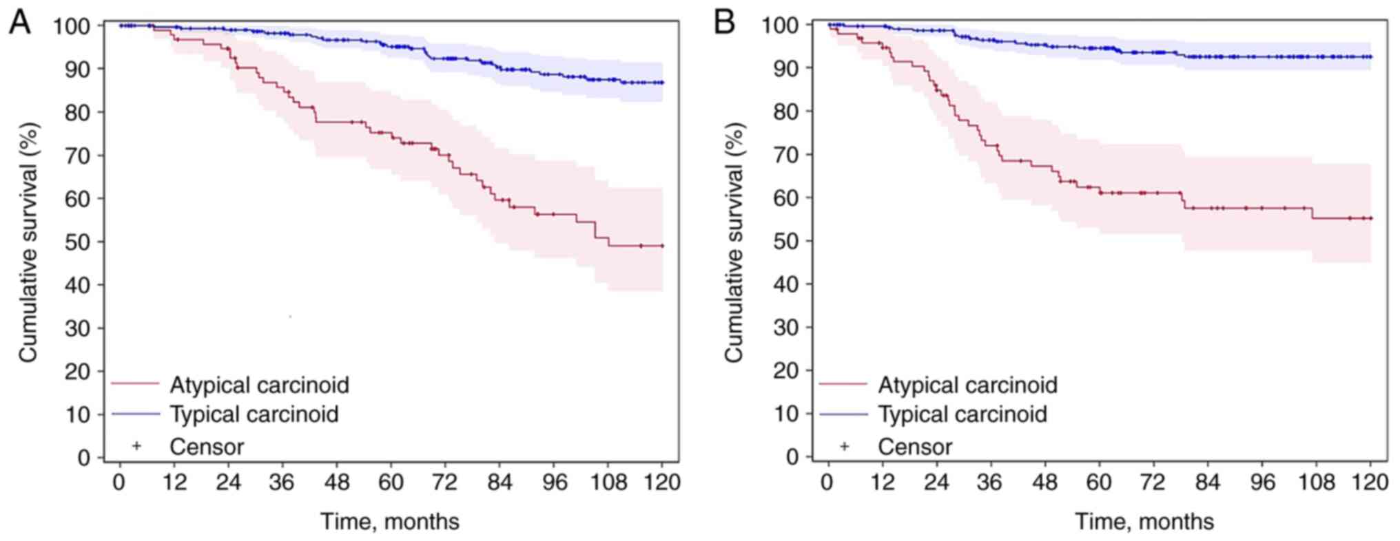
CSS analysis
The CSS of AC was inferior to that of TC; the
10-year CSS rates were 49.1 vs. 86.8% (P<0.001), respectively
(Fig. 2A). Univariate and
multivariate analyses are found in Table II. For AC, univariate analysis of
CSS revealed that age (HR, 1.03; 95% CI, 1.00-1.05; P=0.002), N1-3
stage (HR, 2.39; 95% CI, 1.34-4.26; P=0.002), M1 stage (HR, 2.59;
95% CI, 1.09-6.15; P=0.025), family history of lung cancer (HR,
0.16; 95% CI, 0.02-1.20; P=0.042), pneumonectomy/bi-lobectomy (HR,
2.18; 95% CI, 1.08-4.42; P=0.086) and inadequate N2 lymphadenectomy
(HR, 1.78; 95% CI, 0.99-3.22; P=0.049) were all P<0.10 so were
included in the multivariate analysis. For the multivariate
analysis, age (HR, 1.04; 95% CI, 1.01-1.07; P=0.001) and N1-3 stage
(HR, 2.45; 95% CI, 1.14-5.30; P=0.022) were independent risk
factors. For TC, univariate analysis of CSS determined that age
(HR, 1.06; 95% CI, 1.04-1.09; P<0.001), male sex (HR, 1.86; 95%
CI, 1.06-3.26; P=0.028), T2-4 stage (HR, 1.70; 95% CI, 0.94-3.09;
P=0.077), N1-3 stage (HR, 2.23; 95% CI, 1.15-4.35; P=0.015), M1
stage (HR, 9.99; 95% CI, 4.42-22.66; P<0.001), upper lobe
location (HR, 1.25; 95% CI, 0.68-2.30; P=0.086), cigarette smoking
[former smoker (HR, 2.13; 95% CI, 1.15-3.95); current smoker (HR,
1.86; 95% CI, 0.81-4.27); P=0.044] and inadequate N2
lymphadenectomy (HR, 3.14; 95% CI, 1.74-5.66; P<0.001) were all
P<0.10 so were included in the multivariate analysis. For the
multivariate analysis, age (HR, 1.07; 95% CI, 1.04-1.10;
P<0.001), male sex (HR, 2.03; 95% CI, 1.10-3.75; P=0.026), M1
stage (HR, 4.63; 95% CI, 1.73-12.38; P=0.005), cigarette smoking
[former smoker (HR, 2.14; 95% CI, 1.12-4.11); current smoker (HR,
2.50; 95% CI, 1.03-6.08); P=0.032] and inadequate N2
lymphadenectomy (HR, 3.45; 95% CI, 1.85-6.43; P<0.001) were
significant risk factors.
| Table II.Survival analysis of 404 patients
with pulmonary carcinoid tumors using univariate and multivariate
Cox models. |
Table II.
Survival analysis of 404 patients
with pulmonary carcinoid tumors using univariate and multivariate
Cox models.
|
| Atypical carcinoid
(n=97) | Typical carcinoid
(n=307) |
|---|
|
|
|
|
|---|
|
| Univariate | Multivariate | Univariate | Multivariate |
|---|
|
|
|
|
|
|
|---|
| Factors | HR (95% CI) | P-value | HR (95% CI) | P-value | HR (95% CI) | P-value | HR (95% CI) | P-value |
|---|
| Age | 1.03 | 0.002 | 1.04 | 0.001 | 1.06 | <0.001 | 1.07 | <0.001 |
|
| (1.00-1.05) |
| (1.01-1.07) |
| (1.04-1.09) |
| (1.04-1.10) |
|
| Sex |
| 0.946 |
|
|
| 0.028 |
| 0.026 |
|
Female | - |
|
|
| - |
| - |
|
|
Male | 0.98 |
|
|
| 1.86 |
| 2.03 |
|
|
| (0.51-1.86) |
|
|
| (1.06-3.26) |
| (1.10-3.75) |
|
| BMIa |
| 0.419 |
|
|
| 0.353 |
|
|
|
Underweight/normal | - |
|
|
| - |
|
|
|
|
Overweight/ | 0.68 |
|
|
| 0.73 |
|
|
|
|
obese | (0.37-1.25) |
|
|
| (0.38-1.42) |
|
|
|
| T stage |
| 0.159 |
| 0.483 |
| 0.077 |
| 0.154 |
| T1 | - |
| - |
| - |
| - |
|
|
T2-4 | 1.51 |
| 1.33 |
| 1.70 |
| 1.62 |
|
|
| (0.85-2.67) |
| (0.60-2.91) |
| (0.94-3.09) |
| (0.85-3.11) |
|
| N stage |
| 0.002 |
| 0.022 |
| 0.015 |
| 0.092 |
| N0 | - |
| - |
| - |
| - |
|
|
N1-3 | 2.39 |
| 2.45 |
| 2.23 |
| 2.06 |
|
|
| (1.34-4.26) |
| (1.14-5.30) |
| (1.15-4.35) |
| (0.92-4.60) |
|
| M stage |
| 0.025 |
| 0.604 |
| <0.001 |
| 0.005 |
| M0 | - |
| - |
| - |
| - |
|
| M1 | 2.59 |
| 1.33 |
| 9.99 |
| 4.63 |
|
|
| (1.09-6.15) |
| (0.46-3.82) |
| (4.42-22.66) |
| (1.73-12.38) |
|
| Tumor side |
| 0.852 |
|
|
| 0.167 |
|
|
|
Left | 0.95 |
|
|
| 1.48 |
|
|
|
|
| (0.52-1.70) |
|
|
| (0.84-2.60) |
|
|
|
|
Right | - |
|
|
| - |
|
|
|
| Tumor lobe |
| 0.506 |
|
|
| 0.086 |
| 0.062 |
|
Upper | 0.69 |
|
|
| 1.25 |
| 2.13 |
|
|
| (0.36-1.30) |
|
|
| (0.68-2.30) |
| (1.10-4.13) |
|
|
Middle/main | 0.78 |
|
|
| 0.42 |
| 0.80 |
|
|
bronchus | (0.34-1.76) |
|
|
| (0.16-1.10) |
| (0.28-2.24) |
|
|
Lower | - |
|
|
| - |
| - |
|
| Smoking status |
| 0.740 |
|
|
| 0.044 |
| 0.032 |
| Never
smoker | - |
|
|
| - |
| - |
|
| Former
smoker | 1.27 |
|
|
| 2.13 |
| 2.14 |
|
|
| (0.67-2.42) |
|
|
| (1.15-3.95) |
| (1.12-4.11) |
|
| Current
smoker | 1.22 |
|
|
| 1.86 |
| 2.50 |
|
|
| (0.56-2.67) |
|
|
| (0.81-4.27) |
| (1.03-6.08) |
|
| Family history of
lung cancer |
| 0.042 |
| 0.065 |
| 0.921 |
|
|
| No | - |
| - |
| - |
|
|
|
|
Yes | 0.16 |
| 0.21 |
| 1.05 |
|
|
|
|
| (0.02-1.20) |
| (0.03-1.66) |
| (0.42-2.65) |
|
|
|
| Family history of
other cancer |
| 0.756 |
|
|
| 0.314 |
|
|
| No | - |
|
|
| - |
|
|
|
|
Yes | 1.10 |
|
|
| 0.74 |
|
|
|
|
| (0.60-2.00) |
|
|
| (0.41-1.33) |
|
|
|
| Surgery type |
| 0.086 |
| 0.148 |
| 0.151 |
|
|
|
Pneumo/bi- | 2.18 |
| 2.15 |
| 2.01 |
|
|
|
|
lobectomy | (1.08-4.42) |
| (0.88-5.27) |
| (0.77-5.25) |
|
|
|
|
Lobectomy/sleeve | - |
| - |
| - |
|
|
|
|
Sub-lobectomy | 1.20 |
| 1.50 |
| 1.64 |
|
|
|
|
| (0.56-2.55) |
| (0.61-3.71) |
| (0.88-3.04) |
|
|
|
| N2
Lymphadenectomy |
| 0.049 |
| 0.335 |
| <0.001 |
| <0.001 |
|
Inadequate | 1.78 |
| 1.44 |
| 3.14 |
| 3.45 |
|
|
| (0.99-3.22) |
| (0.69-3.00) |
| (1.74-5.66) |
| (1.85-6.43) |
|
|
Adequate | - |
| - |
| - |
| - |
|
| Comorbidity |
| 0.925 |
|
|
| 0.436 |
|
|
| No | 1.04 |
|
|
| 0.57 |
|
|
|
|
| (0.44-2.47) |
|
|
| (0.14-2.37) |
|
|
|
|
Yes | - |
|
|
| - |
|
|
|
Tumor recurrence analysis
The DFS of AC was inferior to that of TC; the
10-year DFS rates were 55.2 vs. 92.6% (P<0.001), respectively
(Fig. 2B). Univariate and
multivariate analyses are found in Table III. For AC, the univariate
analysis revealed that N1-3 stage (HR, 3.61; 95% CI, 1.86-7.01;
P<0.001), M1 stage (HR, 2.51; 95% CI, 0.97-6.46; P=0.049),
pneumonectomy/bi-lobectomy (HR, 2.42; 95% CI, 1.15-5.12; P=0.030)
and inadequate N2 lymphadenectomy (HR, 1.95; 95% CI, 1.02-3.73;
P=0.039) were all P<0.10 so were included in the multivariate
analysis. For the multivariate analysis, N1-3 stage (HR, 2.62; 95%
CI, 1.16-5.95; P=0.018) and inadequate N2 lymphadenectomy (HR,
2.13; 95% CI, 1.04-4.39; P=0.041) were independent risk factors for
recurrence. For TC, the univariate analysis showed that male sex
(HR, 4.19; 95% CI, 1.59-11.03; P=0.002), overweight/obese BMI (HR,
0.45; 95% CI, 0.17-1.16; P=0.090), N1-3 stage (HR, 2.42; 95% CI,
0.92-6.36; P=0.065) and M1 stage (HR, 31.22; 95% CI, 10. 95–88.99;
P<0.001) were all P<0.10 so were included in the multivariate
analysis. For the multivariate analysis, male sex (HR, 3.72; 95%
CI, 1.33-10.42; P=0.010) and M1 stage (HR, 14.93; 95% CI,
4.77-46.77; P<0.001) were independent risk factors for
recurrence.
| Table III.Recurrence analysis of 404 patients
with pulmonary carcinoid tumors using univariate and multivariate
Cox models. |
Table III.
Recurrence analysis of 404 patients
with pulmonary carcinoid tumors using univariate and multivariate
Cox models.
|
| Atypical carcinoid
(n=97) | Typical carcinoid
(n=307) |
|---|
|
|
|
|
|---|
|
| Univariate | Multivariate | Univariate | Multivariate |
|---|
|
|
|
|
|
|
|---|
| Factors | HR (95% CI) | P-value | HR (95% CI) | P-value | HR (95% CI) | P-value | HR (95% CI) | P-value |
|---|
| Age | 1.01 | 0.173 |
|
| 1.02 | 0.292 |
|
|
|
| (0.98-1.03) |
|
|
| (0.98-1.05) |
|
|
|
| Sex |
| 0.842 |
|
|
| 0.002 |
| 0.010 |
|
Female | - |
|
|
| - |
| - |
|
|
Male | 0.93 |
|
|
| 4.19 |
| 3.72 |
|
|
| (0.46-1.88) |
|
|
| (1.59-11.03) |
| (1.33-10.42) |
|
| BMIa |
| 0.116 |
|
|
| 0.090 |
| 0.163 |
|
Underweight/normal | - |
|
|
| - |
| - |
|
|
Overweight/ | 0.56 |
|
|
| 0.45 |
| 0.47 |
|
|
obese | (0.28-1.10) |
|
|
| (0.17-1.16) |
| (0.17-1.30) |
|
| T stage |
| 0.422 |
| 0.661 |
| 0.414 |
| 0.663 |
| T1 | - |
| - |
| - |
| - |
|
|
T2-4 | 1.30 |
| 0.83 |
| 1.47 |
| 1.26 |
|
|
| (0.68-2.46) |
| (0.37-1.89) |
| (0.58-3.74) |
| (0.45-3.53) |
|
| N stage |
| <0.001 |
| 0.018 |
| 0.065 |
| 0.167 |
| N0 | - |
| - |
| - |
| - |
|
|
N1-3 | 3.61 |
| 2.62 |
| 2.42 |
| 2.14 |
|
|
| (1.86-7.01) |
| (1.16-5.95) |
| (0.92-6.36) |
| (0.76-5.99) |
|
| M stage |
| 0.049 |
| 0.275 |
| <0.001 |
| <0.001 |
| M0 | - |
| - |
| - |
| - |
|
| M1 | 2.51 |
| 1.91 |
| 31.22 |
| 14.93 |
|
|
| (0.97-6.46) |
| (0.63-5.85) |
| (10.95-88.99) |
| (4.77-46.77) |
|
| Tumor side |
| 0.726 |
|
|
| 0.583 |
|
|
|
Left | 1.12 |
|
|
| 14.93 |
|
|
|
|
| (0.59-2.14) |
|
|
| (0.52-3.16) |
|
|
|
|
Right | - |
|
|
| - |
|
|
|
| Tumor lobe |
| 0.198 |
|
|
| 0.618 |
|
|
|
Upper | 0.51 |
|
|
| 0.74 |
|
|
|
|
| (0.24-1.07) |
|
|
| (0.26-2.13) |
|
|
|
|
Middle/main | 0.78 |
|
|
| 0.55 |
|
|
|
|
bronchus | (0.33-1.84) |
|
|
| (0.15-1.98) |
|
|
|
|
Lower | - |
|
|
| - |
|
|
|
| Smoking status |
| 0.809 |
|
|
| 0.369 |
|
|
| Never
smoker | - |
|
|
| - |
|
|
|
| Former
smoker | 1.07 |
|
|
| 1.95 |
|
|
|
|
| (0.53-2.20) |
|
|
| (0.72-5.23) |
|
|
|
| Current
smoker | 1.34 |
|
|
| 1.90 |
|
|
|
|
| (0.56-3.21) |
|
|
| (0.49-7.34) |
|
|
|
| Family history of
lung cancer |
| 0.459 |
|
|
| 0.780 |
|
|
| No | - |
|
|
| - |
|
|
|
|
Yes | 0.59 |
|
|
| 1.23 |
|
|
|
|
| (0.14-2.44) |
|
|
| (0.28-5.33) |
|
|
|
| Family history of
other cancer |
| 0.631 |
|
|
| 0.934 |
|
|
| No | - |
|
|
| - |
|
|
|
|
Yes | 1.18 |
|
|
| 1.04 |
|
|
|
|
| (0.60-2.31) |
|
|
| (0.41-2.65) |
|
|
|
| Surgery type |
| 0.030 |
| 0.355 |
| 0.295 |
|
|
|
Pneumo/bi- | 2.42 |
| 1.88 |
| 1.46 |
|
|
|
|
lobectomy | (1.15-5.12) |
| (0.80-4.41) |
| (0.33-6.37) |
|
|
|
|
Lobectomy/sleeve | - |
| - |
| - |
|
|
|
|
Sub-lobectomy | 0.77 |
| 0.83 |
| 0.37 |
|
|
|
|
| (0.29-2.03) |
| (0.28-2.49) |
| (0.08-1.60) |
|
|
|
| N2
Lymphadenectomy |
| 0.039 |
| 0.041 |
| 0.002 |
| 0.138 |
|
Inadequate | 1.95 |
| 2.13 |
| 2.13 |
| 2.12 |
|
|
| (1.02-3.73) |
| (1.04-4.39) |
| (1.54-9.98) |
| (0.78-5.79) |
|
|
Adequate | - |
| - |
| - |
| - |
|
| Comorbidity |
| 0.485 |
|
|
| 0.807 |
|
|
| No | 1.40 |
|
|
| 0.78 |
|
|
|
|
| (0.54-3.58) |
|
|
| (0.10-5.83) |
|
|
|
|
Yes | - |
|
|
| - |
|
|
|
Discussion
This was a retrospective study, with the objective
to evaluate additional risk factors associated with the survival of
patients with resected PCT. To the best of our knowledge, these
factors are not entirely clarified and are still a matter for
discussion in multidisciplinary tumor boards. Using the HSR Study
Data Management System, the patterns of these tumors were analyzed,
which typically required a long-term follow-up to verify potential
effects on the therapeutic strategies employed and the prognostic
factors identified. It was found that the 10-year CSS and DFS rates
for patients with TC were 86.8 and 92.6%, respectively. In patients
with AC, however, the 10-year CSS and DFS rates were 49.1 and
55.2%, respectively. These findings corroborate the well-known body
of evidence in the literature showing that AC carries a worse
prognosis compared with TC (1,2).
In the present study, it was also found that age and
lymph node involvement were associated with inferior CSS in AC,
while age, male sex, distant metastasis, cigarette smoking and
inadequate mediastinal lymphadenectomy were associated with worse
CSS in TC. Regarding recurrence, lymph node involvement and
inadequate mediastinal lymphadenectomy were associated with
inferior DFS in AC, while male sex and distant metastasis were
associated with inferior DFS in TC. The identified prognostic
factors of the present study are similar to those of previous
reports in the literature. The average age at the time of diagnosis
is ~45 and 55 years for patients with TC and AC, respectively
(8,9). Age (<45 years) is an important
prognostic factor associated with a good prognosis in PCT (10). Moreover, some studies report a
higher prevalence of smoking in patients with AC compared to TC. As
previously published, most patients with TC are female and are
never-smokers (11,12). Furthermore, it has been reported
that the prognosis of female patients with PCT is worse than that
of male patients (13).
TC is an indolent malignancy and excellent long-term
outcomes can be achieved. The 10-year CSS of 86.8% observed in the
present study is very similar to the results previously reported by
other groups (14–16). In the present study, the median age
of diagnosis was 57.0 years, and the majority of the patients
presented with T1 (70.4%) and N0 (83.1%) stage disease. In the
cohort, a 16.9% incidence of nodal involvement (8.8% N2-N3) was
found, which is very similar to the findings reported by Kneuertz
et al (17), where ≥10 lymph
nodes were evaluated. The impact of nodal involvement in TC remains
controversial and, in the present study, it was demonstrated that
nodal involvement was an independent prognostic factor for TC
prognosis. However, other studies have questioned the prognostic
significance of nodal involvement in PCT (18,19).
Martini et al (14) found no
survival difference in patients with N1 or N2 disease. Kneuertz
et al (17) found nodal
involvement to be associated with worse overall survival only in
tumors >2 cm. In a study by Lou et al (16), only 2% of patients with TC without
nodal disease had recurrence, while there was increased recurrence
in positive nodal disease following surgery. Regarding the surgical
approach, the most common type of procedure in patients with TC in
the present study was lobectomy (59.3%), followed by sublobectomy
(24.4%). Despite a previous study demonstrating that sublobar
resection does not seem to compromise oncological outcomes compared
with lobar resection, assuming complete resection and adequate
mediastinal staging is performed (15), no significant difference in the
prognosis of patients with TC who underwent different lung
resections (P>0.05) was found in the present study, which may be
due to the small sample size. Mediastinal lymphadenectomy (≥3
groups) was performed in 67.4% of the patients with TC and was
associated with a better prognosis compared with inadequate
mediastinal lymphadenectomy. Given that lymphadenectomy has not
been a routine practice in patients with TC, most previous studies
did not address the role of lymphadenectomy in this subgroup of
patients. Based on these results, it can be suggested that a
complete lymphadenectomy should always be performed in order
determine the staging and improve the prognosis of patients with
TC.
In two small previous retrospective analyses of TC
and AC, the 10-year survival rates for AC ranged from 59–69%, which
were higher than the CSS rate of 49.1% reported in the present
study, likely owing to the differences in sample size and the
characteristics of the patients enrolled (20,21).
The sample size was larger and there were more patients with lymph
node involvement in the present study, which was also a factor
affecting prognosis. In another larger study (n=507) utilizing the
Surveillance, Epidemiology and End Results database, patients with
AC had a 10-year survival rate of 59% after surgery, which may have
been influenced by a lower proportion of nodal involvement (N1-3,
33.5%) compared with the nodal status of the cohort in the present
study (N1-3, 41.2%) (22). Lymph
node metastases have been shown to be associated with an inferior
survival rate in patients with AC (19). In a recent national study of
surgically managed AC (n=816), no significant difference in
mortality was observed between lobectomy and sublobectomy (23). Similar to TC, it can be suggested
that the extent of lymphadenectomy in AC seems to be more important
than the extent of lung resection.
In a study examining disease recurrence in patients
with TC and AC following surgery, there did not appear to be a
significant difference in the recurrence rate of AC with positive
nodes versus negative nodes (16).
This is in contrast to the findings in the present study, which may
be attributed to the inclusion of patients with higher nodal
status, as well as data regarding inadequate N2 lymphadenectomy,
which was shown to increase recurrence risk. In a retrospective
Spanish study, a univariate analysis found that sublobar resections
in AC were associated with increased rates of recurrence compared
with lobar resections (24). These
observed findings, in conjunction with increased recurrence risk
with inadequate lymphadenectomy, reinforce the suggestion that
mediastinal staging and mediastinal dissection are needed to
improve the prognosis of patients with PCT.
Limitations of the present study largely derive from
its retrospective nature. Additionally, being a tertiary referral
center could result in more patients who are medically complex
and/or with advanced stage PCT. Additional prospective studies are
necessary to validate the findings as well as to determine the
appropriate surgical approach for these patients. Furthermore,
recurrence type may also be associated with prognosis, which is not
the subject of the present study, but is of note. This will be
investigated further in later work.
In conclusion, in the present study, patients with
AC tumors had significantly worse CSS and DFS rates compared with
patients with TC. The degree of nodal involvement in AC was a
prognostic marker, in contrast to that in TC. Inadequate
lymphadenectomy increased the risk of recurrence in AC and
mortality in TC. However, the type of surgery did not have a
significant impact. The present study emphasizes the importance of
mediastinal nodal dissection in patients with PCT.
Acknowledgements
Not applicable.
Funding
This study was supported by grants R01 CA80127 and R01 CA84354
from the National Institutes of Health and the Mayo Clinic
Foundation.
Availability of data and materials
The datasets used and/or analyzed during the current
study are available from the corresponding author on reasonable
request.
Authors' contributions
LD, VE, AL, SES and DEJ designed the study. SDC,
SEB, DW, YHL, JAW and PARS performed the acquisition of data, and
DS, KRS and PY performed analysis and interpretation of data. LD,
VE, AL, SES and DEJ prepared and wrote the original draft of the
manuscript. SDC, SEB, DW, YHL, JAW and PARS reviewed and edited the
manuscript. All authors read and approved the final version of the
manuscript. DS, KRS and PY confirm the authenticity of all the raw
data.
Ethics approval and consent to
participate
Not applicable.
Patient consent for publication
Not applicable.
Competing interests
The authors declare that they have no competing
interests.
References
|
1
|
Rekhtman N: Neuroendocrine tumors of the
lung: An update. Arch Pathol Lab Med. 134:1628–1638. 2010.
View Article : Google Scholar : PubMed/NCBI
|
|
2
|
Yao JC, Hassan M, Phan A, Dagohoy C, Leary
C, Mares JE, Abdalla EK, Fleming JB, Vauthey JN, Rashid A and Evans
DB: One hundred years after ‘carcinoid’: epidemiology of and
prognostic factors for neuroendocrine tumors in 35,825 cases in the
United States. J Clin Oncol. 26:3063–3072. 2008. View Article : Google Scholar : PubMed/NCBI
|
|
3
|
Travis WD, Brambilla E, Nicholson AG,
Yatabe Y, Austin JHM, Beasley MB, Chirieac LR, Dacic S, Duhig E,
Flieder DB, et al: The 2015 World Health Organization
classification of lung tumors: Impact of genetic, clinical and
radiologic advances since the 2004 classification. J Thorac Oncol.
10:1243–1260. 2015. View Article : Google Scholar : PubMed/NCBI
|
|
4
|
Yang P, Allen MS, Aubry MC, Wampfler JA,
Marks RS, Edell ES, Thibodeau S, Adjei AA, Jett J and Deschamps C:
Clinical features of 5,628 primary lung cancer patients: Experience
at Mayo Clinic from 1997 to 2003. Chest. 128:452–462. 2005.
View Article : Google Scholar : PubMed/NCBI
|
|
5
|
Ettinger DS, Wood DE, Aisner DL, Akerley
W, Bauman J, Chirieac LR, D'Amico TA, DeCamp MM, Dilling TJ,
Dobelbower M, et al: Non-small cell lung cancer, version 5.2017,
NCCN clinical practice guidelines in oncology. J Natl Compr Canc
Netw. 15:504–535. 2017. View Article : Google Scholar : PubMed/NCBI
|
|
6
|
U.S. Department of Health and Human
Services, . The health consequences of smoking-50 years of
progress: A report of the surgeon general. Atlanta, GA: U.S.
Department of Health and Human Services, Centers for Disease
Control and Prevention, National Center for Chronic Disease
Prevention and Health Promotion, Office on Smoking and Health;
2014
|
|
7
|
Amin MB, Greene FL, Edge SB, Byrd DR,
Brookland RK, Washington MK, Gershenwald JE, Compton CC, Hess KR,
Sullivan DC, et al: AJCC staging manual. 8th edition. Springer
International Publishing; New York, NY: pp. 1–1024. 2017
|
|
8
|
Cao C, Yan TD, Kennedy C, Hendel N, Bannon
PG and McCaughan BC: Bronchopulmonary carcinoid tumors: Long-term
outcomes after resection. Ann Thorac Surg. 91:339–343. 2011.
View Article : Google Scholar : PubMed/NCBI
|
|
9
|
Skuladottir H, Hirsch FR, Hansen HH and
Olsen JH: Pulmonary neuroendocrine tumors: Incidence and prognosis
of histological subtypes. A population-based study in Denmark. Lung
Cancer. 37:127–135. 2002. View Article : Google Scholar : PubMed/NCBI
|
|
10
|
Stolz A, Harustiak T, Simonek J, Schutzner
J, Polanecky O, Burkert J and Lischke R: Long-term outcomes and
prognostic factors of patients with pulmonary carcinoid tumors.
Neoplasma. 62:478–483. 2015. View Article : Google Scholar : PubMed/NCBI
|
|
11
|
Froudarakis M, Fournel P, Burgard G,
Bouros D, Boucheron S, Siafakas NM and Emonot A: Bronchial
carcinoids. A review of 22 cases. Oncology. 53:153–158. 1996.
View Article : Google Scholar : PubMed/NCBI
|
|
12
|
Fink G, Krelbaum T, Yellin A, Bendayan D,
Saute M, Glazer M and Kramer MR: Pulmonary carcinoid: Presentation,
diagnosis, and outcome in 142 cases in Israel and review of 640
cases from the literature. Chest. 119:1647–1651. 2001. View Article : Google Scholar : PubMed/NCBI
|
|
13
|
Beasley MB, Thunnissen FB, Brambilla E,
Hasleton P, Steele R, Hammar SP, Colby TV, Sheppard M, Shimosato Y,
Koss MN, et al: Pulmonary atypical carcinoid: Predictors of
survival in 106 cases. Hum Pathol. 31:1255–1265. 2000. View Article : Google Scholar : PubMed/NCBI
|
|
14
|
Martini N, Zaman MB, Bains MS, Burt ME,
McCormack PM, Rusch VW and Ginsberg RJ: Treatment and prognosis in
bronchial carcinoids involving regional lymph nodes. J Thorac
Cardiovasc Surg. 107:1–7. 1994. View Article : Google Scholar : PubMed/NCBI
|
|
15
|
Yendamuri S, Gold D, Jayaprakash V, Dexter
E, Nwogu C and Demmy T: Is sublobar resection sufficient for
carcinoid tumors? Ann Thorac Surg. 92:1774–1779. 2011. View Article : Google Scholar : PubMed/NCBI
|
|
16
|
Lou F, Sarkaria I, Pietanza C, Travis W,
Roh MS, Sica G, Healy D, Rusch V and Huang J: Recurrence of
pulmonary carcinoid tumors after resection: Implications for
postoperative surveillance. Ann Thorac Surg. 96:1156–1162. 2013.
View Article : Google Scholar : PubMed/NCBI
|
|
17
|
Kneuertz PJ, Kamel MK, Stiles BM, Lee BE,
Rahouma M, Harrison SW, Altorki NK and Port JL: Incidence and
prognostic significance of carcinoid lymph node metastases. Ann
Thorac Surg. 106:981–988. 2018. View Article : Google Scholar : PubMed/NCBI
|
|
18
|
Johnson R, Trocha S, McLawhorn M, Worley
M, Wheeler G, Thompson L, Schisler N, Schammel D, Schammel C,
Stephenson J and Bolton W: Histology, not lymph node involvement,
predicts long-term survival in bronchopulmonary carcinoids. Am
Surg. 77:1669–1674. 2011. View Article : Google Scholar : PubMed/NCBI
|
|
19
|
Cardillo G, Sera F, Di Martino M, Graziano
P, Giunti R, Carbone L, Facciolo F and Martelli M: Bronchial
carcinoid tumors: Nodal status and long-term survival after
resection. Ann Thorac Surg. 77:1781–1785. 2004. View Article : Google Scholar : PubMed/NCBI
|
|
20
|
Okereke IC, Taber AM, Griffith RC and Ng
TT: Outcomes after surgical resection of pulmonary carcinoid
tumors. J Cardiothorac Surg. 11:352016. View Article : Google Scholar : PubMed/NCBI
|
|
21
|
Ramirez RA, Beyer DT, Diebold AE, Voros
BA, Chester MM, Wang YZ, Boudreaux JP, Woltering EA, Uhlhorn AP,
Ryan P, et al: Prognostic factors in typical and atypical pulmonary
carcinoids. Ochsner J. 17:335–340. 2017.PubMed/NCBI
|
|
22
|
Chen X, Pang Z, Wang Y, Bie F, Zeng Y,
Wang G and Du J: The role of surgery for atypical bronchopulmonary
carcinoid tumor: Development and validation of a model based on
surveillance, epidemiology, and end results (SEER) database. Lung
Cancer. 139:94–102. 2020. View Article : Google Scholar : PubMed/NCBI
|
|
23
|
Walters SL, Canavan ME, Salazar MC, Resio
BJ, Blasberg JD, Mase V and Boffa DJ: A national study of
surgically managed atypical pulmonary carcinoid tumors. Ann Thorac
Surg. 112:921–927. 2021. View Article : Google Scholar : PubMed/NCBI
|
|
24
|
Canizares MA, Matilla JM, Cueto A, Algar
J, Muguruza I, Moreno-Mata N, Moreno-Balsalobre R, Guijarro R,
Arrabal R, Garcia-Fontan E, et al: Atypical carcinoid tumours of
the lung: Prognostic factors and patterns of recurrence. Thorax.
69:648–653. 2014. View Article : Google Scholar : PubMed/NCBI
|