As with phosphorylation, ubiquitination is a highly
reversible post-translational modification that binds ubiquitin
(Ub) proteins to target proteins, modifying their activity and
stability. This is also essential for the editing and recycling of
Ub (1). The critical members
involved in the ubiquitination pathway are Ub-activating (E1s) and
-conjugating enzymes (E2s) and Ub ligases (E3s), which use the
energy supplied by ATP hydrolysis to establish a multi-step
cascade, resulting in Ub binding to the substrate. By contrast, as
anti-ubiquitinase enzymes, deubiquitinases (DUBs) regulate the
target protein degradation and cleave its attached
polyubiquitination chain, thereby regulating target protein
stability (2). The
ubiquitin-proteasome system consists of ubiquitinase enzymes, DUBs
and the 26S proteasome complex, which has a key role in regulating
protein degradation (3). To date,
~100 DUBs have been classified into five families: Ubiquitin
C-terminal hydrolases (UCHs), ovarian tumor-related,
ubiquitin-specific and Machado-Josephin domain proteases and
Jab1/MPN domain-associated metallopeptidases. The UCH family
consists of four members, UCH-L1, UCH-L3, UCH37 and BAP1 (4).
UCH-L1, a protein predominantly expressed in the
brain, has been demonstrated to serve a critical role in
neurodegenerative diseases, such as Parkinson's and Alzheimer's
disease (5–8). There have been some reviews that
discuss UCH-L1 in the brain (9–12), but
to the best of our knowledge, there are few that discuss UCH-L1 in
cancer (13–15). New studies suggest that UCH-L1 has a
role in cancer (16–20). However, the effect of UCH-L1 on
cancer is debatable. Certain research suggests that decreased
UCH-L1 expression is associated with malignancies (21–28).
Due to promoter methylation, UCH-L1 expression is silenced
or decreased, resulting in the progression of numerous types of
cancer, including esophageal (21),
gastric (22), ovarian and
(23), renal cancer (24), head and neck squamous cell (20) and hepatocellular carcinoma (HCC)
(25), breast cancer (26), pancreatic neuroendocrine tumor
(PNET) (27) and nasopharyngeal
carcinoma (NPC) (28). By contrast,
UCH-L1 is considered to be an oncogenic factor promoting the
occurrence, invasion and metastasis of breast (29) and non-small cell lung cancer (NSCLC)
(30), lymphoma (31), parathyroid carcinoma (32,33),
cutaneous squamous cell cancer (34), osteosarcoma (35), uterine serous carcinoma (36) and neuroblastoma (37). However, the specific mechanism of
UCH-L1 in these types of cancer remains unclear.
The molecular structure and function of UCH-L1 are
discussed and summarized in the present review, focusing on its
role in carcinogenesis. Although more evidence is needed to support
UCH-L1 as a marker and therapeutic target for various types
of cancer, the present review details necessary future research to
understand the role of UCH-L1 in cancer. UCH-L1 substrate analysis
will significantly help the research and development of associated
drugs (38).
Although specific molecular functions of UCH-L1 are
not well understood, a number of studies have found that UCH-L1
participates in regulating free Ub pools, lysosomal activity,
signaling molecules and cytoskeletal dynamics (46,47).
UCH-L1 dysregulation contributes to various diseases, including
cancer (48).
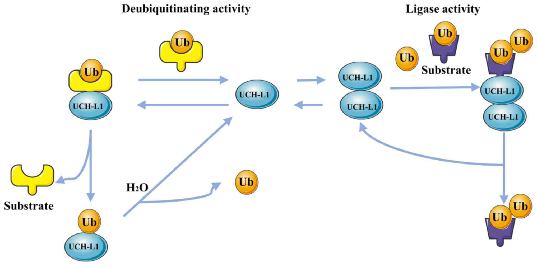
As a member of the UCH family of deubiquitinating
enzymes, UCH-L1 exhibits low-activity hydrolase action that
primarily hydrolyzes Ub chains of small polymeric or unfolded
proteins (42,49). For example, its deubiquitinating
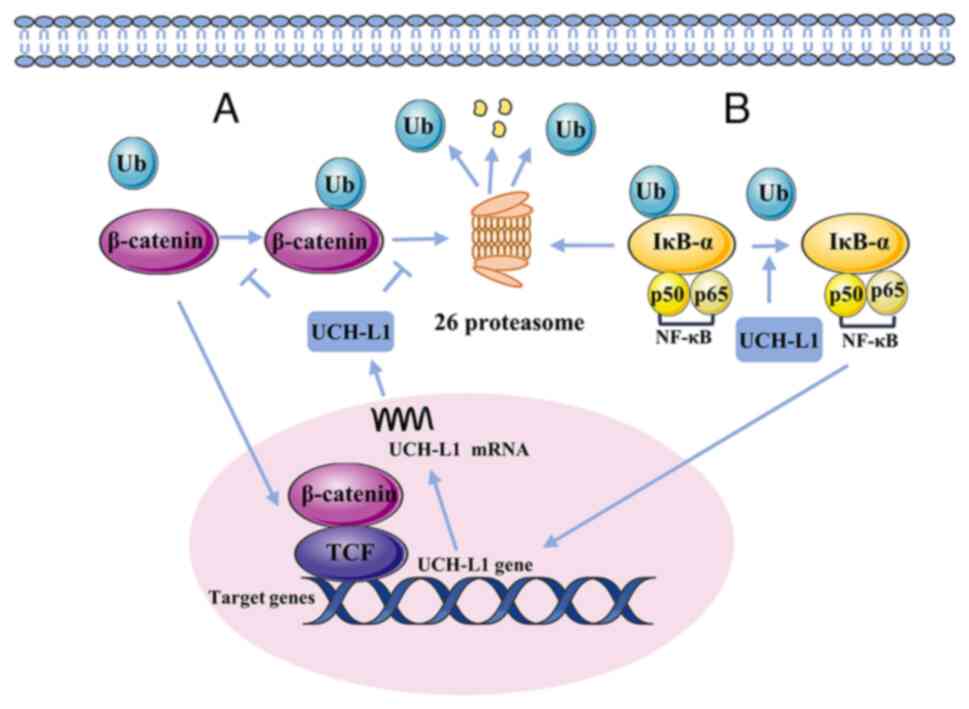
activity controls expression of β-catenin (Fig. 2), which activates the β-catenin/T
cell factor (TCF) pathway and enhances expression of related genes,
including c-myc and c-jun (50).
The β-catenin/TCF signaling pathway promotes UCH-L1
expression (51). Furthermore,
UCH-L1 maintains IκB-α in vascular cells while decreasing
TNF-α-induced NF-κB activation (Fig.
2) (52,53). Thus, UCH-L1 may decrease expression
of NF-κB-driven cytokines, such as inflammatory cytokines or
adhesion molecules. Recently, UCH-L1 has been reported to be
involved in activating the TGF-β/SMAD signaling pathway (54). UCH-L1 has Ub ligase activity in
addition to deubiquitinase activity (55). A recent study suggested that UCH-L1
serves as a Ub ligase enzyme, mediating cortactin (CCTN)
degradation by increasing ubiquitination of K48-linked CCTN
(28). The upregulation of CCTN is
associated with the progression of tumors (56). UCH-L1 can also stabilize the
expression of Ub monomers in vivo (42). UCH-L1, depending on its
ubiquitination enzyme activity, acts on the target protein and
produces free Ub monomers, which participate in the Ub metabolism
of target proteins. These Ub monomers also prevent degradation by
binding to nearby UCH-L1. Furthermore, UCH-L1 stabilization of Ub
monomers is unaffected by its deubiquitination activity (57).
UCH-L1 is associated with skeletal muscle cell
proliferation, sperm formation, angiogenesis and numerous other
biological processes (13). For
example, cell mitosis is inhibited by overexpression of
UCH-L1, resulting in decreased cell proliferation (58). In addition, the C-terminus of UCH-L1
promotes phosphorylation of Akt, which facilitates survival and
metabolic activity of malignant B cells (59). τ protein is the most abundant
microtubule-associated protein in neurons, participating in
regulation of synaptic plasticity, axonal transport and neuronal
survival by promoting microtubule assembly and stabilizing
microtubule networks (60).
However, UCH-L1 inhibition reduces the enzyme activity of
histone deacetylase 6 by reducing the production of K63-linked
ubiquitin chain, leading to abnormal accumulation of τ protein, and
finally affecting the brain (61,62).
Furthermore, UCH-L1 may strengthen the ubiquitination and
degradation of tubulin, arrest proliferation of cells and further
inhibit microtubule formation (63). In addition, UCH-L1 increases the
ubiquitination and degradation of microphthalmia-related
transcription factor (MITF) by binding to ubiquitinated MITF
(64).
Studies have shown that UCH-L1, which also possesses
an antitumor effect, is often deleted or silenced due to promoter
methylation in various types of tumor tissue, such as esophageal
(21) and gastric cancer (22,70),
renal cell carcinoma (24,71), prostate cancer (72), primary head and neck squamous cell
carcinoma (20) and ovarian
(73) and colorectal cancer
(74), resulting in adverse
clinical outcomes. However, the tumor suppressor mechanism of
UCH-L1 remains unclear.
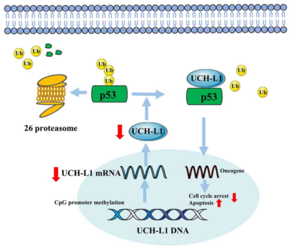
UCH-L1 also blocks the proliferation and induces
apoptosis in breast cancer cells (26,80).
When the expression level of UCH-L1 increases, UCH-L1 further
activates and stabilizes p53 signaling by inhibiting the
degradation of p53, leading to cell cycle arrest at G0/G1 and
apoptosis (Fig. 4) (26). UCH-L1 also induced apoptosis in
breast cancer cells via the PI3K/Akt signaling pathway (80). As a tumor suppressor, UCH-L1
restrains the proliferation of tumor cells, but the silencing or
deletion of UCH-L1 expression caused by promoter methylation
reverses this tumor inhibition. A recent study proposed that for
cancer caused by abnormal methylation of UCH-L1, the
construction of CRISPR-Cas9-based vectors and targeted methylation
of UCH-L1 may regulate expression of UCH-L1 to a basal
level, but this needs to be proven in future research (81).
Breast cancer cell metastasis is the primary cause
of mortality in most patients with breast cancer (83). The high invasiveness of breast
cancer is regulated by abnormal expression of UCH-L1
(84,85). Wang et al (86) transfected MCF7 cells (a highly
invasive breast cancer cell line) with a vector carrying
UCH-L1; UCH-L1 enhanced the invasive ability of breast
cancer by activating the MAPK/Erk signaling pathway. However, the
opposite effect emerged when a vector specific for UCH-L1 small
interfering RNA was transfected into MCF7/Adr cells (a multidrug
resistance breast cancer cell line) (86). In addition, Luo et al
(17) showed that highly expressed
UCH-L1 directly binds to Akt2 to activate the Akt signaling
pathway, resulting in a significant increase in MCF7 cell invasion.
Epithelial-mesenchymal transition (EMT) facilitates cancer cell
invasion and metastasis, which are enhanced by the cytokine TGF-β
(87). In the most aggressive
triple-negative breast cancer [loss of estrogen receptor (ER),
progesterone receptor and HER2 expression], overexpression of
UCH-L1 promotes TGF-β signaling-induced metastasis by
inhibiting degradation of the TGF-β type I receptor and its
downstream effector molecule SMAD2 (29). Therefore, UCH-L1 may be a
therapeutic target for malignant breast cancer.
Multidrug resistance (MDR) is one of the leading
causes of poor treatment outcomes for patients with breast cancer
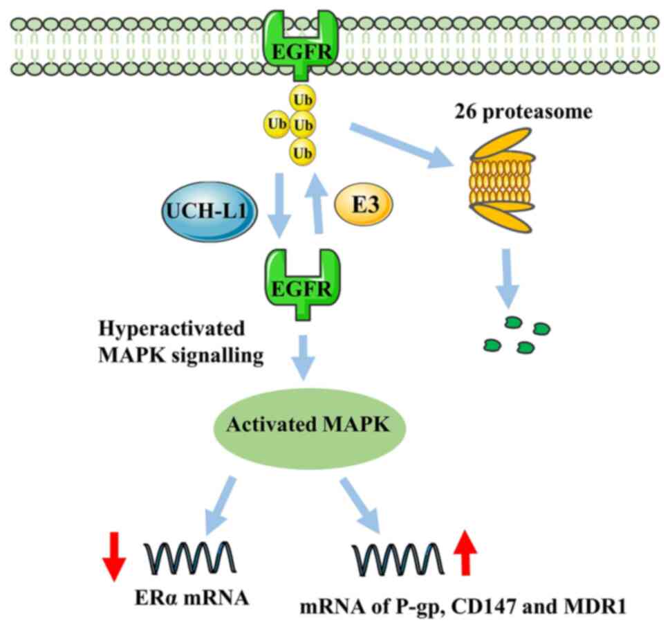
and a severe challenge to managing breast cancer (16). UCH-L1 is associated with the
regulation of chemotherapy resistance in breast cancer.
Immunohistochemistry by Jin et al (88) found that UCH-L1 can inhibit
degradation of EGFR, resulting in high expression of P-glycoprotein
(P-gp), CD147 and matrix metalloproteinase (MMP) in MDR breast
cancer cells. In addition, a study demonstrated that upregulation
of MDR1, CD147 and MMP can also be achieved via activation of the
MAPK/Erk signaling pathway by UCH-L1 (Fig. 5) (86). Notably, high expression of P-gp,
CD147 and MMP has also been proven to be the primary molecular
mechanism mediating MDR (89).
Activation of the UCH-L1/EGFR signaling pathway also inhibits ERα
expression in breast cancer and downregulation of the
deubiquitinase activity of UCH-L1 has the opposite effect, leading
to Erα-breast cancer cells being more sensitive to treatment with
tamoxifen and fulvestrant (90).
Furthermore, ER− breast cancer has a worse prognosis
than ER+ breast cancer (91,92). A
new target for the treatment of EGFR-associated MDR breast cancer
is expected to be identified in further studies of UCH-L1.
UCH-L1 contributes to melanoma development by
decreasing the stability of MITF as well as regulating the PI3K/Akt
signaling pathway (64). A recent
study suggested that UCH-L1 promotes uterine serous carcinoma by
allowing cells to enter mitosis by upregulating expression of
cyclin B, resulting in the proliferation of these cells (36). Therefore, UCH-L1 may be a
novel prognostic marker for uterine serous carcinoma and a
potential therapeutic target. UCH-L1 contributes to lymphatic
metastasis by positively regulating growth arrest specific 2
protein levels, stimulating the migration ability of glioma
(98). Furthermore, a study
reported that UCH-L1 may support the development of distant
metastasis in endometrial cancer (99). The metastasis of prostate cancer is
also enhanced when EMT is promoted by UCH-L1 (100).
Metastasis is the primary cause of poor prognosis
for patients with cancer. UCH-L serves a key role in promoting
metastasis of some malignant tumors such as gastric cancer
(69), breast cancer (86) and endometrial cancer (99). In 2009, UCH-L1 was identified
as an essential factor contributing to tumor metastasis (33). Subsequently, it has been established
that UCH-L1 overexpression further enhances metastasis or
invasiveness of cells by altering cancer cell morphology and
regulating the Akt signaling pathway (85). UCH-L1 is associated with cell
proliferation, invasion and metastasis by activating the Erk1/2 and
Akt pathways via deubiquitination in gastric cancer and liver
metastatic tumor (69).
Hypoxia-inducible factor-1 (HIF-1) supports cancer progression
through a variety of mechanisms including angiogenesis,
proliferation, invasion and metastasis of cells, cancer stem cell
maintenance and treatment resistance (68,101).
The downregulation of HIF-1 target gene expression is hypothesized
to decrease with downregulation of UCH-L1 (102), demonstrating that inhibition of
UCH-L1/HIF-1 pathway activity may be a method to treat distant
metastasis and invasion of tumors.
Recent studies have reported that IMP-1710, an
UCH-L1 inhibitor, has higher quality and selectivity compared with
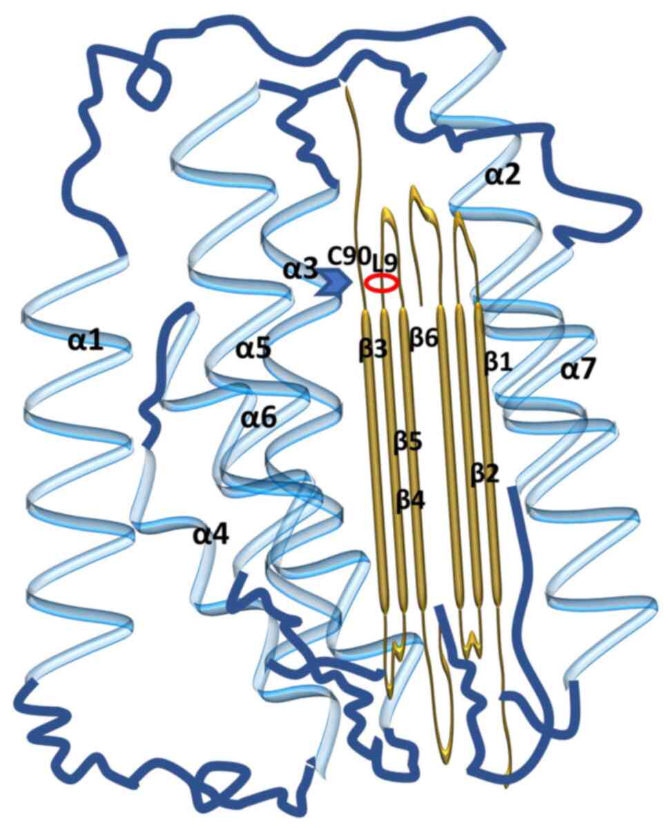
LDN-57444 (111,112). IMP-1710 selectively labels C90 of
UCH-L1 at nanomolar concentrations in a cell model of idiopathic
pulmonary fibrosis to block the profibrotic response (112).
In the present review, the molecular structure of
UCH-L1 and its complex role in cancer were explained. UCH-L1, a
multi-functional protein that is widely expressed in the brain, not
only causes neurodegenerative disease but also serves a complex
role in the occurrence and progression of cancer. UCH-L1
methylation is involved in development of various types of cancer,
such as esophageal (21), gastric
(22), ovarian (23) and renal cancer (24), head and neck squamous cell carcinoma
(20), HCC (25), breast cancer (26), PNET (27) and nasopharyngeal cancer (28,67).
When UCH-L1 expression is restored, UCH-L1 regulates key
cyclin levels (such as p53), inhibits proliferation and promotes
apoptosis of cancer cells (25,26).
Notably, for cancer caused by epigenetic abnormality of
UCH-L1, the CRISPR-Cas9 system, a specific genome editing
technology, may effectively restore gene expression of
UCH-L1 to normal levels and may be expected to be a future
therapeutic option (81). However,
this technology has numerous unsolved challenges, such as
off-target effects and editing efficiency in clinical applications
(113). Moreover, UCH-L1 functions
as an oncogenic factor via PI3K/Akt, MAPK/Erk and other signaling
pathways to induce tumorigeneses numerous types of cancer,
including breast cancer (17,29,88),
NSCLC (19), lymphoma (31), parathyroid carcinoma (32,114),
melanoma (33), cutaneous squamous
cell cancer (34), osteosarcoma
(35), uterine serous carcinoma
(36) and neuroblastoma (37) (Table
I).
UCH-L1 has both Ub hydrolase and ligase activity,
as well as monomeric stabilization effects. The different enzymatic
activities of UCH-L1 regulate stability of different signaling
pathways and cell cycle proteins in cancer cells. UCH-L1 inhibits
or promotes development in different types of cancer, but there is
controversy about the effect of UCH-L1 on cancer and its
mechanism is still not well understood. Therefore, other substrates
of UCH-L1 and specific mechanisms of deubiquitination regulation
are hot spots for further research and targeting UCH-L1 may
be an effective strategy for the treatment of cancer.
Not applicable.
The present review was supported by the National Natural Science
Foundation of China (grant no. 13009038), Project of Science and
Technology Department of Jiangxi Province (grant nos.
20192BAB215024 and 20202BABL206099).
Not applicable.
TO conceived the study and revised the manuscript.
XW drafted and revised the manuscript and constructed the figures.
NZ revised the manuscript. ML, TH and MW reviewed the manuscript.
All authors have read and approved the final manuscript. Data
authentication is not applicable.
Not applicable.
Not applicable.
The authors declare that they have no competing
interests.
|
1
|
Kulathu Y and Komander D: Atypical
ubiquitylation-the unexplored world of polyubiquitin beyond Lys48
and Lys63 linkages. Nat Rev Mol Cell Bio. 13:508–523. 2012.
View Article : Google Scholar : PubMed/NCBI
|
|
2
|
Eldridge AG and O'Brien T: Therapeutic
strategies within the ubiquitin proteasome system. Cell Death
Differ. 17:4–13. 2010. View Article : Google Scholar : PubMed/NCBI
|
|
3
|
Mata-Cantero L, Lobato-Gil S, Aillet F,
Lang V and Rodriguez MS: The ubiquitin-proteasome system (UPS) as a
cancer drug target: Emerging mechanisms and therapeutics. Springer;
Netherlands, Dordrecht: pp. 225–264. 2014
|
|
4
|
Komander D, Clague MJ and Urbé S: Breaking
the chains: Structure and function of the deubiquitinases. Nat Rev
Mol Cell Bio. 10:550–563. 2009. View Article : Google Scholar : PubMed/NCBI
|
|
5
|
Das C, Hoang QQ, Kreinbring CA, Luchansky
SJ, Meray RK, Ray SS, Lansbury PT, Ringe D and Petsko GA:
Structural basis for conformational plasticity of the Parkinson's
disease-associated ubiquitin hydrolase UCH-L1. Proc Natl Acad Sci
USA. 103:4675–4680. 2006. View Article : Google Scholar : PubMed/NCBI
|
|
6
|
Setsuie R and Wada K: The functions of
UCH-L1 and its relation to neurodegenerative diseases. Neurochem
Int. 51:105–111. 2007. View Article : Google Scholar : PubMed/NCBI
|
|
7
|
Doran JF, Jackson P, Kynoch PA and
Thompson RJ: Isolation of PGP 9.5, a new human neurone-specific
protein detected by high-resolution two-dimensional
electrophoresis. J Neurochem. 40:1542–1547. 1983. View Article : Google Scholar : PubMed/NCBI
|
|
8
|
Day INM and Thompson RJ: UCHL1 (PGP 9.5):
Neuronal biomarker and ubiquitin system protein. Prog Neurobiol.
90:327–362. 2010. View Article : Google Scholar : PubMed/NCBI
|
|
9
|
Bishop P, Rocca D and Henley JM: Ubiquitin
C-terminal hydrolase L1 (UCH-L1): Structure, distribution and roles
in brain function and dysfunction. Biochem J. 473:2453–2462. 2016.
View Article : Google Scholar : PubMed/NCBI
|
|
10
|
Wang KK, Yang Z, Sarkis G, Torres I and
Raghavan V: Ubiquitin C-terminal hydrolase-L1 (UCH-L1) as a
therapeutic and diagnostic target in neurodegeneration, neurotrauma
and neuro-injuries. Expert Opin Ther Targets. 21:627–638. 2017.
View Article : Google Scholar : PubMed/NCBI
|
|
11
|
Gong B and Leznik E: The role of ubiquitin
C-terminal hydrolase L1 in neurodegenerative disorders. Drug News
Perspect. 20:365–370. 2007. View Article : Google Scholar : PubMed/NCBI
|
|
12
|
Butterfield DA: Ubiquitin
carboxyl-terminal hydrolase L-1 in brain: Focus on its
oxidative/nitrosative modification and role in brains of subjects
with Alzheimer disease and mild cognitive impairment. Free Radic
Biol Med. 177:278–286. 2021. View Article : Google Scholar : PubMed/NCBI
|
|
13
|
Matuszczak E, Tylicka M, Komarowska MD,
Debek W and Hermanowicz A: Ubiquitin carboxy-terminal hydrolase
L1-physiology and pathology. Cell Biochem Funct. 38:533–540. 2020.
View Article : Google Scholar : PubMed/NCBI
|
|
14
|
Fang Y and Shen X: Ubiquitin
carboxyl-terminal hydrolases: Involvement in cancer progression and
clinical implications. Cancer Metast Rev. 36:669–682. 2017.
View Article : Google Scholar : PubMed/NCBI
|
|
15
|
Fang Y, Fu D and Shen X: The potential
role of ubiquitin c-terminal hydrolases in oncogenesis. Biochim
Biophys Acta. 1806:1–6. 2010.PubMed/NCBI
|
|
16
|
Ning K, Wang T, Sun X, Zhang P, Chen Y,
Jin J and Hua D: UCH-L1-containing exosomes mediate
chemotherapeutic resistance transfer in breast cancer. J Surg
Oncol. 115:932–940. 2017. View Article : Google Scholar : PubMed/NCBI
|
|
17
|
Luo Y, He J, Yang C, Orange M, Ren X,
Blair N, Tan T, Yang JM and Zhu H: UCH-L1 promotes invasion of
breast cancer cells through activating Akt signaling pathway. J
Cell Biochem. 119:691–700. 2018. View Article : Google Scholar : PubMed/NCBI
|
|
18
|
Sharma A, Liu H, Tobar-Tosse F, Chand
Dakal T, Ludwig M, Holz FG, Loeffler KU, Wüllner U and Herwig-Carl
MC: Ubiquitin carboxyl-terminal hydrolases (UCHs): Potential
mediators for cancer and neurodegeneration. Int J Mol Sci.
21:39102020. View Article : Google Scholar : PubMed/NCBI
|
|
19
|
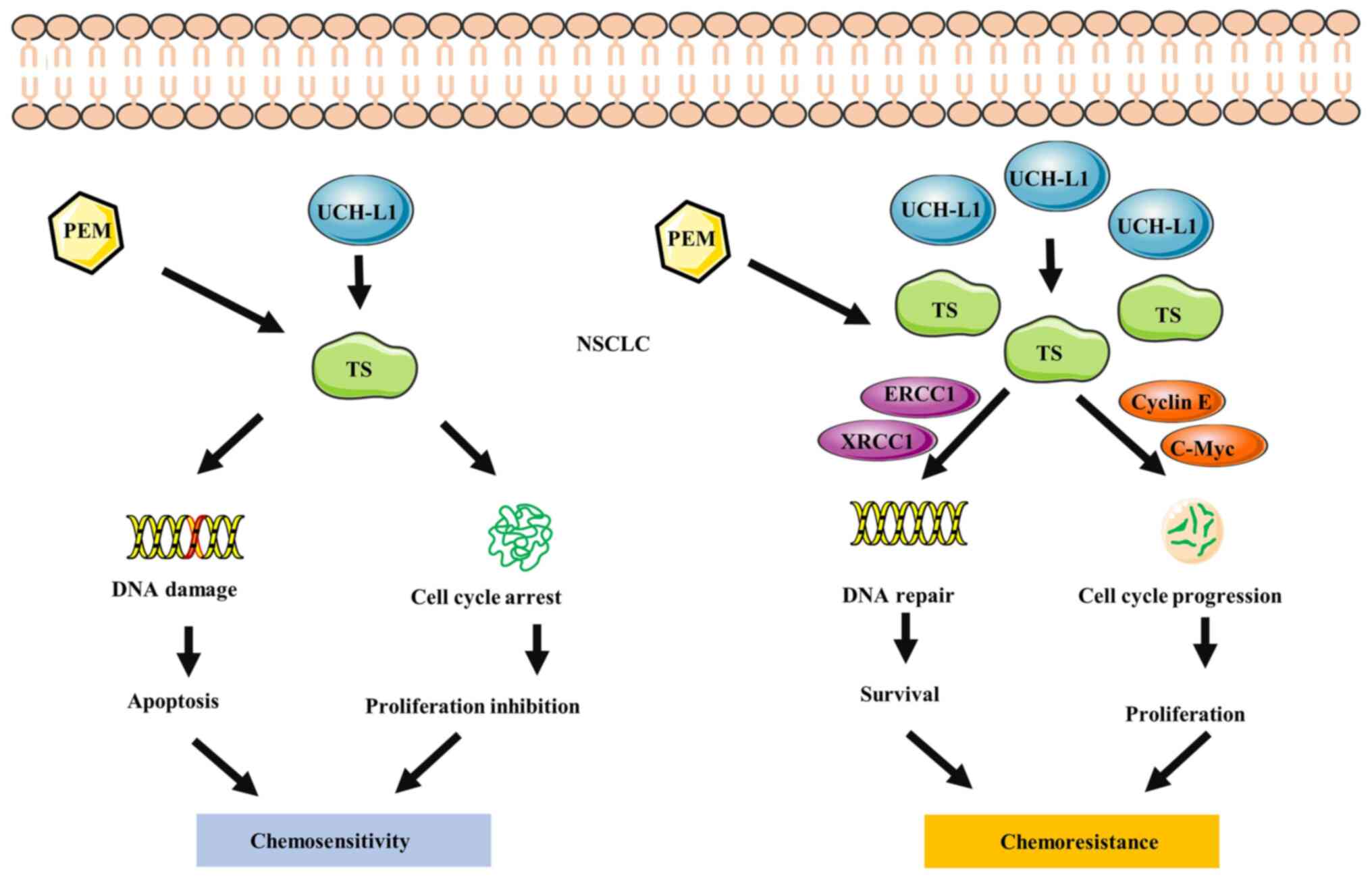
Ding X, Gu Y, Jin M, Guo X, Xue S, Tan C,
Huang J, Yang W, Xue M, Zhou Q, et al: The deubiquitinating enzyme
UCHL1 promotes resistance to pemetrexed in non-small cell lung
cancer by upregulating thymidylate synthase. Theranostics.
10:6048–6060. 2020. View Article : Google Scholar : PubMed/NCBI
|
|
20
|
Tokumaru Y, Yamashita K, Kim MS, Park HL,
Osada M, Mori M and Sidransky D: The role of PGP9.5 as a tumor
suppressor gene in human cancer. Int J Cancer. 123:753–759. 2008.
View Article : Google Scholar : PubMed/NCBI
|
|
21
|
Mandelker DL, Yamashita K, Tokumaru Y,
Mimori K, Howard DL, Tanaka Y, Carvalho AL, Jiang WW, Park HL, Kim
MS, et al: PGP9.5 promoter methylation is an independent prognostic
factor for esophageal squamous cell carcinoma. Cancer Res.
65:4963–4968. 2005. View Article : Google Scholar : PubMed/NCBI
|
|
22
|
Yamashita K, Park HL, Kim MS, Osada M,
Tokumaru Y, Inoue H, Mori M and Sidransky D: PGP9.5 methylation in
diffuse-type gastric cancer. Cancer Res. 66:3921–3927. 2006.
View Article : Google Scholar : PubMed/NCBI
|
|
23
|
Okochi-Takada E, Nakazawa K, Wakabayashi
M, Mori A, Ichimura S, Yasugi T and Ushijima T: Silencing of the
UCHL1 gene in human colorectal and ovarian cancers. Int J Cancer.
119:1338–1344. 2006. View Article : Google Scholar : PubMed/NCBI
|
|
24
|
Kagara I, Enokida H, Kawakami K, Matsuda
R, Toki K, Nishimura H, Chiyomaru T, Tatarano S, Itesako T,
Kawamoto K, et al: CpG hypermethylation of the UCHL1 gene promoter
is associated with pathogenesis and poor prognosis in renal cell
carcinoma. J Urol. 180:343–351. 2008. View Article : Google Scholar : PubMed/NCBI
|
|
25
|
Yu J, Tao Q, Cheung KF, Jin H, Poon FF,
Wang X, Li H, Cheng YY, Röcken C, Ebert MPA, et al: Epigenetic
identification of ubiquitin carboxyl-terminal hydrolase L1 as a
functional tumor suppressor and biomarker for hepatocellular
carcinoma and other digestive tumors. Hepatology. 48:508–518. 2008.
View Article : Google Scholar : PubMed/NCBI
|
|
26
|
Xiang T, Li L, Yin X, Yuan C, Tan C, Su X,
Xiong L, Putti TC, Oberst M, Kelly K, et al: The ubiquitin
peptidase UCHL1 induces G0/G1 cell cycle arrest and apoptosis
through stabilizing p53 and is frequently silenced in breast
cancer. PLoS One. 7:e297832012. View Article : Google Scholar : PubMed/NCBI
|
|
27
|
Finnerty BM, Moore MD, Verma A, Aronova A,
Huang S, Edwards DP, Chen Z, Seandel M, Scognamiglio T, Du YN, et
al: UCHL1 loss alters the cell-cycle in metastatic pancreatic
neuroendocrine tumors. Endocr Relat Cancer. 26:411–423. 2019.
View Article : Google Scholar : PubMed/NCBI
|
|
28
|
Zhao Y, Lei Y, He SW, Li YQ, Wang YQ, Hong
XH, Liang YL, Li JY, Chen Y, Luo WJ, et al: Hypermethylation of
UCHL1 promotes metastasis of nasopharyngeal carcinoma by
suppressing degradation of cortactin (CTTN). Cells. 9:5592020.
View Article : Google Scholar : PubMed/NCBI
|
|
29
|
Liu S, González-Prieto R, Zhang M, Geurink
PP, Kooij R, Iyengar PV, van Dinther M, Bos E, Zhang X, Le Dévédec
SE, et al: Deubiquitinase activity profiling identifies UCHL1 as a
candidate oncoprotein that promotes TGFβ-induced breast cancer
metastasis. Clin Cancer Res. 26:1460–1473. 2020. View Article : Google Scholar : PubMed/NCBI
|
|
30
|
Shimada Y, Kudo Y, Maehara S, Matsubayashi
J, Otaki Y, Kajiwara N, Ohira T, Minna JD and Ikeda N: Ubiquitin
C-terminal hydrolase-L1 has prognostic relevance and is a
therapeutic target for high-grade neuroendocrine lung cancers.
Cancer Sci. 111:610–620. 2020. View Article : Google Scholar : PubMed/NCBI
|
|
31
|
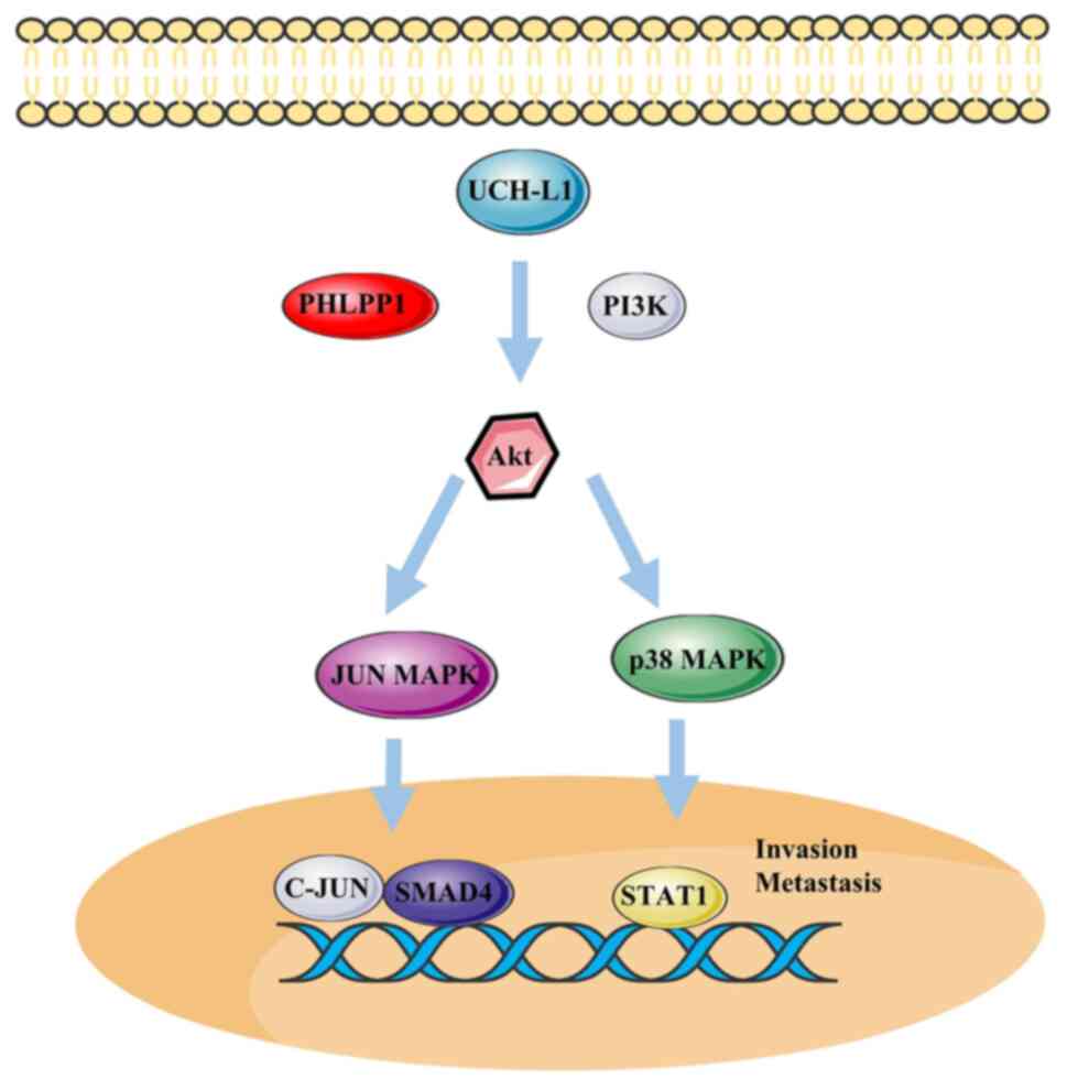
Hussain S, Foreman O, Perkins SL, Witzig
TE, Miles RR, van Deursen J and Galardy PJ: The de-ubiquitinase
UCH-L1 is an oncogene that drives the development of lymphoma in
vivo by deregulating PHLPP1 and Akt signaling. Leukemia.
24:1641–1655. 2010. View Article : Google Scholar : PubMed/NCBI
|
|
32
|
Howell VM, Gill A, Clarkson A, Nelson AE,
Dunne R, Delbridge LW, Robinson BG, Teh BT, Gimm O and Marsh DJ:
Accuracy of combined protein gene product 9.5 and parafibromin
markers for immunohistochemical diagnosis of parathyroid carcinoma.
J Clin Endocrinol Metab. 94:434–441. 2009. View Article : Google Scholar : PubMed/NCBI
|
|
33
|
Kim HJ, Kim YM, Lim S, Nam YK, Jeong J,
Kim HJ and Lee KJ: Ubiquitin C-terminal hydrolase-L1 is a key
regulator of tumor cell invasion and metastasis. Oncogene.
28:117–127. 2009. View Article : Google Scholar : PubMed/NCBI
|
|
34
|
Mastoraki A, Ioannidis E, Patsouris E,
Safioleas M and Aroni K: PGP 9.5 expression in cutaneous
keratoacanthomas and squamous cell carcinomas. Arch Dermatol Res.
301:653–658. 2009. View Article : Google Scholar : PubMed/NCBI
|
|
35
|
Zheng S, Qiao G, Min D, Zhang Z, Lin F,
Yang Q, Feng T, Tang L, Sun Y, Zhao H, et al: Heterogeneous
expression and biological function of ubiquitin carboxy-terminal
hydrolase-L1 in osteosarcoma. Cancer Lett. 359:36–46. 2015.
View Article : Google Scholar : PubMed/NCBI
|
|
36
|
Kwan SY, Au-Yeung CL, Yeung TL,
Rynne-Vidal A, Wong KK, Risinger JI, Lin HK, Schmandt RE, Yates MS,
Mok SC and Lu KH: Ubiquitin carboxyl-terminal hydrolase L1 (UCHL1)
promotes uterine serous cancer cell proliferation and cell cycle
progression. Cancers (Basel). 12:1182020. View Article : Google Scholar : PubMed/NCBI
|
|
37
|
Gu Y, Lv F, Xue M, Chen K, Cheng C, Ding
X, Jin M, Xu G, Zhang Y, Wu Z, et al: The deubiquitinating enzyme
UCHL1 is a favorable prognostic marker in neuroblastoma as it
promotes neuronal differentiation. J Exp Clin Canc Res. 37:2582018.
View Article : Google Scholar : PubMed/NCBI
|
|
38
|
Luchansky SJ, Lansbury PT Jr and Stein RL:
Substrate recognition and catalysis by UCH-L1. Biochemistry.
45:14717–14725. 2006. View Article : Google Scholar : PubMed/NCBI
|
|
39
|
Leroy E, Boyer R, Auburger G, Leube B, Ulm
G, Mezey E, Harta G, Brownstein MJ, Jonnalagada S, Chernova T, et
al: The ubiquitin pathway in Parkinson's disease. Nature.
395:451–452. 1998. View
Article : Google Scholar : PubMed/NCBI
|
|
40
|
Sekiguchi S, Kwon J, Yoshida E, Hamasaki
H, Ichinose S, Hideshima M, Kuraoka M, Takahashi A, Ishii Y, Kyuwa
S, et al: Localization of ubiquitin C-terminal hydrolase L1 in
mouse ova and its function in the plasma membrane to block
polyspermy. Am J Pathol. 169:1722–1729. 2006. View Article : Google Scholar : PubMed/NCBI
|
|
41
|
Day IN and Thompson RJ: Molecular cloning
of cDNA coding for human PGP 9.5 protein. A novel cytoplasmic
marker for neurones and neuroendocrine cells. FEBS Lett.
210:157–160. 1987. View Article : Google Scholar : PubMed/NCBI
|
|
42
|
Larsen CN, Krantz BA and Wilkinson KD:
Substrate specificity of deubiquitinating enzymes: Ubiquitin
C-terminal hydrolases. Biochemistry. 37:3358–3368. 1998. View Article : Google Scholar : PubMed/NCBI
|
|
43
|
Esteve-Rudd J, Campello L, Herrero MT,
Cuenca N and Martin-Nieto J: Expression in the mammalian retina of
parkin and UCH-L1, two components of the ubiquitin-proteasome
system. Brain Res. 1352:70–82. 2010. View Article : Google Scholar : PubMed/NCBI
|
|
44
|
Liu Y, Wu J, Wu H, Wang T, Gan H, Zhang X,
Liu Y, Li R, Zhao Z, Chen Q, et al: UCH-L1 expression of podocytes
in diseased glomeruli and in vitro. J Pathol. 217:642–653. 2009.
View Article : Google Scholar : PubMed/NCBI
|
|
45
|
Johnston SC, Larsen CN, Cook WJ, Wilkinson
KD and Hill CP: Crystal structure of a deubiquitinating enzyme
(human UCH-L3) at 1.8 A resolution. EMBO J. 16:3787–3796. 1997.
View Article : Google Scholar : PubMed/NCBI
|
|
46
|
Grabbe C, Husnjak K and Dikic I: The
spatial and temporal organization of ubiquitin networks. Nat Rev
Mol Cell Biol. 12:295–307. 2011. View Article : Google Scholar : PubMed/NCBI
|
|
47
|
Zhang M, Cai F, Zhang S, Zhang S and Song
W: Overexpression of ubiquitin carboxyl-terminal hydrolase L1
(UCHL1) delays Alzheimer's progression in vivo. Sci Rep.
4:72982014. View Article : Google Scholar : PubMed/NCBI
|
|
48
|
Suong DN, Thao DT, Masamitsu Y and Thuoc
TL: Ubiquitin carboxyl hydrolase L1 significance for human
diseases. Protein Pept Lett. 21:624–630. 2014. View Article : Google Scholar : PubMed/NCBI
|
|
49
|
Wilkinson KD, Lee KM, Deshpande S,
Duerksen-Hughes P, Boss JM and Pohl J: The neuron-specific protein
PGP 9.5 is a ubiquitin carboxyl-terminal hydrolase. Science.
246:670–673. 1989. View Article : Google Scholar : PubMed/NCBI
|
|
50
|
Zhong J, Zhao M, Ma Y, Luo Q, Liu J, Wang
J, Yuan X, Sang J and Huang C: UCHL1 acts as a colorectal cancer
oncogene via activation of the β-catenin/TCF pathway through its
deubiquitinating activity. Int J Mol Med. 30:430–436. 2012.
View Article : Google Scholar : PubMed/NCBI
|
|
51
|
Bheda A, Yue W, Gullapalli A, Whitehurst
C, Liu R, Pagano JS and Shackelford J: Positive reciprocal
regulation of ubiquitin C-terminal hydrolase L1 and
beta-catenin/TCF signaling. PLoS One. 4:e59552009. View Article : Google Scholar : PubMed/NCBI
|
|
52
|
Takami Y, Nakagami H, Morishita R, Katsuya
T, Cui TX, Ichikawa T, Saito Y, Hayashi H, Kikuchi Y, Nishikawa T,
et al: Ubiquitin carboxyl-terminal hydrolase L1, a novel
deubiquitinating enzyme in the vasculature, attenuates NF-kappaB
activation. Arterioscler Thromb Vasc Biol. 27:2184–2190. 2007.
View Article : Google Scholar : PubMed/NCBI
|
|
53
|
Zhang H, Sun Y, Hu R, Luo W, Mao X, Zhao
Z, Chen Q and Zhang Z: The regulation of the UCH-L1 gene by
transcription factor NF-κB in podocytes. Cell Signal. 25:1574–1585.
2013. View Article : Google Scholar : PubMed/NCBI
|
|
54
|
Nagata A, Itoh F, Sasho A, Sugita K,
Suzuki R, Hinata H, Shimoda Y, Suzuki E, Maemoto Y, Inagawa T, et
al: The evolutionarily conserved deubiquitinase UBH1/UCH-L1
augments DAF7/TGF-β signaling, inhibits dauer larva formation, and
enhances lung tumorigenesis. J Biol Chem. 295:9105–9120. 2020.
View Article : Google Scholar : PubMed/NCBI
|
|
55
|
Liu Y, Fallon L, Lashuel HA, Liu Z and
Lansbury PT Jr: The UCH-L1 gene encodes two opposing enzymatic
activities that affect alpha-synuclein degradation and Parkinson's
disease susceptibility. Cell. 111:209–218. 2002. View Article : Google Scholar : PubMed/NCBI
|
|
56
|
Chuma M, Sakamoto M, Yasuda J, Fujii G,
Nakanishi K, Tsuchiya A, Ohta T, Asaka M and Hirohashi S:
Overexpression of cortactin is involved in motility and metastasis
of hepatocellular carcinoma. J Hepatol. 41:629–636. 2004.
View Article : Google Scholar : PubMed/NCBI
|
|
57
|
Osaka H, Wang YL, Takada K, Takizawa S,
Setsuie R, Li H, Sato Y, Nishikawa K, Sun YJ, Sakurai M, et al:
Ubiquitin carboxy-terminal hydrolase L1 binds to and stabilizes
monoubiquitin in neuron. Hum Mol Genet. 12:1945–1958. 2003.
View Article : Google Scholar : PubMed/NCBI
|
|
58
|
Kabuta T, Mitsui T, Takahashi M, Fujiwara
Y, Kabuta C, Konya C, Tsuchiya Y, Hatanaka Y, Uchida K, Hohjoh H
and Wada K: Ubiquitin C-terminal hydrolase L1 (UCH-L1) acts as a
novel potentiator of cyclin-dependent kinases to enhance cell
proliferation independently of its hydrolase activity. J Biol Chem.
288:12615–12626. 2013. View Article : Google Scholar : PubMed/NCBI
|
|
59
|
Hussain S, Bedekovics T, Ali A, Zaid O,
May DG, Roux KJ and Galardy PJ: A cysteine near the C-terminus of
UCH-L1 is dispensable for catalytic activity but is required to
promote AKT phosphorylation, eIF4F assembly, and malignant B-cell
survival. Cell Death Discov. 5:1522019. View Article : Google Scholar : PubMed/NCBI
|
|
60
|
Fulga TA, Elson-Schwab I, Khurana V,
Steinhilb ML, Spires TL, Hyman BT and Feany MB: Abnormal bundling
and accumulation of F-actin mediates tau-induced neuronal
degeneration in vivo. Nat Cell Biol. 9:139–148. 2007. View Article : Google Scholar : PubMed/NCBI
|
|
61
|
Xie M, Han Y, Yu Q, Wang X, Wang S and
Liao X: UCH-L1 inhibition decreases the microtubule-binding
function of tau protein. J Alzheimers Dis. 49:353–363. 2016.
View Article : Google Scholar : PubMed/NCBI
|
|
62
|
Yu Q, Zhang H, Li Y, Liu C, Wang S and
Liao X: UCH-L1 inhibition suppresses tau aggresome formation during
proteasomal impairment. Mol Neurobiol. 55:3812–3821.
2018.PubMed/NCBI
|
|
63
|
Bheda A, Gullapalli A, Caplow M, Pagano JS
and Shackelford J: Ubiquitin editing enzyme UCH L1 and microtubule
dynamics: Implication in mitosis. Cell Cycle. 9:980–994. 2010.
View Article : Google Scholar : PubMed/NCBI
|
|
64
|
Seo EY, Jin SP, Sohn KC, Park CH, Lee DH
and Chung JH: UCHL1 regulates melanogenesis through controlling
MITF stability in human melanocytes. J Invest Dermatol.
137:1757–1765. 2017. View Article : Google Scholar : PubMed/NCBI
|
|
65
|
Rolén U, Freda E, Xie J, Pfirrmann T,
Frisan T and Masucci MG: The ubiquitin C-terminal hydrolase UCH-L1
regulates B-cell proliferation and integrin activation. J Cell Mol
Med. 13:1666–1678. 2009. View Article : Google Scholar : PubMed/NCBI
|
|
66
|
Lohmann F, Sachs M, Meyer TN, Sievert H,
Lindenmeyer MT, Wiech T, Cohen CD, Balabanov S, Stahl RA and
Meyer-Schwesinger C: UCH-L1 induces podocyte hypertrophy in
membranous nephropathy by protein accumulation. Biochim Biophys
Acta. 1842:945–958. 2014. View Article : Google Scholar : PubMed/NCBI
|
|
67
|
Li L, Tao Q, Jin H, van Hasselt A, Poon
FF, Wang X, Zeng MS, Jia WH, Zeng YX, Chan AT and Cao Y: The tumor
suppressor UCHL1 forms a complex with p53/MDM2/ARF to promote p53
signaling and is frequently silenced in nasopharyngeal carcinoma.
Clin Cancer Res. 16:2949–2958. 2010. View Article : Google Scholar : PubMed/NCBI
|
|
68
|
Goto Y, Zeng L, Yeom CJ, Zhu Y, Morinibu
A, Shinomiya K, Kobayashi M, Hirota K, Itasaka S, Yoshimura M, et
al: UCHL1 provides diagnostic and antimetastatic strategies due to
its deubiquitinating effect on HIF-1α. Nat Commun. 6:61532015.
View Article : Google Scholar : PubMed/NCBI
|
|
69
|
Gu Y, Yang M, Zhao M, Luo Q, Yang L, Peng
H, Wang J, Huang SK, Zheng ZX, Yuan XH, et al: The de-ubiquitinase
UCHL1 promotes gastric cancer metastasis via the Akt and Erk1/2
pathways. Tumour Biol. 36:8379–8387. 2015. View Article : Google Scholar : PubMed/NCBI
|
|
70
|
Wang G, Zhang W, Zhou B, Jin C, Wang Z,
Yang Y, Wang Z, Chen Y and Feng X: The diagnosis value of promoter
methylation of UCHL1 in the serum for progression of gastric
cancer. Biomed Res Int. 2015:7410302015. View Article : Google Scholar : PubMed/NCBI
|
|
71
|
Seliger B, Handke D, Schabel E, Bukur J,
Lichtenfels R and Dammann R: Epigenetic control of the ubiquitin
carboxyl terminal hydrolase 1 in renal cell carcinoma. J Transl
Med. 7:902009. View Article : Google Scholar : PubMed/NCBI
|
|
72
|
Mitsui Y, Shiina H, Hiraki M, Arichi N,
Hiraoka T, Sumura M, Honda S, Yasumoto H and Igawa M: Tumor
suppressor function of PGP9.5 is associated with epigenetic
regulation in prostate cancer-novel predictor of biochemical
recurrence after radical surgery. Cancer Epidemiol Biomarkers Prev.
21:487–496. 2012. View Article : Google Scholar : PubMed/NCBI
|
|
73
|
Brait M, Maldonado L, Noordhuis MG, Begum
S, Loyo M, Poeta ML, Barbosa A, Fazio VM, Angioli R, Rabitti C, et
al: Association of promoter methylation of VGF and PGP9.5 with
ovarian cancer progression. PLoS One. 8:e708782013. View Article : Google Scholar : PubMed/NCBI
|
|
74
|
Abdelmaksoud-Dammak R, Saadallah-Kallel A,
Miladi-Abdennadher I, Ayedi L, Khabir A, Sallemi-Boudawara T,
Frikha M, Daoud J and Mokdad-Gargouri R: CpG methylation of
ubiquitin carboxyl-terminal hydrolase 1 (UCHL1) and P53 mutation
pattern in sporadic colorectal cancer. Tumour Biol. 37:1707–1714.
2016. View Article : Google Scholar : PubMed/NCBI
|
|
75
|
Nanok C, Jearanaikoon P, Proungvitaya S
and Limpaiboon T: Aberrant methylation of HTATIP2 and UCHL1 as a
predictive biomarker for cholangiocarcinoma. Mol Med Rep.
17:4145–4153. 2018.PubMed/NCBI
|
|
76
|
Jaferian S, Soleymaninejad M and Daraee H:
Verapamil (VER) enhances the cytotoxic effects of docetaxel and
vinblastine combined therapy against non-small cell lung cancer
cell lines. Drug Res (Stuttg). 68:146–152. 2018. View Article : Google Scholar : PubMed/NCBI
|
|
77
|
Ge N, Yang GS, Zhang TY, Chang N, Kang YH,
Zhou Q and Fan PS: Upregulation of KCNMA1 facilitates the reversal
effect of verapamil on the chemoresistance to cisplatin of
esophageal squamous cell carcinoma cells. Eur Rev Med Pharmacol
Sci. 25:1869–1880. 2021.PubMed/NCBI
|
|
78
|
Li P, Zhong D and Gong PY: Synergistic
effect of paclitaxel and verapamil to overcome multi-drug
resistance in breast cancer cells. Biochem Biophys Res Commun.
516:183–188. 2019. View Article : Google Scholar : PubMed/NCBI
|
|
79
|
Yang G, Fan G, Zhang T, Ma K, Huang J, Liu
M, Teng X, Xu K, Fan P and Cheng D: Upregulation of ubiquitin
carboxyl-terminal hydrolase L1 (UCHL1) mediates the reversal effect
of verapamil on chemo-resistance to adriamycin of hepatocellular
carcinoma. Med Sci Monit. 24:2072–2082. 2018. View Article : Google Scholar : PubMed/NCBI
|
|
80
|
Wang WJ, Li QQ, Xu JD, Cao XX, Li HX, Tang
F, Chen Q, Yang JM, Xu ZD and Liu XP: Over-expression of ubiquitin
carboxy terminal hydrolase-L1 induces apoptosis in breast cancer
cells. Int J Oncol. 33:1037–1045. 2008.PubMed/NCBI
|
|
81
|
Maroufi F, Maali A, Abdollahpour-Alitappeh
M, Ahmadi MH and Azad M: CRISPR-mediated modification of DNA
methylation pattern in the new era of cancer therapy. Epigenomics.
12:1845–1859. 2020. View Article : Google Scholar : PubMed/NCBI
|
|
82
|
Moore MD, Finnerty B, Gray KD, Hoda R, Liu
Y, Soong L, Beninato T, Rao R, Zarnegar R and Fahey TJ III:
Decreased UCHL1 expression as a cytologic biomarker for aggressive
behavior in pancreatic neuroendocrine tumors. Surgery. 163:226–231.
2018. View Article : Google Scholar : PubMed/NCBI
|
|
83
|
Scully OJ, Bay BH, Yip G and Yu Y: Breast
cancer metastasis. Cancer Genomics Proteomics. 9:311–320.
2012.PubMed/NCBI
|
|
84
|
Miyoshi Y, Nakayama S, Torikoshi Y, Tanaka
S, Ishihara H, Taguchi T, Tamaki Y and Noguchi S: High expression
of ubiquitin carboxy-terminal hydrolase-L1 and -L3 mRNA predicts
early recurrence in patients with invasive breast cancer. Cancer
Sci. 97:523–529. 2006. View Article : Google Scholar : PubMed/NCBI
|
|
85
|
Schröder C, Milde-Langosch K, Gebauer F,
Schmid K, Mueller V, Wirtz RM, Meyer-Schwesinger C, Schlüter H,
Sauter G and Schumacher U: Prognostic relevance of ubiquitin
C-terminal hydrolase L1 (UCH-L1) mRNA and protein expression in
breast cancer patients. J Cancer Res Clin. 139:1745–1755. 2013.
View Article : Google Scholar : PubMed/NCBI
|
|
86
|
Wang W, Zou L, Zhou D, Zhou Z, Tang F, Xu
Z and Liu X: Overexpression of ubiquitin carboxyl terminal
hydrolase-L1 enhances multidrug resistance and invasion/metastasis
in breast cancer by activating the MAPK/Erk signaling pathway. Mol
Carcinog. 55:1329–1342. 2016. View Article : Google Scholar : PubMed/NCBI
|
|
87
|
Dongre A and Weinberg RA: New insights
into the mechanisms of epithelial-mesenchymal transition and
implications for cancer. Nat Rev Mol Cell Biol. 20:69–84. 2019.
View Article : Google Scholar : PubMed/NCBI
|
|
88
|
Jin Y, Zhang W, Xu J, Wang H, Zhang Z, Chu
C, Liu X and Zou Q: UCH-L1 involved in regulating the degradation
of EGFR and promoting malignant properties in drug-resistant breast
cancer. Int J Clin Exp Patho. 8:12500–12508. 2015.PubMed/NCBI
|
|
89
|
Li QQ, Wang WJ, Xu JD, Cao XX, Chen Q,
Yang JM and Xu ZD: Up-regulation of CD147 and matrix
metalloproteinase-2, −9 induced by P-glycoprotein substrates in
multidrug resistant breast cancer cells. Cancer Sci. 98:1767–1774.
2007. View Article : Google Scholar : PubMed/NCBI
|
|
90
|
Chen XS, Wang KS, Guo W, Li LY, Yu P, Sun
XY, Wang HY, Guan YD, Tao YG, Ding BN, et al: UCH-L1-mediated
down-regulation of estrogen receptor α contributes to insensitivity
to endocrine therapy for breast cancer. Theranostics. 10:1833–1848.
2020. View Article : Google Scholar : PubMed/NCBI
|
|
91
|
Rochefort H, Glondu M, Sahla ME, Platet N
and Garcia M: How to target estrogen receptor-negative breast
cancer? Endocr Relat Cancer. 10:261–266. 2003. View Article : Google Scholar : PubMed/NCBI
|
|
92
|
Mondal M, Conole D, Nautiyal J and Tate
EW: UCHL1 as a novel target in breast cancer: Emerging insights
from cell and chemical biology. Br J Cancer. 126:24–33. 2022.
View Article : Google Scholar : PubMed/NCBI
|
|
93
|
Yu J, Yu S, Jia M, Sun PL and Gao H:
Ubiquitin C-terminal hydrolase-L1 expression in non-small-cell lung
cancer and its association with clinicopathological features and
prognosis. Virchows Arch. 480:577–585. 2022. View Article : Google Scholar : PubMed/NCBI
|
|
94
|
Sanmamed MF and Chen L: A paradigm shift
in cancer immunotherapy: From enhancement to normalization. Cell.
175:313–326. 2018. View Article : Google Scholar : PubMed/NCBI
|
|
95
|
Mao R, Tan X, Xiao Y, Wang X, Wei Z, Wang
J, Wang X, Zhou H, Zhang L and Shi Y: Ubiquitin C-terminal
hydrolase L1 promotes expression of programmed cell death-ligand 1
in non-small-cell lung cancer cells. Cancer Sci. 111:3174–3183.
2020. View Article : Google Scholar : PubMed/NCBI
|
|
96
|
Hussain S, Bedekovics T, Liu Q, Hu W, Jeon
H, Johnson SH, Vasmatzis G, May DG, Roux KJ and Galardy PJ: UCH-L1
bypasses mTOR to promote protein biosynthesis and is required for
MYC-driven lymphomagenesis in mice. Blood. 132:2564–2574. 2018.
View Article : Google Scholar : PubMed/NCBI
|
|
97
|
Bedekovics T, Hussain S, Feldman AL and
Galardy PJ: UCH-L1 is induced in germinal center B cells and
identifies patients with aggressive germinal center diffuse large
B-cell lymphoma. Blood. 127:1564–1574. 2016. View Article : Google Scholar : PubMed/NCBI
|
|
98
|
Sui R and Piao HZ: UCHL1 enhances the
malignant development of glioma via targeting GAS2. Eur Rev Med
Pharmacol Sci. 24:6195–6203. 2020.PubMed/NCBI
|
|
99
|
Nakao K, Hirakawa T, Suwa H, Kogure K,
Ikeda S, Yamashita S, Minegishi T and Kishi H: High expression of
ubiquitin C-terminal hydrolase L1 Is associated with poor prognosis
in endometrial cancer patients. Int J Gynecol Cancer. 28:675–683.
2018. View Article : Google Scholar : PubMed/NCBI
|
|
100
|
Jang MJ, Baek SH and Kim JH: UCH-L1
promotes cancer metastasis in prostate cancer cells through EMT
induction. Cancer Lett. 302:128–135. 2011. View Article : Google Scholar : PubMed/NCBI
|
|
101
|
Rashid M, Zadeh LR, Baradaran B, Molavi O,
Ghesmati Z, Sabzichi M and Ramezani F: Up-down regulation of HIF-1α
in cancer progression. Gene. 798:1457962021. View Article : Google Scholar : PubMed/NCBI
|
|
102
|
Li X, Hattori A, Takahashi S, Goto Y,
Harada H and Kakeya H: Ubiquitin carboxyl-terminal hydrolase L1
promotes hypoxia-inducible factor 1-dependent tumor cell malignancy
in spheroid models. Cancer Sci. 111:239–252. 2020. View Article : Google Scholar : PubMed/NCBI
|
|
103
|
Kobayashi E, Hwang D, Bheda-Malge A,
Whitehurst CB, Kabanov AV, Kondo S, Aga M, Yoshizaki T, Pagano JS,
Sokolsky M and Shakelford J: Inhibition of UCH-L1 deubiquitinating
activity with two forms of LDN-57444 has anti-invasive effects in
metastatic carcinoma cells. Int J Mol Sci. 20:37332019. View Article : Google Scholar : PubMed/NCBI
|
|
104
|
Liu Y, Lashuel HA, Choi S, Xing X, Case A,
Ni J, Yeh LA, Cuny GD, Stein RL and Lansbury PT Jr: Discovery of
inhibitors that elucidate the role of UCH-L1 activity in the H1299
lung cancer cell line. Chem Biol. 10:837–846. 2003. View Article : Google Scholar : PubMed/NCBI
|
|
105
|
Hussain S, Bedekovics T, Chesi M,
Bergsagel PL and Galardy PJ: UCHL1 is a biomarker of aggressive
multiple myeloma required for disease progression. Oncotarget.
6:40704–40718. 2015. View Article : Google Scholar : PubMed/NCBI
|
|
106
|
D'Arcy P, Wang X and Linder S:
Deubiquitinase inhibition as a cancer therapeutic strategy.
Pharmacol Ther. 147:32–54. 2015. View Article : Google Scholar : PubMed/NCBI
|
|
107
|
Mermerian AH, Case A, Stein RL and Cuny
GD: Structure-activity relationship, kinetic mechanism, and
selectivity for a new class of ubiquitin C-terminal hydrolase-L1
(UCH-L1) inhibitors. Bioorg Med Chem Lett. 17:3729–3732. 2007.
View Article : Google Scholar : PubMed/NCBI
|
|
108
|
Panyain N, Godinat A, Thawani AR,
Lachiondo-Ortega S, Mason K, Elkhalifa S, Smith LM, Harrigan JA and
Tate EW: Activity-based protein profiling reveals deubiquitinase
and aldehyde dehydrogenase targets of a cyanopyrrolidine probe. RSC
Med Chem. 12:1935–1943. 2021. View Article : Google Scholar : PubMed/NCBI
|
|
109
|
Krabill AD, Chen H, Hussain S, Feng C,
Abdullah A, Das C, Aryal UK, Post CB, Wendt MK, Galardy PJ and
Flaherty DP: Ubiquitin C-terminal hydrolase L1: Biochemical and
cellular characterization of a covalent cyanopyrrolidine-based
inhibitor. Chembiochem. 21:712–722. 2020. View Article : Google Scholar : PubMed/NCBI
|
|
110
|
Kooij R, Liu S, Sapmaz A, Xin BT, Janssen
GMC, van Veelen PA, Ovaa H, Dijke PT and Geurink PP: Small-molecule
activity-based probe for monitoring ubiquitin C-terminal hydrolase
L1 (UCHL1) activity in live cells and zebrafish embryos. J Am Chem
Soc. 142:16825–16841. 2020. View Article : Google Scholar : PubMed/NCBI
|
|
111
|
Berkers CR, van Leeuwen FW, Groothuis TA,
Peperzak V, van Tilburg EW, Borst J, Neefjes JJ and Ovaa H:
Profiling proteasome activity in tissue with fluorescent probes.
Mol Pharm. 4:739–748. 2007. View Article : Google Scholar : PubMed/NCBI
|
|
112
|
Panyain N, Godinat A, Lanyon-Hogg T,
Lachiondo-Ortega S, Will EJ, Soudy C, Mondal M, Mason K, Elkhalifa
S, Smith LM, et al: Discovery of a potent and selective covalent
inhibitor and activity-based probe for the deubiquitylating enzyme
UCHL1, with antifibrotic activity. J Am Chem Soc. 142:12020–12026.
2020. View Article : Google Scholar : PubMed/NCBI
|
|
113
|
Roy B, Zhao J, Yang C, Luo W, Xiong T, Li
Y, Fang X, Gao G, Singh CO, Madsen L, et al: CRISPR/cascade
9-mediated genome editing-challenges and opportunities. Front
Genet. 9:2402018. View Article : Google Scholar : PubMed/NCBI
|
|
114
|
Takano T, Miyauchi A, Matsuzuka F, Yoshida
H, Nakata Y, Kuma K and Amino N: PGP9.5 mRNA could contribute to
the molecular-based diagnosis of medullary thyroid carcinoma. Eur J
Cancer. 40:614–618. 2004. View Article : Google Scholar : PubMed/NCBI
|
|
115
|
Sabapathy K and Lane DP: Therapeutic
targeting of p53: All mutants are equal, but some mutants are more
equal than others. Nat Rev Clin Oncol. 15:13–30. 2018. View Article : Google Scholar : PubMed/NCBI
|
|
116
|
Chibaya L, Karim B, Zhang H and Jones SN:
Mdm2 phosphorylation by Akt regulates the p53 response to oxidative
stress to promote cell proliferation and tumorigenesis. Proc Natl
Acad Sci USA. 118:e20031931182021. View Article : Google Scholar : PubMed/NCBI
|
|
117
|
De S, Campbell C, Venkitaraman AR and
Esposito A: Pulsatile MAPK signaling modulates p53 activity to
control cell fate decisions at the G2 checkpoint for DNA damage.
Cell Rep. 30:2083–2093.e5. 2020. View Article : Google Scholar : PubMed/NCBI
|
|
118
|
Brinkmann K, Zigrino P, Witt A, Schell M,
Ackermann L, Broxtermann P, Schüll S, Andree M, Coutelle O,
Yazdanpanah B, et al: Ubiquitin C-terminal hydrolase-L1 potentiates
cancer chemosensitivity by stabilizing NOXA. Cell Rep. 3:881–891.
2013. View Article : Google Scholar : PubMed/NCBI
|
|
119
|
Kabuta T, Setsuie R, Mitsui T, Kinugawa A,
Sakurai M, Aoki S, Uchida K and Wada K: Aberrant molecular
properties shared by familial Parkinson's disease-associated mutant
UCH-L1 and carbonyl-modified UCH-L1. Hum Mol Genet. 17:1482–1496.
2008. View Article : Google Scholar : PubMed/NCBI
|
|
120
|
Kabuta T, Furuta A, Aoki S, Furuta K and
Wada K: Aberrant interaction between Parkinson disease-associated
mutant UCH-L1 and the lysosomal receptor for chaperone-mediated
autophagy. J Biol Chem. 283:23731–23738. 2008. View Article : Google Scholar : PubMed/NCBI
|
|
121
|
Nishikawa K, Li H, Kawamura R, Osaka H,
Wang YL, Hara Y, Hirokawa T, Manago Y, Amano T, Noda M, et al:
Alterations of structure and hydrolase activity of
parkinsonism-associated human ubiquitin carboxyl-terminal hydrolase
L1 variants. Biochem Biophys Res Commun. 304:176–183. 2003.
View Article : Google Scholar : PubMed/NCBI
|
|
122
|
Duffy MJ, Synnott NC and Crown J: Mutant
p53 in breast cancer: Potential as a therapeutic target and
biomarker. Breast Cancer Res Treat. 170:213–219. 2018. View Article : Google Scholar : PubMed/NCBI
|
|
123
|
Sporikova Z, Koudelakova V, Trojanec R and
Hajduch M: Genetic markers in triple-negative breast cancer. Clin
Breast Cancer. 18:e841–e850. 2018. View Article : Google Scholar : PubMed/NCBI
|
|
124
|
Bazarian JJ, Biberthaler P, Welch RD,
Lewis LM, Barzo P, Bogner-Flatz V, Gunnar Brolinson P, Büki A, Chen
JY, Christenson RH, et al: Serum GFAP and UCH-L1 for prediction of
absence of intracranial injuries on head CT (ALERT-TBI): A
multicentre observational study. Lancet Neurol. 17:782–789. 2018.
View Article : Google Scholar : PubMed/NCBI
|
|
125
|
Meyer-Schwesinger C, Meyer TN, Sievert H,
Hoxha E, Sachs M, Klupp EM, Münster S, Balabanov S, Carrier L,
Helmchen U, et al: Ubiquitin C-terminal hydrolase-l1 activity
induces polyubiquitin accumulation in podocytes and increases
proteinuria in rat membranous nephropathy. Am J Pathol.
178:2044–2057. 2011. View Article : Google Scholar : PubMed/NCBI
|
|
126
|
Fang Y, Li F, Qi C, Mao X, Xu Y, Zhao Z,
Wu H and Zhang Z: Plakoglobin is involved in cytoskeletal
rearrangement of podocytes under the regulation of UCH-L1. Biochem
Biophys Res Commun. 529:112–118. 2020. View Article : Google Scholar : PubMed/NCBI
|
|
127
|
Cui JH and Xie X: UCH-L1 expressed by
podocytes: A potentially therapeutic target for lupus nephritis?
Inflammation. 40:657–665. 2017. View Article : Google Scholar : PubMed/NCBI
|
|
128
|
Xu Y, Gao H, Hu Y, Fang Y, Qi C, Huang J,
Cai X, Wu H, Ding X and Zhang Z: High glucose-induced apoptosis and
necroptosis in podocytes is regulated by UCHL1 via RIPK1/RIPK3
pathway. Exp Cell Res. 382:1114632019. View Article : Google Scholar : PubMed/NCBI
|
|
129
|
Ichikawa T, Li J, Dong X, Potts JD, Tang
D, Li DQ, Li DS and Cui T: Ubiquitin carboxyl terminal hydrolase L1
negatively regulates TNFalpha-mediated vascular smooth muscle cell
proliferation via suppressing ERK activation. Biochem Biophys Res
Commun. 391:852–856. 2010. View Article : Google Scholar : PubMed/NCBI
|
|
130
|
Gao X, Wu L, Wang K, Zhou X, Duan M, Wang
X, Zhang Z and Liu X: Ubiquitin carboxyl terminal hydrolase L1
attenuates TNF-α-mediated vascular smooth muscle cell migration
through suppression of NF-κB activation. Int Heart J. 59:1409–1415.
2018. View Article : Google Scholar : PubMed/NCBI
|
|
131
|
Bi HL, Zhang XL, Zhang YL, Xie X, Xia YL,
Du J and Li HH: The deubiquitinase UCHL1 regulates cardiac
hypertrophy by stabilizing epidermal growth factor receptor. Sci
Adv. 6:eaax48262020. View Article : Google Scholar : PubMed/NCBI
|