Introduction
Lung cancer is one of the most deadly tumors, and it
is associated with a high incidence and mortality rate worldwide.
Lung cancer-associated deaths account for 18.7% of all deaths
caused by malignant tumors worldwide (1,2).
Currently, lung cancer is typically divided into non-small cell
lung cancer (NSCLC) and small cell lung cancer (SCLC) according to
pathological type. In a recent study of statistics in the USA,
NSCLC accounted for 82% of all patients with lung cancer, SCLC
accounted for 14%, and the pathological histology of the remaining
3% of patients with lung cancer was unclear (3). The early diagnosis and appropriate
treatment of lung cancer are critical for improving the survival
rate of patients (4,5). Unfortunately, most patients with lung
cancer often delay medical attention in the early stages as the
symptoms are not obvious, resulting in numerous patients reaching a
progressive stage at the time of diagnosis. Only 30% of NSCLC cases
are typically diagnosed at stage I (6). The overall 5-year survival rate for
stage I is 65%, but this rate decreases to 5% for stage IV
(6). Therefore, the early diagnosis
of lung cancer is extremely important.
In the early diagnosis of lung cancer, performing
pathology examinations after biopsy is an important method to
identify the nature of lung lesions. These biopsy methods include
CT-guided percutaneous lung puncture biopsy, bronchoscopic biopsy,
endobronchial ultrasound-guided transbronchial needle aspiration,
and electromagnetic navigation bronchoscopy (ENB) biopsy (7,8).
Electromagnetic navigation bronchoscopy is a diagnostic method that
has been widely used in lung nodule biopsies in recent years. ENB
uses electromagnetic positioning, combined with high-resolution
spiral CT data for the reconstruction of lung structure, and
chooses the best channel to extract the biopsy (9). Previous studies have suggested that
ENB biopsy has a malignant detection rate of 50–85% (10–14).
The majority of previous studies on ENB have assessed a small
number of cases, have used inconsistent practices (e.g.,
differences in the study design and follow-up) and have shown
marked differences in the accuracy of lung nodule biopsy (12,14,15).
Notably, most investigations have focused on the
diagnostic yield, sensitivity and negative predictive value of ENB,
and only a limited number of studies have analyzed the final
diagnoses of non-diagnostic biopsies (16,17).
The final diagnoses of indeterminate diagnoses are often
incomplete, which may result in a delay in treatment. The present
study aimed to explore the actual final diagnosis of pulmonary
lesions with an initial non-diagnostic result on ENB biopsy and
aimed to identify the predictive factors for a non-diagnostic
result.
Materials and methods
Patients and grouping
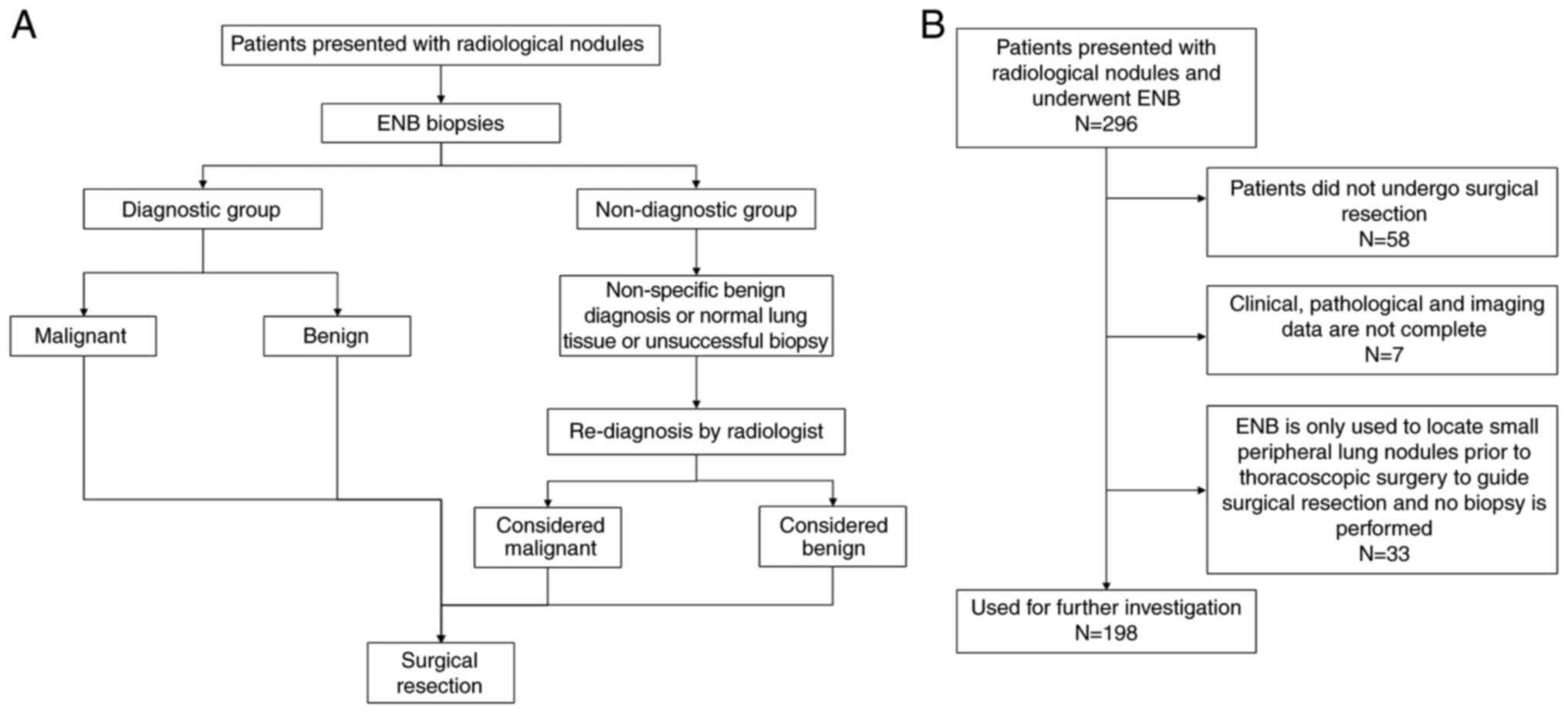
The present study retrospectively analyzed 296
patients who underwent ENB biopsy at The First Affiliated Hospital,
Zhejiang University School of Medicine (Hangzhou, China) between
February 2017 and September 2019. The diagnostic procedure is shown
in Fig. 1A. The eligibility
criteria were as follows: i) Patients were diagnosed with pulmonary
nodules that were suspected to be malignant tumors; ii) patients
underwent ENB; and iii) patients who eventually underwent surgical
resection. The exclusion criteria were as follows: i) Patients who
did not undergo surgical resection; ii) inoperative localization of
pulmonary nodule resection using the ENB technique; and iii)
patients with incomplete clinicopathological data (Fig. 1B). Finally, a total of 198 nodules
from 194 patients that were suspected to be malignant tumors were
included in the present study.
The initial pathology was obtained by ENB biopsy,
and the results of the ENB biopsy were divided into a diagnostic
group and a non-diagnostic group. The diagnostic group contained
cases with biopsy results of adenocarcinoma, squamous cell
carcinoma, other types of primary lung malignant tumors and
metastatic tumors, and other malignant tumors, or of benign lung
diseases, such as inflammatory nodules, fungal nodules and benign
tumors, such as hamartoma and sclerosed hemangioma. The
non-diagnostic group contained cases in which a clear diagnosis of
malignancy or benignity was not obtained after performing ENB. This
usually refers to cases in which the lesion was not successfully
punctured and the pathology was reported as normal lung tissue, no
bright malignant cells were seen, the specimen volume was too small
to be produced and cases where no specific diagnoses have been
made. In cases where the initial biopsy of ENB was non-diagnostic,
the nodules were evaluated again using imaging and a portion of the
patients will undergo surgical resection of malignant tumors or
benign tumors, such as hamartoma. In addition, some patients had
multiple nodules, and at their request, the ipsilateral nodule
together with the lesion considered malignant was removed. The
present study retrospectively reviewed cases of surgical resection
following ENB biopsy. All ENB biopsy procedures were performed by
the same experienced endoscopist, and the lung biopsy slides were
reviewed by two experienced thoracic pathologists to obtain a
pathological diagnosis.
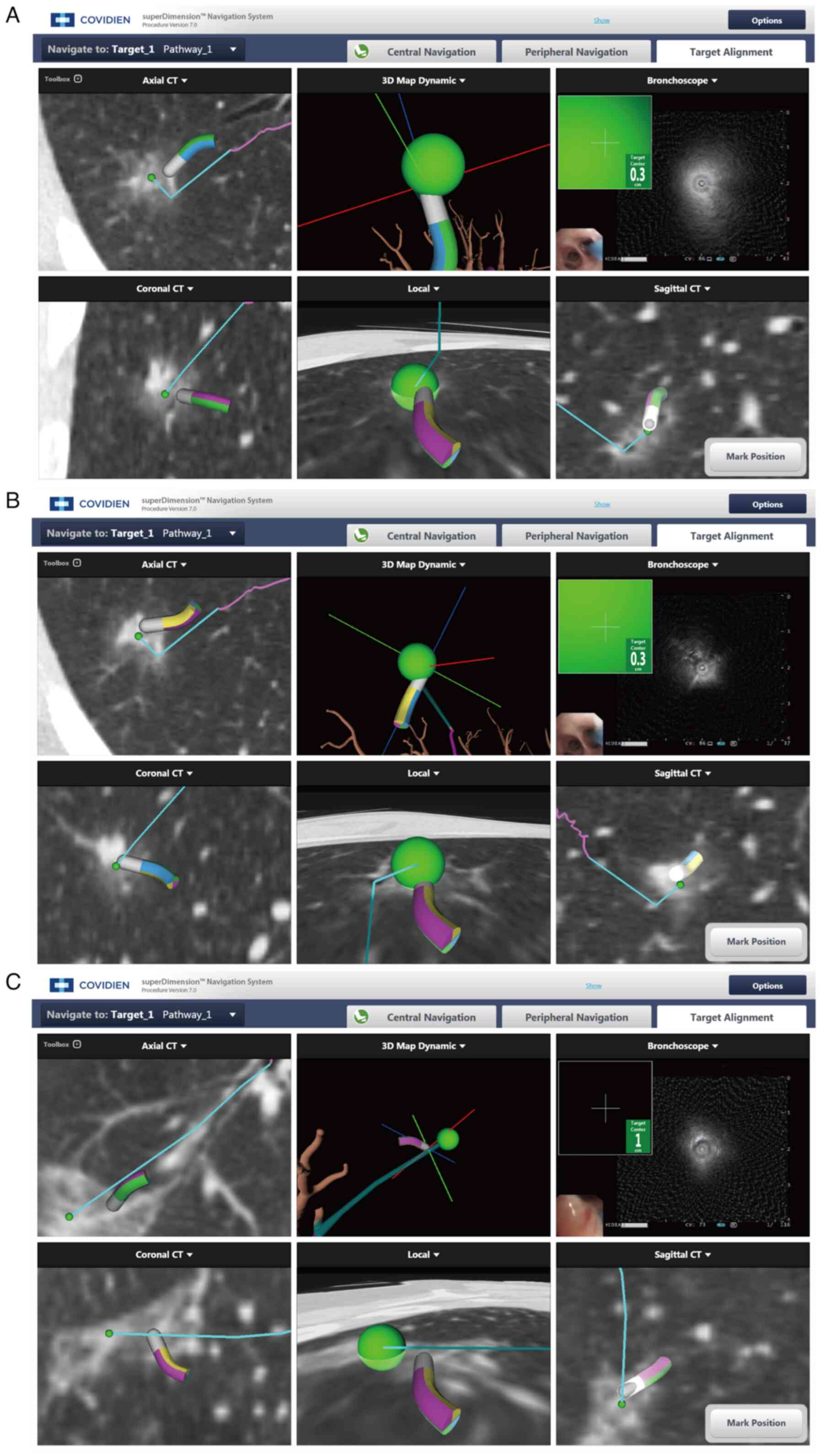
Composition and standard procedure of
ENB
ENB system (V Super Dimension version 7; Medtronic)
components mainly include: i) Electromagnetic positioning plate
that can generate a low-frequency uniform electromagnetic field.
The plate is generally required to be placed under the mattress of
the examination bed (>50 cm from the examined individual) so
that the patient's chest is in the electromagnetic field. ii)
Navigation probe, which is fixed to the tip of a bendable catheter
(diameter, 1 mm; length, 8 mm) and can be rotated 360°. In the
electromagnetic field, the orientation of the probe can be obtained
by the positioning system and transmitted to the computer in
real-time when X, Y and Z axis and tilt and rotation are carried
out in the body of the patient. iii) Extended operation channel,
which can be placed into the relevant operating instruments by the
navigation system to guide the target area for operation. iv) ENB
system hosts and monitors, through receiving and processing
magnetic navigation signals, actual organ images under the
bronchoscope are displayed and virtual navigation 3D tracheal
images are presented through the computer platform, to monitor and
guide the position and direction of the probe.
The ENB standard operation consists of two main
parts: i) Preoperative path planning, a virtual bronchial tree
image is generated by 3D reconstruction of the original CT image
data using ENB software, the target lesion markers are found on the
image, 5–7 anatomical markers are selected, and a navigation path
to the target lesion is generated; and ii) intraoperative
endotracheal navigation, after the patient is anesthetized, the
physician operates the electronic bronchoscope and places the
navigation probe through the working channel. The virtual image is
matched with the actual image by confirming the selected markers on
the virtual image with the actual position of the in vivo
probe. After successful matching, the target lesion is reached via
the preoperative planning path. The navigation ends with exit of
the localization probe, then the needle aspiration and biopsy are
performed via the extended channel (Fig. 2).
Data collection
The clinical, pathological and imaging data of the
included patients were collected through the hospital's electronic
medical record system. The variables, including age, sex and
history of prior malignancy, were extracted from the patients'
records. The radiological variables analyzed for each patient
included the nodule size, nodule type, distance between the pleura
and the nodule, and nodule position.
Statistical analysis
Categorical variables were assessed using the
χ2 test and Fisher's exact test. When continuous
variables obeyed normal distribution, such as age, the data were
analyzed using the independent sample t-test (unpaired parametric
Student's t-test). Nodule size, CT value and distance from pleura
were analyzed using the non-parametric Mann-Whitney U test. Odds
ratio (OR) with a 95% confidence interval (CI) was determined using
binary logistic regression. P<0.05 was considered to indicate a
statistically significant difference. Statistical analysis of all
data was performed using SPSS software (version 26.0; IBM
Corp.).
Results
Distribution of pathological
diagnostic results
Among the 198 nodules, there were 139 cases in the
diagnostic group and 59 (29.8%) cases in the non-diagnostic group.
A total of 165 cases were diagnosed as malignant tumors by surgical
pathology and 33 cases as benign diseases. In the diagnostic group,
there were 119 cases of malignant tumors and 20 cases of benign
diseases. In the non-diagnostic group, there were 46 cases of
malignant tumors and 13 cases of benign diseases, according to
surgical pathology (Table I).
 | Table I.Distribution of pathological
diagnostic results of study subjects. |
Table I.
Distribution of pathological
diagnostic results of study subjects.
| Category | Diagnostic group
(n=139) | Non-diagnostic group
(n=59) | Total (n=198) | P-value |
|---|
| Malignancy | 119 | 46 | 165 | 0.45 |
|
Adenocarcinoma | 112 | 42 |
|
|
| Squamous
carcinoma | 4 | 1 |
|
|
|
Metastasis | 1 | 0 |
|
|
| Other
types | 2 | 3 |
|
|
| Benign | 20 | 13 | 33 | 0.06 |
|
Inflammatory nodules | 10 | 6 |
|
|
|
Hamartoma | 1 | 2 |
|
|
| Fungal
nodules | 3 | 2 |
|
|
| Sclerosed
hemangioma | 1 | 2 |
|
|
| Other
types | 5 | 1 |
|
|
Diagnostic versus non-diagnostic
group
The median lesion size was 1.7 and 1.4 cm for
patients in the diagnostic and non-diagnostic groups, respectively.
Comparing the clinical and imaging data of the patients, there was
no significant difference regarding sex, age, previous history of
malignancy and bronchus signs (Table
II). There was a statistically significant difference between
the groups regarding nodule size (P<0.01), CT value (P<0.01)
and location of the nodules (P=0.04). Compared with subjects in the
diagnostic group, there were more non-solid nodules in the
non-diagnostic group. There were also significant differences in
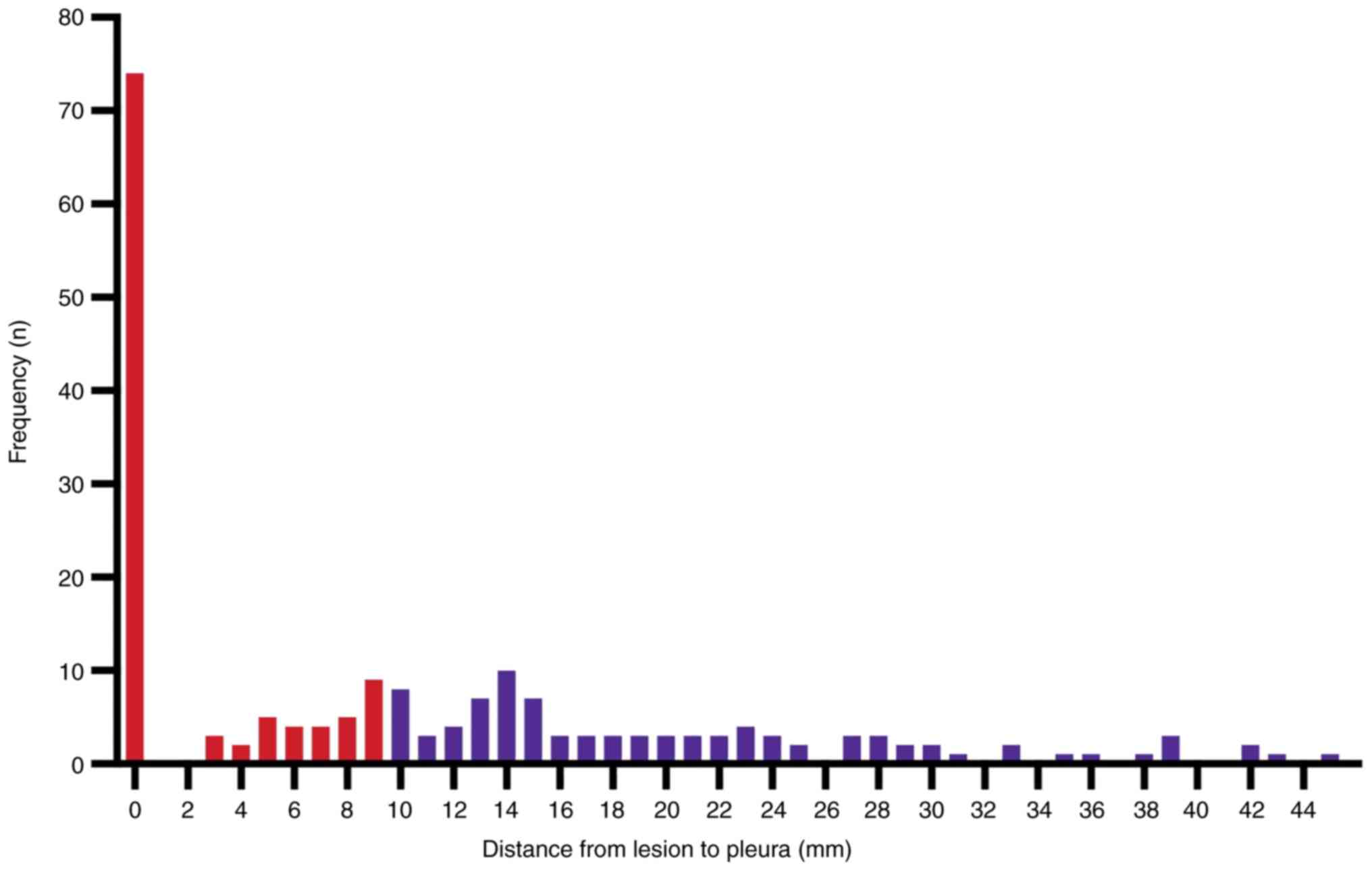
whether the nodules were in contact with the pleura (P=0.02).
Lesions were ≥10 mm from the pleura in 46.5% (92/198) of all cases;
and were on the pleura in 37.4% of all cases (Fig. 3).
| Table II.Clinicopathologic characteristics of
the study population. |
Table II.
Clinicopathologic characteristics of
the study population.
| Category | Diagnostic group
(n=139) | Non-diagnostic group
(n=59) | P-value |
|---|
| Sex |
|
| 0.44 |
| Male, n
(%) | 72 (36.4) | 27 (13.6) |
|
| Female, n
(%) | 67 (33.8) | 32 (16.2) |
|
| Mean age + SD,
years | 61.09±10.87 | 60.22±9.20 | 0.59 |
| History of
malignancy |
|
| 0.50 |
| Yes, n
(%) | 8 (4.0) | 4 (2.0) |
|
| No, n
(%) | 131 (66.2) | 55 (27.8) |
|
| Pathology
results |
|
| 0.19 |
| Benign,
n (%) | 20 (10.1) | 13 (6.6) |
|
|
Malignancy, n (%) | 119 (60.1) | 46 (23.2) |
|
| Median nodule size,
cm (IQS) | 1.7 (1.2) | 1.4 (1.0) | <0.01 |
| Nodule type |
|
| <0.01 |
| Solid,
n (%) | 66 (33.3) | 14 (7.0) |
|
| Pure
GGO, n (%) | 7 (3.5) | 11 (5.6) |
|
| Mixed
GGO, n (%) | 66 (33.3) | 34 (17.2) |
|
| Median CT value, Hu
(IQS) | −120 (−418) | −270 (−432) | <0.01 |
| Median distance
from pleura, cm (IQS) | 0.80 (1.60) | 1.00 (1.70) | 0.12 |
| Pleural
contact |
|
| 0.02 |
| Yes, n
(%) | 57 (28.8) | 14 (7.1) |
|
| No, n
(%) | 82 (41.4) | 45 (22.7) |
|
| Bronchus sign |
|
| 0.18 |
| Yes, n
(%) | 7 (3.5) | 6 (3.0) |
|
| No, n
(%) | 132 (66.7) | 53 (26.8) |
|
| Nodule
position |
|
| 0.04 |
| Upper
left lung, n (%) | 25 (12.6) | 20 (10.1) |
|
| Lower
left lung, n (%) | 17 (8.6) | 10 (5.1) |
|
| Upper
right lung, n (%) | 55 (27.8) | 14 (7.1) |
|
| Middle
lung, n (%) | 18 (9.1) | 4 (2.0) |
|
| Right
lower lung, n (%) | 24 (12.1) | 11 (5.6) |
|
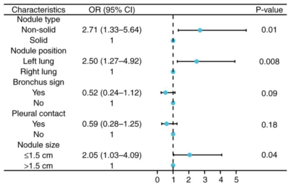
Predictive factor analysis
To explore the non-diagnostic predictors of ENB
biopsy results, a comparison of clinicopathological characteristics
between the two groups was performed using logistic regression.
Following univariate analysis, variables yielding P<0.05 were
analyzed using multivariate logistic regression analysis. The
multivariate analyses demonstrated that nodule size (P=0.04; OR,
2.05; 95% CI, 1.03-4.09), nodule type (P=0.01; OR, 2.71; 95% CI,
1.33-5.64) and nodule position (P=0.008; OR, 2.50; 95% CI,
1.27-4.92) were independent non-diagnostic risk factors for ENB
biopsy (Fig. 4).
Subgroup analysis of the
non-diagnostic group
In the non-diagnostic group, the final pathological
results for 46 cases were malignant tumors, and the final
pathological results of the remaining 13 cases were benign; 78.0%
of non-diagnosed cases were found to be malignant by surgery. The
present study compared and analyzed the data of the two groups and
the results are presented in Table
III. Among them, there was a statistically significant
difference in the nodule type and CT value (both P<0.05).
| Table III.Non-diagnostic subgroup analysis. |
Table III.
Non-diagnostic subgroup analysis.
| Category | Malignant
(n=46) | Benign (n=13) | P-value |
|---|
| Sex |
|
| 0.22 |
| Male,
n | 19 | 8 |
|
| Female,
n | 27 | 5 |
|
| Mean age + SD,
years | 61.28±7.33 | 56.46±7.88 | 0.10 |
| History of
malignancy |
|
| 0.99 |
| Yes,
n | 3 | 1 |
|
| No,
n | 43 | 12 |
|
| Median nodule size,
cm (IQS) | 1.4 (1.0) | 1.3 (0.7) | 0.18 |
| Nodule type |
|
| <0.01 |
| Solid,
n | 5 | 9 |
|
| Pure
GGO, n | 10 | 1 |
|
| Mixed
GGO, n | 31 | 3 |
|
| Median CT value, Hu
(IQS) | −395.00
(−397.50) | −12.00
(−282.00) | <0.01 |
| Median distance
from pleura, cm (IQS) | 1.00 (1.63) | 1.10 (1.20) | 0.61 |
| Pleural
contact |
|
| 0.99 |
| Yes,
n | 11 | 3 |
|
| No,
n | 35 | 10 |
|
| Bronchus sign |
|
| 0.63 |
| Yes,
n | 11 | 3 |
|
| No,
n | 35 | 10 |
|
| Nodule
position |
|
| 0.40 |
| Upper
left lung, n |
| 16 | 4 |
| Lower
left lung, n |
| 7 | 3 |
| Upper
right lung, n |
| 13 | 1 |
| Middle
lung, n |
| 3 | 1 |
| Right
lower lung, n |
| 7 | 4 |
In addition, a multivariate regression model was
used to explore the independent risk factors for the initial
non-diagnosed cases that received a final surgical diagnosis of
malignant tumors. Among them, non-solid nodules (P<0.05; OR,
7.64; 95% CI, 3.11-18.76) was an independent risk factor for
malignant tumors in the initial non-diagnostic group (data not
shown).
Discussion
With the promotion of lung cancer screening, the
detection rate of lung nodules has increased, and the mortality
rate of patients with lung cancer has continued to decline
(18,19). In the age of precision medicine, it
is necessary to formulate precise strategies for every nodule
detected in each patient, either by observation and follow-up,
direct surgical resection or biopsy to determine the pathology. In
particular, individuals at a high risk of lung cancer need to be
treated with caution and different strategies need to be adopted
for different nodules to maximize efficacy (20). Previous studies have reported an 88%
diagnostic yield for large central lesions using bronchoscopy for
tissue sampling, but a significant decrease in diagnostic efficacy
has been identified for small peripheral lung lesions, especially
small nodular lesions below the segmental plane. In one study,
bronchoscopy had a diagnostic yield of only 14% for peripheral
pulmonary lesions <2 cm, and it has been shown that for
peripheral pulmonary lesions, the diagnosis rate of navigation
bronchoscopy is higher than that of non-navigated bronchoscopy
(21). Navigation bronchoscopy is
split into virtual navigation and electromagnetic navigation
(22). Among them, ENB has been
shown to have better specificity than virtual navigation
bronchoscopy (22). ENB was first
applied to the human body in the early 21st century, and it has
been widely used in the diagnosis and treatment of lung cancer by a
number of centers. Previous studies have reported that the overall
accuracy, sensitivity and specificity of ENB biopsy is acceptable
(14,23); however, these studies are limited by
sample size, diagnostic definitions, alternative definitions and
heterogeneity between groups. The results have large volatility and
poor reliability. In 2014, a systematic review and analysis on ENB
biopsy reported that although the overall accuracy of ENB in
diagnosing malignant tumors is considered acceptable, the negative
predictive value of ENB for malignant tumors is only 52.1% (95% CI,
43.5-60.6) (16); in different
research reports, the negative predictive value fluctuates between
25.0 and 89.5% (10,13,21),
but the histological benign diagnosis obtained by an ENB biopsy is
not enough to exclude cancer (16).
The prospective multi-center NAVIGATE study included >1,000
cases to initially evaluate the safety and accuracy of ENB. The
pneumothorax rate published by NAVIGATE was 3.1% and the accuracy
of the ENB initial biopsy was 72.9% (24). Even if the samples obtained show
chronic inflammation or granulomatous inflammation, the negative
predictive value of ENB is still not ideal.
The present study included ~200 biopsy cases and the
final pathology was obtained by lung surgery. The results revealed
that nodules sized ≤1.5 cm, non-solid nodules and nodules in the
left lung were independent non-diagnostic risk factors for ENB
biopsy.
It is generally known that nodule size is one of the
most important factors in the success of ENB biopsy, and the
well-known NAVIGATE study reported that nodule size ≥2 cm was a
significant univariate predictor of diagnostic yield (24). Non-solid nodules contain a large
number of ground glass nodules and are relatively small in diameter
(25). In addition, most do not
have bronchial signs, are relatively difficult to obtain and pose a
great diagnostic challenge to pathologists (23). Notably, the present study revealed
that the presence of nodules in the left lung was also a predictor
of initial non-diagnosis by ENB biopsy; to the best of our
knowledge, this has not been reported previously. The present study
hypothesized that this may be related to the movement of pulmonary
nodules due to respiratory motion of the left, as well as the
right, lung during ENB. It has previously been reported that the
change from full inspiration during a chest CT scan to tidal volume
breathing during a bronchoscopy may significantly affect the
diagnostic rate of ENB (26). The
present study has the limitation that it was a single-center
retrospective study; however, further studies to explore the
factors that influence the success of ENB biopsy will be
conducted.
Non-solid nodules were independent risk factors for
malignant tumors in the ENB non-diagnostic group. The accuracy of
the overall initial biopsy pathology and final pathological
diagnosis in the present study was not high, because the overall
diameter of the lung nodules included in the present study was
relatively small. The median nodule size of the 198 cases was 1.6
cm, and the interquartile range was 1.1 cm, including 90 cases with
nodules ≤1.5 cm in diameter (45.6% of the total cases). Among them,
there were 27 patients with nodules ≤1.0 cm in diameter, including
more ground glass nodules. Usually, if the nodule is in contact
with the pleura, the diagnostic rate of bronchoscopy becomes worse
because there are fewer bronchial signs; however, in the present
study, there was no significant statistical difference between the
two groups in terms of the bronchial signs. The possible reasons
why there was no statistical difference are as follows: First, the
bronchoscopic transparenchymal nodule access technique has been
well established at our center, which is also known as the
tunneling technique; during this technique, a hole is made in the
bronchial wall and a tunnel is created to reach the nodule through
a working channel in the lung parenchyma, allowing theoretical
‘whole lung access’ to the nodule without relying on the natural
bronchial lumen. Moreover, in the present study, the diameter of
the lesion in pleural contact was relatively large, which may cause
a bias in the study.
In the non-diagnostic group, 78.0% of the cases were
finally confirmed as malignant tumors by surgery, which indicates
that clinicians should be cautious in making decisions when the
initial ENB biopsy fails to obtain a clear diagnosis. Options to
improve diagnosis may include combining ENB biopsy with PET-CT,
another ENB biopsy or another biopsy method, in order to prevent
delays in diagnosis and treatment, especially for patients with
ground glass lesions.
In general, ENB biopsy is a safe and effective
technical method; however, it is more difficult to apply if the
nodules have a diameter of <1.5 cm or if the nodules have fewer
solid components. In addition, it is necessary to combine multiple
diagnostic methods to ensure an accurate diagnosis. The present
study explored the related factors of ENB biopsy failure and
inaccurate diagnosis. In addition, the final outcome of the
included cases was obtained through surgery to obtain the
pathological gold standard. Compared with imaging follow-up, the
data quality was more reliable. In addition, in the present study,
more difficult explorations of ENB biopsy of peripheral lung
nodules were conducted and sampling biopsy of smaller nodules and
more ground glass lesions was performed. Notably, the present study
has certain limitations. This study is a single-center
retrospective study, and there is inevitably a selection bias,
which limits the applicability of the conclusions to a certain
extent. In the future, a multicenter, prospective study will be
conducted to explore the diagnosis of ENB biopsy further.
In conclusion, the predictive factors for a
non-diagnostic ENB biopsy were nodule size ≤1.5 cm and non-solid
nodules. It was not rare for patients to finally be diagnosed with
a malignancy in the non-diagnostic group. Therefore, care should be
taken when the results of ENB biopsy are non-diagnostic to prevent
misdiagnosis.
Acknowledgements
Not applicable.
Funding
This work was supported by the Zhejiang Province Major Science
and Technology Special Program Project (grant no. 2020C03058) and
the Zhejiang Province Lung Tumor Diagnosis and Treatment Technology
Research Center (grant no. JBZX-202007).
Availability of data and materials
The datasets used and/or analyzed during the current
study are available from the corresponding author on reasonable
request.
Authors' contributions
WY, HM, BY and JH developed the concept for the
study. ZA, PX, LY and GY contributed to the data collection. WY, WL
and HM conducted the statistical analysis. WY and HM contributed to
the production of the manuscript. All authors contributed to the
article, and read and approved the final manuscript. WY and HM
confirm the authenticity of all the raw data.
Ethics approval and consent to
participate
The present study was approved by the Medical Ethics
Committee and Institutional Review Board of The First Affiliated
Hospital, Zhejiang University (approval no. 2022-764). All
procedures performed in studies involving human participants were
in accordance with the ethical standards of the institutional
and/or national research committee and with The 1964 Declaration of
Helsinki, and its later amendments or comparable ethical standards.
Written informed consent for participation was obtained from all
patients.
Patient consent for publication
Written informed consent for publication was
obtained from all patients.
Competing interests
The authors declare that they have no competing
interests.
References
|
1
|
Siegel RL, Miller KD, Fuchs HE and Jemal
A: Cancer statistics, 2021. CA Cancer J Clin. 71:7–33. 2021.
View Article : Google Scholar : PubMed/NCBI
|
|
2
|
Feng RM, Zong YN, Cao SM and Xu RH:
Current cancer situation in China: Good or bad news from the 2018
global cancer statistics? Cancer Commun (Lond). 39:222019.
View Article : Google Scholar : PubMed/NCBI
|
|
3
|
Miller KD, Nogueira L, Devasia T, Mariotto
AB, Yabroff KR, Jemal A, Kramer J and Siegel RL: Cancer treatment
and survivorship statistics, 2022. CA Cancer J Clin. 72:409–436.
2022. View Article : Google Scholar : PubMed/NCBI
|
|
4
|
Goldstraw P, Chansky K, Crowley J,
Rami-Porta R, Asamura H, Eberhardt WE, Nicholson AG, Groome P,
Mitchell A, Bolejack V, et al: The iaslc lung cancer staging
project: Proposals for revision of the tnm stage groupings in the
forthcoming (Eighth) edition of the TNM classification for lung
cancer. J Thorac Oncol. 11:39–51. 2016. View Article : Google Scholar : PubMed/NCBI
|
|
5
|
Oudkerk M, Liu S, Heuvelmans MA, Walter JE
and Field JK: Lung cancer LDCT screening and mortality
reduction-evidence, pitfalls and future perspectives. Nat Rev Clin
Oncol. 18:135–151. 2021. View Article : Google Scholar : PubMed/NCBI
|
|
6
|
Chansky K, Detterbeck FC, Nicholson AG,
Rusch VW, Vallières E, Groome P, Kennedy C, Krasnik M, Peake M,
Shemanski L, et al: The IASLC lung cancer staging project: External
validation of the revision of the Tnm stage groupings in the eighth
edition of the TNM classification of lung cancer. J Thorac Oncol.
12:1109–1121. 2017. View Article : Google Scholar : PubMed/NCBI
|
|
7
|
Borelli C, Vergara D, Simeone A, Pazienza
L, Castorani G, Graziano P, Micco CD, Quarato CMI and Sperandeo M:
CT-guided transthoracic biopsy of pulmonary lesions: Diagnostic
versus nondiagnostic results. Diagnostics (Basel). 12:3592022.
View Article : Google Scholar : PubMed/NCBI
|
|
8
|
Criner GJ, Eberhardt R, Fernandez-Bussy S,
Gompelmann D, Maldonado F, Patel N, Shah PL, Slebos DJ, Valipour A,
Wahidi MM, et al: Interventional bronchoscopy. Am J Respir Crit
Care Med. 202:29–50. 2020. View Article : Google Scholar : PubMed/NCBI
|
|
9
|
Mehta AC, Hood KL, Schwarz Y and Solomon
SB: The evolutional history of electromagnetic navigation
bronchoscopy: State of the art. Chest. 154:935–947. 2018.
View Article : Google Scholar : PubMed/NCBI
|
|
10
|
Folch EE, Labarca G, Ospina-Delgado D,
Kheir F, Majid A, Khandhar SJ, Mehta HJ, Jantz MA and
Fernandez-Bussy S: Sensitivity and safety of electromagnetic
navigation bronchoscopy for lung cancer diagnosis: Systematic
review and meta-analysis. Chest. 158:1753–1769. 2020. View Article : Google Scholar : PubMed/NCBI
|
|
11
|
Cheng SL and Chu CM: Electromagnetic
navigation bronchoscopy: The initial experience in Hong Kong. J
Thorac Dis. 11:1697–1704. 2019. View Article : Google Scholar : PubMed/NCBI
|
|
12
|
Cho HJ, Roknuggaman M, Han WS, Kang SK and
Kang MW: Electromagnetic navigation bronchoscopy-Chungnam national
university hospital experience. J Thorac Dis. 10 (Suppl
6):S717–S724. 2018. View Article : Google Scholar : PubMed/NCBI
|
|
13
|
Andersen FD, Degn KB and Rasmussen TR:
Electromagnetic navigation bronchoscopy for lung nodule evaluation.
Patient selection, diagnostic variables and safety. Clin Respir J.
14:557–563. 2020. View Article : Google Scholar : PubMed/NCBI
|
|
14
|
Patrucco F, Gavelli F, Daverio M, Antonini
C, Boldorini R, Casadio C and Balbo PE: Electromagnetic navigation
bronchoscopy: Where are we now? Five years of a single-center
experience. Lung. 196:721–727. 2018. View Article : Google Scholar : PubMed/NCBI
|
|
15
|
Baaklini WA, Reinoso MA, Gorin AB,
Sharafkaneh A and Manian P: Diagnostic yield of fiberoptic
bronchoscopy in evaluating solitary pulmonary nodules. Chest.
117:1049–1054. 2000. View Article : Google Scholar : PubMed/NCBI
|
|
16
|
Gex G, Pralong JA, Combescure C, Seijo L,
Rochat T and Soccal PM: Diagnostic yield and safety of
electromagnetic navigation bronchoscopy for lung nodules: A
systematic review and meta-analysis. Respiration. 87:165–176. 2014.
View Article : Google Scholar : PubMed/NCBI
|
|
17
|
Taton O, Bondue B, Gevenois PA, Remmelink
M and Leduc D: Diagnostic yield of combined pulmonary cryobiopsies
and electromagnetic navigation in small pulmonary nodules. Pulm
Med. 2018:60329742018. View Article : Google Scholar : PubMed/NCBI
|
|
18
|
de Koning HJ, van der Aalst CM, de Jong
PA, Scholten ET, Nackaerts K, Heuvelmans MA, Lammers JWJ, Weenink
C, Yousaf-Khan U, Horeweg N, et al: Reduced lung-cancer mortality
with volume CT screening in a randomized trial. New Engl J Med.
382:503–513. 2020. View Article : Google Scholar : PubMed/NCBI
|
|
19
|
Wang Z, Han W, Zhang W, Xue F, Wang Y, Hu
Y, Wang L, Zhou C, Huang Y, Zhao S, et al: Mortality outcomes of
low-dose computed tomography screening for lung cancer in urban
China: A decision analysis and implications for practice. Chin J
Cancer. 36:572017. View Article : Google Scholar : PubMed/NCBI
|
|
20
|
Ghosh S, Mehta AC, Abuquyyas S, Raju S and
Farver C: Primary lung neoplasms presenting as multiple synchronous
lung nodules. Eur Respir Rev. 29:1901422020. View Article : Google Scholar : PubMed/NCBI
|
|
21
|
McGuire AL, Myers R, Grant K, Lam S and
Yee J: The diagnostic accuracy and sensitivity for malignancy of
radial-endobronchial ultrasound and electromagnetic navigation
bronchoscopy for sampling of peripheral pulmonary lesions:
Systematic review and meta-analysis. J Bronchology Interv Pulmonol.
27:106–121. 2020. View Article : Google Scholar : PubMed/NCBI
|
|
22
|
Jiang S, Xie F, Mao X, Ma H and Sun J: The
value of navigation bronchoscopy in the diagnosis of peripheral
pulmonary lesions: A meta-analysis. Thorac Cancer. 11:1191–1201.
2020. View Article : Google Scholar : PubMed/NCBI
|
|
23
|
Ishiwata T, Gregor A, Inage T and Yasufuku
K: Bronchoscopic navigation and tissue diagnosis. Gen Thorac
Cardiovasc Surg. 68:672–678. 2019. View Article : Google Scholar : PubMed/NCBI
|
|
24
|
Folch EE, Pritchett MA, Nead MA, Bowling
MR, Murgu SD, Krimsky WS, Murillo BA, LeMense GP, Minnich DJ,
Bansal S, et al: Electromagnetic navigation bronchoscopy for
peripheral pulmonary lesions: One-year results of the prospective,
multicenter navigate study. J Thorac Oncol. 14:445–458. 2019.
View Article : Google Scholar : PubMed/NCBI
|
|
25
|
Mazzone PJ and Lam L: Evaluating the
patient with a pulmonary nodule: A review. Jama. 327:264–273. 2022.
View Article : Google Scholar : PubMed/NCBI
|
|
26
|
Chen A, Pastis N, Furukawa B and Silvestri
GA: The effect of respiratory motion on pulmonary nodule location
during electromagnetic navigation bronchoscopy. Chest.
147:1275–1281. 2015. View Article : Google Scholar : PubMed/NCBI
|