|
1
|
Louis DN, Perry A, Reifenberger G, von
Deimling A, Figarella-Branger D, Cavenee WK, Ohgaki H, Wiestler OD,
Kleihues P and Ellison DW: The 2016 world health organization
classification of tumors of the central nervous system: A summary.
Acta Neuropathol. 131:803–820. 2016. View Article : Google Scholar : PubMed/NCBI
|
|
2
|
Helseth R, Helseth E, Johannesen TB,
Langberg CW, Lote K, Rønning P, Scheie D, Vik A and Meling TR:
Overall survival, prognostic factors, and repeated surgery in a
consecutive series of 516 patients with glioblastoma multiforme.
Acta Neurol Scand. 122:159–167. 2010. View Article : Google Scholar : PubMed/NCBI
|
|
3
|
Woodworth GF, McGirt MJ, Samdani A,
Garonzik I, Olivi A and Weingart JD: Frameless image-guided
stereotactic brain biopsy procedure: Diagnostic yield, surgical
morbidity, and comparison with the frame-based technique. J
Neurosurg. 104:233–237. 2006. View Article : Google Scholar : PubMed/NCBI
|
|
4
|
Ryu YJ, Choi SH, Park SJ, Yun TJ, Kim JH
and Sohn CH: Glioma: Application of whole-tumor texture analysis of
diffusion-weighted imaging for the evaluation of tumor
heterogeneity. PLoS One. 9:e1083352014. View Article : Google Scholar : PubMed/NCBI
|
|
5
|
Jackson A, O'Connor JP, Parker GJ and
Jayson GC: Imaging tumor vascular heterogeneity and angiogenesis
using dynamic contrast-enhanced magnetic resonance imaging. Clin
Cancer Res. 13:3449–3459. 2007. View Article : Google Scholar : PubMed/NCBI
|
|
6
|
Gillies RJ, Kinahan PE and Hricak H:
Radiomics: Images are more than pictures, they are data. Radiology.
278:563–577. 2016. View Article : Google Scholar : PubMed/NCBI
|
|
7
|
Lubner MG, Smith AD, Sandrasegaran K,
Sahani DV and Pickhardt PJ: CT texture analysis: Definitions,
applications, biologic correlates, and challenges. Radiographics.
37:1483–1503. 2017. View Article : Google Scholar : PubMed/NCBI
|
|
8
|
Lee J, Mulder F, Leeflang M, Wolff R,
Whiting P and Bossuyt PM: QUAPAS: An adaptation of the QUADAS-2
tool to assess prognostic accuracy studies. Ann Intern Med.
175:1010–1018. 2022. View
Article : Google Scholar : PubMed/NCBI
|
|
9
|
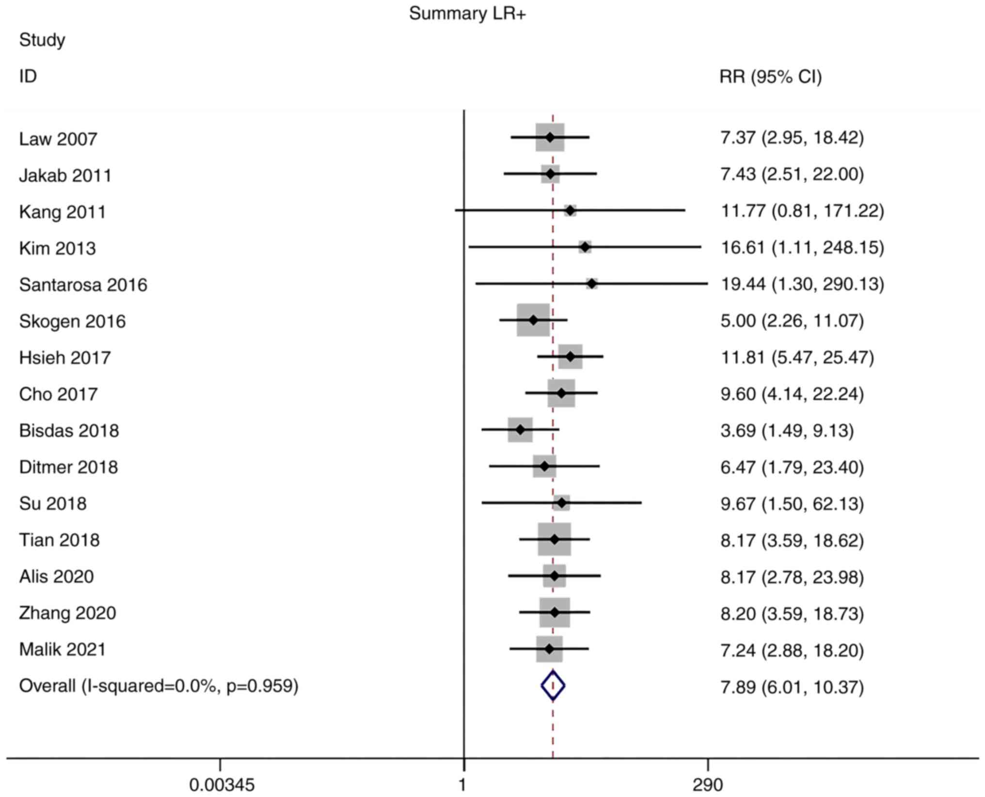
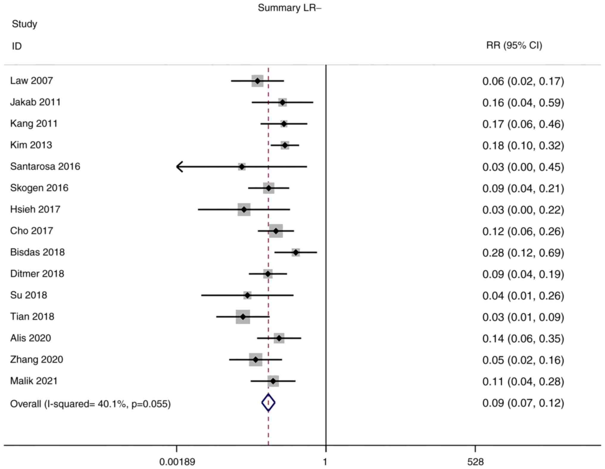
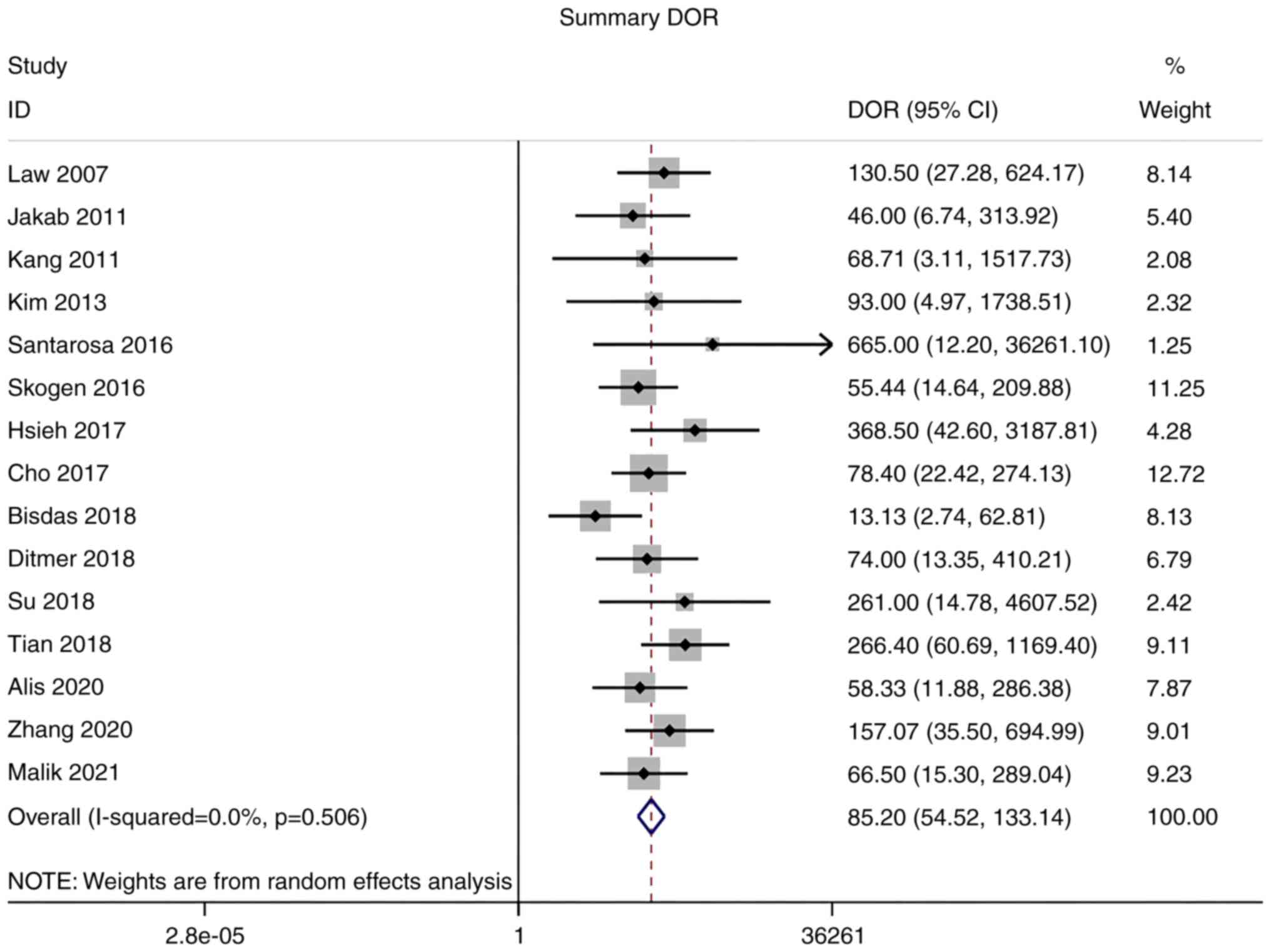
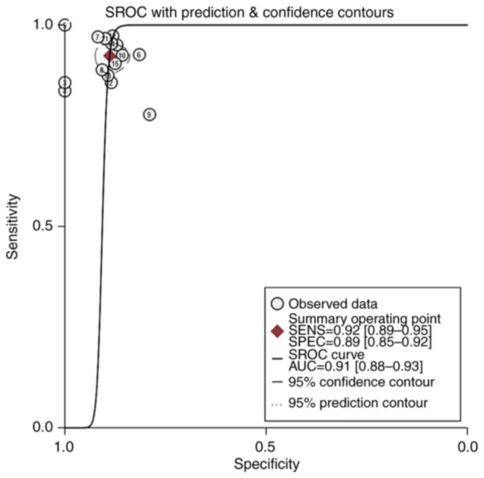
Law M, Young R, Babb J, Pollack E and
Johnson G: Histogram analysis versus region of interest analysis of
dynamic susceptibility contrast perfusion MR imaging data in the
grading of cerebral gliomas. AJNR Am J Neuroradiol. 28:761–766.
2007.PubMed/NCBI
|
|
10
|
Jakab A, Molnár P, Emri M and Berényi E:
Glioma grade assessment by using histogram analysis of diffusion
tensor imaging-derived maps. Neuroradiology. 53:483–491. 2011.
View Article : Google Scholar : PubMed/NCBI
|
|
11
|
Kang Y, Choi SH, Kim YJ, Kim KG, Sohn CH,
Kim JH, Yun TJ and Chang KH: Gliomas: Histogram analysis of
apparent diffusion coefficient maps with standard-or high-b-value
diffusion-weighted MR imaging-correlation with tumor grade.
Radiology. 261:882–890. 2011. View Article : Google Scholar : PubMed/NCBI
|
|
12
|
Kim H, Choi SH, Kim JH, Ryoo I, Kim SC,
Yeom JA, Shin H, Jung SC, Lee AL, Yun TJ, et al: Gliomas:
Application of cumulative histogram analysis of normalized cerebral
blood volume on 3 T MRI to tumor grading. PLoS One. 8:e634622013.
View Article : Google Scholar : PubMed/NCBI
|
|
13
|
Santarosa C, Castellano A, Conte GM,
Cadioli M, Iadanza A, Terreni MR, Franzin A, Bello L, Caulo M,
Falini A and Anzalone N: Dynamic contrast-enhanced and dynamic
susceptibility contrast perfusion MR imaging for glioma grading:
Preliminary comparison of vessel compartment and permeability
parameters using hotspot and histogram analysis. Eur J Radiol.
85:1147–1156. 2016. View Article : Google Scholar : PubMed/NCBI
|
|
14
|
Skogen K, Schulz A, Dormagen JB, Ganeshan
B, Helseth E and Server A: Diagnostic performance of texture
analysis on MRI in grading cerebral gliomas. Eur J Radiol.
85:824–829. 2016. View Article : Google Scholar : PubMed/NCBI
|
|
15
|
Hsieh KLC, Chen CY and Lo CM: Quantitative
glioma grading using transformed gray-scale invariant textures of
MRI. Comput Biol Med. 83:102–108. 2017. View Article : Google Scholar : PubMed/NCBI
|
|
16
|
Cho HH and Park H: Classification of
low-grade and high-grade glioma using multi-modal image radiomics
features. Annu Int Conf IEEE Eng Med Biol Soc. 2017:3081–3084.
2017.PubMed/NCBI
|
|
17
|
Bisdas S, Shen H, Thust S, Katsaros V,
Stranjalis G, Boskos C, Brandner S and Zhang J: Texture
analysis-and support vector machine-assisted diffusional kurtosis
imaging may allow in vivo gliomas grading and IDH-mutation status
prediction: A preliminary study. Sci Rep. 8:61082018. View Article : Google Scholar : PubMed/NCBI
|
|
18
|
Ditmer A, Zhang B, Shujaat T, Pavlina A,
Luibrand N, Gaskill-Shipley M and Vagal A: Diagnostic accuracy of
MRI texture analysis for grading gliomas. J Neurooncol.
140:583–589. 2018. View Article : Google Scholar : PubMed/NCBI
|
|
19
|
Su CQ, Lu SS, Han QY, Zhou MD and Hong XN:
Intergrating conventional MRI, texture analysis of dynamic
contrast-enhanced MRI, and susceptibility weighted imaging for
glioma grading. Acta Radiol. 2019 Jun;60((6)): 777–787. View Article : Google Scholar : PubMed/NCBI
|
|
20
|
Tian Q, Yan LF, Zhang X, Zhang X, Hu YC,
Han Y, Liu ZC, Nan HY, Sun Q, Sun YZ, et al: Radiomics strategy for
glioma grading using texture features from multiparametric MRI. J
Magn Reson Imaging. 48:1518–1528. 2018. View Article : Google Scholar : PubMed/NCBI
|
|
21
|
Alis D, Bagcilar O, Senli YD, Isler C,
Yergin M, Kocer N, Islak C and Kizilkilic O: The diagnostic value
of quantitative texture analysis of conventional MRI sequences
using artificial neural networks in grading gliomas. Clin Radiol.
75:351–357. 2020. View Article : Google Scholar : PubMed/NCBI
|
|
22
|
Zhang Z, Xiao J, Wu S, Lv F, Gong J, Jiang
L, Yu R and Luo T: Deep convolutional radiomic features on
diffusion tensor images for classification of glioma grades. J
Digit Imaging. 33:826–837. 2020. View Article : Google Scholar : PubMed/NCBI
|
|
23
|
Malik N, Geraghty B, Dasgupta A, Maralani
PJ, Sandhu M, Detsky J, Tseng CL, Soliman H, Myrehaug S, Husain Z,
et al: MRI radiomics to differentiate between low grade glioma and
glioblastoma peritumoral region. J Neurooncol. 155:181–191. 2021.
View Article : Google Scholar : PubMed/NCBI
|
|
24
|
Lambin P, Rios-Velazquez E, Leijenaar R,
Carvalho S, van Stiphout RG, Granton P, Zegers CM, Gillies R,
Boellard R, Dekker A and Aerts HJ: Radiomics: Extracting more
information from medical images using advanced feature analysis.
Eur J Cancer. 48:441–446. 2012. View Article : Google Scholar : PubMed/NCBI
|
|
25
|
Sohn CK and Bisdas S: Diagnostic accuracy
of machine learning-based radiomics in grading gliomas: systematic
review and meta-analysis. Contrast Media Mol Imaging.
2020:21270622020. View Article : Google Scholar : PubMed/NCBI
|
|
26
|
Liu C, Zhao W, Xie J, Lin H, Hu X, Li C,
Shang Y, Wang Y, Jiang Y, Ding M, et al: Development and validation
of a radiomics-based nomogram for predicting a major pathological
response to neoadjuvant immunochemotherapy for patients with
potentially resectable non-small cell lung cancer. Front Immunol.
14:11152912023. View Article : Google Scholar : PubMed/NCBI
|
|
27
|
Li J, Xia F, Wang X, Jin Y, Yan J, Wei X
and Zhao Q: Multiclassifier radiomics analysis of ultrasound for
prediction of extrathyroidal extension in papillary thyroid
carcinoma in children. Int J Med Sci. 20:278–286. 2023. View Article : Google Scholar : PubMed/NCBI
|
|
28
|
Berenguer R, Pastor-Juan MDR,
Canales-Vázquez J, Castro-García M, Villas MV, Mansilla Legorburo F
and Sabater S: Radiomics of CT features may be nonreproducible and
redundant: influence of CT acquisition parameters. Radiology.
288:407–415. 2018. View Article : Google Scholar : PubMed/NCBI
|