Introduction
Hepatocellular carcinoma (HCC) is the third most
frequent cause of cancer-associated death worldwide and represents
a major global health challenge (1,2).
Currently, the global five-year survival rate of metastatic HCC is
<20% (3). Great progress has
been made in the diagnosis and treatment of HCC, but the mortality
rate remains unsatisfactory (3,4).
According to the stage of the tumor and the condition of the
patient, there are various treatment options for HCC. A number of
curative treatment options exist for early-stage HCC, including
surgical resection, radiofrequency ablation and liver
transplantation. However, 80% of patients have unresectable HCC and
can only be treated with locoregional therapies such as
radiotherapy, transarterial chemoembolization or transarterial
radioembolization (2). Due to high
relapse rates and drug resistance, the prognosis for most HCC
patients is less than ideal (5,6). It is
therefore imperative to explore new therapeutic targets.
Dysregulated DNA replication in cells is a major
contributor to tumor initiation and progression. Minichromosome
maintenance proteins (MCMs) are essential for DNA replication and
are the subject of considerable research interest (7). The aberrant expression of MCMs has
been detected in numerous malignancies, which can lead to genomic
instability and uncontrolled cell cycle progression (7,8). The
MCM2-7 family is a class of nuclear proteins that contain a highly
conserved nucleoside triphosphate binding motif, and are
evolutionarily and functionally conserved in eukaryotes (9). Members of the MCM2-7 family have been
reported to be upregulated in various cancer tissues and cancer
cell lines, including brain tumors, lymphomas, and breast, lung and
prostate cancers (10–14). For example, MCM2 has been shown to
be associated with the malignant status of lung squamous cell
carcinoma and to regulate the proliferation and cell cycle in this
type of cancer (13).
MCM6 is an important component of the MCM2-7 complex
and is upregulated in a number of tumors, including HCC (15–17).
MCM6 promotes the metastasis of HCC via the MEK/ERK pathway and may
serve as a serum biomarker for early recurrence (15). In addition, MCM6 expression is
inversely correlated with methyl(R217) human antigen R and is a
prognostic marker in non-small cell lung carcinoma (16).
In the present study the role of MCM2-7 in HCC was
investigated and the effect of the knockdown of members of this
family on the proliferation of human HCC cells was evaluated. The
homeostasis of MCM2, MCM3, MCM6 and MCM7 was then investigated.
Also, as only MCM6 was found to be mainly degraded by the
proteasome pathway, it was selected for further study. Ring finger
protein 125 (RNF125) was identified as an E3 ubiquitin ligase for
MCM6 and the relationship of these two proteins in HCC was
investigated.
Materials and methods
Data analysis using public
databases
Gene expression profiling interactive analysis 2
(GEPIA2)
GEPIA2 (http://gepia2.cancer-pku.cn) is a tool for the gene
expression analysis of sequencing data from The Cancer Genome Atlas
and the Tissue Genotype Expression database (18). In the present study, GEPIA2 was used
to evaluate the mRNA expression of MCM2-7 and RNF125 in human liver
HCC (LIHC) and calculate P-values using Student's t-test; an
absolute log2 fold change ≥0.5 and P<0.05 were considered to
indicate a significant difference. GEPIA2 was also used to perform
a prognostic value analysis by the calculation of overall survival
and disease-free survival rates and survival graphs were directly
generated by GEPIA2, with the log-rank test the only option for
analysis. The associations between different MCMs and the clinical
outcomes of LIHC, and the associations between the mRNA expression
of certain MCMs and the pathological stage of LIHC were also
assessed using GEPIA2.
University of alabama cancer database
(UALCAN)
UALCAN (http://ualcan.path.uab.edu) is a comprehensive and
interactive web resource for the analysis of cancer omics data
(19). The protein levels of MCM2-7
in human LIHC were assessed using UALCAN, and Student's t-test was
used to generate P-values.
Plasmid construction
Plasmids containing RNF125 and MCM2-7 were purchased
from Shanghai Cell Researcher Biotech Co., Ltd. and inserted into
empty vectors, which were purchased from Cell Researcher Biotech
Co., Ltd.), such as pCDH, pCDNA3.0-Myc, pET22b or pGEX4T-1.
Briefly, pCDH-MCM2/MCM3/MCM6/MCM7/RNF125, pCDNA3.0-RNF126-Myc,
pET22b-MCM2/MCM3/MCM6/MCM7/RNF125/UBA1/UBCH5A/catalytic core of
human ubiquitin specific peptidase 2 (Usp2cc) and
pGEX4T-1-MCM6/RNF125 were generated. Mutations were generated in
plasmids containing RNF125 or MCM6 by site-directed mutagenesis as
previously described (20). Short
hairpin RNAs (shRNAs) inserted into the Plko.1 vector targeting
RNF125 and MCM2, MCM3, MCM6 and MCM7 were also purchased from
Shanghai Cell Researcher Biotech Co., Ltd., and the specific
sequences of these shRNAs are shown in Table I.
 | Table I.Sequences of the shRNAs targeting
MCM2-7 and RNF125. |
Table I.
Sequences of the shRNAs targeting
MCM2-7 and RNF125.
| shRNA | Target site
sequence (5′-3′) |
|---|
| shNC |
GCGCGATAGCGCTAATAATTT |
| shMCM2-1 |
GCACAAGGTACGTGGTGATAT |
| shMCM2-2 |
CTATCAGAACTACCAGCGTAT |
| shMCM2-3 |
CGCATCCATCTGCGGGACTAT |
| shMCM2-4 |
CGAGGAGTGTGTCTCATTGAT |
| shMCM3-1 |
GCCTCCATTGATGCTACCTAT |
| shMCM3-2 |
GCCACAGATGATCCCAACTTT |
| shMCM3-3 |
CCAGGGAATTTATCAGAGCAA |
| shMCM3-4 |
CGGCAGGTATGACCAGTATAA |
| shMCM6-1 |
CCCGCAGTTTAGAAGTAATTT |
| shMCM6-2 |
CCTAACTACTTGCTCGAAGAT |
| shMCM6-3 |
CCTTTCTTATAGGCTGGTCTT |
| shMCM6-4 |
CCCGATTCGATCTCTTCTTTA |
| shMCM7-1 |
GCGCAGATTTGAGCTGTATTT |
| shMCM7-2 |
GCTAGTAAGGATGCCACCTAT |
| shMCM7-3 |
GTGGACTCAATTTGTGAGAAT |
| shMCM7-4 |
GTGGAGAAAGAAGATGTGAAT |
| shRNF125-1 |
CCGTTTAATACCCGATGAGAA |
| shRNF125-2 |
GAATCACTCGAACACCACATA |
| shRNF125-3 |
GCTTGCTGGATCATTGTATTA |
| shRNF125-4 |
CTGTCCACTTTGCCGTTTAAT |
Cell culture, transfection and
reagents
The human HCC cell lines Huh7 and Hep3B were
purchased from The Cell Bank of Type Culture Collection of The
Chinese Academy of Sciences. These cells were cultured in DMEM
(high glucose) supplemented with 10% fetal bovine serum (Gibco;
Thermo Fisher Scientific, Inc.) and 100 µg/l
penicillin/streptomycin (Gibco; Thermo Fisher Scientific, Inc.).
The plasmids were transfected into the cells using
Lipofectamine® 2000 (Thermo Fisher Scientific, Inc.),
according to the manufacturer's instructions. The ratio of the mass
of nucleic acid (plasmid) to the volume of Lipofectamine 2000 was
1:2 (2 µg:4 µl), which was mixed together at room temperature for
20 min, and then added to cells for continued culture at 37°C.
Stably expressed cell lines were generated using puromycin
selection (2 µg/ml; Beyotime Institute of Biotechnology) for ≥7
days after 48 h transfection. Cycloheximide (CHX; Selleck
Chemicals) was dissolved in dimethyl sulfoxide (Sigma-Aldrich;
Merck KGaA) at a concentration of 100 mM and stored at −40°C. Huh7
and Hep3B cells were treated with 100 µM CHX for 6 h, or at
different time periods, at 37°C. For protein degradation pathway
analysis, Huh7 cells were treated with 1 µM proteasomal inhibitor
bortezomib (BTZ; Selleck Chemicals) or 20 nM autophagy inhibitor
bafilomycin (BAF; Selleck Chemicals) for 6 h at 37°C prior to
western blotting.
Cell proliferation assay
Huh7 and Hep3B cells stably expressing plasmids
containing pCDH-MCMs, pCDH-RNF125 or pLKO.1-shMCMs, were seeded in
96-well plates at 5,000 cells/well. These cells were then incubated
with Cell Counting Kit-8 (CCK-8) solution (Beyotime Institute of
Biotechnology) for 4 h at different time points (0, 24, 48 or 72
h). The 0 h time point was 6 h after the cells were seeded in the
plates. The product was then quantified by spectrophotometry at a
wavelength of 450 nm using a microplate reader (Bio-Rad
Laboratories, Inc.). These experiments were performed with six
replicates and repeated three times.
Colony formation assay
Cells stably expressing plasmids containing
pCDH-MCMs, pCDH-RNF125 or pLKO.1-shMCMs were seeded into 6-well
plates (1,000 cells/well). After 7 days culturing at 37°C, the
cells were fixed with 4% paraformaldehyde (Merck KGaA) for 10 min
at room temperature and then stained with 0.2% crystal violet
(Beyotime Institute of Biotechnology) at room temperature for 15
min. Images were captured using an iPhone 11 camera (Apple, Inc.)
and the number of colonies (≥50 cells) was manually counted using a
light-field microscope (CKX53; Olympus, Inc.).
Yeast two-hybrid screening
The yeast two-hybrid (Y2H) screen was performed as
described previously (21).
Briefly, human MCM6 was used as bait, and its potential E3 ligase
was screened from a library containing ~400 open reading frames
(ORFs) of human E3 ubiquitin ligases. The positive colony was able
to survive in SD-4 medium (deficient in uracil, histidine, leucine
and tryptophan) and could be stained with X-Gal (Sangon Biotech
Co., Ltd.).
Recombinant protein purification
Hexahistidine (His6)- and glutathione S-transferase
(GST)-tagged proteins were purified from the BL21 E. coli
system as described previously (22). Briefly, after induction with
isopropyl-β-d-mercapto-galactopyranoside (Sigma-Aldrich), the cells
transfected with protein-encoding plasmids were centrifuged, lysed
in PBS buffer (137 mM NaCl, 2.7 mM KCl, 8 mM
Na2HPO4 and 2 mM
KH2PO4, pH 7.4), incubated with
Ni2+ or glutathione affinity gels, and eluted with 500
mM imidazole or 25 mM reduced L-glutathione solution (pH 8.0). The
eluate was then dialyzed in PBS buffer containing 10% glycerol for
6 h at 4°C and stored at −70°C.
GST pull-down assay
Purified GST-tagged protein (10 µg), His6-tagged
protein (10 µg) and 25 µl Glutathione Sepharose 4B (Sangon Biotech
Co., Ltd.) were incubated for 4 h at 4°C in 800 µl GST pull-down
buffer [20 mM Tris-Cl, 5 mM MgCl2, 100 mM NaCl, 1 mM
EDTA, 1% NP-40 and fresh 1 mM dithiothreitol (DTT), pH 7.6]
supplemented with fresh 10 mg/ml BSA. The samples were pelleted via
centrifugation at 500 × g for 3 min at 4°C and washed five times
with GST pull-down buffer. The immunoprecipitates were then
denatured in 50 µl of 2X SDS protein loading buffer at 100°C for 10
min before immunoblotting (IB).
In vitro ubiquitination assay
In vitro ubiquitination was performed as
described previously (23).
Briefly, 50 ng recombinant His6-UBA1 (an E1 or ubiquitin-activating
enzyme), 100 ng His6-UBCH5A (an E2 or ubiquitin-conjugating
enzyme), 200 ng GST-RNF125 (E3), 200 ng MCM6-His6 and 50 ng
His6-ubiquitin were added to in vitro ubiquitination buffer
(25 mM Tris-Cl, 100 mM NaCl, 5 mM MgCl2, pH 7.8,
supplemented with 1 mM fresh ATP and 0.5 mM DTT) to a final volume
of 50 µl, and incubated at 37°C for 1.5 h. Subsequently, 50 ng
His6-tagged Usp2cc was added to the tube containing E1, E2, E3 and
MCM6 and incubated at 37°C for a further 30 min. The level of MCM6
ubiquitination was detected by IB analysis using anti-MCM6
antibody.
Co-immunoprecipitation (Co-IP),
immunoprecipitation (IP) and IB
For the Co-IP assay, transfected Huh7 and Hep3B
cells were lysed in 800 µl Co-IP buffer (50 mM Tris-HCl, 1% NP-40,
150 mM NaCl and 5 mM EDTA, pH 7.6) supplemented with fresh protease
inhibitor cocktail (Roche Diagnostics). The cell lysates were then
incubated with anti-MCM6 antibody (1:100 dilution; 13347-2-AP;
Wuhan Sanying Biotechnology) and 25 µl Protein G magnetic beads
(L-1002; Biolinkedin) overnight at 4°C. For the IP assay, Huh7 and
Hep3B cells were lysed in 800 µl RIPA buffer (50 mM Tris-HCl, 150
mM NaCl, 5 mM EDTA, 1% NP-40 and 0.1% SDS, pH 7.6) containing fresh
protease inhibitor cocktail. Then, the cell lysates were incubated
with the anti-MCM6 antibody (1:100 dilution) and 25 µl Protein G
magnetic beads overnight at 4°C. The next day, the
immunoprecipitates were pelleted via centrifugation at 500 × g for
3 min at 4°C and washed five times with Co-IP/IP buffer. Then, the
immunoprecipitates were denaturated for 15 min at 100°C in 50 µl 2X
SDS protein loading buffer. The immunoprecipitates, inputs and
other lysates (10 µl) were subjected to 10% SDS-PAGE and
transferred to polyvinylidene fluoride membranes (MilliporeSigma).
The membranes were blocked with 10% non-fat milk at room
temperature for 45 min before incubation with the following
antibodies: Anti-MCM2 (1:1,000 dilution; 10513-1-AP), anti-MCM3
(1:1,000 dilution; 15597-1-AP), anti-MCM6 (1:1,000 dilution),
anti-MCM7 (1:1,000 dilution; 11225-1-AP), anti-GAPDH (1:6,000
dilution; 60004-1-Ig), anti-RNF125 (1:2,000 dilution; 13290-1-AP),
anti-His (1:2,000 dilution; 66005-1-Ig), anti-GST (1:10,000
dilution; HRP-66001) or anti-ubiquitin (1:2,000, 10201-2-AP), all
from Wuhan Sanying. The membranes were incubated with primary
antibodies overnight at 4°C, and washed three times with TBST (50
mM Tris-HCl, 150 mM NaCl and 0.2% Tween-20; pH 8.0). Subsequently
the membranes were incubated with the following horseradish
peroxidase-labeled secondary antibodies: Goat anti-mouse IgG
(1:10,000 dilution; SA00001-1) or goat anti-rabbit IgG (1:10,000
dilution; SA00001-2), both from Wuhan Sanying, at room temperature
for 2 h and washed three times with TBST. The signals were
visualized using high-signal ECL western blotting substrate (cat.
no. 180-5001; Tanon Science and Technology Co., Ltd.) and a Tanon
5200 imaging system (Tanon Science and Technology Co., Ltd.).
Statistical analysis
Data are expressed as the mean ± SD and were
analyzed by Student's t-test or one-way ANOVA with Tukey's post hoc
test using GraphPad Prism 5 (GraphPad Software; Dotmatics).
P<0.05 was considered to indicate a statistically significant
difference.
Results
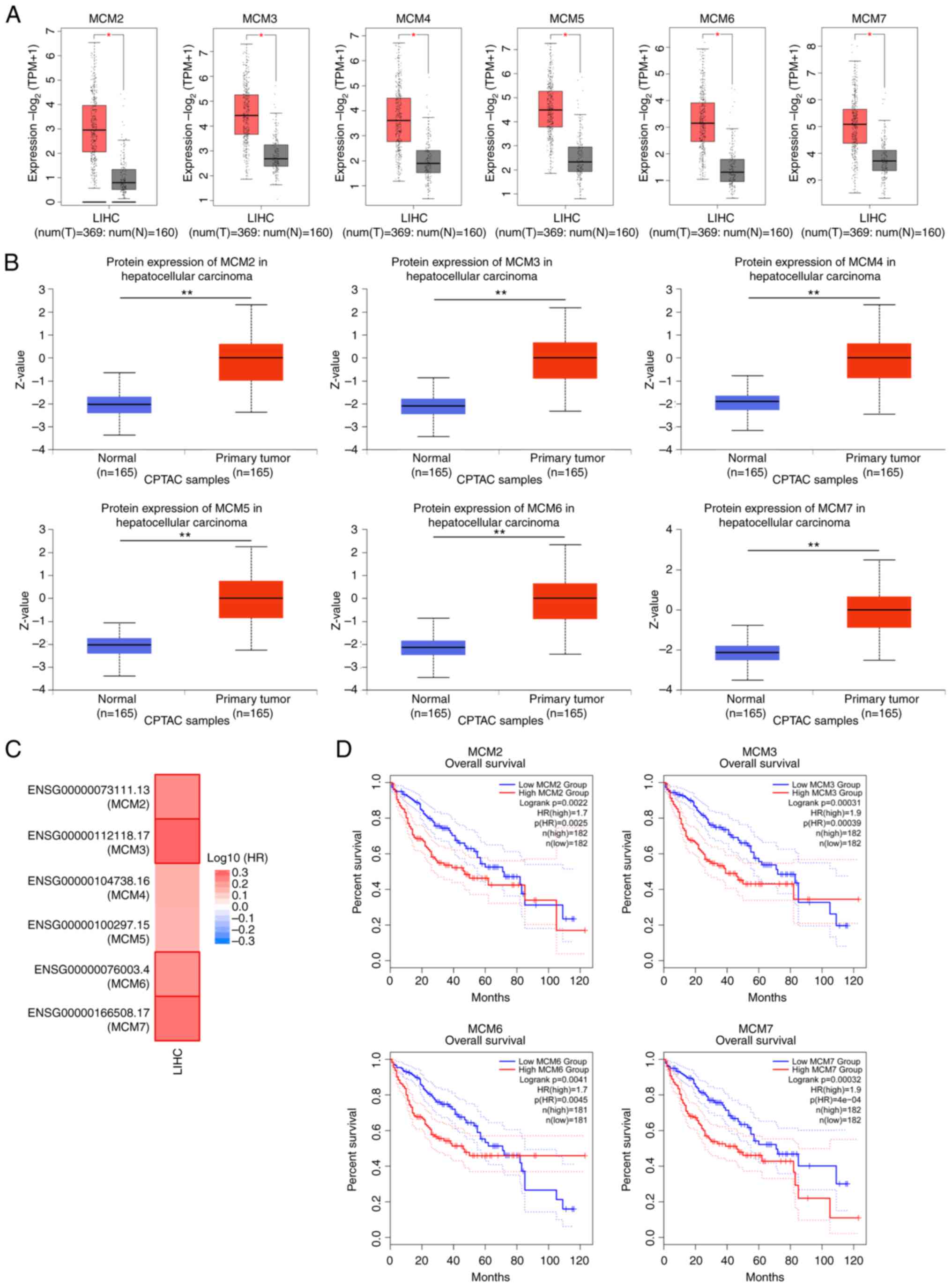
High expression levels of MCM2-7 in
patients with LIHC predict poor overall survival rates
The mRNA and protein expression levels of genes
encoding the MCM2-7 complex in human LIHC were analyzed using the
GEPIA2 and UALCAN databases, respectively, and found to be
significantly upregulated in LIHC compared with the corresponding
normal tissues at the mRNA and protein levels (Fig. 1A and B).
The associations between different MCMs and the
clinical outcomes of LIHC were analyzed using GEPIA2 (Fig. 1C). Stronger associations were
observed for MCM2, MCM3, MCM6 and MCM7 than for MCM4 and 5. The
associations between the mRNA expression of certain MCMs and the
pathological stage of LIHC were also assessed using GEPIA2. The
mRNA expression levels in the MCM2, MCM3, MCM6 and MCM7 groups were
highly variable, and gradually increased with the progression of
LIHC in the first three stages, but then markedly decreased in
stage IV (Fig. S1A). In addition,
patients with LIHC who had high mRNA expression levels of MCM2,
MCM3, MCM6 and MCM7 showed poor overall and disease-free survival
rates (Figs. 1D and S1B). Therefore, these four genes were
selected for further study.
Knockdown of MCMs inhibits the
proliferation of HCC cells
shRNAs targeting MCM2, MCM3, MCM6 and MCM7 were
constructed and stably transfected into human Huh7 and Hep3B cells.
The knockdown efficiencies were detected by IB analysis (Fig. 2A). On the basis of these results,
shMCM2-1 and shMCM2-4 for MCM2, shMCM3-2 and shMCM3-4 for MCM3,
shMCM6-1 and shMCM6-2 for MCM6, and shMCM7-1 and shMCM7-2 for MCM7
were selected for further study. The viability of Huh7 and Hep3B
cells was determined by CCK-8 assay at different time points (0,
24, 48 and 72 h), and cell growth inhibition was observed in the
MCM2/3/6/7-knockdown cells compared with that of the negative
control (scramble) groups (Fig.
2B). Colony formation assays were also performed, the results
of which were highly consistent with those of the CCK-8 assay
(Fig. 2C). In addition, wound
healing assays were performed and MCM2/3/6/7-knockdown exhibited no
evident effect on the migration of Huh7 cells (data not shown).
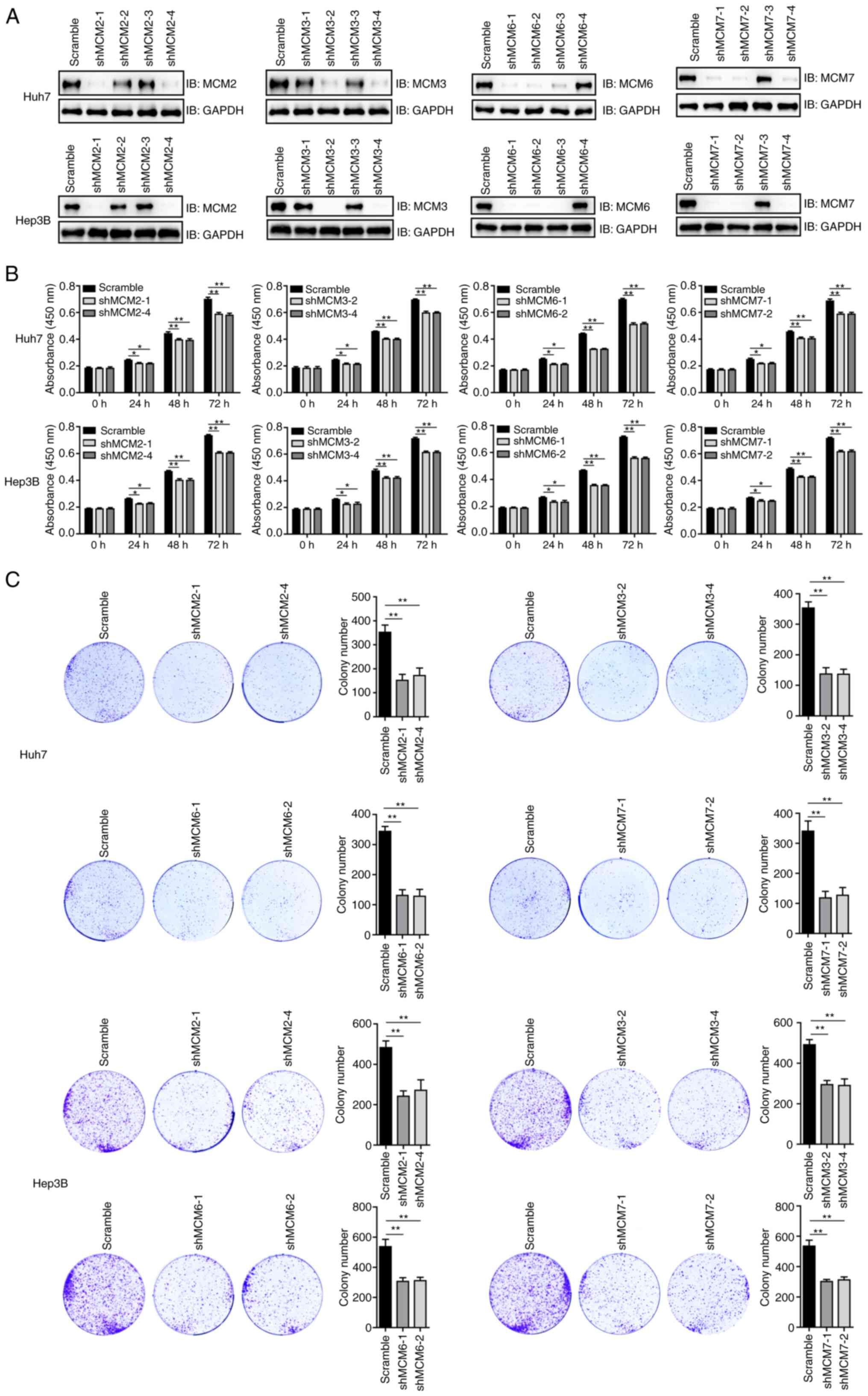 | Figure 2.Knockdown of MCMs inhibits the
proliferation of HCC cells. (A) Knockdown efficiency of shRNAs
targeting MCM2, MCM3, MCM6 and MCM7 as revealed by the IB analysis
of Huh7 and Hep3B HCC cells. (B) Knockdown of MCM2, MCM3, MCM6 or
MCM7 inhibited the proliferation of HCC cells, as revealed by the
cell viability detected using a Cell Counting Kit-8 assay at
different time points. The 0 h time point was defined as 6 h after
cell seeding. This experiment was repeated three times with six
replicates. *P<0.05, **P<0.01. (C) Knockdown of MCM2, MCM3,
MCM6 or MCM7 inhibited the colony formation of HCC cells. Three
samples were tested per group. **P<0.01. MCM, minichromosome
maintenance protein; HCC, hepatocellular carcinoma; sh, short
hairpin; IB, immunoblotting; scramble, negative control shRNA. |
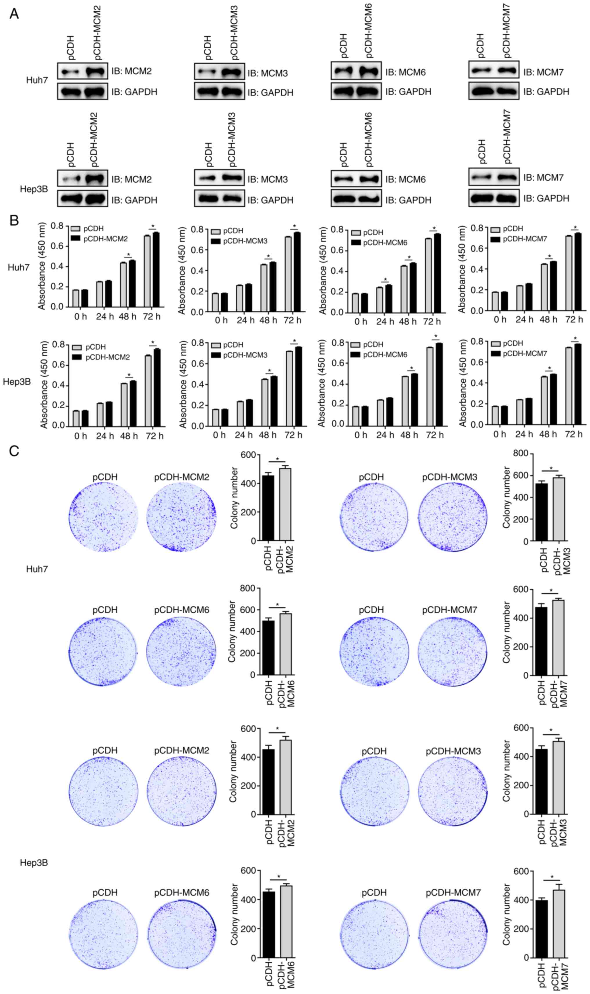
MCMs promote the proliferation of HCC
cells
Huh7 and Hep3B cells were stably transfected with
pCDH or pCDH-MCM2/3/6/7 and the expression of the MDMs was detected
by IB analysis (Fig. 3A). The
viability of the transfected Huh7 and Hep3B cells was then
determined by CCK-8 assay at various time points, and cell growth
promotion was observed in the pCDH-MCM groups compared with the
pCDH control groups (Fig. 3B). The
results of colony formation assays were consistent with the results
of the CCK-8 assay (Fig. 3C).
RNF125 interacts with MCM6
The pathways of MCM2, MCM3, MCM6 and MCM7 protein
degradation were explored. Huh7 cells were treated with the
proteasomal inhibitor BTZ or the autophagy inhibitor BAF for 6 h
before being subjected to IB analysis. The results shown in
Fig. 4A indicate that endogenous
MCM6 protein is degraded mainly by the proteasome pathway, and that
neither of these degradation pathways affect the other three MCMs.
To investigate the regulation of MCM6 protein homeostasis, MCM6 was
used as a bait to screen for its interacting partners using the Y2H
prey library which contains ~400 ORFs of human E3 ubiquitin
ligases. The E3 ligase RNF125 was identified as an interacting
partner for MCM6 and verified in yeast cells (Fig. 4B). A direct interaction between
RNF125 and MCM6 was detected by GST pull-down assay (Fig. 4C), whereas no interaction of RNF125
with MCM2, MCM3 or MCM7 was found (Fig.
4D). Protein-protein interaction domain analysis revealed that
the MCM domain of MCM6 directly interacted with the N-terminal
region (1–135 amino acids) of RNF125 containing the RING domain
(Fig. 4E). Further Co-IP assays
showed that endogenous MCM6 formed a complex with RNF125 in both
Huh7 and Hep3B cells (Fig. 4F).
These data indicate that RNF125 does indeed interact with MCM6.
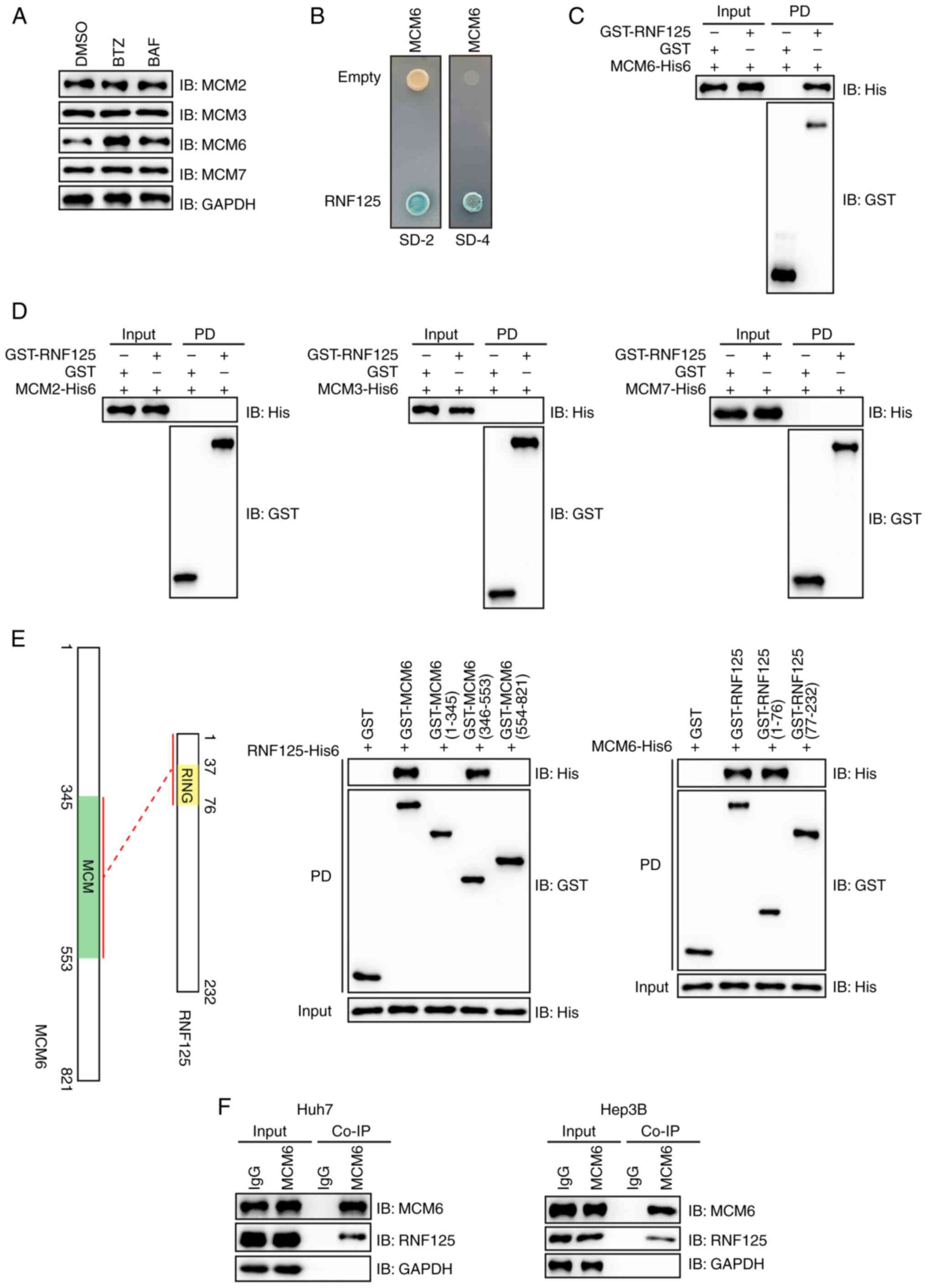 | Figure 4.RNF125 interacts with MCM6. (A)
Treatment of Huh7 cells with proteasome inhibitor BTZ (1 µmol/l) or
autophagy inhibitor BAF (20 nmol/) for 6 h prior to IB analysis
indicates that endogenously expressed MCM6 primarily undergoes
proteasome-dependent degradation. (B) RNF125 is shown to be an
interacting partner for MCM6 using the yeast two-hybrid method,
where SD-2 is deficient in leucine and tryptophan, and SD-4 is
deficient in uracil, histidine, leucine and tryptophan. (C) Direct
interaction between RNF125 and MCM6 was detected by GST PD assay
using recombinant GST-RNF125 and MCM6-His6; the GST PD assay was
evaluated using IB analysis. (D) No interaction of RNF125 with
MCM2, MCM3, and MCM7 was detected by GST pull-down assay with IB
analysis. (E) The N-terminal region (1–76 amino acids) containing
the RING domain of RNF125 directly interacted with the MCM domain
of MCM6. (F) GST PD assays and IB analysis showed that endogenously
expressed MCM6 forms a complex with RNF125. The lysates of Huh7 and
Hep3B cells were immunoprecipitated with IgG or anti-MCM6 antibody
and subjected to IB analysis. RNF125, ring finger protein 125; MCM,
minichromosome maintenance; BTZ, bortezomib; BAF, bafilomycin; IB,
immunoblotting; GST, glutathione S-transferase; PD, pull-down;
Co-IP, co-immunoprecipitation. |
RNF125 promotes the ubiquitination and
degradation of MCM6
To investigate whether RNF125 affects the stability
of the MCM6 protein, Huh7 and Hep3B cells were transfected to
express different amounts of RNF125, and the protein levels of MCM6
were detected by IB analysis. The results shown in Fig. 5A show that RNF125 reduced the
protein levels of MCM6 in a dose-dependent manner. An in
vitro ubiquitination assay was carried out, as shown in
Fig. 5B. In the presence of E1
(UBA1), E2 (UBCH5A) and E3 (RNF125), the ubiquitination of MCM6 was
detectable by IB analysis, and this modification was efficiently
attenuated by Usp2cc. Huh7 and Hep3B cells were stably transfected
with empty vector (pCDH) or pCDH-RNF125 and the upregulation of
RNF125 by pCDH-RNF125 was confirmed by IB analysis (Fig. 5C). The lysates of the transfected
Huh7 and Hep3B cells were immunoprecipitated with anti-MCM6
antibody, and the results revealed that the ubiquitination of MCM6
was markedly increased in cells stably expressing pCDH-RNF125
compared with the control cells (Fig.
5D). Three shRNAs targeting RNF125 were designed and tested in
human Huh7 and Hep3B cells. shRNF125-2 and shRNF125-3 performed
well in the knockdown of RNF125 and were selected for further study
(Fig. 5E). The protein levels of
endogenous MCM6 were increased in the RNF125-knockdown cells
compared with the control cells in the presence of the protein
synthesis inhibitor CHX (Fig. 5F).
In addition, RNF125 knockdown reduced the ubiquitination of MCM6
and concurrently increased MCM6 protein levels in Huh7 and Hep3B
cells (Fig. 5G). These data suggest
that RNF125 is an E3 ligase for MCM6 and promotes its
degradation.
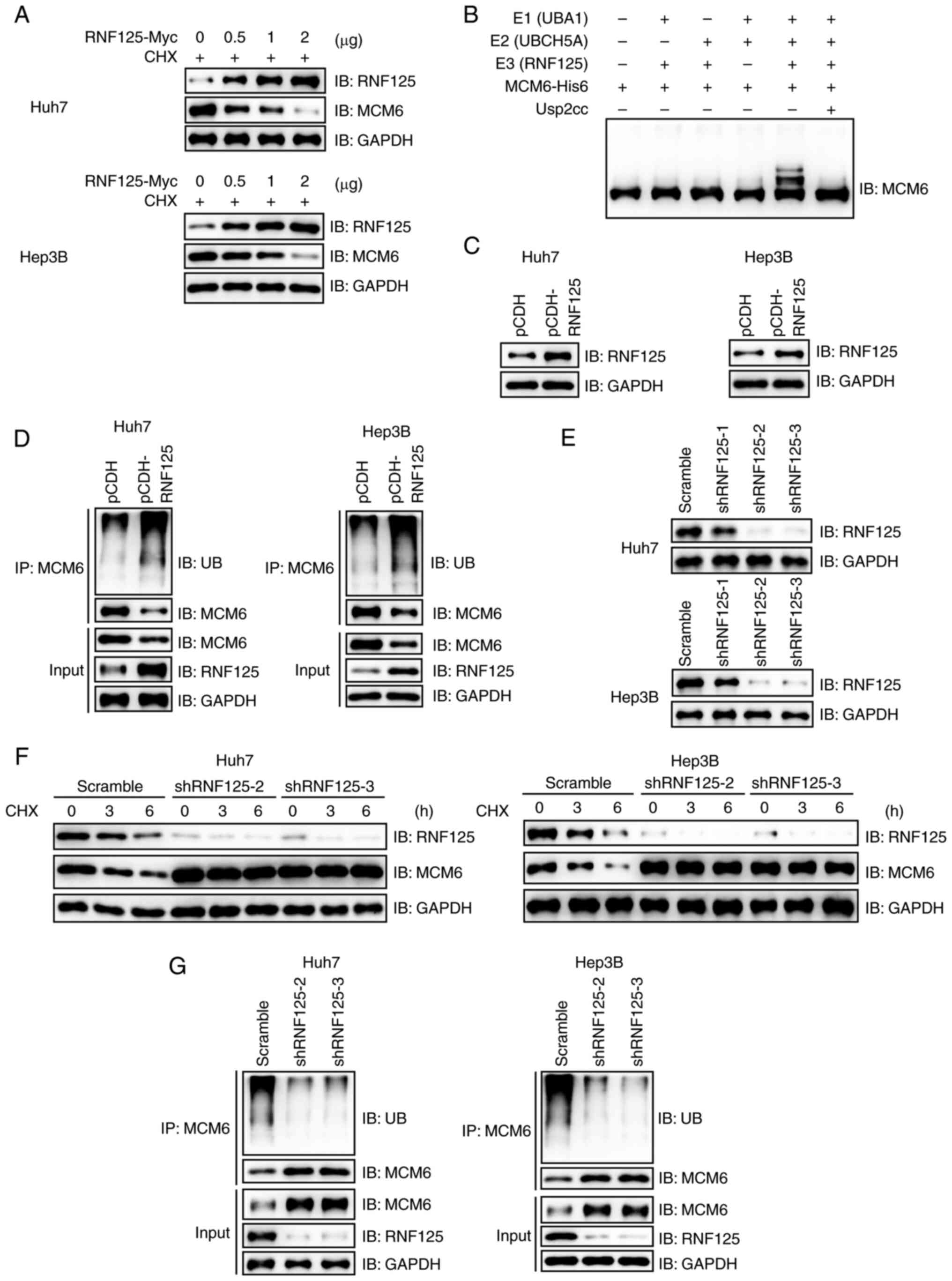 | Figure 5.RNF125 promotes the ubiquitination
and degradation of MCM6. (A) RNF125 promotes the degradation of
MCM6 in a dose-dependent manner, as revealed in Huh7 and Hep3B
cells transiently transfected with different amounts of
pCDNA3.0-RNF125-Myc or empty vectors and subjected to IB analysis.
(B) An in vitro ubiquitination assay was performed, and the
ubiquitination of MCM6 using different ubiquitination enzymes was
detected by IB analysis. The results indicates that RNF125 mediates
the ubiquitination of MCM6 in vitro. (C) Detection of the
protein levels of RNF125 in the lysates of Huh7 and Hep3B cells
stably expressing pCDH or pCDH-RNF125. (D) RNF125 promotes the
ubiquitination of endogenous MCM6, as revealed by
immunoprecipitation of the transfected cells with anti-MCM6
antibody followed by IB analysis. (E) Knockdown efficiency of
shRNAs targeting RNF125 in Huh7 and Hep3B cells detected by IB
analysis. (F) RNF125 knockdown stabilizes MCM6 in Huh7 and Hep3B
cells treated with CHX for different time periods prior to IB
analysis. (G) RNF125 knockdown reduces the ubiquitination of
endogenous MCM6, as demonstrated by the IP of Huh7 and Hep3B cells
stably expressing RNF125 shRNAs with anti-MCM6 antibody prior to IB
analysis. RNF125, ring finger protein 125; MCM, minichromosome
maintenance protein; IB, immunoblotting; sh, short hairpin; CHX,
cycloheximide; IP, immunoprecipitation; Usp2cc, catalytic core of
human ubiquitin specific peptidase 2; scramble, negative
control. |
RNF125 promotes the proliferation of
HCC cells mainly through MCM6
pCDH-RNF125 or empty vector (pCDH) was stably
transfected into Huh7 and Hep3B cells that also stably expressed
scramble shRNA, shMCM6-1 or shMCM6-2, and the protein levels of
RNF125 and MCM6 were detected by IB analysis (Fig. 6A). The results of CCK-8 and colony
formation assays performed using these cells revealed that RNF125
inhibited the proliferation and colony formation of Huh7 and Hep3B
cells with intact MCM6, but not of MCM6 knockdown cells (Fig. 6B and C). These data suggest that
RNF125 promotes the proliferation of HCC cells mainly through MCM6.
The expression of RNF125 in human LIHC was analyzed using the
GEPIA2 database. As shown in Fig.
6D, the expression of RNF125 was downregulated in LIHC compared
with normal tissues at the mRNA level. The association between
RNF125 and the clinical outcome of LIHC was also analyzed, and the
results showed that patients with LIHC and high mRNA expression of
RNF125 had a higher overall survival rate (Fig. 6E). These findings suggest that
RNF125 is a potential therapeutic target for human HCC.
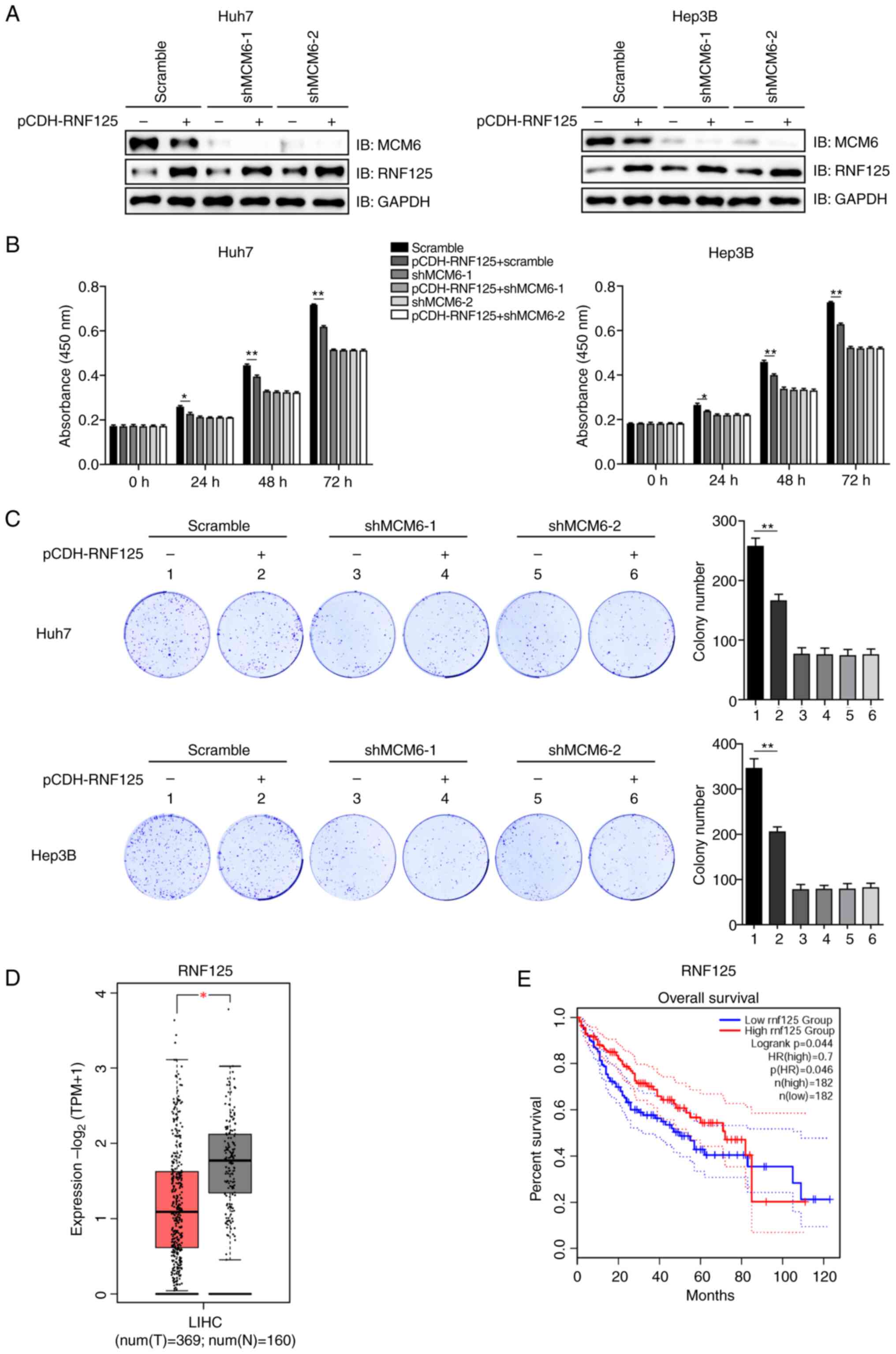 | Figure 6.RNF125 promotes the proliferation of
HCC cells mainly though MCM6. (A) Detection of the protein
expression levels of RNF125 and MCM6 in Huh7 and Hep3B cells stably
expressing scramble or MCM6 shRNAs with or without transfection
with plasmids containing RNF125 using IB analysis. (B) RNF125
promotes the proliferation of HCC cells via MCM6. The viability of
Huh7 and Hep3B cells co-transfected with plasmids expressing RNF125
and shRNAs targeting MCM6 was detected by Cell Counting Kit-8
assay. The experiment was repeated three times with six replicates.
*P<0.05, **P<0.01. (C) RNF125 promotes the colony formation
of HCC cells though MCM6 in Huh7 and Hep3B cells. Three samples
were tested per group. **P<0.01. (D) RNF125 mRNA expression in
human LIHC and corresponding normal tissues as analyzed using the
GEPIA2 database. *P<0.05. (E) Overall survival rates in patients
with LIHC according to the expression level of RNF125 were analyzed
using the GEPIA2 database. RNF125, ring finger protein 125; HCC,
hepatocellular carcinoma; MCM, minichromosome maintenance protein;
scramble, negative control; sh, short hairpin; IB, immunoblotting;
HR, hazard ratio; p(HR), P-value for the HR; TPM, transcript per
million. |
Discussion
In the present study, the expression levels and
prognostic values of MCM2-7 in human HCC were first analyzed using
a public database, and the results suggested that MCM2, MCM3, MCM6
and MCM7 may serve as prognostic markers. The proliferation of
human HCC cells was hindered by the knockdown of any of the four
MCMs, while the promotion of HCC cell proliferation through the
overexpression of these MCMs was shown to have limited effect. It
is hypothesized that this may be due to the fact that the MCM
complexes function as a complete unit (9); thus, increasing only one type has
minimal effects. The homeostasis of these four MCMs was then
investigated using a proteasomal or autophagy inhibitor, and the
results indicated that only MCM6 was degraded by the proteasome
pathway. In addition, RNF125 was identified as an E3 ligase for
MCM6, which promoted its ubiquitination and degradation. Further
experiments showed that RNF125 promoted the proliferation of HCC
cells mainly through MCM6.
The ubiquitin-proteasome pathway is a highly
selective protein degradation pathway, which efficiently degrades
intracellular proteins and plays an important role in cell
proliferation, metabolism and differentiation (24,25).
Dysfunction of ubiquitination has been indicated to lead to
numerous issues, including cancers and neurodegenerative diseases
(22,26,27).
RNF125 is a RING-type E3 ubiquitin ligase that features an
N-terminal RING domain. Various substrates for RNF125 have been
reported, including programmed death-ligand 1 and tripartite motif
containing 14 (28,29). The present study has identified MCM6
as a novel substrate for RNF125. Furthermore, it has established
that RNF125 primarily impedes the proliferation of HCC cells
through MCM6. In addition, analysis using publicly available
databases revealed that the expression of RNF125 was lower in LIHC
compared with corresponding normal tissues. Moreover, a higher
expression level of RNF125 was found to be associated with improved
overall survival in patients with LIHC. These findings are
consistent with those reported in a previous study (30).
To the best of our knowledge, RNF125 is the first E3
ligase identified for MCM6. Various studies have shown that MCM6
can interact with the E3 ligase UBE3A/E6AP (31,32).
However, UBE3A/E6AP is incapable of mediating the ubiquitination of
MCM6 (32). Notably, the present
study demonstrated that RNF125 does not interact with other MCM
family members such as MCM3, MCM2 and MCM7. With the exception of
MCM7 (33), no E3 ligases have been
reported for other MCM family proteins. The knockdown of MCM2, MCM3
or MCM7 also inhibited the proliferation of HCC cells, indicating
that the balance of these proteins is also critical.
The present research has certain limitations.
Firstly, the mechanism by which RNF126-MCM6 ubiquitin signaling
controls the growth of HCC cells was not examined. Secondly, the
findings were not validated in animal models and clinical
specimens. Furthermore, the survival analyses were performed using
GEPIA2 with the log-rank test the only option for analysis;
therefore, it was not possible to exclude the late-stage crossover
from the analysis, which may affect the results of the analysis.
Therefore, endeavors to test the functions of RNF125-MCM6 axis in
animal models and patient samples of LIHC are ongoing. In
conclusion, RNF125 and MCM6 are two promising targets for the
treatment of LIHC.
Supplementary Material
Supporting Data
Acknowledgements
Not applicable.
Funding
The study was financially supported by the Department of
Education of Anhui Province (grant no. 2022AH050774) and the
Foundation of Lu'an People's Hospital (grant no. 2022kykt30).
Availability of data and materials
The data generated in the present study may be
requested from the corresponding author.
Authors' contributions
XF, DS, XL, YL and PJ performed experiments and data
analysis. XF, SW and FL designed and supervised the study. XF and
FL wrote the manuscript and provided financial support. XF and FL
confirm the authenticity of all the raw data. All authors read and
approved the final version of the manuscript.
Ethics approval and consent to
participate
Not applicable.
Patient consent for publication
Not applicable.
Competing interests
The authors declare that they have no competing
interests.
References
|
1
|
Siegel RL, Miller KD, Wagle NS and Jemal
A: Cancer statistics, 2023. CA Cancer J Clin. 73:17–48. 2023.
View Article : Google Scholar : PubMed/NCBI
|
|
2
|
Cho SH, You GR, Park C, Cho SG, Lee JE,
Choi SK, Cho SB and Yoon JH: Acute respiratory distress syndrome
and severe pneumonitis after atezolizumab plus bevacizumab for
hepatocellular carcinoma treatment: A case report. World J
Gastrointest Oncol. 15:892–901. 2023. View Article : Google Scholar : PubMed/NCBI
|
|
3
|
Ferrante ND, Pillai A and Singal AG:
Update on the diagnosis and treatment of hepatocellular carcinoma.
Gastroenterol Hepatol (NY). 16:506–516. 2020.PubMed/NCBI
|
|
4
|
Le DC, Nguyen TM, Nguyen DH, Nguyen DT and
Nguyen LTM: Survival outcome and prognostic factors among patients
with hepatocellular carcinoma: A hospital-based study. Clin Med
Insights Oncol. 17:117955492311781712023. View Article : Google Scholar : PubMed/NCBI
|
|
5
|
Marin JJG, Macias RIR, Monte MJ, Romero
MR, Asensio M, Sanchez-Martin A, Cives-Losada C, Temprano AG,
Espinosa-Escudero R, Reviejo M, et al: Molecular bases of drug
resistance in hepatocellular carcinoma. Cancers (Basel).
12:16632020. View Article : Google Scholar : PubMed/NCBI
|
|
6
|
Saito A, Toyoda H, Kobayashi M, Koiwa Y,
Fujii H, Fujita K, Maeda A, Kaneoka Y, Hazama S, Nagano H, et al:
Prediction of early recurrence of hepatocellular carcinoma after
resection using digital pathology images assessed by machine
learning. Mod Pathol. 34:417–425. 2021. View Article : Google Scholar : PubMed/NCBI
|
|
7
|
Wang Y, Chen H, Zhang J, Cheng ASL, Yu J,
To KF and Kang W: MCM family in gastrointestinal cancer and other
malignancies: From functional characterization to clinical
implication. Biochim Biophys Acta Rev Cancer. 1874:1884152020.
View Article : Google Scholar : PubMed/NCBI
|
|
8
|
Zhou J, Wang M, Zhou Z, Wang W, Duan J and
Wu G: Expression and prognostic value of MCM family genes in
osteosarcoma. Front Mol Biosci. 8:6684022021. View Article : Google Scholar : PubMed/NCBI
|
|
9
|
Tye BK: MCM proteins in DNA replication.
Annu Rev Biochem. 68:649–686. 1999. View Article : Google Scholar : PubMed/NCBI
|
|
10
|
Cai HQ, Cheng ZJ, Zhang HP, Wang PF, Zhang
Y, Hao JJ, Wang MR and Wan JH: Overexpression of MCM6 predicts poor
survival in patients with glioma. Hum Pathol. 78:182–187. 2018.
View Article : Google Scholar : PubMed/NCBI
|
|
11
|
Marnerides A, Vassilakopoulos TP,
Boltetsou E, Levidou G, Angelopoulou MK, Thymara I, Kyrtsonis MC,
Pappi V, Tsopra O, Panayiotidis P, et al: Immunohistochemical
expression and prognostic significance of CCND3, MCM2 and MCM7 in
Hodgkin lymhoma. Anticancer Res. 31:3585–3594. 2011.PubMed/NCBI
|
|
12
|
Wojnar A, Pula B, Piotrowska A, Jethon A,
Kujawa K, Kobierzycki C, Rys J, Podhorska-Okolow M and Dziegiel P:
Correlation of intensity of MT-I/II expression with Ki-67 and MCM-2
proteins in invasive ductal breast carcinoma. Anticancer Res.
31:3027–3033. 2011.PubMed/NCBI
|
|
13
|
Wu W, Wang X, Shan C, Li Y and Li F:
Minichromosome maintenance protein 2 correlates with the malignant
status and regulates proliferation and cell cycle in lung squamous
cell carcinoma. Onco Targets Ther. 11:5025–5034. 2018. View Article : Google Scholar : PubMed/NCBI
|
|
14
|
Stewart PA, Khamis ZI, Zhau HE, Duan P, Li
Q, Chung LWK and Sang QA: Upregulation of minichromosome
maintenance complex component 3 during epithelial-to-mesenchymal
transition in human prostate cancer. Oncotarget. 8:39209–39217.
2017. View Article : Google Scholar : PubMed/NCBI
|
|
15
|
Liu M, Hu Q, Tu M, Wang X, Yang Z, Yang G
and Luo R: MCM6 promotes metastasis of hepatocellular carcinoma via
MEK/ERK pathway and serves as a novel serum biomarker for early
recurrence. J Exp Clin Cancer Res. 37:102018. View Article : Google Scholar : PubMed/NCBI
|
|
16
|
Vigouroux C, Casse JM, Battaglia-Hsu SF,
Brochin L, Luc A, Paris C, Lacomme S, Gueant JL, Vignaud JM and
Gauchotte G: Methyl(R217)HuR and MCM6 are inversely correlated and
are prognostic markers in non small cell lung carcinoma. Lung
Cancer. 89:189–196. 2015. View Article : Google Scholar : PubMed/NCBI
|
|
17
|
Zheng T, Chen M, Han S, Zhang L, Bai Y,
Fang X, Ding SZ and Yang Y: Plasma minichromosome maintenance
complex component 6 is a novel biomarker for hepatocellular
carcinoma patients. Hepatol Res. 44:1347–1356. 2014. View Article : Google Scholar : PubMed/NCBI
|
|
18
|
Tang Z, Li C, Kang B, Gao G, Li C and
Zhang Z: GEPIA: A web server for cancer and normal gene expression
profiling and interactive analyses. Nucleic Acids Res. 45:W98–W102.
2017. View Article : Google Scholar : PubMed/NCBI
|
|
19
|
Chandrashekar DS, Bashel B, Balasubramanya
SAH, Creighton CJ, Ponce-Rodriguez I, Chakravarthi BVSK and
Varambally S: UALCAN: A portal for facilitating tumor subgroup gene
expression and survival analyses. Neoplasia. 19:649–658. 2017.
View Article : Google Scholar : PubMed/NCBI
|
|
20
|
Li C, Lu W, Yang L, Li Z, Zhou X, Guo R,
Wang J, Wu Z, Dong Z, Ning G, et al: MKRN3 regulates the epigenetic
switch of mammalian puberty via ubiquitination of MBD3. Natl Sci
Rev. 7:671–685. 2020. View Article : Google Scholar : PubMed/NCBI
|
|
21
|
Wang CC, Peng H, Wang Z, Yang J, Hu RG, Li
CY and Geng WJ: TRIM72-mediated degradation of the short form of
p62/SQSTM1 rheostatically controls selective autophagy in human
cells. Mil Med Res. 9:352022.PubMed/NCBI
|
|
22
|
Yang Y, Luo Y, Yang C, Hu R, Qin X and Li
C: TRIM25-mediated ubiquitination of G3BP1 regulates the
proliferation and migration of human neuroblastoma cells. Biochim
Biophys Acta Gene Regul Mech. 1866:1949542023. View Article : Google Scholar : PubMed/NCBI
|
|
23
|
Li C, Han T, Li Q, Zhang M, Guo R, Yang Y,
Lu W, Li Z, Peng C, Wu P, et al: MKRN3-mediated ubiquitination of
Poly(A)-binding proteins modulates the stability and translation of
GNRH1 mRNA in mammalian puberty. Nucleic Acids Res. 49:3796–3813.
2021. View Article : Google Scholar : PubMed/NCBI
|
|
24
|
Mansour MA: Ubiquitination: Friend and foe
in cancer. Int J Biochem Cell Biol. 101:80–93. 2018. View Article : Google Scholar : PubMed/NCBI
|
|
25
|
Sheng X, Xia Z, Yang H and Hu R: The
ubiquitin codes in cellular stress responses. Protein Cell.
pwad0452023.(Epub ahead of print). View Article : Google Scholar : PubMed/NCBI
|
|
26
|
Liu Z, Chen P, Gao H, Gu Y, Yang J, Peng
H, Xu X, Wang H, Yang M, Liu X, et al: Ubiquitylation of autophagy
receptor Optineurin by HACE1 activates selective autophagy for
tumor suppression. Cancer Cell. 26:106–120. 2014. View Article : Google Scholar : PubMed/NCBI
|
|
27
|
Xu X, Li C, Gao X, Xia K, Guo H, Li Y, Hao
Z, Zhang L, Gao D, Xu C, et al: Excessive UBE3A dosage impairs
retinoic acid signaling and synaptic plasticity in autism spectrum
disorders. Cell Res. 28:48–68. 2018. View Article : Google Scholar : PubMed/NCBI
|
|
28
|
Jiang C, He L, Xiao S, Wu W, Zhao Q and
Liu F: E3 ubiquitin ligase RNF125 suppresses immune escape in head
and neck squamous cell carcinoma by regulating PD-L1 expression.
Mol Biotechnol. 65:891–903. 2023. View Article : Google Scholar : PubMed/NCBI
|
|
29
|
Jia X, Zhou H, Wu C, Wu Q, Ma S, Wei C,
Cao Y, Song J, Zhong H, Zhou Z and Wang J: The ubiquitin ligase
RNF125 targets innate immune adaptor protein TRIM14 for
ubiquitination and degradation. J Immunol. 198:4652–4658. 2017.
View Article : Google Scholar : PubMed/NCBI
|
|
30
|
Feng Z, Ke S, Wang C, Lu S, Xu Y, Yu H, Li
Z, Yin B, Li X, Hua Y, et al: RNF125 attenuates hepatocellular
carcinoma progression by downregulating SRSF1-ERK pathway.
Oncogene. 42:2017–2030. 2023. View Article : Google Scholar : PubMed/NCBI
|
|
31
|
Martínez-Noël G, Luck K, Kühnle S,
Desbuleux A, Szajner P, Galligan JT, Rodriguez D, Zheng L, Boyland
K, Leclere F, et al: Network analysis of UBE3A/E6AP-associated
proteins provides connections to several distinct cellular
processes. J Mol Biol. 430:1024–1050. 2018. View Article : Google Scholar : PubMed/NCBI
|
|
32
|
Li C, Han T, Guo R, Chen P, Peng C, Prag G
and Hu R: An integrative synthetic biology approach to
interrogating cellular ubiquitin and ufm signaling. Int J Mol Sci.
21:42312020. View Article : Google Scholar : PubMed/NCBI
|
|
33
|
Kühne C and Banks L: E3-ubiquitin
ligase/E6-AP links multicopy maintenance protein 7 to the
ubiquitination pathway by a novel motif, the L2G box. J Biol Chem.
273:34302–34309. 1998. View Article : Google Scholar : PubMed/NCBI
|