Introduction
Lung cancer remains the leading cause of
cancer-related death. In 2022, the number of patients with cancer
in China and in the United States was 4,820,000 and 2,370,000,
respectively, and the number of cancer-related deaths was 3,210,000
and 609,360 respectively (1). A
survey conducted by The American Cancer Society revealed that the
incidence of lung cancer decreased between 2009 and 2018, with 3
and 1% of males and females affected every year, respectively.
Notably, the incidence of lung cancer is declining in females, and
lung cancer occurs in the younger generation due to the increased
incidence of smoking (2). In
clinical practice, small molecule inhibitors, such as anaplastic
lymphoma kinase tyrosine kinase inhibitors and immune checkpoint
inhibitors (ICIs), such as anti-programmed death-1 (PD-1)/PD-ligand
1 (PD-L1) antibodies are used in the treatment of lung cancer, with
the median survival rate of patients with lung cancer increasing
from 8.9 to 12.6 months (3). In
addition, results of a previous study (4) demonstrated that ICIs in combination
with platinum-containing chemotherapy are effective in prolonging
the overall survival (OS) of patients with advanced lung cancer,
and are recommended by relevant guidelines (5) as a standard first-line treatment
option for non-small cell lung cancer (NSCLC). However, monitoring
the efficacy of treatment and prognosis during combination therapy
remains challenging.
Circulating tumor cells (CTCs) derived from tumor in
situ or metastatic lesions (6) are
more representative of tumor heterogeneity than tissue biopsy, and
these can be monitored at different stages of treatment. Notably,
the value of CTCs in treatment monitoring is progressing. PD-L1
levels may be monitored in the clinic to determine the efficacy of
NSCLC treatment using ICIs (7). At
present, PD-L1 testing is performed using immunohistochemistry
(IHC); however, results may be impacted by tumor heterogeneity.
Thus, CTC PD-L1 testing is increasingly considered to be more
accurate in reflecting real-time cancer progression in patients.
Chen et al (8) revealed a
57% concordance rate between the two methods by performing PD-L1
tissue and CTC PD-L1 testing separately in patients with NSCLC;
however, positive PD-L1 expression was detected using a CTC assay.
In addition, Yue et al (9)
used CTC PD-L1 testing to assess the efficacy of IBI308, a PD-1
inhibitor, for the treatment of gastric cancer. The results
demonstrated that changes in CTC PD-L1 levels, and the
high/moderate expression of CTCs were notably associated with
disease progression. In addition, CTC PD-L1 levels at baseline
exhibited potential in predicting the progression-free survival
(PFS) of patients.
The novel CTC detection device, CytoBot, uses a dual
physical and immunological capture mechanism (10). Notably, the efficacy of CytoBot is
yet to be fully elucidated. In a previous study, CytoBot was used
to detect CTCs in patients with advanced lung cancer, and the
potential association between CTCs and other blood markers such as
carcinoembryonic antigen (CEA) 211 and lymphocytes, was determined.
Thus, the effectiveness of CytoBot has been initially validated
(11). In the present study, the
functionality of CytoBot was further determined through CTC PD-L1
detection, and the results were compared with tissue biopsies. In
addition, the prognostic value of CTCs, CTC PD-L1, tissue PD-L1
expression levels, TNM stage, prognostic nutritional index (PNI),
systemic inflammatory score (SIS) and tumor shrinkage at 6 months
of treatment with ICIs in combination with platinum-containing
chemotherapy were determined, and the prognostic values of changes
in PNI and SIS were determined. The present study also aimed to
determine the association between PFS and changes in CTC, CTC PD-L1
and baseline characteristics during treatment, and to determine the
feasibility of clinical applications.
Materials and methods
Patients
In total, 40 male patients diagnosed with advanced
lung cancer at Army Medical Center of PLA (Chongqing Daping
Hospital) from May 2021 to July 2022 were enrolled in the present
study. Based on clinical guidelines, patients that are driver
gene-positive prefer targeted therapy (4); mutations in a panel of driver genes
consisting of EGFR, ALK, ROS1, RET, KRAS, BRAF, HER2, NRAS and MET
were investigated using ARMS/reverse transcription-PCR (Human Lung
Cancer Multi-Gene Assay Kit; Amoy Diagnostics Co., Ltd.) carried
out in the Department of Pathology, Daping Hospital, Army Medical
Center of PLA. Immunotherapy is not used as a first-line treatment.
Asian females notably exhibit more driver gene-positive lung
adenocarcinomas than males (12).
In the present study, 16 adenocarcinomas and 24 squamous carcinomas
were identified using histological analysis (Table I). The inclusion criteria were as
follows: i) Confirmed advanced lung cancer; ii) no relevant
treatment; iii) previous tissue PD-L1 puncture test; iv) complete
patient information, including computed tomography (CT) images; and
v) patient consent to participate in the study. Patients were
excluded from the present study for the following reasons: i)
History of previous lung cancer or immunosuppression; ii) presence
of other cancers; iii) previous surgical resection; and iv)
incomplete patient information.
 | Table I.Baseline and CTC, CTC PD-L1 detection
in the full patient population. |
Table I.
Baseline and CTC, CTC PD-L1 detection
in the full patient population.
| Clinical
characteristics |
|
|---|
| Sex, n (%) |
|
|
Male | 40 (100.0) |
|
Female | 0 (0.0) |
| Age, years
(range) | 62.25 (47–73) |
| Smoking, n (%) |
|
|
Yes | 24 (60.0) |
| No | 16 (40.0) |
| Histology, n
(%) |
|
|
Adenocarcinoma | 16 (40.0) |
|
Squamous | 24 (60.0) |
| T, n (%) |
|
|
T2-T3 | 17 (42.5) |
| T4 | 23 (57.5) |
| N, n (%) |
|
| 0 | 10 (25.0) |
| 2 | 21 (52.5) |
| 3 | 9 (22.5) |
| M, n (%) |
|
| 0 | 10 (25.0) |
| 1 | 30 (75.0) |
| CTC, n (%) |
|
| ≤3 | 25 (62.5) |
|
>3 | 15 (37.5) |
| CTC PD-L1, n
(%) |
|
|
Negative | 34 (85.0) |
|
Positive | 6 (15.0) |
| Number of CTC, n
(range) | 3 (1–7) |
In addition, the efficacy of ICIs with
platinum-containing chemotherapy during first-line treatment for
advanced squamous lung cancer or driver-negative lung
adenocarcinoma was assessed in 26 patients (Fig. S1). The potential association
between PFS and baseline patient characteristics, blood markers
[platelets (PTLs), SIS, red blood cell distribution width (RDW),
neutrophils, hemoglobin (HB), neutrophil-to-lymphocyte ratio (NLR),
lymphocyte-to-mononuclear ratio (LMR), white blood cell (WBC), CEA,
cytokeratin 19 (CTFRA-19), PNI], CTCs, CTC PD-L1 and tissue PD-L1
expression levels, tumor shrinkage at 6 months and inflammatory
changes and maintenance therapy were also determined.
The present study adhered to the Declaration of
Helsinki and approval was obtained from the Ethics Committee of the
Army Medical Center of PLA (Chongqing Daping Hospital, Chongqing,
China; approval no. 2020209). All patients provided written
informed consent.
Mutation tests of driver genes
Mutant Gene Detection Kits (fluorescent PCR method)
were used (AmoyDx), and this detection is based on the ADx-ARMS
method. ADx-ARMS aims to design specific mutation detection primers
for these sites.
For this test, formalin fixation and paraffin
embedding (FFPE) samples were used (range, 5–10 µm). FFPE samples
were first deparaffinized: FFPE samples were taken into 15-ml
centrifuge tubes, 1 ml of xylene (cat. no. 534056, Sigma-Aldrich;
Merck KGaA) was added, the sample was mixed for 10 sec and then
centrifuged for 2 min at room temperature at 13,000 × g, and the
supernatant was removed. A total of 1 ml of anhydrous ethanol (cat.
no. 362808; Sigma-Aldrich; Merck KGaA) was added, the sample was
mixed for 10 sec and then the supernatant was removed. The sample
was left at room temperature for 10 min to allow the ethanol to
fully evaporate. TRIzol® (cat. no. 15596026; Invitrogen;
Thermo Fisher Scientific, Inc.) was added and mixed for 5 min,
chloroform was then added and centrifuged at 4°C and 12,000 × g for
15 min. Isopropanol (0.5 ml; cat. no. 563935; Sigma-Aldrich; Merck
KGaA) was added, mixed at room temperature for 10 min and
centrifuged at 4°C and 12,000 × g for 10 min; following
centrifugation, the supernatant was discarded. A total of 1 ml of
75% ethanol (cat. no. 362808; Sigma-Aldrich; Merck KGaA) was added,
gently washing the precipitate at 4°C and 7,500 × g for 5 min,
followed by discarding the supernatant. A total of 45 µl RNA sample
was used and 5 µl enzyme mixture (hot start enzyme, reverse
transcriptase and uracil glycosylase, was added; the reaction was
vortexed and mixed for 15 sec. A total of 10 µl mixture was added
in the PCR system (primers, probes, magnesium ions, dNTPs), and
rapidly mixed for 15 sec. PCR amplification was carried out using a
Stratagene Mx3000PM (Agilent Technologies, Inc.): Stage 1: 42°C for
5 min, 95°C for 5 min (one cycle); stage 2: 42°C for 5 min, 95°C
for 5 min; stage 3: 42°C for 5 min, 95°C for 5 min; stage 4: 42°C
for 5 min, 95°C for 5 min. Stage 1: 42°C for 5 min, 95°C for 5 min
(one cycle); stage 2: 95°C for 25 sec, 64°C for 20 sec, 72°C for 20
sec (10 cycles); stage 3: 93°C for 25 sec, 60°C for 35 sec, 72°C
for 20 sec (36 cycles). If the 2-ΔΔCq value of the
carboxyfluorescein signal was <35, the sample contained a gene
fusion (i.e. mutation-positive), and conversely, it did not contain
a gene fusion (13).
CTC and CTC PD-L1 measurements
Prior to the CTC test, 4 ml peripheral blood was
obtained from patients using an EDTA blood collection tube with
anticoagulants. The sample was stored at 4°C for <8 h prior to
the test.
The CTC test was performed using the CytoBot kit
(cat. no. H1000; Holosensor Medical Ltd.), according to the
manufacturer's instructions. Briefly, blood samples were diluted
1:1 with PBS (pH 7.0). Subsequently, lymphocytes and tumor cells
were separated from the blood using a density gradient separation
solution, and the obtained cell precipitate was resuspended in 300
µl PBS (pH 7.0). The final cell suspension was added to the cell
separator, and the CTC assay program was selected on the CytoBot to
initiate the test. The 30-min procedure included CTC isolation,
capture and immunofluorescent staining of the nuclei using DAPI
(cat. no. D9542; Sigma-Aldrich; Merck KGaA), staining of the
lymphocyte marker CD45 (cat. no. 304001; BioLegend, Inc.) and
staining of epithelial markers using panCK (cat. no. ab215838;
Abcam). At the end of the procedure, the cell isolators were placed
directly under a fluorescence microscope (RX50M; Sunny Optical
Technology Co., Ltd.) for observation and counting.
The CTC PD-L1 assay was also performed using the
CytoBot, according to the manufacturer's instructions.
Pre-treatment and procedures were performed as previously
described; however, the CTC PD-L1 assay procedure was selected
prior to the test initiation.
Tissue puncture PD-L1 assay
Tissue PD-L1 evaluation was performed using tissue
puncture and IHC assays. Briefly, samples were obtained from the
tumor using a tissue puncture needle. Fixation, dehydration and
embedding: Tissue was placed in 4% paraformaldehyde (cat. no.
1004965000; Sigma-Aldrich; Merck KGaA) for 3–4 h. To dehydrate,
tissue was placed in 75% alcohol (cat. no. 362808; Sigma-Aldrich;
Merck KGaA) for 1.5, 85% alcohol for 1.5 h, 95% alcohol for 1 h,
anhydrous ethanol for 1.5 h, and then embedded with paraffin. The
tissue masses were then cut into pieces of 5 µm as required. The
paraffin sections were incubated at 60°C for 30–60 min, and then
sequentially placed in xylene (cat. no. 534056; Sigma-Aldrich;
Merck KGaA) I, II, and III for 10 min each, an ethanol gradient
(high to low: 100, 95, 80, and 70%) for 2 min each, and washed
three times with PBS. The sections were infiltrated with pre-warmed
containment permeabilization solution [40 ml PBS + 120 ul
TritonX-100 (cat. no. T8787; Sigma-Aldrich; Merck KGaA) + 400 µl
30% H2O2 (1.08597; Sigma-Aldrich; Merck KGaA)
for 30 min (to prevent light). Sections were placed in a solution
of antigen repair solution (0.01 M sodium citrate; pH 6.0; cat. no.
SAB5702489; Sigma-Aldrich; Merck KGaA) and then incubated room
temperature for 20 min. The endogenous enzyme activity was
deactivated by incubation with 3% H2O2 for 10
min at room temperature. After washing, non-specific loci were
blocked by goat serum blocking solution (cat. no. WE0320; Beijing
Baiolaibo Technology Co., Ltd.) for 30 min at 37°C. Diluted
anti-PD-L1 antibody (50 µl; 1:1,000; cat. no. 11146105; Dako) was
added and incubated at 37°C for 1–2 h. A total of 20 µl diluted
secondary antibody (HRP Anti-Rabbit IgG antibody; 1:500; cat. no.
ab288151; Abcam) was added after washing and incubated at 37°C for
1–2 h. Chromogenic Reaction: DAB (cat. no. D12384; Sigma-Aldrich;
Merck KGaA) was added for 10 min and the reaction was observed and
confirmed by microscopy. For observation, the sample was re-stained
with hematoxylin (cat. no. H3136; Sigma-Aldrich; Merck KGaA) for 30
sec, washed with water, differentiated with 1% hydrochloric acid
alcohol (cat. no. 123864-74-4; Byxbio) for 2 sec, and rinsed with
tap water for 3 min. For dehydration, ethanol gradients (from low
to high: 50, 70, 95, 100%) were used for 2 min each. Xylene was
used for 5 min to make the sections transparent, and then neutral
gum was added to seal the sections. Results were observed under a
light microscope, ×200 (RX50M; Sunny Optical Technology Co.,
Ltd.).
Statistical analysis
SPSS (version, 24.0; IBM Corp.) was used for
statistical analysis. A χ2-test and Kaplan-Meier
analyses were performed, and receiver operating characteristic
(ROC) curves were used for determining the predictive value of CTC
PD-L1. Pearson's correlation coefficient was used for assessing the
correlation between CTCs in the blood and the level of tissue PD-L1
expression. The baseline data and clinical test indicators of the
patients were included before and after treatment as variables in
the analysis, and were then analyzed and explored separately under
different subgroups. The impact of these blood biochemistry indexes
on prognosis were evaluated using Cox regression analysis and
visualized using a forest plot. The results are presented by
subgroup hazard ratio (HR) and P-value. P<0.05 was considered to
indicate a statistically significant difference.
Results
Baseline characteristics of patients
with advanced NSCLC
A total number of 40 male patients with advanced
NSCLC were enrolled in the present study, aged 47–73 years. CTC
detection was positive in all patients at initial diagnosis. In
total, 26 patients received first-line therapy with ICIs
(tislelizumab or carrilizumab) plus platinum-containing dual-drug
chemotherapy for a total of 4–6 cycles. Thereafter, ICIs were
maintained every 3 weeks, and clinical response and survival
outcomes were assessed. In total, 21, two and three patients were
evaluated as partial response (PR), stable disease (SD) and
progressive disease (PD), respectively. Treatment was decreased or
delayed in three cases due to the presence of adverse events (AEs),
including two cases of grade 3 interstitial pneumonia and one case
of myocarditis. Notably, all AEs were manageable. In addition, CTC
and CTC PD-L1 evaluation was repeated at 6 months in 11 patients,
and the potential association between changes in CTCs and CTC
PD-L1, and the efficacy of NSCLC immunotherapy was determined.
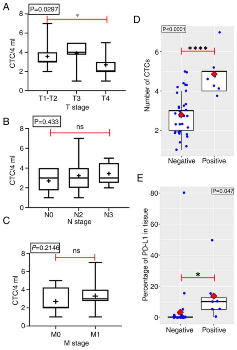
CTC and CTC PD-L1 detection in
patients with advanced NSCLC
CTC detection was performed in all 40 patients with
advanced NSCLC, and all patients exhibited a CTC count of 1–7
CTCs/4 ml blood. Of these, 25 patients exhibited a CTC count of ≤3
(62.5% of patients; Table I). Tumor
size was significantly associated with CTC number in all 40
patients (Fig. 1A), while lymph
node invasiveness and tumor metastasis were not associated with
tumor size (Fig. 1B and C). The
number of CTCs in patients with positive tissue PD-L1 expression
was significantly higher than that in patients with negative tissue
PD-L1 expression (Fig. 1D). The
percentage of positive tissue PD-L1 expression in patients with
positive CTC PD-L1 was significantly higher than that in patients
with negative CTC PD-L1 (Fig.
1E).
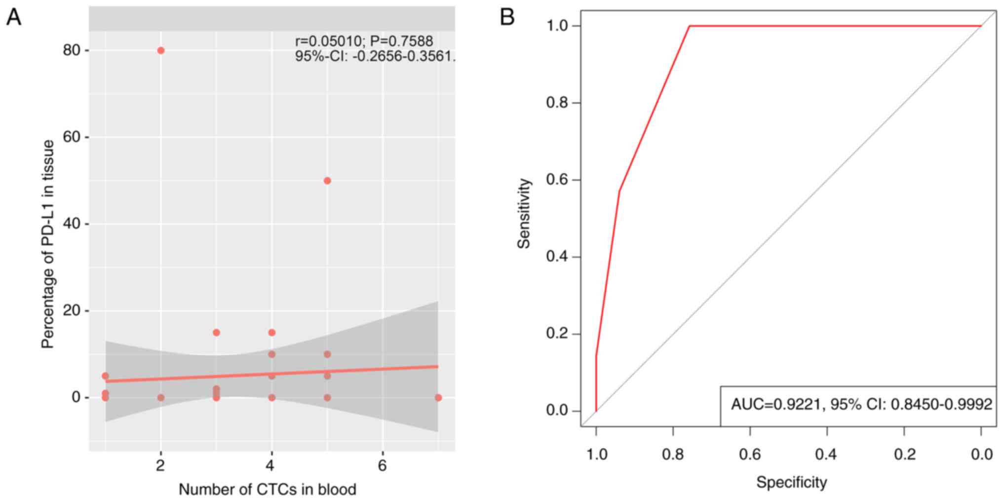
There was no correlation between CTC counts in the
blood and tissue PD-L1 expression in 40 patients with NSCLC
[Fig. 2A; r=0.05010, 95% confidence
interval (CI),-0.2656–0.3561]. However, these results may have been
impacted by the small sample size included in the present study.
The area under the ROC curve (AUC) was 0.9221 (95% CI,
0.8450–0.9992), indicating that CTC number detection influenced the
rate of positive CTC PD-L1 detection, and CTC detection was most
effective in identifying patients with negative PD-L1 expression
when the cut-off value was ≥3.5, with a sensitivity and specificity
of 100 and 73.5%, respectively (Fig.
2B).
Prognostic value of baseline
characteristics and blood indicators in patients with NSCLC
The baseline characteristics of patients were
collected, including age, histological classification, a history of
smoking, TNM stage, maintenance treatment, tumor volume and
shrinkage, blood panel including PTL, SIS, RDW, HB, NLR, LMR, CEA,
PNI, neutrophils, lymphocytes, monocytes, CYFRA19, albumin, CTCs
and CTC PD-L1. A correlation analysis between patient
characteristics and PFS of patients was performed. Our data shown
that the expression of PD-L1 in the tumor tissue prior to ICI
treatment was significantly associated with PFS (Fig. 3A), and also the level of CEA
(Fig. 3C). However, the rest of
indicators were significantly associated with PFS (Fig. 3B and C).
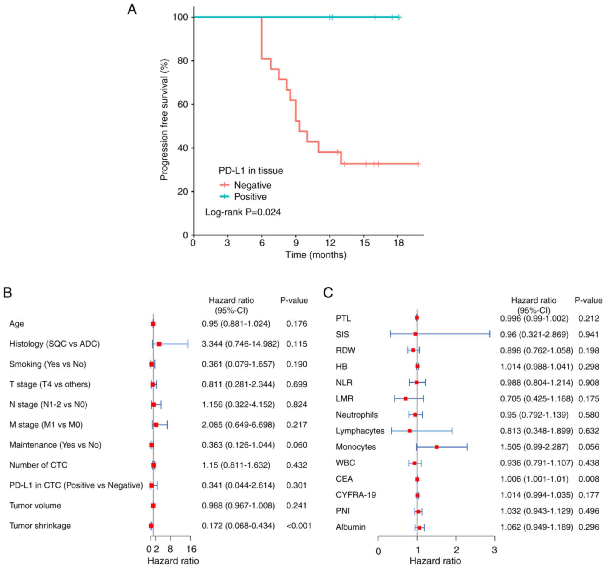 | Figure 3.Prognostic value of baseline
characteristics and CTCs in 26 patients with NSCLC. Association
between PFS and (A) tissue PD-L1 levels, (B) baseline
characteristics, CTCs and CTC PD-L1, and (C) other blood
indicators. CTCs, circulating tumor cells; NSCLC, non-small cell
lung cancer; PFS, progression-free survival; PD-L1, programmed
death-ligand 1; SQC, squamous-cell carcinoma; ADC, adenocarcinoma;
PTL, platelet; SIS, systemic inflammatory score; RDW, red blood
cell distribution width; WBC, white blood cell; HB, hemoglobin;
NLR, neutrophil-to-lymphocyte ratio; LMR, lymphocyte-to-mononuclear
ratio; CEA, carcinoembryonic antigen; CYFRA-19, cytokeratin 19;
PNI, prognostic nutritional index. |
Prognostic factors of NSCLC during
treatment with ICIs plus platinum-containing chemotherapy
To identify accessible indicators of disease
prognosis, 26 patients with advanced NSCLC were treated with a
combination of ICIs, and platinum-containing chemotherapy (Table II). In total, only 11/26 enrolled
patients underwent re-testing of CTC PD-L1 after 6 months of
treatment due to segregation because of the COVID-19 pandemic. We
investigated whether the rate of decline in blood indicators was
correlated with PFS during combination therapy. Data shown that SIS
(Table SI), PNI (Table SII; Fig. 4A) and blood indicators (Fig. 4A) were not associated with PFS in
treated patients. However, a decline in the rate of blood CYFRA19
(Table SII; Fig. 4A) was associated with PFS. Moreover,
it was revealed that tumor shrinkage (Fig. 4B) was significantly associated with
PFS, as the high decrease of tumor shrinkage was associated with
better prognosis. Following the exclusion of three patients due to
the presence of AEs, it was revealed that maintenance treatment
[ICIs + platinum-containing chemotherapy (200 mg tislelizumab or
camrelizumab, carboplatin for AUC=5) administered intravenously
every 3 weeks] was significantly associated with PFS (Fig. 4C). Notably, maintenance treatment
may exhibit potential in predicting treatment outcomes and the
prognosis of patients in clinical practice.
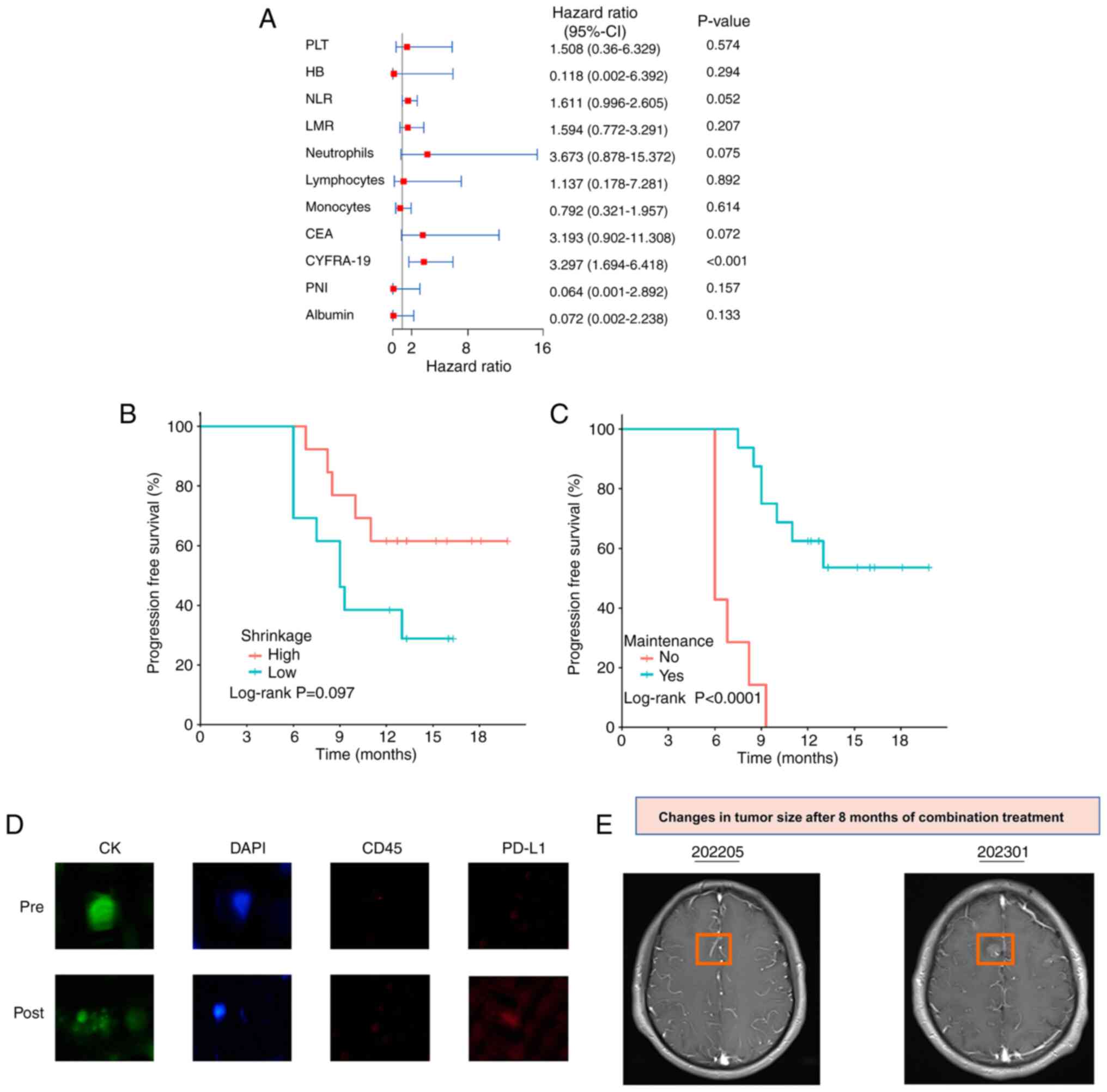 | Figure 4.Prognostic value of blood indicators
in 11 patients with NSCLC following treatment. (A) Association
between PFS and blood indicators in 11 patients with NSCLC.
Association between PFS and (B) treatment maintenance or (C) tumor
shrinkage. (D) CTC PD-L1 expression following treatment with ICIs
in combination with platinum-containing compounds. (E) Tumor
metastasis. NSCLC, non-small cell lung cancer; PFS,
progression-free survival; PD-L1, programmed death-ligand 1; CTCs,
circulating tumor cells; ICIs, immune checkpoint inhibitors; PLT,
platelet; HB, hemoglobin; NLR, neutrophil-to-lymphocyte ratio; LMR,
lymphocyte-to-mononuclear ratio; CEA, carcinoembryonic antigen;
CYFRA-19, cytokeratin 19; PNI, prognostic nutritional index; CK,
cytokeratin. |
| Table II.Patient characteristics and CTC, CTC
PD-L1 after combination treatment. |
Table II.
Patient characteristics and CTC, CTC
PD-L1 after combination treatment.
| Clinical
characteristics | n (%) |
|---|
| Sex |
|
|
Male | 26 (100.0) |
|
Female | 0 (0.0) |
| Histology |
|
|
Adenocarcinoma | 8 (30.8) |
|
Squamous | 18 (69.2) |
| Smoking |
|
| No | 2 (7.7) |
|
Yes | 24 (92.3) |
| Maintenance
ICB |
|
| No | 10 (38.5) |
|
Yes | 16 (61.5) |
| T |
|
|
T2-T3 | 11 (42.3) |
| T4 | 15 (57.7) |
| N |
|
| 0 | 6 (23.1) |
| 2 | 16 (61.5) |
| 3 | 4 (15.4) |
| M |
|
| 0 | 10 (38.5) |
| 1 | 16 (61.5) |
| Line ICB |
|
| 1 | 26 (100.0) |
| CTC |
|
| ≤3 | 17 (65.4) |
|
>3 | 9 (34.6) |
| CTC PD-L1 |
|
|
Negative | 23 (88.5) |
|
Positive | 3 (11.5) |
| PFS |
|
| 0 | 12 (46.2) |
| 1 | 14 (53.8) |
In addition, one patient with negative CTC PD-L1
expression prior to treatment exhibited positive PD-L1 expression
during ICI maintenance therapy (Fig.
4D). Notably, CT imaging revealed new intracranial lesions and
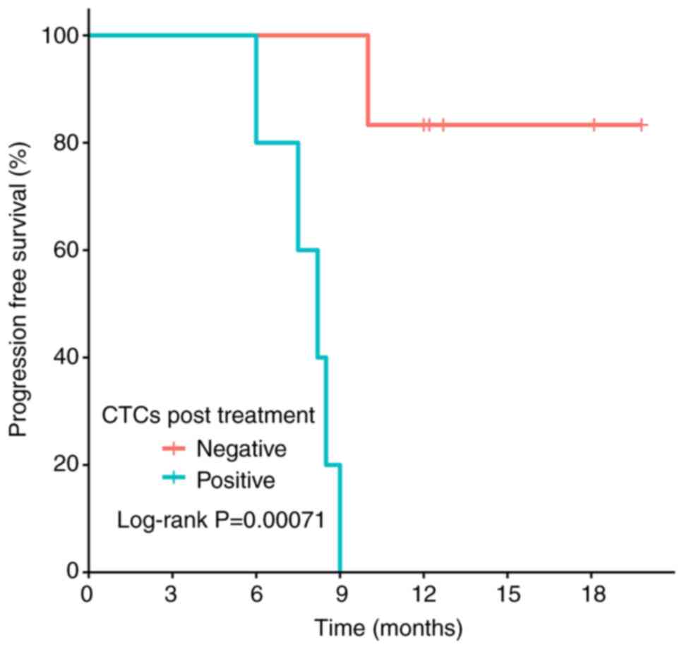
tumor progression (Fig. 4E). By the
median follow-up time of 13 months, the median PFS for patients
with ≥1 CTC at 6 months of treatment was 8.2 months (95% CI,
7.5-NA), while the median PFS for patients with no detectable CTCs
was not reached (Fig. 5, n=11).
These results highlighted that the patients with no detectable CTCs
at 6 months of treatment had a longer PFS and an improved
prognosis.
Discussion
ICIs combined with platinum-containing drugs are the
first-line treatment option for patients with NSCLC and positive
PD-L1 expression, and clinical trials have demonstrated that this
treatment regimen is effective in prolonging patient OS (4,14).
However, the roles of certain markers in predicting treatment
outcomes are yet to be determined. A novel tumor marker is required
to determine the effectiveness of ICIs in combination with
platinum-containing drugs for the treatment of advanced lung
cancer. Results of the present study demonstrated that positive CTC
PD-L1 expression was detected during disease progression following
treatment of NSCLC, and may be indicative of immune escape and drug
resistance.
Results of previous studies highlighted the
predictive role of CTCs and CTC PD-L1 in tumor prognosis. Yue et
al (9) revealed that CTC PD-L1
levels at baseline were notably predictive of PFS. Moreover, Kong
et al (15) systematically
analyzed the association between CTC surface PD-L1 expression
levels and patient prognosis, and the results demonstrated that CTC
PD-L1 expression was associated with shorter OS and PFS of
patients. In total, 40 male patients with advanced lung cancer were
included in the present study. The results revealed no significant
association between CTC PD-L1 detection and IHC results, and the
number of CTCs and CTC PD-L1 in the peripheral blood of patients
was not significantly associated with either advanced cancer
progression or PFS. A newly developed CTC separation device,
CytoBot, was used in the present study, which was validated for the
diagnosis of breast cancer (10)
and lung cancer (11). However, the
value of CytoBot in determining patient prognosis was not
demonstrated in the present study. Results of the present study
revealed that the number of CTCs significantly impacted the
detection rate of CTC PD-L1, perhaps due to tumor heterogeneity.
Notably, a previous study revealed that PD-L1 positivity was
notably higher on CTCs than on tissues (83 vs. 41%); however, there
was no association between CTC PD-L1 and tissue PD-L1 expression
(16). Janning et al
(7) compared the efficiency of the
EpCAM-based CellSearch® detection system (Veridex, LLC)
and the epitope-independent Parsortix® system (ANGLE
plc) in detecting CTC PD-L1, and the results demonstrated that
tumor heterogeneity notably affected CTC PD-L1 detection, and the
concordance of CTC PD-L1 detection with clinical puncture results.
In addition, tumor heterogeneity also affects the results of tissue
puncture analyses, and a small number of tissue samples obtained
through puncture do not represent the entire tumor profile.
However, the tissue puncture remains the gold standard of clinical
testing. In addition, results of a previous study demonstrated that
acquired PD-L1 positivity (conversion to positive expression
following initial negative expression during treatment) also occurs
in the clinic (17). Notably,
acquired PD-L1expression was associated with enhanced metastasis
and drug resistance. Peng et al (18) demonstrated that hepatocyte growth
factor, MET amplification and EGFR-T790M upregulate PD-L1
expression in NSCLC, and enhance immune escape of tumor cells
through various mechanisms. Further clinical trials are required to
improve the accuracy of CTC detection tests.
At present, markers and patient characteristics that
may aid in predicting the efficacy of clinical treatment with ICIs
in combination with platinum-containing compounds are limited.
Thus, novel methods for real-time monitoring during treatment are
required. In addition, these methods should be reproducible, less
invasive and more cost-effective. Routine blood tests are easily
obtainable. Results of a previous study revealed that SIS may
exhibit potential in predicting patient prognosis, and a
combination of SIS and NLR exhibits prognostic value in advanced
pancreatic adenocarcinoma (19).
Zaitsu et al (20)
retrospectively analyzed the clinical characteristics of 73
patients who received immunotherapy for lung cancer. The results
demonstrated that ≥50% PD-L1 expression, ≤1 SIS and <5.6 NLR
were notably associated with longer PFS in patients, indicating
that SIS may exhibit potential as a prognostic marker for
immunotherapy in lung cancer. A previous meta-analysis demonstrated
that patients with high PNI prior to treatment exhibited a longer
OS, disease-free survival/relapse-free survival and PFS (21). In addition, a previous study
revealed that PNI exhibited potential in predicting the OS of
patients with extended-stage SCLC following first-line treatment
(22). Tumor shrinkage is a
relatively common prognostic indicator in chemotherapy, but it is
not often used in the context of novel treatment options, such as
immunotherapy. Notably, immunotherapy or other treatments may cause
cancer tissue to enlarge for a short period prior to shrinkage
(23). The potential association
between PFS and the clinical characteristics of patients,
maintenance treatment, NLR, LMR, CEA and CYFRA-19 was determined in
the present study, before and after 6 months of treatment. Results
of the present study revealed that only tumor shrinkage and
maintenance therapy were significantly associated with patient
prognosis. Blood and tissue samples were collected a long time
apart, some even for months, and it is hypothesized that this may
lead to inaccurate analysis of tissue PD-L1 and blood PD-L1
association. Blood samples were immediately taken from all
patients, which was usually used for testing within 1 week.
Meanwhile, it was anticipated that these patients would be
undergoing long-term treatment, so patients were screened for
negative driver genes based on their clinical characteristics,
including sex, no pleural effusion, type of tumor, molecular
phenotype, which was at least 1 week earlier than waiting for the
genetic testing results. One of the limitations of the present
study was the in-contain of female patients.
CTCs have previously been used as biomarkers for
monitoring response to cancer treatment in numerous clinical trials
(24,25), either alone, or in combination with
imaging tests or serum biomarkers, like microRNA. In some cases,
CTCs are more sensitive than imaging tests, and researchers may
prefer to use CTCs in the evaluation of treatment efficacy
(26). CTC detection is
non-invasive, and may aid in reducing the frequent radiation
exposure during imaging. In addition, the potential association
between PFS and the aforementioned factors before and after
treatment was analyzed in the present study. The current study
revealed that only changes in CYFRA-19 before and after treatment
were associated with PFS. This result may be due to the small
sample size included in the present study, including 26/40 patients
with advanced lung cancer who received conventional drug therapy
and maintained their current treatment regimen. Thus, further
clinical trials with larger sample sizes are required.
Novel therapeutic prognostic markers have been
identified through the development of analytical techniques. CD8A
is mainly expressed in cytotoxic T lymphocytes (CTLs) (27), and CD8A expression levels are
closely associated with tumor stage, with a higher expression
observed at early-stage lung adenocarcinoma than late-stage disease
(28). Ock et al (29) demonstrated that CD8A is a
quantifiable indicator for assessment of CD8+ CTL
recruitment or activity, and exhibits potential in predicting the
response to anti-PD-1/PD-L1 therapy. Moreover, CD8A exhibits a high
predictive value for immunotherapy outcomes, with an AUC of >0.5
in 21/25 patients treated with ICIs (28). Thus, CD8A may exhibit potential as a
biomarker of tumor microenvironment remodeling and as a promising
predictor of immunotherapy efficacy. Carbohydrate Antigen 72–4
(CA72-4) is a routine serum tumor marker used in clinical practice
(30). Results of a previous study
(31) revealed that CA72-4 may
increase sensitivity and specificity when combined with other
biomarkers, like CEA. Moreover, Su et al (32) demonstrated the superior predictive
efficacy of CA72-4 when combined with CA125 in 137 patients with
SCLC treated with first-line chemotherapy. There are other markers
that have an important role in cancer development, such as RANKL
and insulin receptor substrate-4 (33). In cancer cells, RANKL expression is
frequently increased, leading to increased bone resorption and
development of bone metastases. The specific role of the
RANK-RANKL-OPG system in cancer development is thoroughly discussed
by De Leon-Oliva et al (34). However, the relationship between
these markers and treatment with ICIs plus platinum-containing drug
treatment has not been reported.
Neoplastic transformation is caused by the
accumulation of somatic mutations in the DNA of affected cells.
There are notable differences in the frequency of genetic mutations
between individual tumors and between different types of tumors.
Notably, tumor mutation burden (TMB) may be used to predict ICI
efficacy (35). Rizvi et al
(36) performed whole-exome
sequencing in patients with NSCLC treated with pembrolizumab, and
the results demonstrated that a higher burden of non-synonymous
mutations in tumors was associated with improved response,
persistent clinical benefits and improved PFS. These results
suggested that TMB exhibits potential as a biomarker for certain
cancers, and in identifying patients who may benefit from
immunotherapy. Alternate biomarkers for ICI treatment, such as
tumor infiltrating lymphocytes (37), the gene expression profile of T
cells (38), the immune gene
expression profile (39) and
microbiome characterization (40),
exhibit potential in predicting therapeutic outcomes.
The present study retrospectively analyzed the
expression of PD-L1 on CTCs in the peripheral blood of 40 patients
with advanced lung cancer, and compared the detection tests with
IHC assays. Moreover, the association between PFS and CTCs, CTC
PD-L1 and other clinical features was evaluated before and after
treatment with ICIs in combination with platinum-containing
compounds. The current standard treatment protocol for patients
with driver-negative advanced NSCLC involves ICIs combined with
platinum-containing chemotherapy. However, CTC counts and CTC PDL-1
expression are not associated with PFS at initial diagnosis.
Undetectable CTC at 6 months of treatment may be indicative of good
prognosis, whereas the transition of negative CTC PD-L1 expression
to positive may be indicative of poor prognosis. Notably, the
nature of the present study was observational, and there are
numerous limitations. Thus, additional ROC analyses are required
with increased sample sizes. The present study provided a novel
theoretical basis for the detection of CTC PD-L1 in predicting
patient prognosis.
Supplementary Material
Supporting Data
Supporting Data
Acknowledgements
Not applicable.
Funding
The present study was funded by the Chongqing Science and Health
Joint Medical Research Project (grant no. 2021MSXM327) and the
special subject of improving scientific and technological
innovation ability of the Military Medical University (grant no.
2020XQN21).
Availability of data and materials
The datasets used and/or analyzed during the current
study are available from the corresponding author on reasonable
request.
Authors' contributions
XS, CZ and HZ conceived and designed the study. HZ
coordinated the present study and provided administrative support.
CZ and QC provided the study materials or patients. SC, QC and QM
collected and assembled the data. HX, QC and CZ analyzed and
interpreted the data. XS and HZ confirm the authenticity of all the
raw data. All authors contributed to the writing of the manuscript.
All authors have read and approved the final version of the
manuscript.
Ethics approval and consent to
participate
The present study adheres to the Declaration of
Helsinki and has received approval from the Ethics Committee of
Army Medical Center of PLA (Chongqing Daping Hospital, Chongqing,
China; approval no. 2020209). All participants provided written
informed consent.
Patient consent for publication
Not applicable.
Competing interests
The authors declare that they have no competing
interests.
References
|
1
|
Siegel RL, Miller KD, Fuchs HE and Jemal
A: Cancer statistics, 2022. CA Cancer J Clin. 72:7–33. 2022.
View Article : Google Scholar : PubMed/NCBI
|
|
2
|
Siegel RL, Miller KD, Fuchs HE and Jemal
A: Cancer Statistics, 2021. CA Cancer J Clin. 71:7–33. 2021.
View Article : Google Scholar : PubMed/NCBI
|
|
3
|
Antonia SJ, López-Martin JA, Bendell J,
Ott PA, Taylor M, Eder JP, Jäger D, Pietanza MC, Le DT, de Braud F,
et al: Nivolumab alone and nivolumab plus ipilimumab in recurrent
small-cell lung cancer (CheckMate 032): A multicentre, open-label,
phase 1/2 trial. Lancet Oncol. 17:883–895. 2016. View Article : Google Scholar : PubMed/NCBI
|
|
4
|
Singh N, Temin S, Baker S Jr, Blanchard E,
Brahmer JR, Celano P, Duma N, Ellis PM, Elkins IB, Haddad RY, et
al: Therapy for Stage IV Non-Small-Cell lung cancer with driver
alterations: ASCO living guideline. J Clin Oncol. 40:3310–3322.
2022. View Article : Google Scholar : PubMed/NCBI
|
|
5
|
Jones GS and Baldwin DR: Recent advances
in the management of lung cancer. Clin Med (Lond). 18 (Suppl
2):S41–S46. 2018. View Article : Google Scholar : PubMed/NCBI
|
|
6
|
Ricciuti B, Wang X, Alessi JV, Rizvi H,
Mahadevan NR, Li YY, Polio A, Lindsay J, Umeton R, Sinha R, et al:
Association of high tumor mutation burden in Non-Small cell lung
cancers with increased immune infiltration and improved clinical
outcomes of PD-L1 blockade across PD-L1 expression levels. JAMA
Oncol. 8:1160–1168. 2022. View Article : Google Scholar : PubMed/NCBI
|
|
7
|
Janning M, Kobus F, Babayan A, Wikman H,
Velthaus JL, Bergmann S, Schatz S, Falk M, Berger LA, Bottcher LM,
et al: Determination of PD-L1 expression in circulating tumor cells
of NSCLC patients and correlation with response to PD-1/PD-L1
inhibitors. Cancers (Basel). 11:8352019. View Article : Google Scholar : PubMed/NCBI
|
|
8
|
Chen YL, Huang WC, Lin FM, Hsieh HB, Hsieh
CH, Hsieh RK, Chen KW, Yen MH, Lee J, Su S, et al: Novel
circulating tumor cell-based blood test for the assessment of PD-L1
protein expression in treatment-naive, newly diagnosed patients
with non-small cell lung cancer. Cancer Immunol Immunother.
68:1087–1094. 2019. View Article : Google Scholar : PubMed/NCBI
|
|
9
|
Yue C, Jiang Y, Li P, Wang Y, Xue J, Li N,
Li D, Wang R, Dang Y, Hu Z, et al: Dynamic change of PD-L1
expression on circulating tumor cells in advanced solid tumor
patients undergoing PD-1 blockade therapy. Oncoimmunology.
7:e14381112018. View Article : Google Scholar : PubMed/NCBI
|
|
10
|
Wang J, Dallmann R, Lu R, Yan J and
Charmet J: Flow Rate-Independent multiscale liquid biopsy for
precision oncology. ACS Sens. 8:1200–1210. 2023. View Article : Google Scholar : PubMed/NCBI
|
|
11
|
Mei ZF, Yan J, Qian L, He YC, Feng JJ,
Wang J, Jie ZJ and Zou H: Enrichment of circulating tumor cells of
lung cancer and correlation with serum leukomonocyte and tumor
biomarkers: A retrospective study. Technol Cancer Res Treat.
22:20710575812023. View Article : Google Scholar : PubMed/NCBI
|
|
12
|
Ahn MJ, Mendoza MJL, Pavlakis N, Kato T,
Soo RA, Kim DW, Liam CK, Hsia TC, Lee CK, Reungwetwattana T, et al:
Asian Thoracic Oncology Research Group (ATORG) Expert Consensus
Statement on MET Alterations in NSCLC: Diagnostic and Therapeutic
Considerations. Clin Lung Cancer. 23:670–685. 2022. View Article : Google Scholar : PubMed/NCBI
|
|
13
|
Livak KJ and Schmittgen TD: Analysis of
relative gene expression data using real-time quantitative PCR and
the 2(−Delta Delta C(T)) method. Methods. 25:402–408. 2001.
View Article : Google Scholar : PubMed/NCBI
|
|
14
|
Esposito G, Palumbo G, Carillio G, Manzo
A, Montanino A, Sforza V, Costanzo R, Sandomenico C, La Manna C,
Martucci N, et al: Immunotherapy in small cell lung cancer. Cancers
(Basel). 12:25222020. View Article : Google Scholar : PubMed/NCBI
|
|
15
|
Kong D, Zhang W, Yang Z, Li G, Cheng S,
Zhang K and Feng L: Correlation between PD-L1 expression ON CTCs
and prognosis of patients with cancer: A systematic review and
meta-analysis. Oncoimmunology. 10:19384762021. View Article : Google Scholar : PubMed/NCBI
|
|
16
|
Guibert N, Delaunay M, Lusque A, Boubekeur
N, Rouquette I, Clermont E, Mourlanette J, Gouin S, Dormoy I, Favre
G, et al: PD-L1 expression in circulating tumor cells of advanced
non-small cell lung cancer patients treated with nivolumab. Lung
Cancer. 120:108–112. 2018. View Article : Google Scholar : PubMed/NCBI
|
|
17
|
Dill EA, Gru AA, Atkins KA, Friedman LA,
Moore ME, Bullock TN, Cross JV, Dillon PM and Mills AM: PD-L1
Expression and intratumoral heterogeneity across breast cancer
subtypes and stages: An assessment of 245 primary and 40 metastatic
tumors. Am J Surg Pathol. 41:334–342. 2017. View Article : Google Scholar : PubMed/NCBI
|
|
18
|
Peng S, Wang R, Zhang X, Ma Y, Zhong L, Li
K, Nishiyama A, Arai S, Yano S and Wang W: EGFR-TKI resistance
promotes immune escape in lung cancer via increased PD-L1
expression. Mol Cancer. 18:1652019. View Article : Google Scholar : PubMed/NCBI
|
|
19
|
Ma LX, Wang Y, Espin-Garcia O, Allen MJ,
Jang GH, Zhang A, Dodd A, Ramotar S, Hutchinson S, Tehfe M, et al:
Systemic inflammatory prognostic scores in advanced pancreatic
adenocarcinoma. Br J Cancer. 128:1916–1921. 2023. View Article : Google Scholar : PubMed/NCBI
|
|
20
|
Zaitsu J, Yamashita Y, Ishikawa A, Saito
A, Kagimoto A, Mimura T, Hirakawa T, Mito M, Fukuhara K, Senoo T,
et al: Systemic inflammatory score predicts response and prognosis
in patients with lung cancer treated with immunotherapy. Anticancer
Res. 41:3673–3682. 2021. View Article : Google Scholar : PubMed/NCBI
|
|
21
|
Wang Z, Wang Y, Zhang X and Zhang T:
Pretreatment prognostic nutritional index as a prognostic factor in
lung cancer: Review and meta-analysis. Clin Chim Acta. 486:303–310.
2018. View Article : Google Scholar : PubMed/NCBI
|
|
22
|
Bahceci A, Kotek SA and Isik D: The
prognostic values of prognostic nutritional index in
extensive-stage small-cell lung cancer. Anticancer Drugs.
33:e534–e540. 2022. View Article : Google Scholar : PubMed/NCBI
|
|
23
|
Ayoub M, Eleneen Y and Colen RR: Cancer
Imaging in Immunotherapy. Adv Exp Med Biol. 1244:309–324. 2020.
View Article : Google Scholar : PubMed/NCBI
|
|
24
|
Magbanua M, Savenkov O, Asmus EJ, Ballman
KV, Scott JH, Park JW, Dickler M, Partridge A, Carey LA, Winer EP
and Rugo HS: Clinical significance of circulating tumor cells in
hormone receptor-positive metastatic breast cancer patients who
received letrozole with or without bevacizumab. Clin Cancer Res.
26:4911–4920. 2020. View Article : Google Scholar : PubMed/NCBI
|
|
25
|
Graf RP, Hullings M, Barnett ES, Carbone
E, Dittamore R and Scher HI: Clinical Utility of the
Nuclear-localized AR-V7 biomarker in circulating tumor cells in
improving physician treatment choice in castration-resistant
prostate cancer. Eur Urol. 77:170–177. 2020. View Article : Google Scholar : PubMed/NCBI
|
|
26
|
Yu M, Bardia A, Wittner BS, Stott SL, Smas
ME, Ting DT, Isakoff SJ, Ciciliano JC, Wells MN, Shah AM, et al:
Circulating breast tumor cells exhibit dynamic changes in
epithelial and mesenchymal composition. Science. 339:580–584. 2013.
View Article : Google Scholar : PubMed/NCBI
|
|
27
|
Ma K, Qiao Y, Wang H and Wang S:
Comparative expression analysis of PD-1, PD-L1, and CD8A in lung
adenocarcinoma. Ann Transl Med. 8:14782020. View Article : Google Scholar : PubMed/NCBI
|
|
28
|
Niu D, Chen Y, Mi H, Mo Z and Pang G: The
epiphany derived from T-cell-inflamed profiles: Pan-cancer
characterization of CD8A as a biomarker spanning clinical
relevance, cancer prognosis, immunosuppressive environment, and
treatment responses. Front Genet. 13:9744162022. View Article : Google Scholar : PubMed/NCBI
|
|
29
|
Ock CY, Keam B, Kim S, Lee JS, Kim M, Kim
TM, Jeon YK, Kim DW, Chung DH and Heo DS: Pan-Cancer immunogenomic
perspective on the tumor microenvironment based on PD-L1 and CD8
T-Cell infiltration. Clin Cancer Res. 22:2261–2270. 2016.
View Article : Google Scholar : PubMed/NCBI
|
|
30
|
Wang SL, Yu GY, Yao J, Li ZS, Mao AR and
Bai Y: Diagnostic role of carbohydrate antigen 72-4 for
gastrointestinal malignancy screening in Chinese patients: A
prospective study. J Dig Dis. 19:685–692. 2018. View Article : Google Scholar : PubMed/NCBI
|
|
31
|
Xu Y, Zhang P, Zhang K and Huang C: The
application of CA72-4 in the diagnosis, prognosis, and treatment of
gastric cancer. Biochim Biophys Acta Rev Cancer. 187:1886342021.
View Article : Google Scholar : PubMed/NCBI
|
|
32
|
Su Y, Lu C, Zheng S, Zou H, Shen L, Yu J,
Weng Q, Wang Z, Chen M, Zhang R, et al: Precise prediction of the
sensitivity of platinum chemotherapy in SCLC: Establishing and
verifying the feasibility of a CT-based radiomics nomogram. Front
Oncol. 13:10061722023. View Article : Google Scholar : PubMed/NCBI
|
|
33
|
Guijarro LG, Justo BF, Boaru DL, De
Castro-Martinez P, De Leon-Oliva D, Fraile-Martinez O,
Garcia-Montero C, Alvarez-Mon M, Toledo-Lobo M and Ortega MA: Is
Insulin Receptor Substrate4 (IRS4) a platform involved in the
activation of several oncogenes? Cancers (Basel). 15:46512023.
View Article : Google Scholar : PubMed/NCBI
|
|
34
|
De Leon-Oliva D, Barrena-Blazquez S,
Jimenez-Alvarez L, Fraile-Martinez O, Garcia-Montero C,
Lopez-Gonzalez L, Torres-Carranza D, Garcia-Puente LM, Carranza ST,
Alvarez-Mon MA, et al: The RANK-RANKL-OPG System: A multifaceted
regulator of homeostasis, immunity, and cancer. Medicina (Kaunas).
59:17522023. View Article : Google Scholar : PubMed/NCBI
|
|
35
|
Chan TA, Yarchoan M, Jaffee E, Swanton C,
Quezada SA, Stenzinger A and Peters S: Development of tumor
mutation burden as an immunotherapy biomarker: utility for the
oncology clinic. Ann Oncol. 30:44–56. 2019. View Article : Google Scholar : PubMed/NCBI
|
|
36
|
Rizvi NA, Hellmann MD, Snyder A, Kvistborg
P, Makarov V, Havel JJ, Lee W, Yuan J, Wong P, Ho TS, et al: Cancer
immunology. Mutational landscape determines sensitivity to PD-1
blockade in non-small cell lung cancer. Science. 348:124–128. 2015.
View Article : Google Scholar : PubMed/NCBI
|
|
37
|
Tumeh PC, Harview CL, Yearley JH, Shintaku
IP, Taylor EJ, Robert L, Chmielowski B, Spasic M, Henry G, Ciobanu
V, et al: PD-1 blockade induces responses by inhibiting adaptive
immune resistance. Nature. 515:568–571. 2014. View Article : Google Scholar : PubMed/NCBI
|
|
38
|
Cristescu R, Mogg R, Ayers M, Albright A,
Murphy E, Yearley J, Sher X, Liu XQ, Lu H, Nebozhyn M, et al:
Pan-tumor genomic biomarkers for PD-1 checkpoint blockade-based
immunotherapy. Science. 362:eaar35932018. View Article : Google Scholar : PubMed/NCBI
|
|
39
|
Auslander N, Zhang G, Lee JS, Frederick
DT, Miao B, Moll T, Tian T, Wei Z, Madan S, Sullivan RJ, et al:
Robust prediction of response to immune checkpoint blockade therapy
in metastatic melanoma. Nat Med. 24:1545–1549. 2018. View Article : Google Scholar : PubMed/NCBI
|
|
40
|
Roy S and Trinchieri G: Microbiota: A key
orchestrator of cancer therapy. Nat Rev Cancer. 17:271–285. 2017.
View Article : Google Scholar : PubMed/NCBI
|