Spandidos Publications Logo
  • About
    • About Spandidos
    • Aims and Scopes
    • Abstracting and Indexing
    • Editorial Policies
    • Reprints and Permissions
    • Job Opportunities
    • Terms and Conditions
    • Contact
  • Journals
    • All Journals
    • Oncology Letters
      • Oncology Letters
      • Information for Authors
      • Editorial Policies
      • Editorial Board
      • Aims and Scope
      • Abstracting and Indexing
      • Bibliographic Information
      • Archive
    • International Journal of Oncology
      • International Journal of Oncology
      • Information for Authors
      • Editorial Policies
      • Editorial Board
      • Aims and Scope
      • Abstracting and Indexing
      • Bibliographic Information
      • Archive
    • Molecular and Clinical Oncology
      • Molecular and Clinical Oncology
      • Information for Authors
      • Editorial Policies
      • Editorial Board
      • Aims and Scope
      • Abstracting and Indexing
      • Bibliographic Information
      • Archive
    • Experimental and Therapeutic Medicine
      • Experimental and Therapeutic Medicine
      • Information for Authors
      • Editorial Policies
      • Editorial Board
      • Aims and Scope
      • Abstracting and Indexing
      • Bibliographic Information
      • Archive
    • International Journal of Molecular Medicine
      • International Journal of Molecular Medicine
      • Information for Authors
      • Editorial Policies
      • Editorial Board
      • Aims and Scope
      • Abstracting and Indexing
      • Bibliographic Information
      • Archive
    • Biomedical Reports
      • Biomedical Reports
      • Information for Authors
      • Editorial Policies
      • Editorial Board
      • Aims and Scope
      • Abstracting and Indexing
      • Bibliographic Information
      • Archive
    • Oncology Reports
      • Oncology Reports
      • Information for Authors
      • Editorial Policies
      • Editorial Board
      • Aims and Scope
      • Abstracting and Indexing
      • Bibliographic Information
      • Archive
    • Molecular Medicine Reports
      • Molecular Medicine Reports
      • Information for Authors
      • Editorial Policies
      • Editorial Board
      • Aims and Scope
      • Abstracting and Indexing
      • Bibliographic Information
      • Archive
    • World Academy of Sciences Journal
      • World Academy of Sciences Journal
      • Information for Authors
      • Editorial Policies
      • Editorial Board
      • Aims and Scope
      • Abstracting and Indexing
      • Bibliographic Information
      • Archive
    • International Journal of Functional Nutrition
      • International Journal of Functional Nutrition
      • Information for Authors
      • Editorial Policies
      • Editorial Board
      • Aims and Scope
      • Abstracting and Indexing
      • Bibliographic Information
      • Archive
    • International Journal of Epigenetics
      • International Journal of Epigenetics
      • Information for Authors
      • Editorial Policies
      • Editorial Board
      • Aims and Scope
      • Abstracting and Indexing
      • Bibliographic Information
      • Archive
    • Medicine International
      • Medicine International
      • Information for Authors
      • Editorial Policies
      • Editorial Board
      • Aims and Scope
      • Abstracting and Indexing
      • Bibliographic Information
      • Archive
  • Articles
  • Information
    • Information for Authors
    • Information for Reviewers
    • Information for Librarians
    • Information for Advertisers
    • Conferences
  • Language Editing
Spandidos Publications Logo
  • About
    • About Spandidos
    • Aims and Scopes
    • Abstracting and Indexing
    • Editorial Policies
    • Reprints and Permissions
    • Job Opportunities
    • Terms and Conditions
    • Contact
  • Journals
    • All Journals
    • Biomedical Reports
      • Information for Authors
      • Editorial Policies
      • Editorial Board
      • Aims and Scope
      • Abstracting and Indexing
      • Bibliographic Information
      • Archive
    • Experimental and Therapeutic Medicine
      • Information for Authors
      • Editorial Policies
      • Editorial Board
      • Aims and Scope
      • Abstracting and Indexing
      • Bibliographic Information
      • Archive
    • International Journal of Epigenetics
      • Information for Authors
      • Editorial Policies
      • Editorial Board
      • Aims and Scope
      • Abstracting and Indexing
      • Bibliographic Information
      • Archive
    • International Journal of Functional Nutrition
      • Information for Authors
      • Editorial Policies
      • Editorial Board
      • Aims and Scope
      • Abstracting and Indexing
      • Bibliographic Information
      • Archive
    • International Journal of Molecular Medicine
      • Information for Authors
      • Editorial Policies
      • Editorial Board
      • Aims and Scope
      • Abstracting and Indexing
      • Bibliographic Information
      • Archive
    • International Journal of Oncology
      • Information for Authors
      • Editorial Policies
      • Editorial Board
      • Aims and Scope
      • Abstracting and Indexing
      • Bibliographic Information
      • Archive
    • Medicine International
      • Information for Authors
      • Editorial Policies
      • Editorial Board
      • Aims and Scope
      • Abstracting and Indexing
      • Bibliographic Information
      • Archive
    • Molecular and Clinical Oncology
      • Information for Authors
      • Editorial Policies
      • Editorial Board
      • Aims and Scope
      • Abstracting and Indexing
      • Bibliographic Information
      • Archive
    • Molecular Medicine Reports
      • Information for Authors
      • Editorial Policies
      • Editorial Board
      • Aims and Scope
      • Abstracting and Indexing
      • Bibliographic Information
      • Archive
    • Oncology Letters
      • Information for Authors
      • Editorial Policies
      • Editorial Board
      • Aims and Scope
      • Abstracting and Indexing
      • Bibliographic Information
      • Archive
    • Oncology Reports
      • Information for Authors
      • Editorial Policies
      • Editorial Board
      • Aims and Scope
      • Abstracting and Indexing
      • Bibliographic Information
      • Archive
    • World Academy of Sciences Journal
      • Information for Authors
      • Editorial Policies
      • Editorial Board
      • Aims and Scope
      • Abstracting and Indexing
      • Bibliographic Information
      • Archive
  • Articles
  • Information
    • For Authors
    • For Reviewers
    • For Librarians
    • For Advertisers
    • Conferences
  • Language Editing
Login Register Submit
  • This site uses cookies
  • You can change your cookie settings at any time by following the instructions in our Cookie Policy. To find out more, you may read our Privacy Policy.

    I agree
Search articles by DOI, keyword, author or affiliation
Search
Advanced Search
presentation
Oncology Letters
Join Editorial Board Propose a Special Issue
Print ISSN: 1792-1074 Online ISSN: 1792-1082
Journal Cover
June-2024 Volume 27 Issue 6

Full Size Image

Sign up for eToc alerts
Recommend to Library

Journals

International Journal of Molecular Medicine

International Journal of Molecular Medicine

International Journal of Molecular Medicine is an international journal devoted to molecular mechanisms of human disease.

International Journal of Oncology

International Journal of Oncology

International Journal of Oncology is an international journal devoted to oncology research and cancer treatment.

Molecular Medicine Reports

Molecular Medicine Reports

Covers molecular medicine topics such as pharmacology, pathology, genetics, neuroscience, infectious diseases, molecular cardiology, and molecular surgery.

Oncology Reports

Oncology Reports

Oncology Reports is an international journal devoted to fundamental and applied research in Oncology.

Experimental and Therapeutic Medicine

Experimental and Therapeutic Medicine

Experimental and Therapeutic Medicine is an international journal devoted to laboratory and clinical medicine.

Oncology Letters

Oncology Letters

Oncology Letters is an international journal devoted to Experimental and Clinical Oncology.

Biomedical Reports

Biomedical Reports

Explores a wide range of biological and medical fields, including pharmacology, genetics, microbiology, neuroscience, and molecular cardiology.

Molecular and Clinical Oncology

Molecular and Clinical Oncology

International journal addressing all aspects of oncology research, from tumorigenesis and oncogenes to chemotherapy and metastasis.

World Academy of Sciences Journal

World Academy of Sciences Journal

Multidisciplinary open-access journal spanning biochemistry, genetics, neuroscience, environmental health, and synthetic biology.

International Journal of Functional Nutrition

International Journal of Functional Nutrition

Open-access journal combining biochemistry, pharmacology, immunology, and genetics to advance health through functional nutrition.

International Journal of Epigenetics

International Journal of Epigenetics

Publishes open-access research on using epigenetics to advance understanding and treatment of human disease.

Medicine International

Medicine International

An International Open Access Journal Devoted to General Medicine.

Journal Cover
June-2024 Volume 27 Issue 6

Full Size Image

Sign up for eToc alerts
Recommend to Library

  • Article
  • Citations
    • Cite This Article
    • Download Citation
    • Create Citation Alert
    • Remove Citation Alert
    • Cited By
  • Similar Articles
    • Related Articles (in Spandidos Publications)
    • Similar Articles (Google Scholar)
    • Similar Articles (PubMed)
  • Download PDF
  • Download XML
  • View XML
Case Report Open Access

Precision CT‑guided marking with India ink and microcoils for laparoscopic resection of a lateral lymph node recurrence of rectal cancer: A case report

  • Authors:
    • Shunsuke Furukawa
    • Masatsugu Hiraki
    • Takeshi Oda
    • Yukihiko Takahashi
    • Ryuichiro Samejima
  • View Affiliations / Copyright

    Affiliations: Department of Surgery, Japanese Red Cross Society Karatsu Red Cross Hospital, Karatsu, Saga 847‑8588, Japan, Department of Radiology, Japanese Red Cross Society Karatsu Red Cross Hospital, Karatsu, Saga 847‑8588, Japan
    Copyright: © Furukawa et al. This is an open access article distributed under the terms of Creative Commons Attribution License.
  • Article Number: 266
    |
    Published online on: April 12, 2024
       https://doi.org/10.3892/ol.2024.14399
  • Expand metrics +
Metrics: Total Views: 0 (Spandidos Publications: | PMC Statistics: )
Metrics: Total PDF Downloads: 0 (Spandidos Publications: | PMC Statistics: )
Cited By (CrossRef): 0 citations Loading Articles...

This article is mentioned in:


Abstract

Although rarely used in laparoscopic surgery, computed tomography (CT)‑guided marking is useful for targeting small lesions. The present study describes the performance of laparoscopic resection with preoperative CT‑guided marking for lateral lymph node recurrence of rectal cancer. A 48‑year‑old man underwent laparoscopic low anterior resection with D3 lymph node dissection for rectal cancer (postoperative diagnosis, stage IIIb). Postoperative adjuvant chemotherapy was then administered. Solitary lymph node recurrence was observed in the left lateral region after 6 months. Systemic chemotherapy reduced the size of the metastatic lymph nodes; however, 3.5 years after the surgery, the diameter increased by 10 mm, and laparoscopic resection was thus planned. Because the target lesion was small and located deep in the pelvis, preoperative CT‑guided marking was performed with India ink injection and embolization microcoil implantation. The markers were clearly identified and the lesion was successfully resected with adequate margins. The approach described in the present study is thus considered potentially useful for the detection of small lesions.

Introduction

Lateral lymph node metastasis is a form of postoperative recurrence in lower rectal cancer. Local resection, radiotherapy, and systemic chemotherapy are indicated for the local recurrence of rectal cancer. Regarding solitary lateral lymph node recurrence, it is reported that it may offer a favorable long-term prognosis when treated with local resection (1). However, small tumors or lymph nodes can be difficult to detect due to their location in the deep pelvic region and the impact of reoperation. Furthermore, the resection margin is difficult to determine due to the presence of surrounding tissues. Therefore, CT-guided marking techniques, which are commonly used in pulmonary surgery, but are only rarely applied in abdominal surgeries (2), should be considered. India ink marking has been widely used and it is considered safe and effective (3). In the past, the effectiveness of CT-guided marking with India ink has been reported in abdominal surgery (4). In addition, CT-guided marking using microcoils has been reported to be safe and effective (5–7). Microcoil localization has the advantages of accuracy, high success rate, low complications, and good tolerance (8–10).

We herein report a case of laparoscopic resection of lateral lymph node recurrence of rectal cancer that was successfully treated with preoperative CT-guided marking with India ink and microcoils.

Case report

A 48-year-old man had undergone laparoscopic low anterior resection with D3 lymph node dissection and left lateral lymph node dissection for rectal cancer in September 2019 at the Japanese Red Cross Society Karatsu Red Cross Hospital (Karatsu, Japan). The tumor had mainly been located in the upper rectum above the peritoneal reflection. A digital rectal examination palpated the inferior margin of the tumor, 7 cm from the anus. The final findings were pT3N1bM0 and pStage IIIb according to the 8th edition of the UICC classification (11). Postoperative adjuvant chemotherapy consisting of capecitabine was administered for 6 months. Oxaliplatin was not administered due to occupational reasons. However, at 6 months postoperatively, a solitary mass (diameter: 10 mm) was found in the left lateral region on enhanced abdominal computed tomography (CT), and he was diagnosed to have local recurrence because of an accompanying increase in the tumor marker levels. Systemic chemotherapy (irinotecan, S-1, and bevacizumab) was administered for 2 years. Stable disease persisted as the lesion decreased to 8 mm, and the carcinoembryonic antigen (CEA) and carbohydrate antigen 19-9 (CA19-9) levels fell to within the normal range. Fluorodeoxyglucose (FDG) positron emission tomography CT showed no accumulation of 18F-FDG (early SUVmax, 1.3; delayed SUVmax, 2.0). However, the 
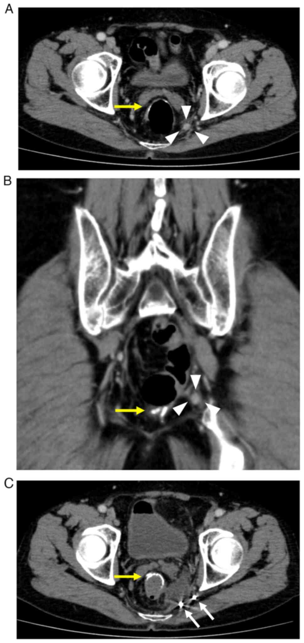
lesion again increased in size to 10 mm at 3.5 years after surgery (Fig. 1A and B). FDG positron emission tomography CT showed the minor accumulation of 18F-FDG (early SUVmax 1.6, delayed SUVmax, 2.8). The patient's CEA and CA19-9 levels were within the normal ranges. No palpable tumorous lesions were observed on a digital rectal examination. The lesion had mildly increased in size, and PET-CT showed a minor accumulation. As a result, a relapse was suspected. Hence, percutaneous CT-guided biopsy was not considered due to the risk of dissemination. Resection of the solitary left lateral lymph node was considered, because no other new lesions had appeared. A high degree of operative difficulty was expected due to reoperation, the tumor location in the deep pelvis, and visibility problems regarding lesion identification. Therefore, preoperative marking with CT guidance was performed before surgery to ensure a secure and safe resection. Written informed consent was obtained for the CT-guided marking and surgical procedures. The patient was placed in a prone position on the CT table, and a 21-gauge needle was percutaneously inserted into the pelvis near the tumor. After local anesthesia, saline solution (2.0 ml) was topically injected near the tumor using a fine needle to maintain the space for placement of the microcoil so as not to injure the surrounding nerve and vasculature. India ink (1.0 ml) was injected between the tumor and the pelvic side, and two microcoils, originally designed for vascular embolization (Hilal Embolization Microcoil™, Cook Medical Japan), were implanted by a radiologist. The two coils were placed on either side (i.e., the interior and exterior) of the tumor on the morning of the surgery (Fig. 1C). The ureteral stent was placed by a urologist to avoid injury to the left ureter. Subsequently, 
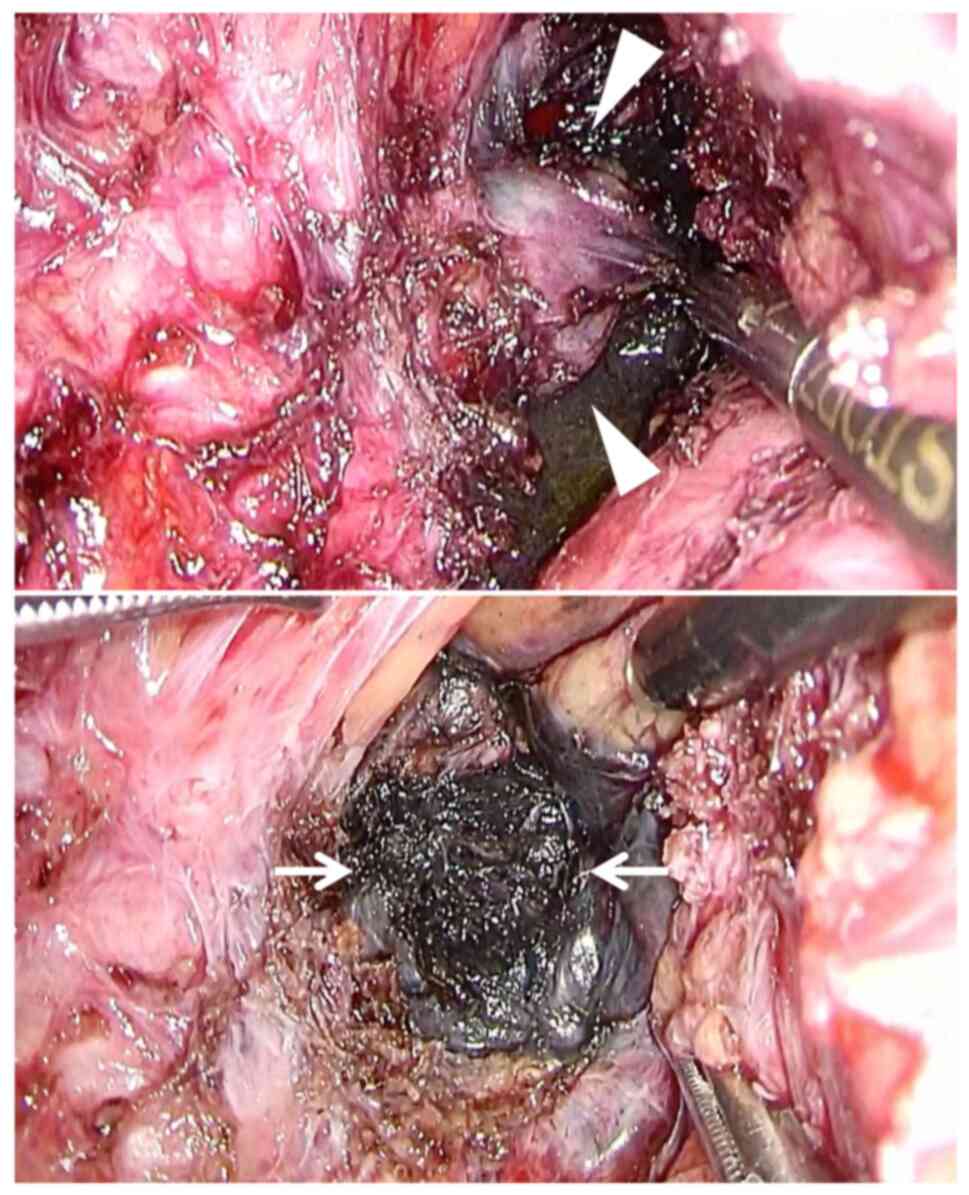
laparoscopic surgery was performed. After the sharp dissection of adhesions between the large intestine and the pelvic wall, the left ureter was identified and preserved. A rectal examination was performed intraoperatively to avoid any rectal injury. After 
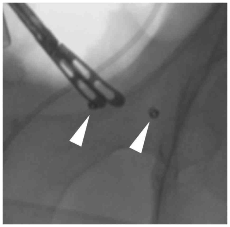
careful dissection, the targeted tumor was identified intraoperatively using the two embolization microcoils as landmarks and intraoperative fluoroscopy (Fig. 2). The embolization microcoils were not visible, but they were useful for identifying the extent of resection. The tumor was identified between the two coils and thus was successfully exfoliated with the surrounding fat. During resection, black ink was visibly identified (Fig. 3). Laparoscopic lymphadenectomy was completed within 5 h and 7 min with 100 ml of blood loss. The resected specimen was a solid nodule 
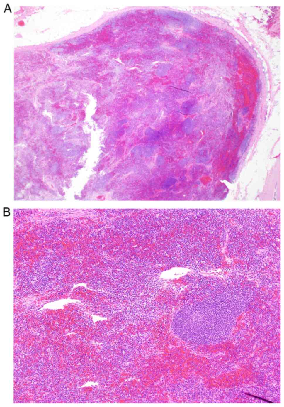
measuring 10 mm in size. The pathological diagnosis revealed no viable tumor cells in the dissected lymph nodes (Fig. 4). The patient was discharged on postoperative day 5 without any complications and remained disease-free for 7 months postoperatively.

Figure 1.

Recurrent solitary lymph node on the left lateral pelvic region in computed tomography images. Yellow arrow and white arrowhead indicate the anastomosis of the low anterior resection and lateral lymph node tumor, respectively. (A) Horizontal plane. (B) Coronal plane. (C) Two microcoils (white arrows) were implanted near the interior and exterior of the lateral lymph node tumor. CT, computed tomography.

Figure 2.

The two microcoils (white arrowheads) were clearly identified by fluoroscopy, and the tumor was recognized to be located between them.

Figure 3.

The tumor (white arrowheads) was present in front of the India ink. After resecting the tumor, the India ink was identified (white arrows). The rectal wall is situated to the right of the laparoscopic forceps.

Figure 4.

The pathological diagnosis revealed no viable tumor cells. (A) H&E; magnification, ×10. (B) H&E; magnification, ×100. H&E, hematoxylin and eosin.

Discussion

Postoperative pelvic recurrence is a clinical condition experienced by some patients after curative resection for rectal cancer. The indications for treatment are based on a comprehensive consideration of the number of recurrences, location, and postoperative period. Regarding lateral lymph node recurrence, it has been reported that even if lateral lymph node metastasis occurs, the disease can be controlled if there are <2 metastatic lymph nodes (12). Local resection can achieve a favorable long-term prognosis in patients with solitary lateral lymph node metastasis (1). Thus, surgical resection may be considered if the disease is localized and no other recurrent lesions are observed. However, surgical resection involves various procedural difficulties associated with reoperation, a location in the deep pelvis, and the visual identification of lesions.

Preoperative CT-guided marking for tumor identification is frequently reported to be effective for intraoperative identification of small tumors in the lung and for accurate surgical resection in respiratory surgery (13). However, there have been few reports on CT-guided marking in abdominal surgery, and its effectiveness and indications have not yet been established. The key issue is that lateral lymph node metastasis sometimes occurs in the deep pelvic space and after surgery. In this case, we considered that CT-guided marking may be effective for recognizing the target and resecting the tumor with sufficient surgical margins, as it was very small and the dissection layer was difficult to identify due to reoperation. A search of PubMed with the key words ‘preoperative’ and ‘CT-guided marking’ revealed one case report on preoperative CT-guided marking in abdominal surgery, which reported the efficacy of CT-guided marking with India ink and iopamidol for the recurrence of ascending colon cancer (diameter: 2 cm) in the right pararenal region (4).

Hookwire, ICG, India ink, and Lipiodol are often used for markings (14,15). India ink and microcoils were used in this study. India ink has been conventionally used, and its effectiveness and safety have been established (3). Because the ink is black, it is considered to have excellent visibility. However, there is concern that the ink may spread to the surrounding tissue and thereby obscure the objective. Moreover, such markings may not be visible during surgery. Therefore, microcoils were used to increase the chance of intraoperative confirmation and to accurately resect the tumor. Microcoils have been used for vascular embolization and have shown good tissue compatibility. In addition, such microcoils are soft and thus do not cause significant damage to the tissue after implantation into the body (16,17). CT-guided microcoils have been reported to be safe and effective tools for easy and fast video-assisted thoracoscopic surgery (5–7). Therefore, we decided to use microcoils to treat this patient after obtaining sufficient written informed consent. As previously reported, iopamidol was used instead of the microcoils (4). However, there was a concern that it may not be visible on fluoroscopy because it could spread to the surrounding tissue and become diluted because it is a liquid. In addition, when performing preoperative CT-guided marking, careful examination is required to avoid complications such as bleeding and infection (18). As a result, X-ray fluoroscopy made it easy to recognize the markings and accurately resect the tumor laparoscopically. CT-guided marking with India ink and microcoils using fluoroscopy is useful for abdominal surgery. Although few reports have previously described this method, it may be useful for the curative resection of small tumors.

In conclusion, CT-guided marking with India ink and microcoils is useful for ensuring curative surgery for small tumors. To our knowledge, this is the first English case report of successful laparoscopic surgery using CT-guided marking with India ink and microcoils.

Acknowledgements

Not applicable.

Funding

Funding: No funding was received.

Availability of data and materials

All data generated or analyzed during this study are included in this published article.

Authors' contributions

All of the authors contributed to the diagnosis and treatment of the patient. SF and MH drafted the manuscript. TO, YT and RS edited the manuscript. RS supervised the study and approved the final version of the manuscript. MH and RS confirm the authenticity of all the raw data. All authors read and approved the final version of the manuscript.

Ethics approval and consent to participate

Not applicable.

Patient consent for publication

Written informed consent was obtained from the patient for publication of this case report and accompanying images.

Competing interests

The authors declare that they have no competing interests.

Glossary

Abbreviations

Abbreviations:

CT

computed tomography

CEA

carcinoembryonic antigen

CA19-9

carbohydrate antigen 19-9

FDG

fluorodeoxyglucose

References

1 

Miura T, Tsunenari T, Sasaki T, Yokoyama T and Fukuhara K: A curatively resected case of lateral lymph node metastasis five-years after initial surgery for rectal cancer. Gan To Kagaku Ryoho. 44:1405–1407. 2017.(In Japanese). PubMed/NCBI

2 

Mun M, Matsuura Y, Nakao M, Ichinose J, Nakagawa K and Okumura S: Noninvasive computed tomography-guided marking technique for peripheral pulmonary nodules. J Thorac Dis. 8 (Suppl 9):S672–S676. 2016. View Article : Google Scholar : PubMed/NCBI

3 

McArthur C, Roayaie S and Waye J: Safety of preoperation endoscopic tattoo with india ink for identification of colonic lesions. Surg Endosc. 13:397–400. 1999. View Article : Google Scholar

4 

Koyama H, Noma S, Tamaki Y, Goto K, Kitamura E, Maeda T, Matsumoto S, Sano A and Sugimura K: CT localisation of small pulmonary nodules prior to thorascopic resection: Evaluation of a point marker system. Eur J Radiol. 65:468–472. 2008. View Article : Google Scholar

5 

Liu L, Zhang LJ, Chen B, Cao JM, Lu GM, Yuan L, Li K and Xu J: Novel CT-guided coil localization of peripheral pulmonary nodules prior to videoassisted thoracoscopic surgery: A pilot study. Acta Radiol. 55:699–706. 2014. View Article : Google Scholar

6 

Huang ZG, Wang CL, Sun HL, Li CD, Gao BX, Chen H and Yang MX: CT-guided microcoil localization of small peripheral pulmonary nodules to direct video-assisted thoracoscopic resection without the aid of intraoperative fluoroscopy. Korean J Radiol. 22:1124–1131. 2021. View Article : Google Scholar

7 

An J, Dong Y, Li Y, Han X, Niu H, Zou Z, Wu J, Tian Y and Chen Z: CT-guided placement of microcoil end in the pleural cavity for video-assisted thoracic surgical resection of ground-glass opacity: A retrospective study. J Cardiothorac Surg. 17:3162022. View Article : Google Scholar : PubMed/NCBI

8 

Powell TI, Jangra D, Clifton JC, Lara-Guerra H, Church N, English J, Evans K, Yee J, Coxson H, Mayo JR and Finley RJ: Peripheral lung nodules: Fluoroscopically guided video-assisted thoracoscopic resection after computed tomography-guided localization using platinum microcoils. Ann Surg. 240:481–488. 2004. View Article : Google Scholar : PubMed/NCBI

9 

Mayo JR, Clifton JC, Powell TI, English JC, Evans KG, Yee J, McWilliams AM, Lam SC and Finley RJ: Lung nodules: CT-guided placement of microcoils to direct video-assisted thoracoscopic surgical resection. Radiology. 250:576–585. 2009. View Article : Google Scholar : PubMed/NCBI

10 

Lempel JK, Raymond DP, Ahmad U, O'Malley S, Bolen MA, Graham R, Azok JT, Bullen J, Raja S and Murthy S: Video-assisted thoracic surgery resection without intraoperative fluoroscopy after CT-guided microcoil localization of peripheral pulmonary nodules. J Vasc Interv Radiol. 29:1423–1428. 2018. View Article : Google Scholar

11 

UICC, . TNM Classification of Malignant Tumours. 8th edition. John Wiley & Sons Ltd.; New York, NY: 2017

12 

Homma Y, Hamano T, Otsuki Y, Shimizu S and Kobayashi Y: Total number of lymph node metastases is a more significant risk factor for poor prognosis than positive lateral lymph node metastasis. Surg Today. 45:168–174. 2015. View Article : Google Scholar : PubMed/NCBI

13 

Ueki H, Fujimoto T, Okuno M, Kusuda Y, Taguchi I, Itou Y, Kiyonaka S and Kawabata G: The use of CT-guided marking for the laparoscopic resection of a solitary retroperitoneal metastasis of colon cancer. J Endourol Case Rep. 4:120–123. 2018. View Article : Google Scholar : PubMed/NCBI

14 

Ashida R, Yamao K, Okubo K, Sawaki A, Mizuno N, Nakamura T, Tajika M, Kawai H and Shimizu Y: Indocyanine green is an ideal dye for endoscopic ultrasound-guided fine-needle tattooing of pancreatic tumors. Endoscopy. 38:190–192. 2006. View Article : Google Scholar : PubMed/NCBI

15 

Kang DY, Kim HK, Kim YK, Yong HS, Kang EY and Choi YH: Needlescopy-assisted resection of pulmonary nodule after dual localization. Eur Respir J. 37:13–17. 2011. View Article : Google Scholar : PubMed/NCBI

16 

Hwang S, Kim TG and Song YG: Comparison of hook wire versus coil localization for video-assisted thoracoscopic surgery. Thorac Cancer. 9:384–389. 2018. View Article : Google Scholar : PubMed/NCBI

17 

Hu L, Gao J, Chen C, Zhi X, Liu H and Hong N: Comparison between the application of microcoil and hookwire for localizing pulmonary nodules. Eur Radiol. 29:4036–4043. 2019. View Article : Google Scholar

18 

Fumimoto S, Sato K, Koyama M, Yamamoto K, Narumi Y, Hanaoka N and Katsumata T: Combined lipiodol marking and video-assisted thoracoscopic surgery in a hybrid operating room. J Thorac Dis. 10:2940–2947. 2018. View Article : Google Scholar : PubMed/NCBI

Related Articles

  • Abstract
  • View
  • Download
  • Twitter
Copy and paste a formatted citation
Spandidos Publications style
Furukawa S, Hiraki M, Oda T, Takahashi Y and Samejima R: Precision CT‑guided marking with India ink and microcoils for laparoscopic resection of a lateral lymph node recurrence of rectal cancer: A case report. Oncol Lett 27: 266, 2024.
APA
Furukawa, S., Hiraki, M., Oda, T., Takahashi, Y., & Samejima, R. (2024). Precision CT‑guided marking with India ink and microcoils for laparoscopic resection of a lateral lymph node recurrence of rectal cancer: A case report. Oncology Letters, 27, 266. https://doi.org/10.3892/ol.2024.14399
MLA
Furukawa, S., Hiraki, M., Oda, T., Takahashi, Y., Samejima, R."Precision CT‑guided marking with India ink and microcoils for laparoscopic resection of a lateral lymph node recurrence of rectal cancer: A case report". Oncology Letters 27.6 (2024): 266.
Chicago
Furukawa, S., Hiraki, M., Oda, T., Takahashi, Y., Samejima, R."Precision CT‑guided marking with India ink and microcoils for laparoscopic resection of a lateral lymph node recurrence of rectal cancer: A case report". Oncology Letters 27, no. 6 (2024): 266. https://doi.org/10.3892/ol.2024.14399
Copy and paste a formatted citation
x
Spandidos Publications style
Furukawa S, Hiraki M, Oda T, Takahashi Y and Samejima R: Precision CT‑guided marking with India ink and microcoils for laparoscopic resection of a lateral lymph node recurrence of rectal cancer: A case report. Oncol Lett 27: 266, 2024.
APA
Furukawa, S., Hiraki, M., Oda, T., Takahashi, Y., & Samejima, R. (2024). Precision CT‑guided marking with India ink and microcoils for laparoscopic resection of a lateral lymph node recurrence of rectal cancer: A case report. Oncology Letters, 27, 266. https://doi.org/10.3892/ol.2024.14399
MLA
Furukawa, S., Hiraki, M., Oda, T., Takahashi, Y., Samejima, R."Precision CT‑guided marking with India ink and microcoils for laparoscopic resection of a lateral lymph node recurrence of rectal cancer: A case report". Oncology Letters 27.6 (2024): 266.
Chicago
Furukawa, S., Hiraki, M., Oda, T., Takahashi, Y., Samejima, R."Precision CT‑guided marking with India ink and microcoils for laparoscopic resection of a lateral lymph node recurrence of rectal cancer: A case report". Oncology Letters 27, no. 6 (2024): 266. https://doi.org/10.3892/ol.2024.14399
Follow us
  • Twitter
  • LinkedIn
  • Facebook
About
  • Spandidos Publications
  • Careers
  • Cookie Policy
  • Privacy Policy
How can we help?
  • Help
  • Live Chat
  • Contact
  • Email to our Support Team