Colorectal cancer (CRC) is a prevalent malignancy of
the gastrointestinal tract, with high rates of morbidity and
mortality. In 2018, the GLOBOCAN database published an analysis of
the incidence and mortality rates for 36 cancers across 185
countries, and the results revealed that CRC is the third most
common cancer and the second leading cause of cancer-associated
mortalities worldwide (1,2). The etiology of CRC is associated with
various factors, including age, sex, inflammatory bowel disease,
lifestyle and environmental factors (3–5).
Current treatment options for CRC include surgery, chemotherapy,
radiotherapy, immunotherapy and targeted biological therapies
(6). However, the absence of highly
specific biomarkers for the diagnosis of CRC and the development of
chemoresistance in advanced stages of treatment may impact the
quality of life of patients with late-stage disease (7). Thus, research is focused on the
development of novel effective targeted therapies for CRC. Notably,
ferroptosis may exhibit potential as an emerging strategy for CRC
treatment.
Regulated cell death, including apoptosis,
pyroptosis, necroptosis, ferroptosis, autophagy-dependent cell
death and neoplastic cell death, is a crucial mechanism for
maintaining the internal homeostasis of the human body and
preserving tissue function and morphology (8,9). The
primary method for distinguishing between different forms of cell
death is based on the associated morphological characteristics
(Table I).
Ferroptosis, initially described by Dixon in 2012,
is a novel form of regulated cell death (9). It is distinct from other forms of
regulated cell death in terms of morphology, biochemical features
and gene expression (10). Notably,
ferroptosis is characterized by unique morphological features,
including mitochondrial shrinkage, increased mitochondrial membrane
density, reduced or absent mitochondrial cristae, rupture of the
outer mitochondrial membrane, and preservation of nuclear integrity
(11,12). The main morphological
characteristics of apoptosis include cell shrinkage, nuclear
condensation and the maintenance of plasma membrane integrity,
while its biochemical features are primarily DNA fragmentation and
macromolecule synthesis (13). The
key morphological traits of pyroptosis involve cell swelling, the
formation of numerous bubbles on the pyroptotic cells, and
chromatin degradation, leading to the formation of inflammasomes
(14–16). The primary morphological
characteristics of necroptosis include increased cell membrane
permeability, cell swelling and loss of organelle integrity, while
its biochemical hallmark is a decrease in adenosine triphosphate
levels (17,18). Autophagy is characterized by the
formation of double-membraned autophagosomes, and its biochemical
feature is an increase in lysosomal activity (19).
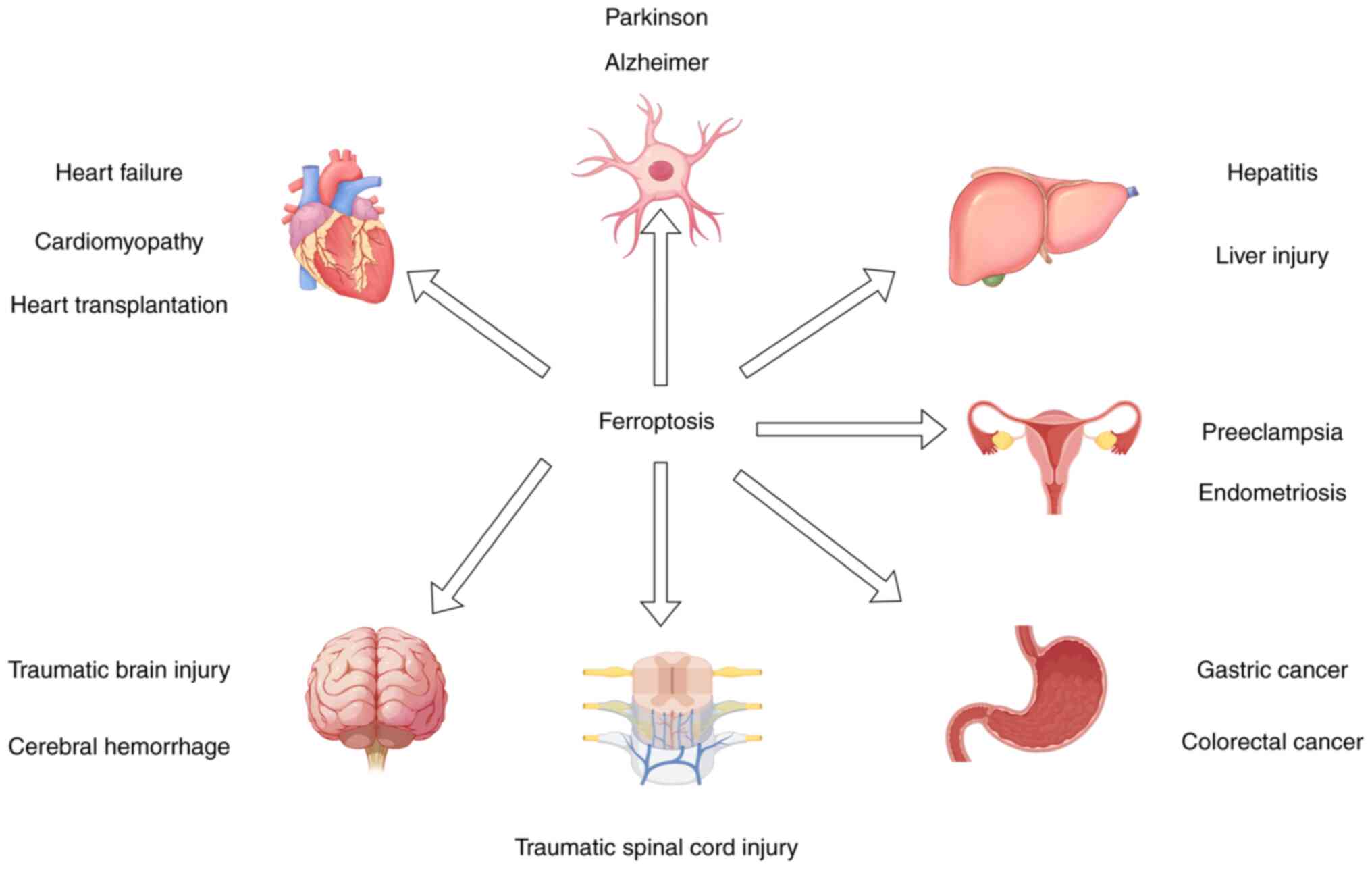
Ferroptosis has been investigated in a variety of
diseases, including Parkinson's disease (20,21),
Alzheimer's disease (22,23), liver injury (24,25),
brain injury (26,27), spinal cord injury (28–30),
kidney injury (31,32), cardiovascular disease (33–35)
and gynecological disorders (36,37)
(Fig. 1). At present, research is
focused on the role of ferroptosis in the treatment of cancer. Yan
et al (38) demonstrated
that ferroptosis plays a key role in the development of CRC, breast
cancer, ovarian cancer, renal cancer, lymphoma and melanoma
(39). However, results of a
previous study revealed that ferroptosis may also play a role in
CRC progression and metastasis, through the activation of different
signaling pathways (40). Thus,
previous studies highlight that ferroptosis may exert contrasting
effects in CRC, demonstrating the requirement for further
investigations. In addition, the development of novel treatment
approaches is required to mitigate the adverse effects of
ferroptosis on CRC. This article systematically reviews the
mechanisms and signaling pathways of ferroptosis in CRC, the role
of ferroptosis within the tumor microenvironment, and the progress
in research on ferroptosis in reducing drug resistance. It aims to
provide new insights for the diagnosis and treatment of CRC.
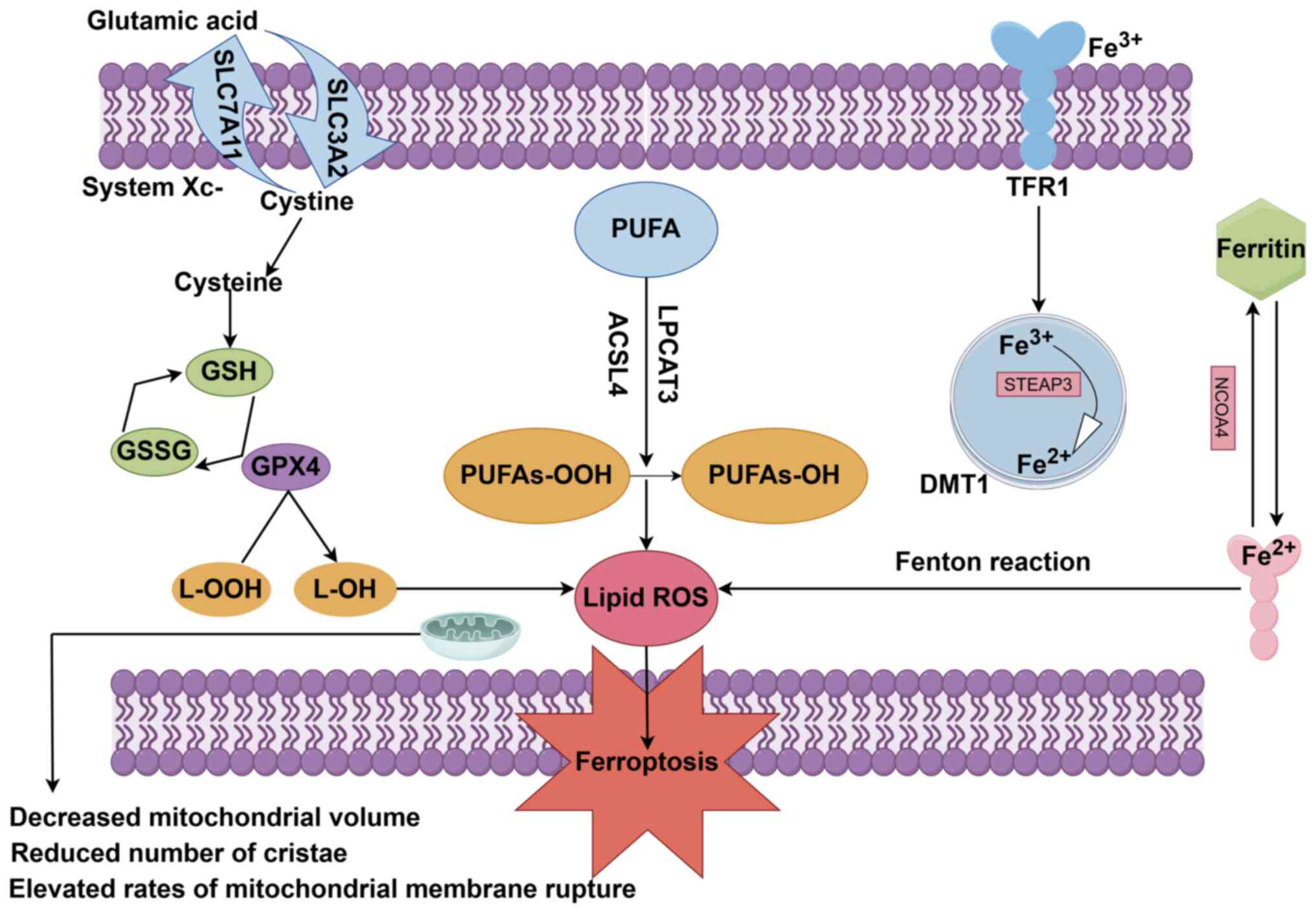
Ferroptosis is primarily characterized by the
excessive accumulation of iron and lipid reactive oxygen species
(10). Previous studies have
identified mechanisms that may be involved with ferroptosis,
including amino acid, lipid and iron metabolism (41). Key mechanisms involved in
ferroptosis are displayed in Fig.
2.
System Xc- is a critical component of the cellular
antioxidant system that is widely distributed in the phospholipid
bilayer. System Xc- is a heterodimer composed of two subunits:
Namely, solute carrier family 7 member 11 (SLC7A11) and SLC3A2.
System Xc- facilitates the 1:1 exchange of cystine and glutamate
across the cell membrane (9). Once
inside the cell, cystine is utilized to synthesize glutathione,
which, in the presence of glutathione peroxidase, reduces the
production of lipid reactive oxygen species. Therefore, inhibition
of System Xc- leads to reduced glutathione peroxidase activity,
decreased cellular antioxidant capacity, accumulation of lipid
reactive oxygen species and ferroptosis (9). A well-established oncogene, P53, may
induce ferroptosis via downregulation of SLC7A11 expression,
thereby inhibiting System Xc- activity (42,43).
Moreover, sorafenib (44),
sulfasalazine (45) and erastin
(46) induce ferroptosis via
inhibition of System Xc-.
GPX4 plays a critical role in the cellular
peroxidase system, inhibiting the formation of peroxides and thus
acting as a key regulator of ferroptosis. GPX4 catalyzes the
conversion of glutathione to oxidized glutathione and reduces
cytotoxic lipid peroxides to their corresponding alcohols (12). Yang et al (47) demonstrated that reduced GPX4
expression may lead to the induction of ferroptosis, whereas
increased GPX4 expression may inhibit ferroptosis. RAS-selective
lethal 3 (RSL3) is a widely established inhibitor of GPX4 and an
inducer of ferroptosis. RSL3 directly targets GPX4, thereby
inhibiting its activity and leading to reductions in cellular
antioxidant capacity, the accumulation of lipid reactive oxygen
species and the induction of ferroptosis (48). Costa et al (49) identified ML162 and ML210 as GPX4
inhibitors that induce ferroptosis, through a mechanism that is
comparable with that of RSL3.
Lipid peroxidation is critical in the induction of
ferroptosis. Lipids are essential components of cell membranes, and
lipid peroxidation disrupts these membranes, thereby triggering
ferroptosis (50). Polyunsaturated
fatty acids (PUFAs), such as arachidonic acid and adrenic acid, are
long-chain fatty acids with multiple double bonds that are highly
susceptible to lipid peroxidation. Phosphatidylethanolamine
containing arachidonic and adrenic acids is a key phospholipid in
the induction of cellular ferroptosis. There are two
lipid-metabolizing enzymes associated with ferroptosis; namely,
acyl-CoA synthetase long-chain family member 4 (ACSL4) and
lysophosphatidylcholine acyltransferase 3, which are involved in
the biosynthesis of phosphatidylethanolamine. These enzymes
activate PUFAs and impact their transmembrane properties, leading
to ferroptosis (51). In addition,
cytochrome P450 oxidoreductase-mediated lipid peroxidation may play
a key role in the induction of ferroptosis (52). Cytochrome P450 oxidoreductase
transfers electrons from reduced nicotinamide adenine dinucleotide
phosphate to oxygen, producing hydrogen peroxide. Hydrogen peroxide
subsequently reacts with iron to generate reactive hydroxyl
radicals, which peroxidize the polyunsaturated fatty acid chains of
membrane phospholipids. This process disrupts the integrity of
cellular membranes during iron accumulation, ultimately leading to
ferroptosis (53,54).
Iron accumulation is closely associated with
ferroptosis, and primarily involves iron absorption and reduction
processes (55). Iron is a critical
raw material for the production of hemoglobin and myoglobin.
Moreover, iron plays a vital role in various cellular metabolic
processes, such as oxygen storage and transport, DNA and RNA
synthesis, cellular differentiation, and enzymatic reactions
(56). Healthy individuals exhibit
iron levels of 3–5 g, with >50% circulating in red blood cells
as hemoglobin or stored as ferritin. In addition, small amounts of
iron bind to transferrin in the plasma (57,58).
Intracellular iron homeostasis is maintained through
a balance of iron absorption, utilization, storage and excretion.
Fe3+ is absorbed in the duodenum and jejunum, and
transported into cells via transferrin receptor 1, where it is
reduced to Fe2+ by metal reductase in the endoplasmic
reticulum. For biological activation, a small amount of
Fe2+ is released into a labile iron pool in the
cytoplasm via the divalent metal transporter 1, while the remainder
is recycled or stored as ferritin (59,60).
The results of a previous study revealed that cancer cells require
higher levels of iron than healthy cells, and are more susceptible
to iron depletion, also known as iron addiction (61). Notably, in the presence of a large
number of cancer cells, ferritin is degraded through autophagy.
This process is carried out via autophagy-related proteins 5 and 7,
and the nuclear receptor coactivator 4 (NCOA4) signaling pathways.
NCOA4 binds to lysosomes, degrades ferritin and releases
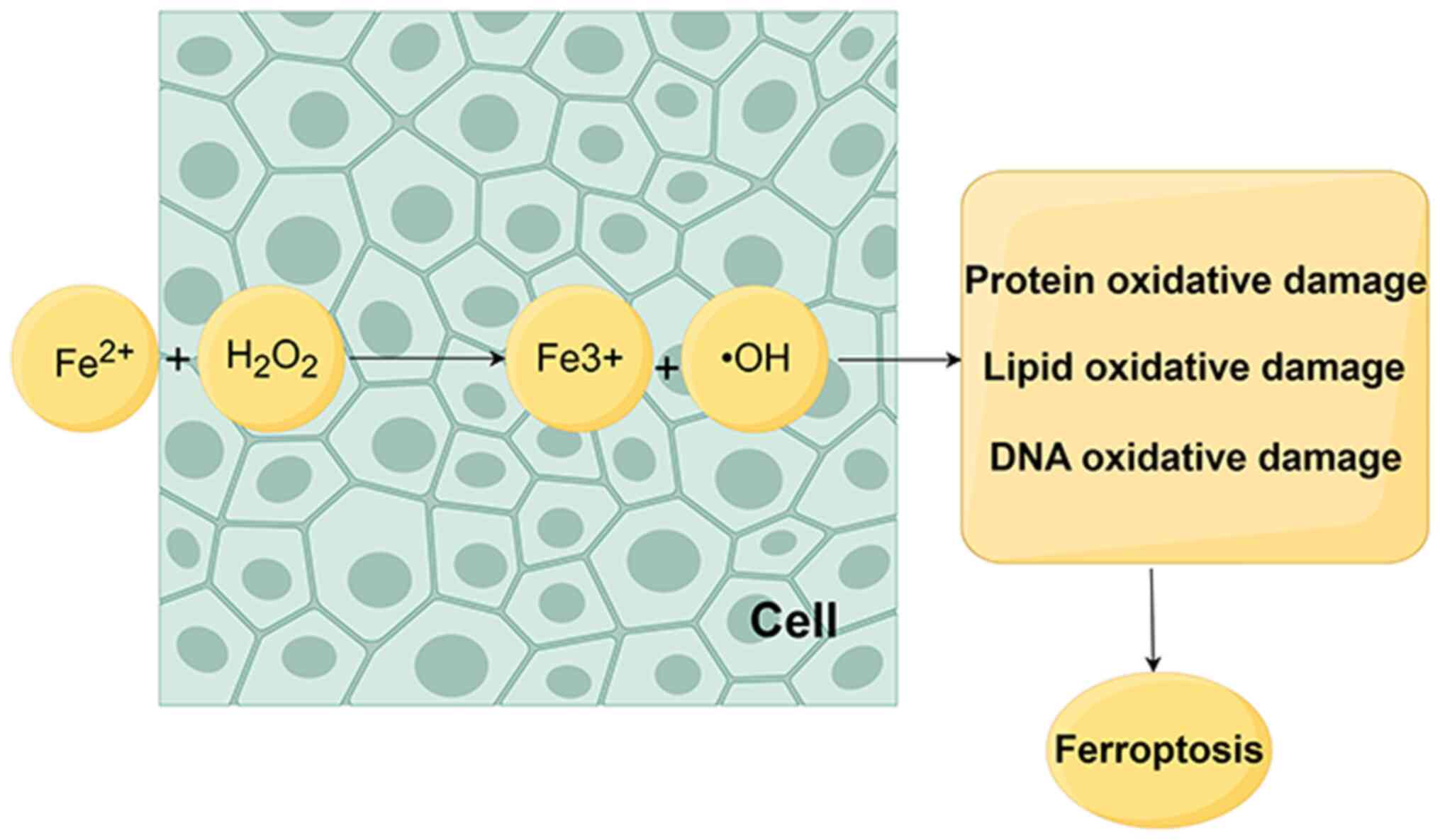
Fe2+, leading to an abnormal increase in Fe2+
levels (62–64). Excessive Fe2+
accumulation triggers the Fenton reaction (Fig. 3), initiating ferroptosis and further
contributing to the accumulation of reactive oxygen species
(65).
In addition, herbs and plant extracts may induce
ferroptosis. Emodin, a natural anthraquinone derivative extracted
from various herbs, may induce ferroptosis in CRC cells through
NCOA4-mediated ferritin autophagy and NF-κB pathways, thereby
inhibiting CRC progression (71).
Ginsenoside Rh3, a semi-natural product isolated from Panax
ginseng, may induce ferroptosis in CRC cells via the Stat3/p53/NRF2
axis, demonstrating potential in the treatment of cancer (72). Moreover, curcumin may inhibit CRC
through the induction of ferroptosis. Results of a previous study
demonstrated that curcumin played a role in the regulation of
oncogenes, such as P53, and the SLC7A11/glutathione/GPX4 axis
(73). The combination of curcumin
and Andrographis paniculata may induce ferroptosis in CRC,
through the downregulation of GPX4 and iron regulatory protein 1
(74). In addition, esculin induces
endoplasmic reticulum stress through the regulation of eukaryotic
translation initiation factor 2α/CHOP and Nrf2/HO-1 pathways via
the PERK signaling pathway; thus, promoting apoptosis and
ferroptosis in CRC cells, ultimately inhibiting CRC occurrence and
progression (75). Results of a
previous study revealed that baicalein promotes ferroptosis through
inhibition of the JAK2/STAT3/GPX4 signaling pathway; thus, exerting
an inhibitory effect on CRC (76).
Kruppel-like factor 2 (KLF2) is an oncogene that may
also inhibit CRC progression. Notably, KLF2 suppresses the PI3K/AKT
signaling pathway, thereby inducing ferroptosis (77). Results of a previous study
demonstrated that bromelain inhibits the proliferation of
KRAS-mutant CRC cells and induces ferroptosis via ACSL4 (78). Wei et al (79) revealed that Tagitinin C, a novel
inducer of ferroptosis, acts as a potent chemosensitizer that
enhances the efficacy of chemotherapeutic agents. Notably,
Tagitinin C induces ferroptosis through the PERK/Nrf2/HO-1
signaling pathway. TIGAR, a TP53-induced regulator of glycolysis
and apoptosis, plays a crucial role in energy metabolism,
autophagy, stem cell differentiation and cell survival (80). Liu et al (81) demonstrated that TIGAR inhibits CRC
progression through increasing the sensitivity of CRC cells to
ferroptosis via the ROS/AMPK/SCD1 signaling pathway. Chaudhary
et al (82) revealed that
lipid carrier protein 2 inhibits ferroptosis through upregulation
of GPX4 expression and the cystine/glutamate antiporter component,
xCT; thus, promoting CRC progression. Moreover, increased
expression of lipid carrier protein 2 may lead to resistance to
5-fluorouracil in CRC cells, further contributing to
chemotherapeutic resistance. In a recent study, it has been
reported that TRIM36-mediated FOXA2 promotes colorectal cancer by
inhibiting the Nrf2/GPX4 signaling pathway and suppressing
ferroptosis. However, the specific mechanism by which FOXA2
regulates Nrf2, whether directly or indirectly, remains unclear
(83). Results of a previous study
demonstrated that uridine-cytidine kinase-like 1 (UCKL1) enhances
the proliferation and metastasis of CRC cells via the
UCKL1/Nrf2/SLC7A11 axis, ultimately inhibiting ferroptosis and
promoting CRC development and progression (84). These findings suggest that reduced
lipid carrier protein 2, FOXA2 and UCKL1 expression levels may
promote ferroptosis; thus, acting as an effective strategy for the
treatment of CRC.
MicroRNAs (miRNAs/miRs) may play a role in the
regulation of ferroptosis. Results of a previous study have
revealed that miR-148a-3p acts as a tumor suppressor in CRC through
targeting SLC7A11 and activating ferroptosis (85). In addition, miR-509-5p promotes
ferroptosis through targeting SLC7A1 (86), while miR-15a-3p induces ferroptosis
through the direct targeting of GPX4 (87). The oncogenic miRNA, miR-19a,
promotes the proliferation, migration and invasion of CRC cells.
Iron-responsive element binding protein 2 (IREB2) is a direct
target of miR-19a. Thus, targeting IREB2 may lead to the inhibition
of ferroptosis via miR-19a, exhibiting potential as a novel target
for CRC treatment (88). In
addition, results of a previous study have revealed that miR-545
suppresses transferrin, promoting CRC cell survival through the
inhibition of ferroptosis (89).
Long non-coding RNAs (lncRNAs) may also regulate
ferroptosis. The results of a previous study have revealed that
LINC00239 may act as a ferroptosis inhibitor in CRC, through
interaction with Kelch-like ECH-associated protein 1. This leads to
a reduction in the antitumor activity of Erastin and RSL3, and the
promotion of CRC progression (90).
The TME is a complex environment containing tumor
cells, stromal cells, immune cells, adipocytes, endothelial cells
and the extracellular matrix (92).
Dai et al (93) revealed
that autophagic degradation-mediated ferroptosis leads to the
release of cancer cell components into the TME and tumor-associated
macrophage polarization. Moreover, Ma et al (94) revealed that CD36 mediates fatty acid
uptake via CD8+ T cells within the TME induced
ferroptosis, leading to a reduction in CD8+ T cell
effector function and antitumor activity. In addition,
cancer-associated fibroblasts in the TME may impair the antitumor
capacity of natural killer cells through the induction of
ferroptosis (95).
Chemotherapy is the most common post-operative
treatment for patients with CRC. The results of our previous study
demonstrated that oxaliplatin and 5-fluorouracil are highly
associated with ferroptosis in CRC (82,100)
(Table V).
The results of a previous study revealed that the
induction of ferroptosis through the inhibition of the
KIF20A/NUAK1/Nrf2/GPX4 signaling pathway enhances sensitivity to
oxaliplatin, thereby improving the quality of life of patients with
CRC (100). In addition,
inhibition of cysteine desulfurase expression leads to increased
intracellular reactive oxygen species levels, the promotion of
ferroptosis and reduced levels of resistance to oxaliplatin
(101). Overexpression of
RNA-binding motif single-stranded interacting protein 1 (RBMS1)
inhibits ferroptosis; therefore, suppressing RBMS1 expression may
increase the sensitivity of CRC cells to oxaliplatin (102). Inhibition of Candida
nucleata also promotes ferroptosis and decreases CRC cell
resistance to oxaliplatin (103).
The results of a previous study also demonstrated that oxaliplatin
resistance is reversed following inhibition of cyclin-dependent
kinase 1 expression (104).
Overexpression of lipid transport protein 2 leads to
inhibition of ferroptosis, which ultimately increases the
resistance to 5-fluorouracil. Therefore, targeting lipid transport
protein 2 levels may attenuate resistance to 5-fluorouracil
(82). In addition, serine protease
1 is associated with chemotherapeutic resistance in CRC. This
protein is highly expressed in CRC cells and is negatively
associated with the prognosis of patients. Moreover, Liu et
al (105) demonstrated that
serine protease 1 interacts with SLC7A11 to increase expression
levels, ultimately inhibiting ferroptosis, leading to increased
resistance to 5-fluorouracil. Thus, targeting serine protease 1 may
exhibit potential in the treatment of CRC.
When targeting cancer cells, chemotherapy may also
target healthy cells. Thus, targeted therapy is considered an
alternative treatment option to chemotherapy. Mu et al
(106) demonstrated that
3-bromopyruvic acid and cetuximab may induce ferroptosis; thus,
exhibiting potential in mitigating cetuximab resistance in CRC. In
addition, the combination of β-elemene, a natural product derived
from turmeric, and cetuximab may exert effects on metastatic CRC
cells with Kras mutations, inducing ferroptosis and reducing
cetuximab resistance (107).
At present, research is focused on the role of
ferroptosis in the physiological and pathological processes of
numerous diseases, leading to the development of novel treatment
approaches. The present study aimed to review the specific
mechanisms underlying ferroptosis in CRC, including drug-, gene-,
protein- and RNA-induced ferroptosis. The aforementioned forms of
ferroptosis play distinct roles in CRC. For example, various
signaling pathways directly facilitate drug-induced ferroptosis,
thereby impeding CRC onset and progression. By contrast, gene-,
protein- and RNA-induced ferroptosis involve specific signaling
pathways and mechanisms that may exhibit potential as effective
strategies for targeted CRC treatment. Notably, mechanisms may
include targeting of GPX4 expression and xCT, the Nrf2/GPX4
signaling pathway, the UCKL1/Nrf2/SLC7A11 signaling pathway,
miR-19a, miR-545, and lncRNA LINC00239. In addition, the
association between ferroptosis and the TME further highlighted
that induction of ferroptosis may attenuate drug resistance in
CRC.
Results of the present study demonstrated that
induction of ferroptosis may inhibit CRC progression. In addition,
genes, proteins and RNAs that inhibit ferroptosis may promote CRC
development and progression. However, the clinical application of
ferroptosis is limited at present, as the specific mechanisms and
signaling pathways through which ferroptosis promotes or inhibits
CRC development are yet to be fully elucidated. Thus, further
investigations into the specific role of ferroptosis in CRC are
required for the development of effective targeted therapies.
In conclusion, ferroptosis may exhibit potential in
the treatment of CRC. Further investigations are required to
elucidate the signaling pathways involved in ferroptosis, and to
identify the genes, proteins and signaling pathways that inhibit
ferroptosis in CRC. Moreover, further investigations should focus
on determining alternative therapeutic modalities that may increase
the therapeutic effects of ferroptosis, and on assessing the
specific anti-tumor effects of ferroptosis in CRC progression. An
increased understanding of ferroptosis in CRC may lead to the
effective implementation of treatment in clinical practice.
The figures were created using Figdraw.
This article is funded by Shandong Province's Traditional
Chinese Medicine Science and Technology Development Project (grant
no. 2019-0187) and Qilu Chinese Medicine Advantageous Specialty
Cluster Construction Project (grant no. YWC2022ZKJQ0003).
Not applicable.
ZG, HZ and XS contributed to the conception and the
main idea of the work. ZG and HZ drafted the manuscript, figures
and tables. XS reviewed and modified the manuscript. All authors
read and approved the final version of the manuscript. Data
authentication is not applicable.
Not applicable.
Not applicable.
All authors declare that they have no competing
interests.
|
1
|
Favoriti P, Carbone G, Greco M, Pirozzi F,
Pirozzi RE and Corcione F: Worldwide burden of colorectal cancer: A
review. Updates Surg. 68:7–11. 2016. View Article : Google Scholar : PubMed/NCBI
|
|
2
|
Bray F, Ferlay J, Soerjomataram I, Siegel
RL, Torre LA and Jemal A: Global cancer statistics 2018: GLOBOCAN
estimates of incidence and mortality worldwide for 36 cancers in
185 countries. CA Cancer J Clin. 68:394–424. 2018. View Article : Google Scholar : PubMed/NCBI
|
|
3
|
Bouvard V, Loomis D, Guyton KZ, Grosse Y,
Ghissassi FE, Benbrahim-Tallaa L, Guha N, Mattock H and Straif K;
International Agency for Research on Cancer Monograph Working
Group, : Carcinogenicity of consumption of red and processed meat.
Lancet Oncol. 16:1599–1600. 2015. View Article : Google Scholar : PubMed/NCBI
|
|
4
|
Johnson CH, Dejea CM, Edler D, Hoang LT,
Santidrian AF, Felding BH, Ivanisevic J, Cho K, Wick EC,
Hechenbleikner EM, et al: Metabolism links bacterial biofilms and
colon carcinogenesis. Cell Metab. 21:891–897. 2015. View Article : Google Scholar : PubMed/NCBI
|
|
5
|
Dejea CM, Wick EC, Hechenbleikner EM,
White JR, Mark Welch JL, Rossetti BJ, Peterson SN, Snesrud EC,
Borisy GG, Lazarev M, et al: Microbiota organization is a distinct
feature of proximal colorectal cancers. Proc Natl Acad Sci USA.
111:18321–18326. 2014. View Article : Google Scholar : PubMed/NCBI
|
|
6
|
Benson AB, Venook AP, Al-Hawary MM,
Cederquist L, Chen YJ, Ciombor KK, Cohen S, Cooper HS, Deming D,
Engstrom PF, et al: NCCN guidelines insights: Colon cancer, version
2.2018. J Natl Compr Canc Netw. 16:359–369. 2018. View Article : Google Scholar : PubMed/NCBI
|
|
7
|
Guo J, Xu B, Han Q, Zhou H, Xia Y, Gong C,
Dai X, Li Z and Wu G: Ferroptosis: A novel Anti-tumor action for
cisplatin. Cancer Res Treat. 50:445–460. 2018. View Article : Google Scholar : PubMed/NCBI
|
|
8
|
Stockwell BR, Friedmann Angeli JP, Bayir
H, Bush AI, Conrad M, Dixon SJ, Fulda S, Gascón S, Hatzios SK,
Kagan VE, et al: Ferroptosis: A regulated cell death nexus linking
metabolism, redox biology, and disease. Cell. 171:273–285. 2017.
View Article : Google Scholar : PubMed/NCBI
|
|
9
|
Dixon SJ, Lemberg KM, Lamprecht MR, Skouta
R, Zaitsev EM, Gleason CE, Patel DN, Bauer AJ, Cantley AM, Yang WS,
et al: Ferroptosis: An iron-dependent form of nonapoptotic cell
death. Cell. 149:1060–1072. 2012. View Article : Google Scholar : PubMed/NCBI
|
|
10
|
Xu S, He Y, Lin L, Chen P, Chen M and
Zhang S: The emerging role of ferroptosis in intestinal disease.
Cell Death Dis. 12:2892021. View Article : Google Scholar : PubMed/NCBI
|
|
11
|
Yagoda N, von Rechenberg M, Zaganjor E,
Bauer AJ, Yang WS, Fridman DJ, Wolpaw AJ, Smukste I, Peltier JM,
Boniface JJ, et al: RAS-RAF-MEK-dependent oxidative cell death
involving voltage-dependent anion channels. Nature. 447:865–869.
2007. View Article : Google Scholar
|
|
12
|
Li J, Cao F, Yin HL, Huang ZJ, Lin ZT, Mao
N, Sun B and Wang G: Ferroptosis: Past, present and future. Cell
Death Dis. 11:882020. View Article : Google Scholar : PubMed/NCBI
|
|
13
|
Ketelut-Carneiro N and Fitzgerald KA:
Apoptosis, pyroptosis, and Necroptosis-oh my! The many ways a cell
can die. J Mol Biol. 434:1673782022. View Article : Google Scholar : PubMed/NCBI
|
|
14
|
Aglietti RA and Dueber EC: Recent insights
into the molecular mechanisms underlying pyroptosis and gasdermin
family functions. Trends Immunol. 38:261–271. 2017. View Article : Google Scholar : PubMed/NCBI
|
|
15
|
Fang Y, Tian S, Pan Y, Li W, Wang Q, Tang
Y, Yu T, Wu X, Shi Y, Ma P and Shu Y: Pyroptosis: A new frontier in
cancer. Biomed Pharmacother. 121:1095952020. View Article : Google Scholar : PubMed/NCBI
|
|
16
|
Liu X, Zhang Z, Ruan J, Pan Y, Magupalli
VG, Wu H and Lieberman J: Inflammasome-activated gasdermin D causes
pyroptosis by forming membrane pores. Nature. 535:153–158. 2016.
View Article : Google Scholar : PubMed/NCBI
|
|
17
|
Sun L, Wang H, Wang Z, He S, Chen S, Liao
D, Wang L, Yan J, Liu W, Lei X and Wang X: Mixed lineage kinase
domain-like protein mediates necrosis signaling downstream of RIP3
kinase. Cell. 148:213–227. 2012. View Article : Google Scholar : PubMed/NCBI
|
|
18
|
Negroni A, Colantoni E, Cucchiara S and
Stronati L: Necroptosis in intestinal inflammation and cancer: New
concepts and therapeutic perspectives. Biomolecules. 10:14312020.
View Article : Google Scholar : PubMed/NCBI
|
|
19
|
Liu S, Yao S, Yang H, Liu S and Wang Y:
Autophagy: Regulator of cell death. Cell Death Dis. 14:6482023.
View Article : Google Scholar : PubMed/NCBI
|
|
20
|
Guiney SJ, Adlard PA, Bush AI, Finkelstein
DI and Ayton S: Ferroptosis and cell death mechanisms in
Parkinson's disease. Neurochem Int. 104:34–48. 2017. View Article : Google Scholar : PubMed/NCBI
|
|
21
|
Do Van B, Gouel F, Jonneaux A, Timmerman
K, Gelé P, Pétrault M, Bastide M, Laloux C, Moreau C, Bordet R, et
al: Ferroptosis, a newly characterized form of cell death in
Parkinson's disease that is regulated by PKC. Neurobiol Dis.
94:169–178. 2016. View Article : Google Scholar : PubMed/NCBI
|
|
22
|
Yan N and Zhang J: Iron metabolism,
ferroptosis, and the links with Alzheimer's disease. Front
Neurosci. 13:14432020. View Article : Google Scholar : PubMed/NCBI
|
|
23
|
Cong L, Dong X, Wang Y, Deng Y, Li B and
Dai R: On the role of synthesized hydroxylated chalcones as dual
functional amyloid-β aggregation and ferroptosis inhibitors for
potential treatment of Alzheimer's disease. Eur J Med Chem.
166:11–21. 2019. View Article : Google Scholar : PubMed/NCBI
|
|
24
|
Deng G, Li Y, Ma S, Gao Z, Zeng T, Chen L,
Ye H, Yang M, Shi H, Yao X, et al: Caveolin-1 dictates ferroptosis
in the execution of acute immune-mediated hepatic damage by
attenuating nitrogen stress. Free Radic Biol Med. 148:151–161.
2020. View Article : Google Scholar : PubMed/NCBI
|
|
25
|
Park SJ, Cho SS, Kim KM, Yang JH, Kim JH,
Jeong EH, Yang JW, Han CY, Ku SK, Cho IJ and Ki SH: Protective
effect of sestrin2 against iron overload and ferroptosis-induced
liver injury. Toxicol Appl Pharmacol. 379:1146652019. View Article : Google Scholar : PubMed/NCBI
|
|
26
|
Zhang Z, Wu Y, Yuan S, Zhang P, Zhang J,
Li H, Li X, Shen H, Wang Z and Chen G: Glutathione peroxidase 4
participates in secondary brain injury through mediating
ferroptosis in a rat model of intracerebral hemorrhage. Brain Res.
1701:112–125. 2018. View Article : Google Scholar : PubMed/NCBI
|
|
27
|
Kenny EM, Fidan E, Yang Q, Anthonymuthu
TS, New LA, Meyer EA, Wang H, Kochanek PM, Dixon CE, Kagan VE and
Bayir H: Ferroptosis contributes to neuronal death and functional
outcome after traumatic brain injury. Crit Care Med. 47:410–418.
2019. View Article : Google Scholar : PubMed/NCBI
|
|
28
|
Yao X, Zhang Y, Hao J, Duan HQ, Zhao CX,
Sun C, Li B, Fan BY, Wang X, Li WX, et al: Deferoxamine promotes
recovery of traumatic spinal cord injury by inhibiting ferroptosis.
Neural Regen Res. 14:5322019. View Article : Google Scholar : PubMed/NCBI
|
|
29
|
Zhang Y, Sun C, Zhao C, Hao J, Zhang Y,
Fan B, Li B, Duan H, Liu C, Kong X, et al: Ferroptosis inhibitor
SRS 16–86 attenuates ferroptosis and promotes functional recovery
in contusion spinal cord injury. Brain Res. 1706:48–57. 2019.
View Article : Google Scholar : PubMed/NCBI
|
|
30
|
Shi Z, Yuan S, Shi L, Li J, Ning G, Kong X
and Feng S: Programmed cell death in spinal cord injury
pathogenesis and therapy. Cell Prolif. 54:e129922021. View Article : Google Scholar : PubMed/NCBI
|
|
31
|
Martin-Sanchez D, Ruiz-Andres O, Poveda J,
Carrasco S, Cannata-Ortiz P, Sanchez-Niño MD, Ruiz Ortega M, Egido
J, Linkermann A, Ortiz A and Sanz AB: Ferroptosis, but not
necroptosis, is important in nephrotoxic folic Acid-induced AKI. J
Am Soc Nephrol. 28:218–229. 2017. View Article : Google Scholar : PubMed/NCBI
|
|
32
|
Müller T, Dewitz C, Schmitz J, Schröder
AS, Bräsen JH, Stockwell BR, Murphy JM, Kunzendorf U and Krautwald
S: Necroptosis and ferroptosis are alternative cell death pathways
that operate in acute kidney failure. Cell Mol Life Sci.
74:3631–3645. 2017. View Article : Google Scholar : PubMed/NCBI
|
|
33
|
Liu B, Zhao C, Li H, Chen X, Ding Y and Xu
S: Puerarin protects against heart failure induced by pressure
overload through mitigation of ferroptosis. Biochem Biophys Res
Commun. 497:233–240. 2018. View Article : Google Scholar : PubMed/NCBI
|
|
34
|
Chen X, Xu S, Zhao C and Liu B: Role of
TLR4/NADPH oxidase 4 pathway in promoting cell death through
autophagy and ferroptosis during heart failure. Biochem Biophys Res
Commun. 516:37–43. 2019. View Article : Google Scholar : PubMed/NCBI
|
|
35
|
Li D, Pi W, Sun Z, Liu X and Jiang J:
Ferroptosis and its role in cardiomyopathy. Biomed Pharmacother.
153:1132792022. View Article : Google Scholar : PubMed/NCBI
|
|
36
|
Ng SW, Norwitz SG and Norwitz ER: The
impact of iron overload and ferroptosis on reproductive disorders
in humans: Implications for preeclampsia. Int J Mol Sci.
20:32832019. View Article : Google Scholar : PubMed/NCBI
|
|
37
|
Ng SW, Norwitz SG, Taylor HS and Norwitz
ER: Endometriosis: The role of iron overload and ferroptosis.
Reprod Sci. 27:1383–1390. 2020. View Article : Google Scholar : PubMed/NCBI
|
|
38
|
Yan HF, Zou T, Tuo QZ, Xu S, Li H, Belaidi
AA and Lei P: Ferroptosis: Mechanisms and links with diseases.
Signal Transduct TargetTher. 6:492021. View Article : Google Scholar
|
|
39
|
Yan H, Talty R, Aladelokun O, Bosenberg M
and Johnson CH: Ferroptosis in colorectal cancer: A future target?
Br J Cancer. 128:1439–1451. 2023. View Article : Google Scholar : PubMed/NCBI
|
|
40
|
Wu T, Wan J, Qu X, Xia K, Wang F, Zhang Z,
Yang M, Wu X, Gao R, Yuan X, et al: Nodal promotes colorectal
cancer survival and metastasis through regulating SCD1-mediated
ferroptosis resistance. Cell Death Dis. 14:2292023. View Article : Google Scholar : PubMed/NCBI
|
|
41
|
Wang Y, Zhang Z, Sun W, Zhang J, Xu Q,
Zhou X and Mao L: Ferroptosis in colorectal cancer: Potential
mechanisms and effective therapeutic targets. Biomed Pharmacother.
153:1135242022. View Article : Google Scholar : PubMed/NCBI
|
|
42
|
Jiang L, Hickman JH, Wang SJ and Gu W:
Dynamic roles of p53-mediated metabolic activities in ROS-induced
stress responses. Cell Cycle. 14:2881–2885. 2015. View Article : Google Scholar : PubMed/NCBI
|
|
43
|
Jiang L, Kon N, Li T, Wang SJ, Su T,
Hibshoosh H, Baer R and Gu W: Ferroptosis as a p53-mediated
activity during tumour suppression. Nature. 520:57–62. 2015.
View Article : Google Scholar : PubMed/NCBI
|
|
44
|
Li Q, Chen K, Zhang T, Jiang D, Chen L,
Jiang J, Zhang C and Li S: Understanding Sorafenib-induced
ferroptosis and resistance mechanisms: Implications for cancer
therapy. Eur J Pharmacol. 955:1759132023. View Article : Google Scholar : PubMed/NCBI
|
|
45
|
Chen X, Kang R, Kroemer G and Tang D:
Broadening horizons: The role of ferroptosis in cancer. Nat Rev
Clin Oncol. 18:280–296. 2021. View Article : Google Scholar : PubMed/NCBI
|
|
46
|
Wang L, Liu Y, Du T, Yang H, Lei L, Guo M,
Ding HF, Zhang J, Wang H, Chen X and Yan C: ATF3 promotes
Erastin-induced ferroptosis by suppressing system Xc. Cell Death
Differ. 27:662–675. 2020. View Article : Google Scholar : PubMed/NCBI
|
|
47
|
Yang WS and Stockwell BR: Synthetic lethal
screening identifies compounds activating Iron-dependent,
nonapoptotic cell death in oncogenic-RAS-harboring cancer cells.
Chem Biol. 15:234–245. 2008. View Article : Google Scholar : PubMed/NCBI
|
|
48
|
Sui X, Zhang R, Liu S, Duan T, Zhai L,
Zhang M, Han X, Xiang Y, Huang X, Lin H and Xie T: RSL3 drives
ferroptosis through GPX4 inactivation and ROS production in
colorectal cancer. Front Pharmacol. 9:13712018. View Article : Google Scholar : PubMed/NCBI
|
|
49
|
Costa I, Barbosa DJ, Benfeito S, Silva V,
Chavarria D, Borges F, Remião F and Silva R: Molecular mechanisms
of ferroptosis and their involvement in brain diseases. Pharmacol
Ther. 244:1083732023. View Article : Google Scholar : PubMed/NCBI
|
|
50
|
Harayama T and Riezman H: Understanding
the diversity of membrane lipid composition. Nat Rev Mol Cell Biol.
19:281–296. 2018. View Article : Google Scholar : PubMed/NCBI
|
|
51
|
Kagan VE, Mao G, Qu F, Angeli JP, Doll S,
Croix CS, Dar HH, Liu B, Tyurin VA, Ritov VB, et al: Oxidized
arachidonic and adrenic PEs navigate cells to ferroptosis. Nat Chem
Biol. 13:81–90. 2017. View Article : Google Scholar : PubMed/NCBI
|
|
52
|
Koppula P, Zhuang L and Gan B: Cytochrome
P450 reductase (POR) as a ferroptosis fuel. Protein Cell.
12:675–679. 2021. View Article : Google Scholar : PubMed/NCBI
|
|
53
|
Yan B, Ai Y, Sun Q, Ma Y, Cao Y, Wang J,
Zhang Z and Wang X: Membrane damage during ferroptosis is caused by
oxidation of phospholipids catalyzed by the oxidoreductases POR and
CYB5R1. Mol Cell. 81:355–369.e10. 2021. View Article : Google Scholar : PubMed/NCBI
|
|
54
|
Zou Y, Li H, Graham ET, Deik AA, Eaton JK,
Wang W, Sandoval-Gomez G, Clish CB, Doench JG and Schreiber SL:
Cytochrome P450 oxidoreductase contributes to phospholipid
peroxidation in ferroptosis. Nat Chem Biol. 16:302–309. 2020.
View Article : Google Scholar : PubMed/NCBI
|
|
55
|
Ning X, Qi H, Yuan Y, Li R, Wang Y, Lin Z
and Yin Y: Identification of a new small molecule that initiates
ferroptosis in cancer cells by inhibiting the system Xc−
to deplete GSH. Eur J Pharmacol. 934:1753042022. View Article : Google Scholar : PubMed/NCBI
|
|
56
|
Fan X, Li A, Yan Z, Geng X, Lian L, Lv H,
Gao D and Zhang J: From iron metabolism to ferroptosis: Pathologic
changes in coronary heart disease. Oxid Med Cell Longev.
2022:62918892022. View Article : Google Scholar : PubMed/NCBI
|
|
57
|
Zhou L, Zhao B, Zhang L, Wang S, Dong D,
Lv H and Shang P: Alterations in cellular iron metabolism provide
more therapeutic opportunities for cancer. Int J Mol Sci.
19:15452018. View Article : Google Scholar : PubMed/NCBI
|
|
58
|
Basak T and Kanwar RK: Iron imbalance in
cancer: Intersection of deficiency and overload. Cancer Med.
11:3837–3853. 2022. View Article : Google Scholar : PubMed/NCBI
|
|
59
|
Chifman J, Laubenbacher R and Torti SV: A
systems biology approach to iron metabolism. Adv Exp Med Biol.
844:201–225. 2014. View Article : Google Scholar : PubMed/NCBI
|
|
60
|
Han C, Liu Y, Dai R, Ismail N, Su W and Li
B: Ferroptosis and its potential role in human diseases. Front
Pharmacol. 11:2392020. View Article : Google Scholar : PubMed/NCBI
|
|
61
|
Manz DH, Blanchette NL, Paul BT, Torti FM
and Torti SV: Iron and cancer: Recent insights. Ann N Y Acad Sci.
1368:149–161. 2016. View Article : Google Scholar : PubMed/NCBI
|
|
62
|
Zhou B, Liu J, Kang R, Klionsky DJ,
Kroemer G and Tang D: Ferroptosis is a type of Autophagy-dependent
cell death. Semin Cancer Biol. 66:89–100. 2020. View Article : Google Scholar : PubMed/NCBI
|
|
63
|
Mancias JD, Wang X, Gygi SP, Harper JW and
Kimmelman AC: Quantitative proteomics identifies NCOA4 as the cargo
receptor mediating ferritinophagy. Nature. 509:105–109. 2014.
View Article : Google Scholar : PubMed/NCBI
|
|
64
|
Hou W, Xie Y, Song X, Sun X, Lotze MT, Zeh
HJ III, Kang R and Tang D: Autophagy promotes ferroptosis by
degradation of ferritin. Autophagy. 12:1425–1428. 2016. View Article : Google Scholar : PubMed/NCBI
|
|
65
|
Xie Y, Hou W, Song X, Yu Y, Huang J, Sun
X, Kang R and Tang D: Ferroptosis: Process and function. Cell Death
Differ. 23:369–379. 2016. View Article : Google Scholar : PubMed/NCBI
|
|
66
|
Yang J, Mo J, Dai J, Ye C, Cen W, Zheng X,
Jiang L and Ye L: Cetuximab promotes RSL3-induced ferroptosis by
suppressing the Nrf2/HO-1 signalling pathway in KRAS mutant
colorectal cancer. Cell Death Dis. 12:10792021. View Article : Google Scholar : PubMed/NCBI
|
|
67
|
Tian X, LI S and Ge G: Apatinib promotes
ferroptosis in colorectal cancer cells by targeting ELOVL6/ACSL4
Signaling. Cancer Manag Res. 13:1333–1342. 2021. View Article : Google Scholar : PubMed/NCBI
|
|
68
|
Zhu JF, Liu Y, Li WT, Li MH, Zhen CH, Sun
PW, Chen JX, Wu WH and Zeng W: Ibrutinib facilitates the
sensitivity of colorectal cancer cells to ferroptosis through
BTK/NRF2 pathway. Cell Death Dis. 14:1512023. View Article : Google Scholar : PubMed/NCBI
|
|
69
|
Zhao X and Chen F: Propofol induces the
ferroptosis of colorectal cancer cells by downregulating STAT3
expression. Oncol Lett. 22:7672021. View Article : Google Scholar : PubMed/NCBI
|
|
70
|
Chen H, Qi Q, Wu N, Wang Y, Feng Q, Jin R
and Jiang L: Aspirin promotes RSL3-induced ferroptosis by
suppressing mTOR/SREBP-1/SCD1-mediated lipogenesis in PIK3CA-mutant
colorectal cancer. Redox Biol. 55:1024262022. View Article : Google Scholar : PubMed/NCBI
|
|
71
|
Shen Z, Zhao L, Yoo SA, Lin Z, Zhang Y,
Yang W and Piao J: Emodin induces ferroptosis in colorectal cancer
through NCOA4-mediated ferritinophagy and NF-κb pathway
inactivation. Apoptosis. May 5–2024.doi: 10.1007/s10495-024-01973-2
(Epub ahead of print). View Article : Google Scholar
|
|
72
|
Wu Y, Pi D, Zhou S, Yi Z, Dong Y, Wang W,
Ye H, Chen Y, Zuo Q and Ouyang M: Ginsenoside Rh3 induces
pyroptosis and ferroptosis through the Stat3/p53/NRF2 axis in
colorectal cancer cells. Acta Biochim Biophys Sin (Shanghai).
55:587–600. 2023. View Article : Google Scholar : PubMed/NCBI
|
|
73
|
Ming T, Lei J, Peng Y, Wang M, Liang Y,
Tang S, Tao Q, Wang M, Tang X, He Z, et al: Curcumin suppresses
colorectal cancer by induction of ferroptosis via regulation of p53
and solute carrier family 7 member 11/glutathione/glutathione
peroxidase 4 signaling axis. Phytother Res. 38:3954–3972. 2024.
View Article : Google Scholar : PubMed/NCBI
|
|
74
|
Miyazaki K, Xu C, Shimada M and Goel A:
Curcumin and andrographis exhibit Anti-tumor effects in colorectal
cancer via activation of ferroptosis and dual suppression of
glutathione Peroxidase-4 and ferroptosis suppressor Protein-1.
Pharmaceuticals (Basel). 16:3832023. View Article : Google Scholar : PubMed/NCBI
|
|
75
|
Ji X, Chen Z, Lin W, Wu Q, Wu Y, Hong Y,
Tong H, Wang C and Zhang Y: Esculin induces endoplasmic reticulum
stress and drives apoptosis and ferroptosis in colorectal cancer
via PERK regulating eIF2α/CHOP and Nrf2/HO-1 cascades. J
Ethnopharmacol. 328:1181392024. View Article : Google Scholar : PubMed/NCBI
|
|
76
|
Lai JQ, Zhao LL, Hong C, Zou QM, Su JX, Li
SJ, Zhou XF, Li ZS, Deng B, Cao J and Qi Q: Baicalein triggers
ferroptosis in colorectal cancer cells via blocking the
JAK2/STAT3/GPX4 axis. Acta Pharmacol Sin. 45:1715–1726. 2024.
View Article : Google Scholar : PubMed/NCBI
|
|
77
|
Li J, Jiang JL, Chen YM and Lu WQ: KLF2
inhibits colorectal cancer progression and metastasis by inducing
ferroptosis via the PI3K/AKT signaling pathway. J Pathol Clin Res.
9:423–435. 2023. View Article : Google Scholar : PubMed/NCBI
|
|
78
|
Park S, Oh J, Kim M and Jin EJ: Bromelain
effectively suppresses Kras-mutant colorectal cancer by stimulating
ferroptosis. Anim Cells Syst (Seoul). 22:334–340. 2018. View Article : Google Scholar : PubMed/NCBI
|
|
79
|
Wei R, Zhao Y, Wang J, Yang X, Li S, Wang
Y, Yang X, Fei J, Hao X, Zhao Y, et al: Tagitinin C induces
ferroptosis through PERK-Nrf2-HO-1 signaling pathway in colorectal
cancer cells. Int J Biol Sci. 17:2703–2717. 2021. View Article : Google Scholar : PubMed/NCBI
|
|
80
|
Ko YH, Domingo-Vidal M, Roche M, Lin Z,
Whitaker-Menezes D, Seifert E, Capparelli C, Tuluc M, Birbe RC,
Tassone P, et al: TP53-inducible glycolysis and apoptosis regulator
(TIGAR) metabolically reprograms carcinoma and stromal cells in
breast cancer. J Biol Chem. 291:26291–26303. 2016. View Article : Google Scholar : PubMed/NCBI
|
|
81
|
Liu MY, Li HM, Wang XY, Xia R, Li X, Ma
YJ, Wang M and Zhang HS: TIGAR drives colorectal cancer ferroptosis
resistance through ROS/AMPK/SCD1 pathway. Free Radic Biol Med.
182:219–231. 2022. View Article : Google Scholar : PubMed/NCBI
|
|
82
|
Chaudhary N, Choudhary BS, Shah SG,
Khapare N, Dwivedi N, Gaikwad A, Joshi N, Raichanna J, Basu S,
Gurjar M, et al: Lipocalin 2 expression promotes tumor progression
and therapy resistance by inhibiting ferroptosis in colorectal
cancer. Int J Cancer. 149:1495–1511. 2021. View Article : Google Scholar : PubMed/NCBI
|
|
83
|
Liu X, Yan C, Chang C, Meng F, Shen W,
Wang S and Zhang Y: FOXA2 suppression by TRIM36 exerts Anti-tumor
role in colorectal cancer via inducing NRF2/GPX4-Regulated
ferroptosis. Adv Sci (Weinh). 10:e23045212023. View Article : Google Scholar : PubMed/NCBI
|
|
84
|
Wu W, Zhao Y, Qin B, Jiang X, Wang C, Hu
R, Ma R, Lee MH, Liu H, Li K and Yuan P: Non-canonical role of
UCKL1 on ferroptosis defence in colorectal cancer. EBioMedicine.
93:1046502023. View Article : Google Scholar : PubMed/NCBI
|
|
85
|
Martino E, Balestrieri A, Aragona F,
Bifulco G, Mele L, Campanile G, Balestrieri ML and D'Onofrio N:
MiR-148a-3p promotes colorectal cancer cell ferroptosis by
targeting SLC7A11. Cancers (Basel). 15:43422023. View Article : Google Scholar : PubMed/NCBI
|
|
86
|
Elrebehy MA, Abdelghany TM, Elshafey MM,
Gomaa MH and Doghish AS: miR-509-5p promotes colorectal cancer cell
ferroptosis by targeting SLC7A11. Pathol Res Pract. 247:1545572023.
View Article : Google Scholar : PubMed/NCBI
|
|
87
|
Liu L, Yao H, Zhou X, Chen J, Chen G, Shi
X, Wu G, Zhou G and He S: MiR-15a-3p regulates ferroptosis via
targeting glutathione peroxidase GPX4 in colorectal cancer. Mol
Carcinog. 61:301–310. 2022. View Article : Google Scholar : PubMed/NCBI
|
|
88
|
Fan H, Ai R, Mu S, Niu X, Guo Z and Liu L:
MiR-19a suppresses ferroptosis of colorectal cancer cells by
targeting IREB2. Bioengineered. 13:12021–12029. 2022. View Article : Google Scholar : PubMed/NCBI
|
|
89
|
Zheng S, Hu L, Song Q, Shan Y, Yin G, Zhu
H, Kong W and Zhou C: miR-545 promotes colorectal cancer by
inhibiting transferring in the non-normal ferroptosis signaling.
Aging. 13:26137–26147. 2021. View Article : Google Scholar : PubMed/NCBI
|
|
90
|
Han Y, Gao X, Wu N, Jin Y, Zhou H, Wang W,
Liu H, Chu Y, Cao J, Jiang M, et al: Long noncoding RNA LINC00239
inhibits ferroptosis in colorectal cancer by binding to Keap1 to
stabilize Nrf2. Cell Death Dis. 13:7422022. View Article : Google Scholar : PubMed/NCBI
|
|
91
|
Binnewies M, Roberts EW, Kersten K, Chan
V, Fearon DF, Merad M, Coussens LM, Gabrilovich DI,
Ostrand-Rosenberg S, Hedrick CC, et al: Understanding the tumor
immune microenvironment (TIME) for effective therapy. Nat Med.
24:541–550. 2018. View Article : Google Scholar : PubMed/NCBI
|
|
92
|
Chen F, Zhuang X, Lin L, Yu P, Wang Y, Shi
Y, Hu G and Sun Y: New horizons in tumor microenvironment biology:
Challenges and opportunities. BMC Med. 13:452015. View Article : Google Scholar : PubMed/NCBI
|
|
93
|
Dai E, Han L, Liu J, Xie Y, Kroemer G,
Klionsky DJ, Zeh HJ, Kang R, Wang J and Tang D: Autophagy-dependent
ferroptosis drives tumor-associated macrophage polarization via
release and uptake of oncogenic KRAS protein. Autophagy.
16:2069–2083. 2020. View Article : Google Scholar : PubMed/NCBI
|
|
94
|
Ma X, Xiao L, Liu L, Ye L, Su P, Bi E,
Wang Q, Yang M, Qian J and Yi Q: CD36-mediated ferroptosis dampens
intratumoral CD8+ T cell effector function and impairs their
antitumor ability. Cell Metab. 33:1001–1012.e5. 2021. View Article : Google Scholar : PubMed/NCBI
|
|
95
|
Yao L, Hou J, Wu X, Lu Y, Jin Z, Yu Z, Yu
B, Li J, Yang Z, Li C, et al: Cancer-associated fibroblasts impair
the cytotoxic function of NK cells in gastric cancer by inducing
ferroptosis via iron regulation. Redox Biol. 67:1029232023.
View Article : Google Scholar : PubMed/NCBI
|
|
96
|
St Paul M and Ohashi PS: The roles of CD8+
T cell subsets in antitumor immunity. Trends Cell Biol. 30:695–704.
2020. View Article : Google Scholar : PubMed/NCBI
|
|
97
|
Tang R, Xu J, Zhang B, Liu J, Liang C, Hua
J, Meng Q, Yu X and Shi S: Ferroptosis, necroptosis, and pyroptosis
in anticancer immunity. J Hematol Oncol. 13:1102020. View Article : Google Scholar : PubMed/NCBI
|
|
98
|
Han J, Khatwani N, Searles TG, Turk MJ and
Angeles CV: Memory CD8+ T cell responses to cancer. Semin Immunol.
49:1014352020. View Article : Google Scholar : PubMed/NCBI
|
|
99
|
Lin Z, Zou S and Wen K: The crosstalk of
CD8+ T cells and ferroptosis in cancer. Front Immunol.
14:12554432024. View Article : Google Scholar : PubMed/NCBI
|
|
100
|
Yang C, Zhang Y, Lin S, Liu Y and Li W:
Suppressing the KIF20A/NUAK1/Nrf2/GPX4 signaling pathway induces
ferroptosis and enhances the sensitivity of colorectal cancer to
oxaliplatin. Aging (Albany NY). 13:13515–13534. 2021. View Article : Google Scholar : PubMed/NCBI
|
|
101
|
Lin JF, Hu PS, Wang YY, Tan YT, Yu K, Liao
K, Wu QN, Li T, Meng Q, Lin JZ, et al: Phosphorylated NFS1 weakens
oxaliplatin-based chemosensitivity of colorectal cancer by
preventing PANoptosis. Signal Transduct Target Ther. 7:542022.
View Article : Google Scholar : PubMed/NCBI
|
|
102
|
Xu Y, Hao J, Chen Q, Qin Y, Qin H, Ren S,
Sun C, Zhu Y, Shao B, Zhang J and Wang H: Inhibition of the
RBMS1/PRNP axis improves ferroptosis resistance-mediated
oxaliplatin chemoresistance in colorectal cancer. Mol Carcinog.
63:224–237. 2024. View Article : Google Scholar : PubMed/NCBI
|
|
103
|
Li B, Wei Z, Wang Z, Xu F, Yang J, Lin B,
Chen Y, Wenren H, Wu L, Guo X, et al: Fusobacterium nucleatum
induces oxaliplatin resistance by inhibiting ferroptosis through
E-cadherin/β-catenin/GPX4 axis in colorectal cancer. Free Radic
Biol Med. 220:125–138. 2024. View Article : Google Scholar : PubMed/NCBI
|
|
104
|
Zeng K, Li W, Wang Y, Zhang Z, Zhang L,
Zhang W, Xing Y and Zhou C: Inhibition of CDK1 Overcomes
oxaliplatin resistance by regulating ACSL4-mediated ferroptosis in
colorectal cancer. Adv Sci (Weinh). 10:23010882023. View Article : Google Scholar : PubMed/NCBI
|
|
105
|
Liu W, Liu C, Xiao J, Qian C, Chen Z, Lin
W, Zhang Y, Wu J, Zhou R and Zhao L: HTRA1 interacts with SLC7A11
to modulate colorectal cancer chemosensitivity by inhibiting
ferroptosis. Cell Death Discovery. 10:2282024. View Article : Google Scholar : PubMed/NCBI
|
|
106
|
Mu M, Zhang Q, Zhao C, Li X, Chen Z, Sun X
and Yu J: 3-Bromopyruvate overcomes cetuximab resistance in human
colorectal cancer cells by inducing Autophagy-dependent
ferroptosis. Cancer Gene Therapy. 30:1414–1425. 2023. View Article : Google Scholar : PubMed/NCBI
|
|
107
|
Chen P, Li X, Zhang R, Liu S, Xiang Y,
Zhang M, Chen X, Pan T, Yan L, Feng J, et al: Combinative treatment
of β-elemene and cetuximab is sensitive to KRAS mutant colorectal
cancer cells by inducing ferroptosis and inhibiting
Epithelial-mesenchymal transformation. Theranostics. 10:5107–5119.
2020. View Article : Google Scholar : PubMed/NCBI
|


