Introduction
Gastric cancer (GC) is now widely recognized as one
of the most common cancer types, with the sixth highest incidence
(5.6%) and causing the third most cancer deaths (7.7%) worldwide
(1). As a rare subtype of GC,
α-fetoprotein (AFP)-producing gastric carcinoma (AFPGC) was first
described by Bourreille et al (2) in 1970. AFP is typically considered an
ideal clinical serum biomarker for screening and monitoring
hepatocellular carcinoma, noncancerous liver diseases, yolk sac
tumors and tumors of gonadal origin (3–8). The
elevation of serum AFP levels has also been observed in certain
other types of cancers, including cancer of the stomach, lung,
pancreas, colon and bladder (9–15).
Among all these organs, the stomach is believed to be the most
common site of occurrence (16–18).
The proportion of AFPGC among all GC cases is controversial:
Reports usually estimate it to be between 1.3–7.1%, (17,19–22),
whilst certain data suggest it is as high as 15% (23).
Hepatoid adenocarcinoma of the stomach (HAS),
another rarer subtype of GC, was first described by Ishikura et
al (24,25) in the 1980s to describe GC cases with
hepatoid features. The proportion of HAS among all GC cases was
previously estimated to be between 1.7–15.0‰ (26,27).
Several factors have been noted to have a
significant impact on the increased risk of developing GC,
including family history, diet, alcohol consumption, smoking, H.
pylori and Epstein-Barr virus infections (28). There is no further evidence
indicating that specific non-genetic factors are more inclined to
predispose to specific subtypes such as AFPGC or HAS.
For all subtypes of GC, surgical resection remains
the primary treatment strategy, including conventional surgery and
endoscopic resection for early-stage lesions. Postoperative
adjuvant radiotherapy, chemotherapy and targeted therapy are also
utilized as supplementary treatment modalities (29).
The present report provides a description of the
clinical and pathological findings, upper gastrointestinal
endoscopy and enhanced chest-abdominal computed tomography (CT)
images, and the outcome of surgery for an elderly patient with GC,
and AFPGC and HAS features in serum test and pathology,
respectively. As there are certain contemporary inconsistencies in
the definitions of AFPGC and related concepts, the present report
also proposes a new classification method for relevant diseases and
pertinent literature is reviewed.
Case report
A 75-year-old woman presented to the General Clinic,
Yancheng No. 1 People's Hospital (Yancheng, China) in November 2023
(day 0), with a chief complaint of dizziness for 1 day and a
history of polydipsia and polyuria for over a decade. The patient
was admitted to the ward of the Department of General Medicine with
an initial diagnosis of type 2 diabetes mellitus (T2DM) and
hypertension. Routine laboratory tests on admission suggested a
positive fecal occult blood (OB) test, positive serum H.
pylori IgG antibodies and positive H. pylori current
infection marker. The patient also had mild anemia with a blood
hemoglobin level of 91 g/l (reference range, 130–175 g/l). Further
gastrointestinal (GI) tumor marker tests indicated an elevated
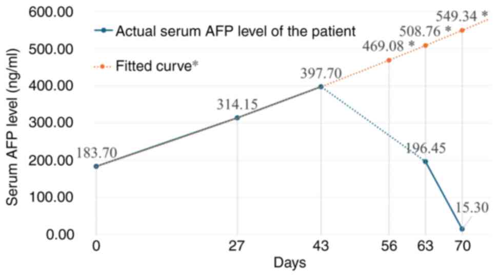
serum AFP level of 183.70 ng/ml (reference range, 0–7 ng/ml). The
results of other related laboratory tests, including liver function
tests, Hepatitis B indicators and other GI tumor markers, were all
within normal limits. Abdominal CT also revealed no significant
hepatic abnormalities. Following treatment for acid suppression and
gastric protection with omeprazole and sucralfate, the fecal OB
test turned negative. The patient was discharged upon their request
as the symptoms had subsided.
Follow-up at 1-month post-discharge (day 27)
revealed a marked increase in serum AFP to 314.15 ng/ml at Qingdun
Town Healthcare Center (Yancheng, China). A total of 2 weeks later,
the patient presented to the General Clinic at Yancheng No. 1
People's Hospital again and was readmitted to the ward of the
Department of General Medicine in December 2023 (day 43). Further
tests indicated an ulteriorly elevated serum AFP level at 397.70
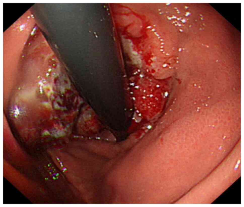
ng/ml. An upper gastrointestinal endoscopy revealed irregular
elevations and depressions from the esophagus (40 cm from incisors)
to the cardia, covered with a white coating on the surface
presumably mainly made up of necrotic tissue and mucus, with a
crater-like elevated nodule, indicating the presence of ulcerative
GC (Borrmann II). The lesion tissue was brittle and prone to
hemorrhage (Fig. 1). The endoscopic
diagnosis indicated esophageal cardia cancer. Subsequent enhanced
chest-abdominal CT demonstrated thickening and enhancement of the
gastric wall lateral to the lesser curvature of the cardia and
gastric fundus, further clarifying the extent of the lesion
(Fig. 2). Endoscopic biopsy sample
collected was fixed with 4% formaldehyde solution for 6 h at 25°C.
Paraffin-embedded tissue sections (4 µm) were deparaffinized with
xylene and rehydrated with a series of anhydrous ethanol, 95%
ethanol, 70% ethanol and PBS. For pathological examination, part of
the sections were stained with hematoxylin for 3 min at 25°C and
eosin for 45 sec at 25°C. For immunohistochemistry (IHC), part of
the sections underwent blocking of endogenous peroxidase using 3%
hydrogen peroxide for 10 min at 25°C, followed by blocking of
unspecific protein binding using 5% bovine serum albumin (cat. no.
GC305010; Servicebio Ltd) for 1 h at 37°C. IHC sections underwent
heat-mediated antigen retrieval with sodium citrate buffer (pH=6)
for 10 min at 97°C and were then incubated overnight at 4°C with
AFP antibody (1:100 dilution; cat. no. RMA-1069; Maxim
Biotechnologies, Ltd), hepatocyte paraffin (Hep Par) 1 antibody
(1:100 dilution; cat. no. MAB-1034; Maxim Biotechnologies, Ltd),
cytokeratin (CK)19 antibody (1:100 dilution; cat. no. MAB-0829;
Maxim Biotechnologies, Ltd) or caudal type homeobox (CDX)2 antibody
(1:100 dilution; cat. no. RMA-0631; Maxim Biotechnologies, Ltd).
Subsequently, IHC sections were treated with HRP-conjugated goat
anti-rabbit IgG (H+L) (1:200 dilution; cat. no. GB23303; Servicebio
Ltd) as the secondary antibody for 20 min at 25°C, and
visualization was performed using a DAB color development kit (cat.
no. G1212-200T; Servicebio Ltd), followed by counterstaining using
hematoxylin for 3 min at 25°C. All sections were sealed with
neutral resin and scanned with a digital slide scanner (NanoZoomer
S20; Hamamatsu Photonics KK). Pathological examination confirmed
the diagnosis of poorly differentiated cancer and focal tissue
showed liver-like features (Fig.
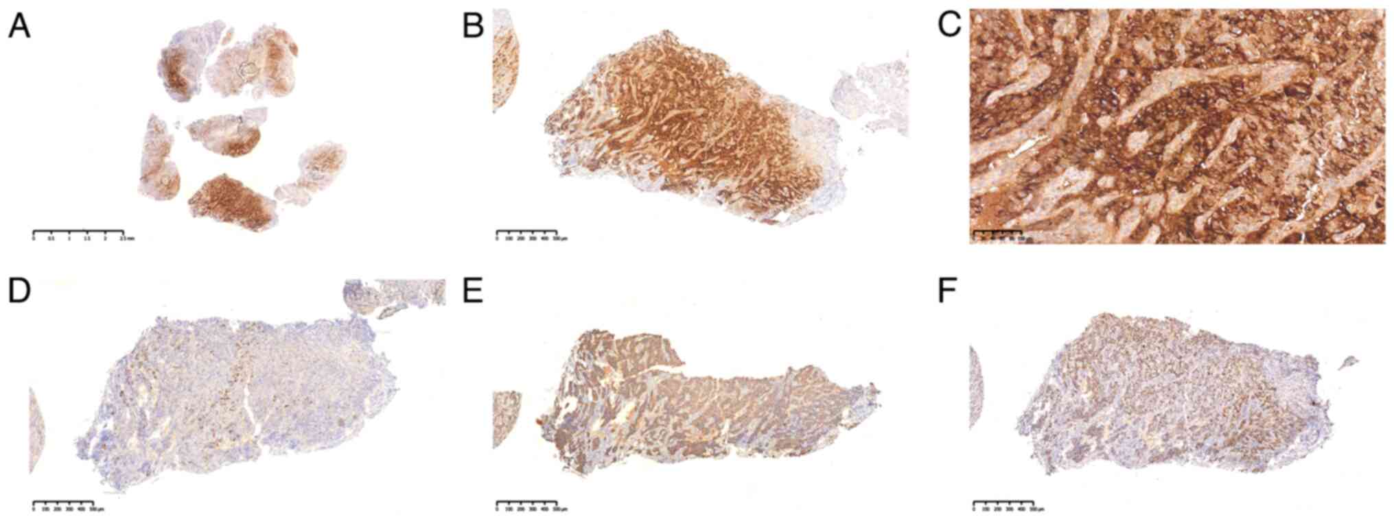
3). IHC results revealed that the endoscopic biopsy samples
were immunopositive for AFP (partial), Hep Par 1 (focal), CK19
(patchy) and CDX2 (homogeneous) (Fig.
4). Based on the elevation in serum AFP and positive AFP
staining outcome, the patient was diagnosed with AFPGC.
Furthermore, based on the liver-like pathology features, the
patient was diagnosed with HAS.
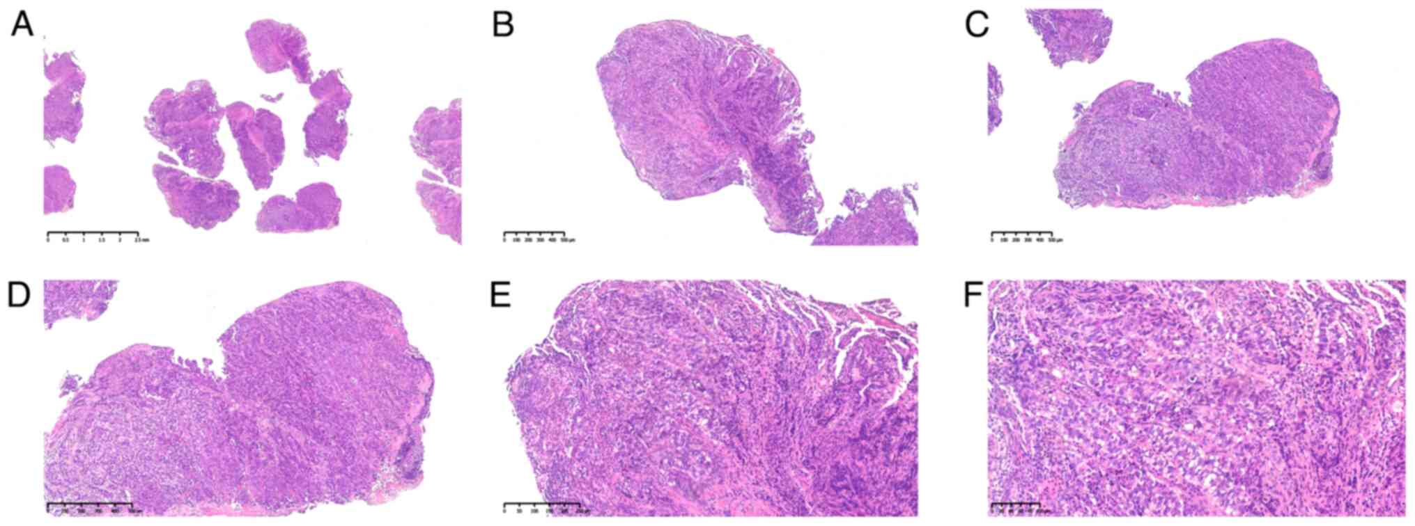 | Figure 3.Pathological images. Hematoxylin and
eosin stain at the following magnifications: (A) ×60, (B) ×200, (C)
×200, (D) ×280, (E) ×500 and (F) ×800. Under low magnification
(A-C), the tumor cells demonstrate infiltrative growth, and the
histological pattern consists of a combination of tubular
adenocarcinoma and solid arrangement, with a gradual migratory
process between the two. Under high magnification (D-F), the cancer
cells are large, with plentiful cytoplasm appearing either
eosinophilic or hyaline and containing visible intracytoplasmic
vacuoles. The nuclei of these cells are either round or irregular
in shape. The medullary or striated structures comprise
eosinophilic polygonal cells, and the cancer cells exhibit
different degrees of differentiation towards hepatocytes. The
interstitium is rich in blood vessels and sinuses, with narrow
fibrous interstitial compartments. |
The patient showed no obvious GI-related symptoms
throughout the whole observation period before surgery. The patient
was then referred to Jiangsu Provincial People's Hospital (Nanjing,
China) for radical gastric cancer surgery and the discharge record
for this hospitalization was obtained from the patient through a
follow-up visit. The patient underwent a 3D laparoscopic-assisted
radical total gastrectomy and esophagojejunal Roux-en-Y anastomosis
in January 2024 (day 56). The postoperative pathologic resection
specimen revealed a lesion located in the lesser curvature of the
cardia with a lesion size of 6.5×6.0×2.0 cm, which was styled as
Borrmann III in vision and low-differentiated (G3) diffuse tubular
adenocarcinoma [International Classification of Diseases for
Oncology type 8211/3 (30); T3N0M0
(31)] in the histology. The lesion
infiltrated the sub-plasma layer, cancer emboli were seen in the
vasculature, there was no clear invasion of nerves, no cancer
metastasis in the lymph nodes, and no cancer involvement in the
fatty-fibrous connective tissue (data not shown). IHC results
revealed that the postoperative resection specimen was
immunopositive for human epiderminal growth gator receptor 2 (2+),
Ki67 (90%+), postmeiotic segregation increased 2 (partial), MutL
homolog 1, MutS homolog (MSH)2, MSH6, AFP, Sal-like protein 4
(partial) and Glypican-3, and negative for in situ
hybridization Epstein-Barr encoding region and p53 (data not
shown). Post-surgery, the serum AFP level reduced to the normal
range (Fig. 5).
The patient was followed up with remotely by
telephone in April 2024 (day 148). At the time of this follow-up,
the patient was alive and in good postoperative condition.
Discussion
Since the first report in 1970, the definition of
AFPGC has remained ambiguous. In the original case reported by
Bourreille et al (2), AFPGC
was initially defined as GC with excessive serum AFP levels. With
the further development of IHC techniques, certain researchers
preferred to redefine AFPGC as one type of GC with positive IHC
staining for AFP (32). To resolve
this divergence, recent guidelines have suggested that AFPGC be
defined as GC with both elevated serum AFP levels and positive IHC
staining for AFP (33). However, a
recent case reported, in which there were GC-related serum AFP
elevation and negative IHC staining for AFP, has challenged the
definitions from the guidelines (34).
In the present case, as the pathological findings
revealed hepatoid features, there was another related disease to
discuss. HAS is another rare but aggressive GC-related disease; HAS
was first reported by Ishikura et al (24,25) in
the 1980s after the observation of certain AFPGC cases with
hepatoid features. Subsequent studies have reported that AFP
expression is not necessarily observed in certain cases of HAS,
further relaxing the definition of HAS as GC with foci of
hepatocellular differentiation (Fig.
6) (18,35).
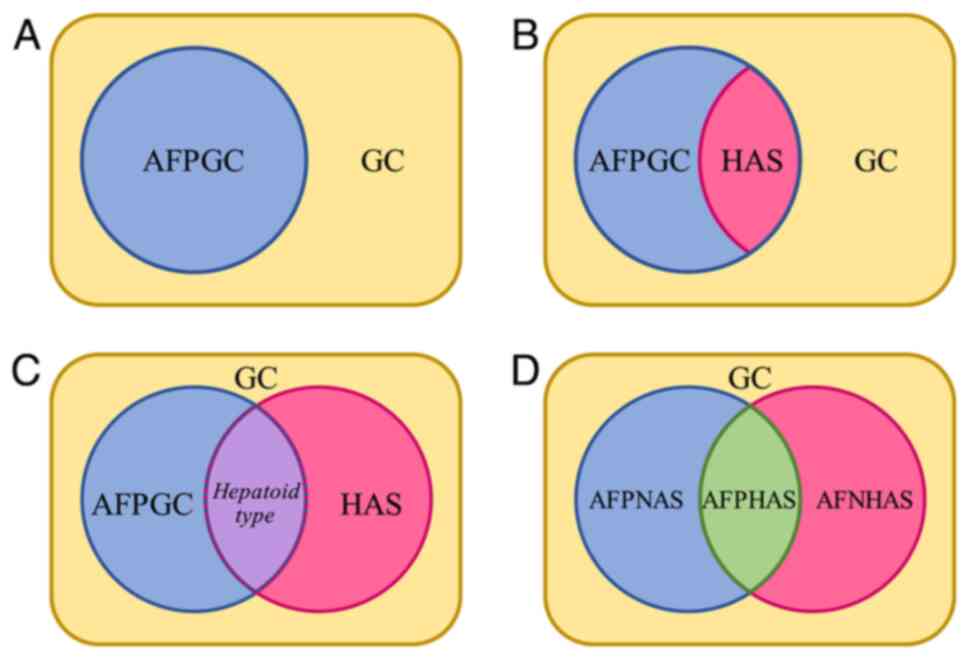 | Figure 6.History of changes in the
relationship between AFPGC and HAS. (A) Bourreille et al
(2) proposed the original
definition of AFPGC in 1970. (B) Ishikura et al (24,25)
proposed the original definition of HAS as a subtype of AFPGC in
the 1980s. (C) Expansion of the definition of HAS. The expanded HAS
concept now encompasses non-AFPGC components in addition to its
original scope. Some researchers redefined this cross part of AFPGC
and expanded HAS (the range of the original HAS concept) as the
hepatoid type (16,37), but certain researchers considered
this component no longer part of AFPGC (39). (D) The proposal in the present
report of a separate definition of AFPNAS, AFPHAS and AFNHAS
assigned to each component to avoid misunderstandings and
disagreements resulting from differences in definitions. AFP,
α-fetoprotein; AFPGC, AFP-producing gastric carcinoma; HAS,
hepatoid adenocarcinoma of the stomach; AFPNAS, AFP-positive
non-hepatoid adenocarcinoma of the stomach; AFPHAS, AFP-positive
hepatoid adenocarcinoma of the stomach; GC, gastric cancer. |
Due to the many histological overlaps between AFPGC
and HAS, this ambiguous space of definition often confuses the two,
and this confusion has already led to their misuse in certain cases
(36). Indeed, it is essential to
further clarify the distinction between AFPGC and HAS. During fetal
development, AFP is rationally synthesized not only by the liver
but also by the yolk sac and gastrointestinal tract. Therefore, the
tissues producing AFP in GC can also have different morphologies.
Motoyama et al (37) first
advocated the typing of AFPGC based on histological features. They
proposed three subtypes: i) Hepatoid type; ii) yolk sac tumor-like
type; and iii) fetal gastrointestinal type, which has also been
referred to as enteroblastic type in classifications by Kinjo et
al (16). This classification
further highlights the differences and connections between AFPGC
and HAS. After finding certain cases that showed only features of
common adenocarcinoma, Kinjo et al (16) proposed a fourth type, the common
adenocarcinoma type, in 2012. These four subtypes can appear alone
or in combination. Recently, a rare case of adenocarcinoma
coexisting with squamous cell carcinoma has been reported (34). This may require an expansion of the
existing classification.
The diagnosis of AFPGC is extremely heterogeneous
and a single diagnosis of only AFPGC may result in a loss of
critical information. Certain researchers have tried to eliminate
the concept of AFPGC and advocated for a diagnostic system based on
histological criteria instead (38), and this viewpoint was supported by
the fifth edition of the World Health Organization (WHO)
classification of tumors, which includes only HAS and not AFPGC
(30).
However, it should be noted that the distinction
between AFP-positive and -negative types remains critical when
identifying histological type. Chen et al (39) revealed that a group with higher AFP
expression had a higher frequency of liver metastasis and worse
overall survival compared with those with lower AFP expression
among patients with HAS. Moreover, a large-scale epidemiological
study has reported the importance of serum AFP levels in patients
with GC (40). Therefore, the
present report proposes a new classification method to take both
histological and AFP-producing features into account. GCs with an
elevation in serum AFP levels are defined as serum-positive, and
those with positive results in IHC staining for AFP in pathological
tissues as tissue-positive. Either serum-positive or
tissue-positive should be considered as AFP-positive, and specific
serum and IHC manifestations should be marked in the diagnosis. The
criteria for serum AFP positivity are widely defined by different
values in different studies. Generally, a serum AFP level of >20
ng/ml can be considered as clinically significant and serum AFP
positive (40,41). However, certain researchers
preferred 100 ng/ml as the cutoff (21,39,42).
Nevertheless, there is insufficient evidence for any of the two
cutoffs. Considering both cutoffs may be a better option to avoid
including cases with physiological variations or focusing too late
on potential cases. In many studies, a cutoff value of 300 ng/ml is
also used to distinguish very high levels of AFP from others
(41,43). Therefore, 20, 100 and 300 ng/ml
could be used as cutoffs to grade serum AFP levels, which may be
helpful to include more cases previously missed and eliminate the
confusion introduced by different definitions of AFP positive cases
(Table I). Similarly, a grading
system to assess the IHC staining results of AFP is also proposed
(Table II). The four-level grading
method based on staining intensity is a novel, simple and
reproducible method widely used to evaluate or interpret the IHC
staining results of many indicators (44–47).
 | Table I.Serum positivity according to the
level of serum α-fetoprotein. |
Table I.
Serum positivity according to the
level of serum α-fetoprotein.
| Serum AFP level,
ng/ml | n≤20 |
20<n≤100a |
100<n≤300b |
n>300c |
|---|
| Serum-positivity
grade | S0 | S1+ | S2+ | S3+ |
 | Table II.Tissue positivity according to the
immunohistochemistry staining intensity. |
Table II.
Tissue positivity according to the
immunohistochemistry staining intensity.
|
|
| Faint |
|
|
|---|
|
|
|
|
|
|
|---|
| Staining
intensity | Complete
absence | <10% tumor
cells | >10% tumor
cells | Moderate | Strong |
|---|
| Tissue-positivity
grade | T0 | T1+ | T2+ | T3+ |
Under the aforementioned definition of AFP
positivity, the concepts of AFPGC and HAS were combined to
introduce three related new diagnoses: i) AFP-positive hepatoid
adenocarcinoma of the stomach (AFPHAS); ii) AFP-positive
non-hepatoid adenocarcinoma of the stomach (AFPNAS); and iii)
AFP-negative hepatoid adenocarcinoma of the stomach (AFNHAS). This
new diagnostic classification would be able to cover all cases
encompassed by AFPGC and HAS under all definition methods and
establish a fixed definition for each case. This may help prevent
confusion and data misapplication in statistical analysis arising
from similar cases being classified under different diagnoses with
different definition methods.
Furthermore, if further evidence shows that other
histological types significantly differ in prognosis or other
aspects, AFPNAS may be further categorized into more specific
diagnosis groups, including but not limited to AFP-positive
yolk-sac-tumor-like adenocarcinoma of the stomach, AFP-positive
fetal-gastrointestinal adenocarcinoma of the stomach and
AFP-positive common adenocarcinoma of the stomach. AFP-negative
cases with other histological types can also be categorized as
AFP-negative yolk-sac-tumor-like adenocarcinoma of the stomach,
AFP-negative fetal-gastrointestinal adenocarcinoma of the stomach
and AFP-negative common adenocarcinoma of the stomach. If needed,
AFP-positive squamous-cell carcinoma of the stomach and
AFP-negative squamous-cell carcinoma of the stomach can also be
discussed.
In the present case, the patient was finally
diagnosed as AFPHAS (S3+T3+) according to the proposed new
classification system. Several existing meta-analyses (48,49)
have conducted preliminary statistical evaluations of various
pathologic features, serum AFP levels and survival rates in
patients. It is anticipated that a new classification could be
further implemented into these data to reveal the impact of serum
AFP grading and newly proposed subtypes on patient prognosis.
Unfortunately, due to the paucity of literature focusing on
differences in the degree of AFP staining in tissues, assessing the
impact of tissue AFP grading on patient prognosis remains a task of
future work to consider after the present report.
Generally, existing published data is limited by
small sample sizes in individual studies and a lack of AFP-level
grading. Therefore, whilst these studies (40,41,50)
suggest that AFP levels and pathological type impact prognosis, the
exact extent of this impact remains unclear. The new classification
system proposed in the present report could enable researchers to
further categorize both reported and unreported cases, which will
allow the exploration of the specific influence of AFP levels and
pathological type on survival expectations in patients with GC in
more depth. Ultimately, this could help clinicians refine a more
precise assessment of the survival expectations of patients, and
possibly influence future decisions regarding additional
postoperative treatment options.
There is often a genetic factor in the development
of cancer, and this may be true for AFPGC cases as well. A
whole-exome sequencing study (51)
revealed the association between certain genes and AFPGC that AFPGC
cases with mutations of multiple genes such as cyclin E (CCNE) 1,
Cyclin D1, EGFR, Erb-b2 receptor tyrosine kinase (ERBB) 2, ERBB3,
ERBB4, Aurora kinase A, AXL receptor tyrosine, B-cell lymphoma 6,
breast cancer gene 2, vascular endothelial growth factor receptor
1, fibroblast growth factor receptor 2, cellular myelocytomatosis
oncogene and myeloid cell leukemia 1, tend to have a worse OS rate
and exhibit more aggressive behavior than normal GC subtypes, due
to the activation of core signal pathways such as RTK/RAS/PI(3)K,
p53/cell cycle and JAK/STAT. Based on these findings, CCNE1- and
ERBB2-targeted medications may have potential in future AFPGC
therapy, which may help to offer differentiated options for
patients with AFPGC beyond conventional GC therapies.
Unfortunately, the study (51) only
used tissue IHC positivity as the inclusion criterion, it did not
differentiate between histological types and it omitted
serum-positive-only cases. With the new proposed classification
system in the present report, researchers in the future could
further refine the relationships between mutations, pathways and
pathological typing.
Moreover, the patient in the present case was
infected with H. pylori and had a history of T2DM. H.
pylori is categorized as a group 1 carcinogen by the WHO and is
now widely recognized as a primary risk factor for GC (52–55).
It is not fully elucidated how H. pylori causes GC, but
studies have revealed that the mechanism is related to multiple
virulence factors, including cytotoxin-associated gene A,
vacuolating cytotoxin A and outer membrane proteins (56,57).
Only one previous study explored whether H. pylori serves a
different role in different GC subtypes, and this study failed to
demonstrate an association between the H. pylori-positive
rate and the presence of AFP-positive or hepatoid features in
patients with GC (58).
Furthermore, compared with H. pylori, the effect of T2DM on
the development of GC does not seem to be precise. Certain earlier
studies reported an increased risk of GC with pre-existing T2DM
(59–61). However, recent research highlight
that different study types can lead to different conclusions on
this issue. Statistical significance between T2DM history and GC
risk has not been demonstrated in prospective studies (62). In addition, a later large-scale
study reported that T2DM is unrelated to GC overall but may be
associated with excess cardia GC risk (63). In the present case, the tumor was
located around the cardia of the patient. If the patient had been
able to eradicate H. pylori earlier and received more
regular glucose management, they may have been not developed GC.
This demonstrates the importance of routine screening for H.
pylori and long-term regular glucose monitoring in the primary
health care system.
In conclusion, the present report describes the case
of an elderly patient with AFPGC and HAS with hepatoid features in
the tumor and with both elevated serum AFP levels and positive IHC
AFP staining. Although the case could have been diagnosed as both
AFPGC and HAS using any existing definition, it was found that the
current parallelism of conflicting definition methods of AFPGC and
HAS was already a barrier to scientific progress. Therefore, the
present report proposed a new classification considering both
histological and AFP-producing features, and both expressing
features in serum and tissue, to eliminate this obstacle, covering
all cases encompassed by AFPGC and HAS under all definition methods
and establishing a fixed definition for each case. Moreover, there
is currently minimal variation in the treatment approaches for most
types of GC, including AFPGC and HAS. Nonetheless, it is crucial to
recognize that both AFP level (41)
and pathological type (50) serve
as independent prognostic factors for GC. The new classification
proposed in the present report not only holds significance for
pathologists but also extends its relevance to other clinicians by
highlighting the importance of AFP levels and pathological type in
GC cases. This dual focus is anticipated to remarkably enhance the
precision of survival prognosis evaluations. Furthermore, it
carries the potential to impact subsequent choices concerning
adjunctive postoperative treatments, thereby broadening the scope
of personalized patient care strategies in gastric oncological
settings. However, further studies are required to elucidate the
associations between each specific subtype and
genetic-environmental factors such as genes, H. pylori
infection and chronic health history, under the new classification
method proposed.
Acknowledgements
Not applicable.
Funding
This study was funded by the Science and Technology Development
Fund, Macau SAR (grant nos. 0098/2021/A2 and 0048/2023/AFJ), and
the Chinese Medicine Guangdong Laboratory (grant no.
HQCML-C-2024007).
Availability of data and materials
The data generated in the present study are not
publicly available due to patient privacy protection but may be
requested from the corresponding author.
Authors' contributions
ZYY proposed the new classification and drafted the
manuscript. CHX and ZMS performed the pathology and IHC
examinations and prepared Figs. 3
and 4. WC performed the CT scan and
prepared Fig. 2. XZ and CNW
developed the treatment plan for the patient, collected the
clinical history, and wrote part of the manuscript text. JLT and
XDW performed the endoscopy and prepared Fig. 1. XSL and XW searched for relevant
literature and refined the new classification. QQY, YH and XYX
performed the followed-up of the patient, obtained the
post-discharge record from the patient and analyzed patient data.
XDW and QBW conceived the idea, refined the new classification and
contributed to supervision. ZYY and XDW checked and confirmed the
authenticity of all the raw data. All authors have read and
approved the final manuscript.
Ethics approval and consent to
participate
All procedures performed involving human
participants were in accordance with the ethical standards of the
institutional and/or national research committee and with the 1964
Helsinki Declaration and its later amendments or comparable ethical
standards.
Patient consent for publication
Written informed consent was obtained from the
patient for the case information and images to be published in the
present case report.
Competing interests
The authors declare that they have no competing
interests.
Authors' information
ORCID of Authors:
Dr Zhen-yu Ye, https://orcid.org/0000-0001-5068-2204
Prof Xu-dong Wu, https://orcid.org/0000-0002-2881-954X
Prof Qi-biao Wu, https://orcid.org/0000-0002-1670-1050
Glossary
Abbreviations
Abbreviations:
|
AFP
|
α-fetoprotein
|
|
AFPGC
|
AFP-producing gastric carcinoma
|
|
AFPHAS
|
AFP-positive hepatoid adenocarcinoma
of the stomach
|
|
AFPNAS
|
AFP-positive non-hepatoid
adenocarcinoma of the stomach
|
|
CDX
|
caudal type homeobox
|
|
CK
|
cytokeratin
|
|
CT
|
computed tomography
|
|
GC
|
gastric cancer
|
|
GI
|
gastrointestinal
|
|
HAS
|
hepatoid adenocarcinoma of the
stomach
|
|
Hep Par
|
hepatocyte paraffin
|
|
IHC
|
immunohistochemistry
|
|
OB
|
occult blood
|
|
T2DM
|
type 2 diabetes mellitus
|
|
WHO
|
World Health Organization
|
References
|
1
|
Chhikara BS and Parang K: Global cancer
statistics 2022: The trends projection analysis. Chem Biol Lett.
10:4512023.
|
|
2
|
Bourreille J, Metayer P, Sauger F, Matray
F and Fondimare A: Existence of alpha-fetoprotein during
gastric-origin secondary cancer of the liver. Presse Med (1893).
78:1277–1278. 1970.(In French). PubMed/NCBI
|
|
3
|
Daniele B, Bencivenga A, Megna AS and
Tinessa V: Alpha-fetoprotein and ultrasonography screening for
hepatocellular carcinoma. Gastroenterology. 127 (5 Suppl
1):S108–S112. 2004. View Article : Google Scholar : PubMed/NCBI
|
|
4
|
O'Conor GT, Tatarinov YS, Abelev GI and
Uriel J: A collaborative study for the evaluation of a serologic
test for primary liver cancer. Cancer. 25:1091–1098. 1970.
View Article : Google Scholar : PubMed/NCBI
|
|
5
|
Babalı A, Çakal E, Purnak T, Bıyıkoğlu I,
Cakal B, Yüksel O and Köklü S: Serum α-fetoprotein levels in liver
steatosis. Hepatol Int. 3:551–555. 2009. View Article : Google Scholar : PubMed/NCBI
|
|
6
|
Motoyama T, Watanabe H, Yamamoto T and
Sekiguchi M: Production of alpha-fetoprotein by human germ cell
tumors in vivo and in vitro. Acta Pathol Jpn. 37:1263–1277.
1987.PubMed/NCBI
|
|
7
|
Ezaki T, Yukaya H, Ogawa Y, Chang YC and
Nagasue N: Elevation of alpha-fetoprotein level without evidence of
recurrence after hepatectomy for hepatocellular carcinoma. Cancer.
61:1880–1883. 1988. View Article : Google Scholar : PubMed/NCBI
|
|
8
|
Ganjei P, Nadji M, Albores-Saavedra J and
Morales AR: Histologic markers in primary and metastatic tumors of
the liver. Cancer. 62:1994–1998. 1988. View Article : Google Scholar : PubMed/NCBI
|
|
9
|
Yasunami R, Hashimoto Z, Ogura T, Hirao F
and Yamamura Y: Primary lung cancer producing alpha-fetoprotein: A
case report. Cancer. 47:926–929. 1981. View Article : Google Scholar : PubMed/NCBI
|
|
10
|
Saito S, Hatano T, Hayakawa M, Koyama Y,
Ohsawa A and Iwamasa T: Studies on alpha-fetoprotein produced by
renal cell carcinoma. Cancer. 63:544–549. 1989. View Article : Google Scholar : PubMed/NCBI
|
|
11
|
Yamagata T, Yamagata Y, Nakanishi M,
Matsunaga K, Minakata Y and Ichinose M: A case of primary lung
cancer producing alpha-fetoprotein. Can Respir J. 11:504–506. 2004.
View Article : Google Scholar : PubMed/NCBI
|
|
12
|
Matsueda K, Yamamoto H, Yoshida Y and
Notohara K: Hepatoid carcinoma of the pancreas producing protein
induced by vitamin K absence or antagonist II (PIVKA-II) and
alpha-fetoprotein (AFP). J Gastroenterol. 41:1011–1019. 2006.
View Article : Google Scholar : PubMed/NCBI
|
|
13
|
Itoh T, Kishi K, Tojo M, Kitajima N,
Kinoshita Y, Inatome T, Fukuzaki H, Nishiyama N, Tachibana H and
Takahashi H: Acinar cell carcinoma of the pancreas with elevated
serum alpha-fetoprotein levels: A case report and a review of 28
cases reported in Japan. Gastroenterol Jpn. 27:785–791. 1992.
View Article : Google Scholar : PubMed/NCBI
|
|
14
|
Kato K, Matsuda M, Ingu A, Imai M, Kasai
S, Mito M and Kobayashi T: Colon cancer with a high serum
alpha-fetoprotein level. Am J Gastroenterol. 91:1045–1046.
1996.PubMed/NCBI
|
|
15
|
Yachida S, Fukushima N, Nakanishi Y, Akasu
T, Kitamura H, Sakamoto M and Shimoda T:
Alpha-fetoprotein-producing carcinoma of the colon: Report of a
case and review of the literature. Dis Colon Rectum. 46:826–831.
2003. View Article : Google Scholar : PubMed/NCBI
|
|
16
|
Kinjo T, Taniguchi H, Kushima R, Sekine S,
Oda I, Saka M, Gotoda T, Kinjo F, Fujita J and Shimoda T:
Histologic and immunohistochemical analyses of
α-fetoprotein-producing cancer of the stomach. Am J Surg Pathol.
36:56–65. 2012. View Article : Google Scholar : PubMed/NCBI
|
|
17
|
Liu X, Cheng Y, Sheng W, Lu H, Xu Y, Long
Z, Zhu H and Wang Y: Clinicopathologic features and prognostic
factors in alpha-fetoprotein-producing gastric cancers: Analysis of
104 cases. J Surg Oncol. 102:249–255. 2010. View Article : Google Scholar : PubMed/NCBI
|
|
18
|
Nagai E, Ueyama T, Yao T and Tsuneyoshi M:
Hepatoid adenocarcinoma of the stomach. A clinicopathologic and
immunohistochemical analysis. Cancer. 72:1827–1835. 1993.
View Article : Google Scholar : PubMed/NCBI
|
|
19
|
Wang D, Li C, Xu Y, Xing Y, Qu L, Guo Y,
Zhang Y, Sun X and Suo J: Clinicopathological characteristics and
prognosis of alpha-fetoprotein positive gastric cancer in Chinese
patients. Int J Clin Exp Pathol. 8:6345–6355. 2015.PubMed/NCBI
|
|
20
|
Hirajima S, Komatsu S, Ichikawa D, Kubota
T, Okamoto K, Shiozaki A, Fujiwara H, Konishi H, Ikoma H and Otsuji
E: Liver metastasis is the only independent prognostic factor in
AFP-producing gastric cancer. World J Gastroenterol. 19:6055–6061.
2013. View Article : Google Scholar : PubMed/NCBI
|
|
21
|
Kono K, Amemiya H, Sekikawa T, Iizuka H,
Takahashi A, Fujii H and Matsumoto Y: Clinicopathologic features of
gastric cancers producing alpha-fetoprotein. Dig Surg. 19:359–365.
2002. View Article : Google Scholar : PubMed/NCBI
|
|
22
|
Chang YC, Nagasue N, Abe S, Taniura H,
Kumar DD and Nakamura T: Comparison between the clinicopathologic
features of AFP-positive and AFP-negative gastric cancers. Am J
Gastroenterol. 87:321–325. 1992.PubMed/NCBI
|
|
23
|
Sun N, Sun Q, Liu Q, Zhang T, Zhu Q, Wang
W, Cao M and Zang QI: α-fetoprotein-producing gastric carcinoma: A
case report of a rare subtype and literature review. Oncol Lett.
11:3101–3104. 2016. View Article : Google Scholar : PubMed/NCBI
|
|
24
|
Ishikura H, Fukasawa Y, Ogasawara K,
Natori T, Tsukada Y and Aizawa M: An AFP-producing gastric
carcinoma with features of hepatic differentiation. A case report.
Cancer. 56:840–848. 1985. View Article : Google Scholar : PubMed/NCBI
|
|
25
|
Ishikura H, Kirimoto K, Shamoto M,
Miyamoto Y, Yamagiwa H, Itoh T and Aizawa M: Hepatoid
adenocarcinomas of the stomach. An analysis of seven cases. Cancer.
58:119–126. 1986. View Article : Google Scholar : PubMed/NCBI
|
|
26
|
Lin JX, Wang ZK, Hong QQ, Zhang P, Zhang
ZZ, He L, Wang Q, Shang L, Wang LJ, Sun YF, et al: Assessment of
clinicopathological characteristics and development of an
individualized prognostic model for patients with hepatoid
adenocarcinoma of the stomach. JAMA Netw Open. 4:e21282172021.
View Article : Google Scholar : PubMed/NCBI
|
|
27
|
Shi J, Liu L and Chen YP: Research
progress of prognostic analysis and clinicopathological
characteristics of hepatoid adenocarcinoma of the stomach. Zhonghua
Zhong Liu Za Zhi. 43:104–107. 2021.(In Chinese). PubMed/NCBI
|
|
28
|
Machlowska J, Baj J, Sitarz M, Maciejewski
R and Sitarz R: Gastric cancer: Epidemiology, risk factors,
classification, genomic characteristics and treatment strategies.
Int J Mol Sci. 21:40122020. View Article : Google Scholar : PubMed/NCBI
|
|
29
|
Wang FH, Zhang XT, Tang L, Wu Q, Cai MY,
Li YF, Qu XJ, Qiu H, Zhang YJ, Ying JE, et al: The Chinese society
of clinical oncology (CSCO): Clinical guidelines for the diagnosis
and treatment of gastric cancer, 2023. Cancer Commun (Lond).
44:127–172. 2024. View Article : Google Scholar : PubMed/NCBI
|
|
30
|
WHO Classification of Tumours Editorial
Board, . WHO classification of tumours. Digestive System Tumours.
5th edition. World Health Organization; Lyon: 2019
|
|
31
|
Amin MB, Edge SB, Greene FL and Brierley
JD: AJCC cancer staging manual. 8th edition. Springer; New York,
NY: 2017
|
|
32
|
Kodama T, Kameya T, Hirota T, Shimosato Y,
Ohkura H, Mukojima T and Kitaoka H: Production of
alpha-fetoprotein, normal serum proteins, and human chorionic
gonadotropin in stomach cancer: Histologic and immunohistochemical
analyses of 35 cases. Cancer. 48:1647–1655. 1981. View Article : Google Scholar : PubMed/NCBI
|
|
33
|
Chinese Medical Association Oncology, .
Clinical diagnosis and treatment guideline of gastric cancer of
Chinese Medical Association (2021 edition). Natl Med J Chin.
102:1169–1189. 2022.(In Chinese).
|
|
34
|
Sun L, Wei JJ, An R, Cai HY, Lv Y, Li T,
Shen XF, Du JF and Chen G: Gastric adenosquamous carcinoma with an
elevated serum level of alpha-fetoprotein: A case report. World J
Gastrointest Surg. 15:2357–2361. 2023. View Article : Google Scholar : PubMed/NCBI
|
|
35
|
Trompetas V, Varsamidakis N, Frangia K,
Polimeropoulos V and Kalokairinos E: Gastric hepatoid
adenocarcinoma and familial investigation: Does it always produce
alpha-fetoprotein? Eur J Gastroenterol Hepatol. 15:1241–1244. 2003.
View Article : Google Scholar : PubMed/NCBI
|
|
36
|
Jiao XH, Dong LC, Tian GL, Zheng YY and
Shao YY: 2 cases of alpha-fetoprotein-elevated gastric cancer and
literature review. J Qiqihar Med Univ. 23:1752010.(In Chinese).
|
|
37
|
Motoyama T, Aizawa K, Watanabe H, Fukase M
and Saito K: Alpha-fetoprotein producing gastric carcinomas: A
comparative study of three different subtypes. Acta Pathol Jpn.
43:654–661. 1993.PubMed/NCBI
|
|
38
|
Yang XS, Wang AQ, Wu Y, Li Z, Bu Z and Ji
J: Hepatoid adenocarcinoma of stomach: Current research, progress,
and controversy. Chin J Clin Oncol. 50:679–684. 2023.(In
Chinese).
|
|
39
|
Chen EB, Wei YC, Liu HN, Tang C, Liu ML,
Peng K and Liu T: Hepatoid adenocarcinoma of stomach: Emphasis on
the clinical relationship with alpha-fetoprotein-positive gastric
cancer. Biomed Res Int. 2019:67104282019. View Article : Google Scholar : PubMed/NCBI
|
|
40
|
Chen Y, Qu H, Jian M, Sun G and He Q: High
level of serum AFP is an independent negative prognostic factor in
gastric cancer. Int J Biol Markers. 30:e387–e393. 2015. View Article : Google Scholar : PubMed/NCBI
|
|
41
|
Lin HJ, Hsieh YH, Fang WL, Huang KH and Li
AF: Clinical manifestations in patients with
alpha-fetoprotein-producing gastric cancer. Curr Oncol.
21:e394–e399. 2014. View Article : Google Scholar : PubMed/NCBI
|
|
42
|
Ooi A, Nakanishi I, Sakamoto N, Tsukada Y,
Takahashi Y, Minamoto T and Mai M: Alpha-fetoprotein
(AFP)-producing gastric carcinoma. Is it hepatoid differentiation?
Cancer. 65:1741–1747. 1990. View Article : Google Scholar : PubMed/NCBI
|
|
43
|
Gong W, Shou D and Gong P: Extremely high
expression of serum alpha-fetoprotein level of gastric
adenocarcinoma: A rare case with an unexpected well-prognosis.
Springerplus. 5:20562016. View Article : Google Scholar : PubMed/NCBI
|
|
44
|
Yu J, Kane S, Wu J, Benedettini E, Li D,
Reeves C, Innocenti G, Wetzel R, Crosby K, Becker A, et al:
Mutation-specific antibodies for the detection of EGFR mutations in
non-small-cell lung cancer. Clin Cancer Res. 15:3023–3028. 2009.
View Article : Google Scholar : PubMed/NCBI
|
|
45
|
Simonetti S, Molina MA, Queralt C, de
Aguirre I, Mayo C, Bertran-Alamillo J, Sanchez JJ, Gonzalez-Larriba
JL, Jimenez U, Isla D, et al: Detection of EGFR mutations with
mutation-specific antibodies in stage IV non-small-cell lung
cancer. J Transl Med. 8:1352010. View Article : Google Scholar : PubMed/NCBI
|
|
46
|
Hofman P, Ilie M, Hofman V, Roux S, Valent
A, Bernheim A, Alifano M, Leroy-Ladurie F, Vaylet F, Rouquette I,
et al: Immunohistochemistry to identify EGFR mutations or ALK
rearrangements in patients with lung adenocarcinoma. Ann Oncol.
23:1738–1743. 2012. View Article : Google Scholar : PubMed/NCBI
|
|
47
|
Guo R, Ma L, Bai X, Miao L, Li Z and Yang
J: A scoring method for immunohistochemical staining on Ki67. Appl
Immunohistochem Mol Morphol. 29:e20–e28. 2021. View Article : Google Scholar : PubMed/NCBI
|
|
48
|
Li XD, Wu CP, Ji M, Wu J, Lu B, Shi HB and
Jiang JT: Characteristic analysis of α-fetoprotein-producing
gastric carcinoma in China. World J Surg Oncol. 11:2462013.
View Article : Google Scholar : PubMed/NCBI
|
|
49
|
Su JS, Chen YT, Wang RC, Wu CY, Lee SW and
Lee TY: Clinicopathological characteristics in the differential
diagnosis of hepatoid adenocarcinoma: A literature review. World J
Gastroenterol. 19:321–327. 2013. View Article : Google Scholar : PubMed/NCBI
|
|
50
|
Jiang J, Ding Y, Lu J, Chen Y, Chen Y,
Zhao W, Chen W, Kong M, Li C, Teng X, et al: Integrative analysis
reveals a clinicogenomic landscape associated with liver metastasis
and poor prognosis in hepatoid adenocarcinoma of the stomach. Int J
Biol Sci. 18:5554–5574. 2022. View Article : Google Scholar : PubMed/NCBI
|
|
51
|
Lu J, Ding Y, Chen Y, Jiang J, Chen Y,
Huang Y, Wu M, Li C, Kong M, Zhao W, et al: Whole-exome sequencing
of alpha-fetoprotein producing gastric carcinoma reveals genomic
profile and therapeutic targets. Nat Commun. 12:39462021.
View Article : Google Scholar : PubMed/NCBI
|
|
52
|
Crowe SE: Helicobacter pylori infection. N
Engl J Med. 380:1158–1165. 2019. View Article : Google Scholar : PubMed/NCBI
|
|
53
|
Bornschein J, Selgrad M, Warnecke M,
Kuester D, Wex T and Malfertheiner P: H. pylori infection is a key
risk factor for proximal gastric cancer. Dig Dis Sci. 55:3124–3131.
2010. View Article : Google Scholar : PubMed/NCBI
|
|
54
|
Kumar S, Metz DC, Ellenberg S, Kaplan DE
and Goldberg DS: Risk factors and incidence of gastric cancer after
detection of Helicobacter pylori infection: A large cohort study.
Gastroenterology. 158:527–536.e7. 2020. View Article : Google Scholar : PubMed/NCBI
|
|
55
|
Shirani M, Pakzad R, Haddadi MH, Akrami S,
Asadi A, Kazemian H, Moradi M, Kaviar VH, Zomorodi AR, Khoshnood S,
et al: The global prevalence of gastric cancer in Helicobacter
pylori-infected individuals: A systematic review and meta-analysis.
BMC Infect Dis. 23:5432023. View Article : Google Scholar : PubMed/NCBI
|
|
56
|
Alipour M: Molecular mechanism of
Helicobacter pylori-induced gastric cancer. J Gastrointest Cancer.
52:23–30. 2021. View Article : Google Scholar : PubMed/NCBI
|
|
57
|
Takahashi-Kanemitsu A, Lu MX, Knight CT,
Yamamoto T, Hayash T, Mii Y, Ooki T, Kikuchi I, Kikuchi A, Barker
N, et al: The Helicobacter pylori CagA oncoprotein disrupts Wnt/PCP
signaling and promotes hyperproliferation of pyloric gland base
cells. Sci Signal. 16:eabp90202023. View Article : Google Scholar : PubMed/NCBI
|
|
58
|
Liu DR, Li BB, Yan B, Liu L, Jia Y, Wang
Y, Ma X and Yang F: The clinicopathological features and prognosis
of serum AFP positive gastric cancer: A report of 16 cases. Int J
Clin Exp Pathol. 13:2439–2446. 2020.PubMed/NCBI
|
|
59
|
Ge Z, Ben Q, Qian J, Wang Y and Li Y:
Diabetes mellitus and risk of gastric cancer: A systematic review
and meta-analysis of observational studies. Eur J Gastroenterol
Hepatol. 23:1127–1135. 2011. View Article : Google Scholar : PubMed/NCBI
|
|
60
|
Yoon JM, Son KY, Eom CS, Durrance D and
Park SM: Pre-existing diabetes mellitus increases the risk of
gastric cancer: A meta-analysis. World J Gastroenterol. 19:936–945.
2013. View Article : Google Scholar : PubMed/NCBI
|
|
61
|
Sekikawa A, Fukui H, Maruo T, Tsumura T,
Okabe Y and Osaki Y: Diabetes mellitus increases the risk of early
gastric cancer development. Eur J Cancer. 50:2065–2071. 2014.
View Article : Google Scholar : PubMed/NCBI
|
|
62
|
Bae JM: Diabetes history and gastric
cancer risk: Different results by types of follow-up studies. Asian
Pac J Cancer Prev. 23:1523–1528. 2022. View Article : Google Scholar : PubMed/NCBI
|
|
63
|
Dabo B, Pelucchi C, Rota M, Jain H,
Bertuccio P, Bonzi R, Palli D, Ferraroni M, Zhang ZF,
Sanchez-Anguiano A, et al: The association between diabetes and
gastric cancer: Results from the stomach cancer pooling project
consortium. Eur J Cancer Prev. 31:260–269. 2022. View Article : Google Scholar : PubMed/NCBI
|


