Introduction
Primary cutaneous B-cell lymphomas (PCBCLs) are
low-grade malignant extranodal B-cell non-Hodgkin's lymphomas that
are confined to the skin without extracutaneous manifestations
(1). The three main subtypes of
PCBCL include primary cutaneous follicle center lymphoma (PCFCL),
primary cutaneous marginal zone lymphoma (PCMZL) and primary
cutaneous diffuse large BCL (PCDLBCL) (2). Although PCFCL is the most common type
among them, accounting for ~55% of all PCBCL cases, it accounts for
<1% of cases of B-cell lymphoma (3). PCFCL is an indolent lymphoma with an
incidence rate that has been increasing exponentially over the past
few decades (4).
PCFCL usually presents as limited skin damage to the
head or trunk (5), with no
extracutaneous involvement of lymph nodes, bone marrow or internal
organs at the time of diagnosis (6). Clinically, the limited skin lesions in
PCFCL appear morphologically as single or multiple slow-growing
pink or purplish-red plaques, papules, nodules or tumors (7). The present study reports the case of a
young patient with PCFCL and no extracutaneous manifestations.
Case report
In August 2024, a 32-year-old man presented to The
Affiliated Yantai Yuhuanding Hospital of Qingdao University
(Yantai, China) with a skin swelling on the left upper arm that had
been present for 2 years. The patient experienced occasional
itching without night sweats, pain or fever. The swelling was hard
and was gradually increasing in size. A physical examination showed
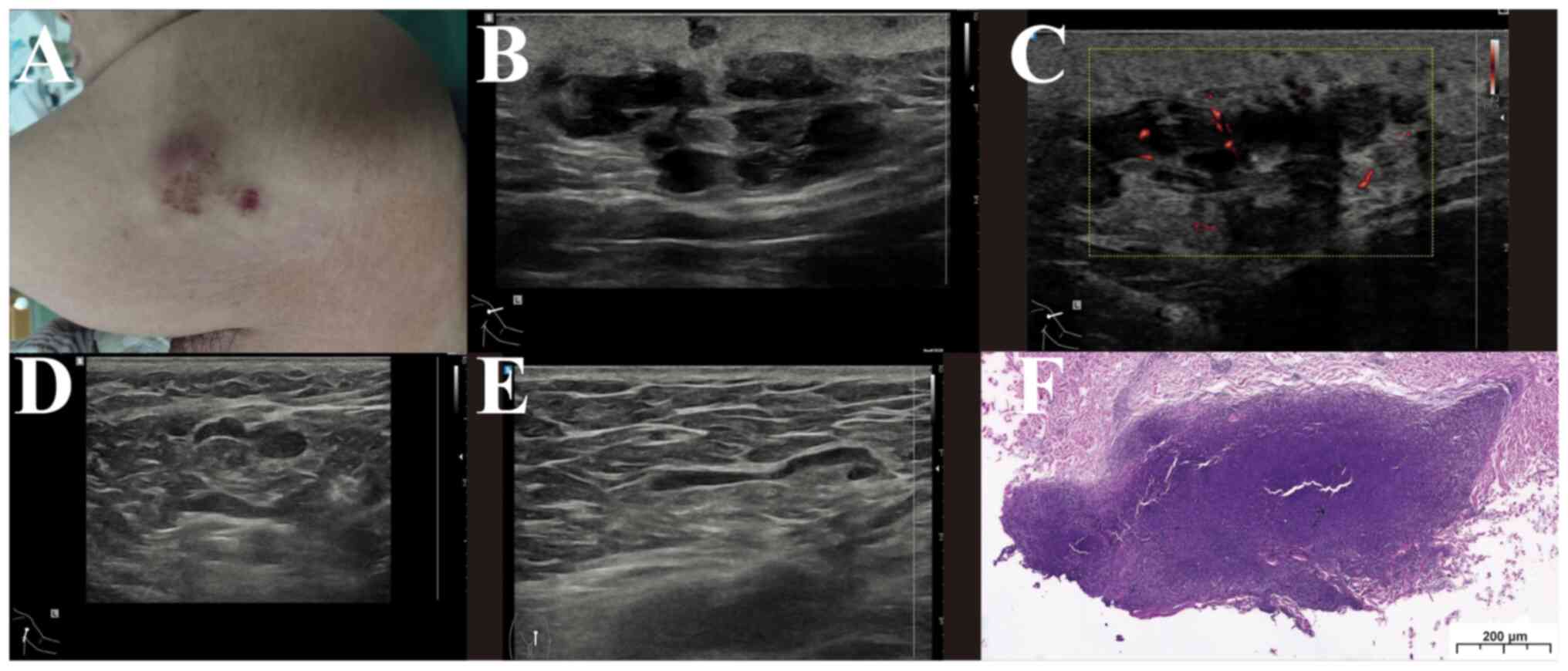
a purplish-red nodule of the skin of the left upper arm that was
hard and measured ~4×3 cm, with poor mobility and no obvious
redness, swelling or pressure pain (Fig. 1A). The superficial lymph nodes were
not palpably enlarged. Ultrasound revealed a subcutaneous
hypoechoic mass in the left upper arm, measuring ~4.0×1.6×3.7 cm,
with poorly defined borders, irregular morphology, heterogeneous
internal echogenicity, grid-like internal changes, unclear
demarcation from the skin and a longitudinal to transverse ratio of
<1 (Fig. 1B). A rich blood flow
signal was seen within the mass on color Doppler flow imaging
(CDFI) (Fig. 1C); lymph nodes were
detected bilaterally in the axillary and inguinal areas with clear
borders, acceptable morphology, clear corticomedullary demarcation
and a gated blood flow signal (Fig. 1D
and E). The diagnosis from the ultrasound was a subcutaneous
solid mass on the left upper arm, with possible malignancy.
Bilateral axillary and inguinal lymph nodes exhibited no obvious
swelling. Positron emission tomography-computed tomography (PET-CT)
showed an irregular mass-like soft-tissue density shadow with
abnormally high fluorodeoxyglucose uptake, a maximum standardized
uptake value of 12.3, a size of ~4.0×1.9 cm and poor demarcation
from the adjacent skin in the subcutaneous area of the left lateral
upper arm. The mass was considered to be a lymphoma. Blood
biochemistry indicated lactate dehydrogenase levels within the
normal range.
A skin biopsy was performed. Tissues were fixed in
4% neutral buffer formaldehyde solution at room temperature for 24
h, and then sectioned to a thickness of 3 µm. The sections were
stained with hematoxylin and eosin at room temperature for 3 min
and then observed using an optical microscope. The results showed a
partial proliferation of oversized sheets of heterogeneous lymphoid
cells in the dermis of the skin tissue (Fig. 1F). Incubation with primary
antibodies was performed at 37°C for 8 min. Horseradish
peroxidase-conjugated secondary antibodies (ultraView Universal
HRP; cat. no. SN 2884795; 1:500; Roche Diagnostics) were added and
incubated at 37°C for 8 min. All antibodies were supplied by
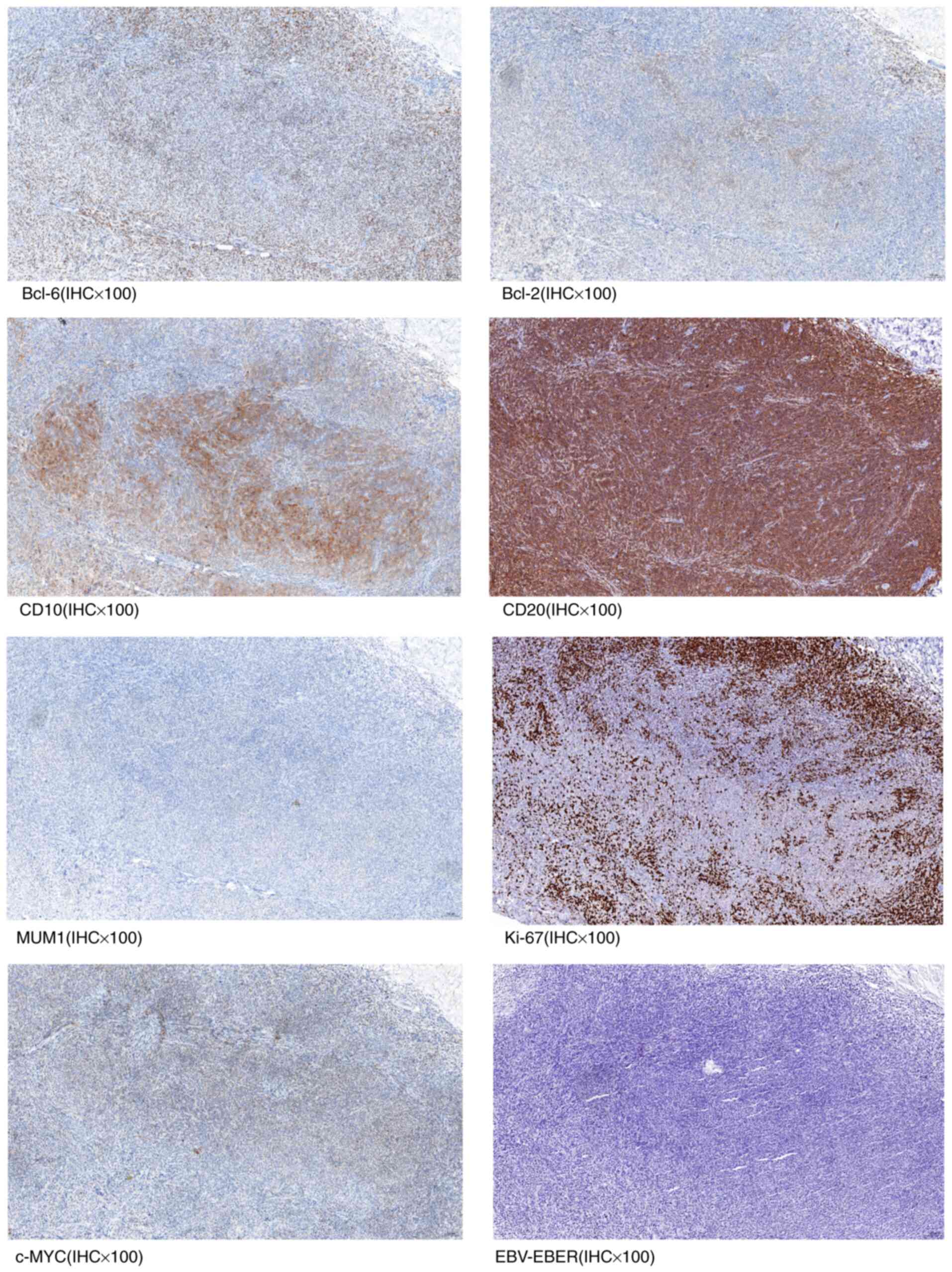
Beijing Zhongshan Jinqiao Biotechnology Co., Ltd. A pathological
slide scanner was used to obtain the following immunohistochemistry
results: Bcl-6(+) (cat. no. ZM-0011; clone LN22), Bcl-2(some weak
+) (cat. no. ZA-0536; clone OTIR1H2), CD10(+) (cat. no. ZM-0283;
clone UMAB235), CD20(+) (cat. no. ZM-0039; clone L26;), MUM1(−)
(cat. no. ZA-0583; clone OTIR1D1O), Ki-67(hotspot area 60% +) (cat.
no. ZM-0166; clone UMAB107) and c-MYC(30% weak +) (cat. no.
ZA-0555; clone EP121) (Fig. 2).
Using in situ hybridization, the sample was determined to be
negative for Epstein-Barr virus-encoded small RNAs (EBER) (Fig. 2). The EBER in situ
hybridization kit (cat. no. ISH-7001UM) was developed by Beijing
Zhongshan Jinqiao Biotechnology Co., Ltd. The EBER probe is a
single-stranded DNA probe that binds specifically to EBER
sequences, and can detect EBER1 and EBER2 at the same time. The
detection method for gene rearrangement was PCR combined with
fragment analysis. Detection was performed using the InVivoScribe
Lymphocyte Gene Rearrangement Detection Kit (InVivoScribe Co.,
Ltd.). Analysis was performed using the ABI3500 Dx Genetic Analyzer
(Thermo Fisher Scientific, Inc.). Positive gene rearrangement was
detected in B lymphocytes. Immunoglobulin gene rearrangement
testing revealed positivity for IGK (VK-JK) (cat. no. 4-088-0370)
and IGK (VK-Kde+intron-Kde) (cat. no. 4-088-0370), and negativity
for IGH (Fr1-JH, cat. no. 4-088-1750; Fr2-JH, cat. no. 4-088-1750;
and Fr3-JH, cat. no. 4-088-1090) and IGL (cat. no. 4-088-0550).
Combined with the immunohistochemistry and immunoglobulin molecular
rearrangement results, the lesion was consistent with PCFCL. A bone
marrow aspiration biopsy showed hypoproliferative subcortical
myelopoiesis (<5%) and no lymphoma involvement.
Ultrasound-guided puncture biopsy of the bilateral axillary and
inguinal lymph nodes showed that the samples were consistent with
reactive hyperplasia of lymphoid tissues, and no lymphoma
involvement was observed.
Due to the limited extent of the lesion, the patient
underwent radiotherapy combined with chemoimmunotherapy (700 mg
rituximab on day 0, 1.5 g cyclophosphamide on day 1, 4 mg
vincristine on day 1 and 100 mg prednisone on days 1–5). Low-dose
radiotherapy was administered with a total dose of 30 Gy in 15
fractions (2 Gy per fraction, once daily) and with a treatment
field margin of 1–1.5 cm. Four cycles of R-CVP were completed. At
post-treatment follow-up, the lesion size had reduced to 2×1
cm.
Discussion
PCFCL is predominantly found in male patients. The
age of onset for PCFCL patients is usually middle-age and older
(>50 years), and it is less common for patients to be young
males. The patient in this case was a young male. The lesion in
this case, a slow-growing purplish-red nodule on the patient's left
upper arm, matched the typical clinical presentation of PCFCL.
In the present study, ultrasound of the PCFCL lesion
showed a nodular lesion with extremely low echogenicity and a
raster-like pattern, no necrosis, no calcification, and posterior
unaccompanied acoustic shadows within it. CDFI revealed an abundant
blood supply. The possibility of a lymphatic origin should
therefore be considered (8,9). A diagnosis also requires a combination
of complete histopathological and immunohistochemical analyses
after a skin biopsy to determine the nature of the nodules at the
lesions. This is combined with a thorough general examination,
medical history review, biochemical tests (lactate dehydrogenase),
PET-CT and ultrasound-guided puncture biopsy to rule out
extracutaneous organ and lymph node involvement.
Histologically, the tumors of PCFCL are composed of
large central cells derived from B cells in the germinal center and
include three growth patterns: Follicular, diffuse and mixed.
Immunohistochemistry shows positive CD20 and BCL-6 expression, and
mostly negative results for BCL-2 and MUM1.Rarely, co-expression of
CD10 and BCL-2 indicates a high likelihood of recurrence and poor
prognosis. Co-expression is also strong in an alternative diagnosis
of systemic follicular lymphoma (FL) involving the skin (10). In this case, BCL-2 did not show
strong positive expression, which was consistent with the common
manifestations of PCFCL.
The histopathological and immunohistochemical
phenotypes can be distinguished from FL by the fact that most
PCFCLs do not show the t(14;18) translocation at the BCL-2 locus,
and their immunohistochemistry will be consistent with tumors
derived from germinal center B cells showing BCL-6 positivity, MUM1
negativity and varying degrees of CD10 expression (11,12).
Positive immunoglobulin gene rearrangement results confirm the
clonal nature of the infiltration and also differentiate it from
reactive lymphoid tissue hyperplasia (13). PCFCL also needs to be differentiated
from other subtypes of PCBCL. PCMZL occurs in adolescents (<20
years of age) without a sex preference, and histologically shows
small centrocyte-like or mononuclear lymphocytes surrounding
reactive germinal centers. Immunohistochemistry shows positive
results for CD20, CD79a and BCL-2 expression, and negative results
for CD10 and BCL-6 (14). PCDLBCL
usually shows high expression of BCL-2, MUM1 and MYC (15).
The International Society for Cutaneous Lymphomas
(ISCL) has established a Tumor-Node-Metastasis staging system
(16) for primary cutaneous
lymphomas, which is based on the number and extent of cutaneous
lesions, lymph node involvement and the presence or absence of
organ involvement, and can effectively assess the patient's
prognosis and formulate the correct treatment plan (16). Since PCFCL is an inert lymphoma and
low-dose radiotherapy reduces toxicity to achieve better outcomes
with remission rates approaching 100%, localized low-dose
radiotherapy is recommended for patients with single lesions or
single irradiated fields. Patients with multiple localized lesions
can be treated with multiple fields and patients with generalized
lesions can be treated extensively with systemic rituximab.
Although the complete remission rate of surgical resection is also
close to 100% for the treatment of small lesions, its recurrence
rate is high compared with low-dose radiotherapy (11,17,18).
In a retrospective study, Wang et al (19) reported a complete remission rate of
100% and a relapse rate of 20% in patients with PCFCL treated with
chemoimmunotherapy (R-CVP). Rituximab-based chemoimmunotherapy is
highly effective in treating and preventing relapse. In this case,
the patient received low-dose radiation therapy combined with
chemoimmunotherapy to prolong clinical remission and reduce
recurrence risk. In conclusion, PCFCL is relatively rare among
young individuals. The current report presents the case of a
32-year-old male patient with PCFCL, including its diagnosis and
treatment, with the intention to inform the diagnosis and treatment
of the disease.
Acknowledgements
Not applicable.
Funding
Funding: No funding was received.
Availability of data and materials
The data generated in the present study may be
requested from the corresponding author.
Authors' contributions
XY was responsible for data collection and writing
the manuscript. YW and YC were responsible for collecting images
and data. XC made substantial contributions to conception and
design, revising the manuscript critically for important
intellectual content. All authors have read and approved the
manuscript. XY and XC confirm the authenticity of all the raw
data.
Ethics approval and consent to
participate
The studies involving human subjects were reviewed
and approved by the Ethics Committee of the Affiliated Yantai
Yuhuanding Hospital of Qingdao University (Yantai, China; approval
no. 2025-616).
Patient consent for publication
The patient provided written informed consent for
publication.
Competing interests
The authors declare that they have no competing
interests.
References
|
1
|
Kim MJ, Hong ME, Maeng CH, Jung HA, Hong
JY, Choi MK, Kim SJ, Ko YH and Kim WS: Clinical features and
treatment outcomes of primary cutaneous B-cell lymphoma: A
single-center analysis in South Korea. Int J Hematol. 101:273–278.
2015. View Article : Google Scholar : PubMed/NCBI
|
|
2
|
Swerdlow SH, Campo E, Pileri SA, Harris
NA, Stein H, Siebert R, Advani R, Ghielmini M, Salles GA, Zelenetz
AD and Jaffe E: The 2016 revision of the World Health Organization
classification of lymphoid neoplasms. Blood. 127:2375–2390. 2016.
View Article : Google Scholar : PubMed/NCBI
|
|
3
|
Suárez AL, Pulitzer M, Horwitz S,
Moskowitz A, Querfeld C and Myskowski PL: Primary cutaneous B-cell
lymphomas: Part I. Clinical features, diagnosis, and
classification. J Am Acad Dermatol. 69:329.e1–e13. 341–342. 2023.
View Article : Google Scholar : PubMed/NCBI
|
|
4
|
Korgavkar K and Weinstock MA: Changing
incidence trends of cutaneous B-cell lymphoma. J Invest Dermatol.
134:840–842. 2014. View Article : Google Scholar : PubMed/NCBI
|
|
5
|
Willemze R, Cerroni L, Kempf W, Berti E,
Facchetti F, Swerdlow SH and Jaffe ES: The 2018 update of the
WHO-EORTC classification for primary cutaneous lymphomas. Blood.
133:1703–1714. 2019. View Article : Google Scholar : PubMed/NCBI
|
|
6
|
Correia E, Cha J, Krishnasamy S, Donnell
MO, 'Shi W, Porcu P and Nikbakht N: A case series of primary
cutaneous B-cell lymphomas with atypical presentations: Diagnostic
and therapeutic challenges. Haematologica. 107:1014–1016. 2022.
View Article : Google Scholar : PubMed/NCBI
|
|
7
|
Niu WY, Yan XS, Qiao H, Sun YJ, Gu HY, Li
GL, Cui ZG and Du J: An adolescent with primary cutaneous follicle
center lymphoma: A case report and literature review. Front Oncol.
13:12737192023. View Article : Google Scholar : PubMed/NCBI
|
|
8
|
Wu X and Lei L: Ultrasonic manifestations
of primary axillary cutaneous follicular central lymphoma in the
case. Chin J Ultras Med. 38:4692022.(In Chinese).
|
|
9
|
Mandava A, Koppula V, Wortsman X, Catalano
O and Alfageme F: The clinical value of imaging in primary
cutaneous lymphomas: Role of high resolution ultrasound and PET-CT.
Brit J Radiol. 92:201809042019. View Article : Google Scholar : PubMed/NCBI
|
|
10
|
Pham-Ledard A, Cowppli-Bony A, Doussau A,
Prochazkova-Carlotti M, Laharanne E, Jouary T, Belaud-Rotureau MA,
Vergier B, Merlio JP and Beylot-Barry M: Diagnostic and prognostic
value of BCL2 rearrangement in 53 patients with follicular lymphoma
presenting as primary skin lesions. Am J Clin Pathol. 143:362–373.
2015. View Article : Google Scholar : PubMed/NCBI
|
|
11
|
Hristov AC, Tejasvi T and Wilcox RA:
Cutaneous B-cell lymphomas: 2023 Update on diagnosis,
risk-stratification, and management. Am J Hematol. 98:1326–1332.
2023. View Article : Google Scholar : PubMed/NCBI
|
|
12
|
Saksena A, Jain A, Pack SD, Kim J, Lee I,
Tyagi M, Xi L, Pittaluga S, Raffeld M and Jaffe ES: Follicle center
lymphoma (FCL) of the lower female genital tract (LFGT): A novel
variant of primary cutaneous follicle center lymphoma (PCFCL). Am J
Surg Pathol. 47:409–419. 2023. View Article : Google Scholar : PubMed/NCBI
|
|
13
|
Al Harbi SM, Al Natour S, Al Saif NM, Al
Saif N and Al Bayat MI: Primary cutaneous follicle center lymphoma
presenting as a solitary nodule on the forearm of an adolescent
girl: A case report and literature review. Clin Cosmet Investig
Dermatol. 16:167–172. 2023. View Article : Google Scholar : PubMed/NCBI
|
|
14
|
Ceppi F, Pope E, Ngan B and Abla O:
Primary cutaneous lymphomas in children and adolescents. Pediatr
Blood Cancer. 63:1886–1894. 2016. View Article : Google Scholar : PubMed/NCBI
|
|
15
|
Uccella S, Goteri G, Maiorana A, Donati V,
Tibiletti MG, Magnoli F, Facchi S, Merchiori D, Morsia E, Papotti
R, et al: Clinicopathological, cytogenetic, and molecular profiles
of primary cutaneous diffuse large B-cell lymphomas. Hum Pathol.
136:44–55. 2023. View Article : Google Scholar : PubMed/NCBI
|
|
16
|
Olsen EA: Evaluation, diagnosis, and
staging of cutaneous lymphoma. Dermatol Clin. 33:643–654. 2015.
View Article : Google Scholar : PubMed/NCBI
|
|
17
|
Vitiello P, Sica A, Ronchi A, Caccavale S,
Franco R and Argenziano G: Primary cutaneous B-cell lymphomas: An
update. Front Oncol. 10:6512020. View Article : Google Scholar : PubMed/NCBI
|
|
18
|
Specht L, Dabaja B, Illidge T, Wilson LD
and Hoppe RT; International Lymphoma Radiation Oncology Group, :
Modern radiation therapy for primary cutaneous lymphomas: Field and
dose guidelines from the international lymphoma radiation oncology
Group. Int J Radiat Oncol Biol Phys. 92:32–39. 2015. View Article : Google Scholar : PubMed/NCBI
|
|
19
|
Wang S, Perlmutter JW, Johnston J, Nugent
Z and Wiseman M: Rituximab treatment of primary cutaneous follicle
center lymphoma: A retrospective review. J Cutan Med Surg.
26:604–612. 2022. View Article : Google Scholar : PubMed/NCBI
|