Spandidos Publications Logo
  • About
    • About Spandidos
    • Aims and Scopes
    • Abstracting and Indexing
    • Editorial Policies
    • Reprints and Permissions
    • Job Opportunities
    • Terms and Conditions
    • Contact
  • Journals
    • All Journals
    • Oncology Letters
      • Oncology Letters
      • Information for Authors
      • Editorial Policies
      • Editorial Board
      • Aims and Scope
      • Abstracting and Indexing
      • Bibliographic Information
      • Archive
    • International Journal of Oncology
      • International Journal of Oncology
      • Information for Authors
      • Editorial Policies
      • Editorial Board
      • Aims and Scope
      • Abstracting and Indexing
      • Bibliographic Information
      • Archive
    • Molecular and Clinical Oncology
      • Molecular and Clinical Oncology
      • Information for Authors
      • Editorial Policies
      • Editorial Board
      • Aims and Scope
      • Abstracting and Indexing
      • Bibliographic Information
      • Archive
    • Experimental and Therapeutic Medicine
      • Experimental and Therapeutic Medicine
      • Information for Authors
      • Editorial Policies
      • Editorial Board
      • Aims and Scope
      • Abstracting and Indexing
      • Bibliographic Information
      • Archive
    • International Journal of Molecular Medicine
      • International Journal of Molecular Medicine
      • Information for Authors
      • Editorial Policies
      • Editorial Board
      • Aims and Scope
      • Abstracting and Indexing
      • Bibliographic Information
      • Archive
    • Biomedical Reports
      • Biomedical Reports
      • Information for Authors
      • Editorial Policies
      • Editorial Board
      • Aims and Scope
      • Abstracting and Indexing
      • Bibliographic Information
      • Archive
    • Oncology Reports
      • Oncology Reports
      • Information for Authors
      • Editorial Policies
      • Editorial Board
      • Aims and Scope
      • Abstracting and Indexing
      • Bibliographic Information
      • Archive
    • Molecular Medicine Reports
      • Molecular Medicine Reports
      • Information for Authors
      • Editorial Policies
      • Editorial Board
      • Aims and Scope
      • Abstracting and Indexing
      • Bibliographic Information
      • Archive
    • World Academy of Sciences Journal
      • World Academy of Sciences Journal
      • Information for Authors
      • Editorial Policies
      • Editorial Board
      • Aims and Scope
      • Abstracting and Indexing
      • Bibliographic Information
      • Archive
    • International Journal of Functional Nutrition
      • International Journal of Functional Nutrition
      • Information for Authors
      • Editorial Policies
      • Editorial Board
      • Aims and Scope
      • Abstracting and Indexing
      • Bibliographic Information
      • Archive
    • International Journal of Epigenetics
      • International Journal of Epigenetics
      • Information for Authors
      • Editorial Policies
      • Editorial Board
      • Aims and Scope
      • Abstracting and Indexing
      • Bibliographic Information
      • Archive
    • Medicine International
      • Medicine International
      • Information for Authors
      • Editorial Policies
      • Editorial Board
      • Aims and Scope
      • Abstracting and Indexing
      • Bibliographic Information
      • Archive
  • Articles
  • Information
    • Information for Authors
    • Information for Reviewers
    • Information for Librarians
    • Information for Advertisers
    • Conferences
  • Language Editing
Spandidos Publications Logo
  • About
    • About Spandidos
    • Aims and Scopes
    • Abstracting and Indexing
    • Editorial Policies
    • Reprints and Permissions
    • Job Opportunities
    • Terms and Conditions
    • Contact
  • Journals
    • All Journals
    • Biomedical Reports
      • Information for Authors
      • Editorial Policies
      • Editorial Board
      • Aims and Scope
      • Abstracting and Indexing
      • Bibliographic Information
      • Archive
    • Experimental and Therapeutic Medicine
      • Information for Authors
      • Editorial Policies
      • Editorial Board
      • Aims and Scope
      • Abstracting and Indexing
      • Bibliographic Information
      • Archive
    • International Journal of Epigenetics
      • Information for Authors
      • Editorial Policies
      • Editorial Board
      • Aims and Scope
      • Abstracting and Indexing
      • Bibliographic Information
      • Archive
    • International Journal of Functional Nutrition
      • Information for Authors
      • Editorial Policies
      • Editorial Board
      • Aims and Scope
      • Abstracting and Indexing
      • Bibliographic Information
      • Archive
    • International Journal of Molecular Medicine
      • Information for Authors
      • Editorial Policies
      • Editorial Board
      • Aims and Scope
      • Abstracting and Indexing
      • Bibliographic Information
      • Archive
    • International Journal of Oncology
      • Information for Authors
      • Editorial Policies
      • Editorial Board
      • Aims and Scope
      • Abstracting and Indexing
      • Bibliographic Information
      • Archive
    • Medicine International
      • Information for Authors
      • Editorial Policies
      • Editorial Board
      • Aims and Scope
      • Abstracting and Indexing
      • Bibliographic Information
      • Archive
    • Molecular and Clinical Oncology
      • Information for Authors
      • Editorial Policies
      • Editorial Board
      • Aims and Scope
      • Abstracting and Indexing
      • Bibliographic Information
      • Archive
    • Molecular Medicine Reports
      • Information for Authors
      • Editorial Policies
      • Editorial Board
      • Aims and Scope
      • Abstracting and Indexing
      • Bibliographic Information
      • Archive
    • Oncology Letters
      • Information for Authors
      • Editorial Policies
      • Editorial Board
      • Aims and Scope
      • Abstracting and Indexing
      • Bibliographic Information
      • Archive
    • Oncology Reports
      • Information for Authors
      • Editorial Policies
      • Editorial Board
      • Aims and Scope
      • Abstracting and Indexing
      • Bibliographic Information
      • Archive
    • World Academy of Sciences Journal
      • Information for Authors
      • Editorial Policies
      • Editorial Board
      • Aims and Scope
      • Abstracting and Indexing
      • Bibliographic Information
      • Archive
  • Articles
  • Information
    • For Authors
    • For Reviewers
    • For Librarians
    • For Advertisers
    • Conferences
  • Language Editing
Login Register Submit
  • This site uses cookies
  • You can change your cookie settings at any time by following the instructions in our Cookie Policy. To find out more, you may read our Privacy Policy.

    I agree
Search articles by DOI, keyword, author or affiliation
Search
Advanced Search
presentation
Oncology Letters
Join Editorial Board Propose a Special Issue
Print ISSN: 1792-1074 Online ISSN: 1792-1082
Journal Cover
April-2026 Volume 31 Issue 4

Full Size Image

Sign up for eToc alerts
Recommend to Library

Journals

International Journal of Molecular Medicine

International Journal of Molecular Medicine

International Journal of Molecular Medicine is an international journal devoted to molecular mechanisms of human disease.

International Journal of Oncology

International Journal of Oncology

International Journal of Oncology is an international journal devoted to oncology research and cancer treatment.

Molecular Medicine Reports

Molecular Medicine Reports

Covers molecular medicine topics such as pharmacology, pathology, genetics, neuroscience, infectious diseases, molecular cardiology, and molecular surgery.

Oncology Reports

Oncology Reports

Oncology Reports is an international journal devoted to fundamental and applied research in Oncology.

Experimental and Therapeutic Medicine

Experimental and Therapeutic Medicine

Experimental and Therapeutic Medicine is an international journal devoted to laboratory and clinical medicine.

Oncology Letters

Oncology Letters

Oncology Letters is an international journal devoted to Experimental and Clinical Oncology.

Biomedical Reports

Biomedical Reports

Explores a wide range of biological and medical fields, including pharmacology, genetics, microbiology, neuroscience, and molecular cardiology.

Molecular and Clinical Oncology

Molecular and Clinical Oncology

International journal addressing all aspects of oncology research, from tumorigenesis and oncogenes to chemotherapy and metastasis.

World Academy of Sciences Journal

World Academy of Sciences Journal

Multidisciplinary open-access journal spanning biochemistry, genetics, neuroscience, environmental health, and synthetic biology.

International Journal of Functional Nutrition

International Journal of Functional Nutrition

Open-access journal combining biochemistry, pharmacology, immunology, and genetics to advance health through functional nutrition.

International Journal of Epigenetics

International Journal of Epigenetics

Publishes open-access research on using epigenetics to advance understanding and treatment of human disease.

Medicine International

Medicine International

An International Open Access Journal Devoted to General Medicine.

Journal Cover
April-2026 Volume 31 Issue 4

Full Size Image

Sign up for eToc alerts
Recommend to Library

  • Article
  • Citations
    • Cite This Article
    • Download Citation
    • Create Citation Alert
    • Remove Citation Alert
    • Cited By
  • Similar Articles
    • Related Articles (in Spandidos Publications)
    • Similar Articles (Google Scholar)
    • Similar Articles (PubMed)
  • Download PDF
  • Download XML
  • View XML
Case Report Open Access

Gliosarcoma of the right cerebellar hemisphere and parahippocampal region: A case report and literature review

  • Authors:
    • Fa Wu
    • Yulin Yang
    • Zaiqing Wang
    • Rui Jiang
    • Tingting Wu
    • Ting Yu
    • Peng Wang
  • View Affiliations / Copyright

    Affiliations: Department of Radiology, The General Hospital of Western Theater Command, Chengdu, Sichuan 610083, P.R. China, Department of Ultrasound, Longquanyi District First People's Hospital, Chengdu, Sichuan 610000, P.R. China, Department of Radiology, Chengdu Bayi Orthopedic Hospital, Chengdu, Sichuan 610100, P.R. China, Department of Pathology, The General Hospital of Western Theater Command, Chengdu, Sichuan 610083, P.R. China
    Copyright: © Wu et al. This is an open access article distributed under the terms of Creative Commons Attribution License.
  • Article Number: 147
    |
    Published online on: February 25, 2026
       https://doi.org/10.3892/ol.2026.15500
  • Expand metrics +
Metrics: Total Views: 0 (Spandidos Publications: | PMC Statistics: )
Metrics: Total PDF Downloads: 0 (Spandidos Publications: | PMC Statistics: )
Cited By (CrossRef): 0 citations Loading Articles...

This article is mentioned in:


Abstract

Intracranial gliosarcoma (GSM) is a rare and aggressive variant of glioblastoma, characterized by a dismal prognosis and high rates of early recurrence and metastasis. Preoperative differentiation from other neoplasms based on imaging features remains a significant clinical challenge. The current study reports the case of a 55‑year‑old female who presented with a headache as the primary clinical symptom. The patient underwent brain MRI due to progressively worsening symptoms of diplopia, intermittent dull headaches localized to the right occipital region and an unsteady gait. Preoperative magnetic resonance imaging revealed a large, lobulated mass in the right cerebellar hemisphere extending into the parahippocampal region. The diagnosis of WHO Grade IV GSM was confirmed by postoperative histopathological and immunohistochemical analysis, which revealed a biphasic pattern with glial fibrillary acidic protein‑positive glial and smooth muscle actin/vimentin‑positive sarcomatous components, alongside a high Ki‑67 proliferation index of 30%. The patient underwent a subtotal resection followed by adjuvant radiotherapy with concurrent temozolomide chemotherapy. Despite this multimodal treatment, follow‑up imaging demonstrated tumor recurrence at 2 months, with significant further progression and brain herniation observed at the 12‑month follow‑up. The present case underscores the diagnostic challenges and aggressive clinical course of GSM, particularly when located in the cerebellum. The rapid recurrence despite combined‑modality therapy highlights the need for improved diagnostic strategies and more effective treatment protocols for this formidable disease.

View Figures

Figure 1

Magnetic resonance imaging findings of
gliosarcoma in the right cerebellar hemisphere and right
parahippocampal region. (A) In the right cerebellar hemisphere and
right parahippocampal region, patchy slightly hyperintense and
isointense signals on T1-weighted imaging were observed. (B) On
T2-weighted imaging, slightly hyperintense signals were noted,
mixed with small patchy slightly hypointense signals. (C)
Susceptibility-weighted imaging demonstrates punctate hypointense
foci within the lesion. (D) Fluid-attenuated inversion recovery
imaging shows hyperintensity. The lesions exhibited obvious
inhomogeneous enhancement on (E) axial and (F) coronal images. The
lesion involves the right portion of the brainstem and cerebellar
vermis, with compression of the adjacent fourth ventricle and
brainstem. (G) The peak value of NAA in the lesion area was
decreased, the peak value of Cho was significantly increased, and
the peak value of Cr remained basically unchanged. MRI, magnetic
resonance imaging; NAA, N-acetylaspartate; Cr, creatine; TSE/M,
turbo spin echo/magnetization.

Figure 2

Recurrence of gliosarcoma on 2-month
follow-up magnetic resonance imaging. A lobulated mixed-signal mass
predominantly with (A) slightly long T1 and (B) slightly long T2
signals is present in the right cerebellar hemisphere and right
parahippocampal region. (C) Fluid-attenuated inversion recovery
imaging shows hyperintensity. After enhancement, the (D) axial, (E)
sagittal and (F) coronal images of the lesion exhibited obvious
inhomogeneous enhancement and annular enhancement. The lesion is
growing across the tentorium, with thickening and significant
enhancement of the adjacent right cerebellar tentorium; it involves
the right portion of the brainstem, causing compression of the
adjacent fourth ventricle and brainstem. (G) Magnetic resonance
spectroscopy of the recurrent lesion reveals a decreased NAA peak
and an increased Cho peak in the lesion area. The left panel shows
the voxel placement (yellow grid). The middle and right panels
display the spectrum without pseudo-color mapping, likely due to
extremely low metabolite concentrations, incomplete water
suppression (note the prominent water peak on the right), and/or
post-processing thresholds. NAA, N-acetylaspartate; Cr, creatine;
TSE/M, turbo spin echo/magnetization.

Figure 3

Enlargement of the recurrent
gliosarcoma with brain herniation formation on 1-year follow-up
magnetic resonance imaging. The dura mater in the right occipital
region was thickened with obvious enhancement. The right cerebellar
hemisphere, right parahippocampal region and right temporal lobe
exhibited patchy mixed signal shadows. (A) On the T1WI sequence,
slightly longer T1 signals were observed, and (B) on the T2WI
sequence, slightly longer T2 signals were observed. (C)
Fluid-attenuated inversion recovery imaging shows isointense and
slightly hyperintense signals. After enhancement, the (D) axial,
(E) sagittal and (F) coronal images of the lesion exhibited obvious
inhomogeneous enhancement and annular enhancement. The lesion grows
across the tentorium, with thickening and significant enhancement
of the adjacent right cerebellar tentorium; it involves the right
portion of the brainstem, causing compression of the adjacent
fourth ventricle, right lateral ventricle and brainstem, with
leftward shift of the midline structures. (G) Magnetic resonance
spectroscopy reveals a decreased NAA peak and an increased Cho peak
in the lesion area. NAA, N-acetylaspartate; Cr, creatine; TSE/M,
turbo spin echo/magnetization.

Figure 4

Pathological images of the resected
gliosarcoma. (A) The tumor displays an irregular morphology with a
grey-brown cut surface and firm consistency. (B and C) Hematoxylin
and eosin-stained tissue samples at (B) ×40 and (C) ×100
magnification. The tumor is composed of dual components: Glial and
sarcomatous. The sarcomatous component exhibits a spindle cell
sarcoma pattern, characterized by densely packed long fascicles of
spindle cells arranged in fascicular or small cluster-like
configurations, abundant cytoplasm and prominent stromal vascular
proliferation. (D) Glial fibrillary acidic protein positivity (×200
magnification). (E) Weak positivity for S-100 (×200 magnification).
(F) Smooth muscle actin positivity (×200 magnification). (G)
Vimentin positivity (×200 magnification). (H) Ki-67 positivity
(30%) (×200 magnification). (I) Caldesmon negativity (×200
magnification). (J) CD34 negativity (×200 magnification). (K)
Cytokeratin negativity (×200 magnification). (L) Epithelial
membrane antigen negativity (×200 magnification). (M) Progesterone
receptor negativity (×200 magnification).

Figure 5

Intracranial lesion radiotherapy
treatment plan. (A) Target delineation: The GTVs were defined as
GTV-65 (intracranial lesions outside the brainstem) and GTV-53
(brainstem lesions). The planning target volumes were generated by
expanding the respective GTVs by 0.8 cm, and the brainstem
interface was not expanded. (B) The dose distribution was shown on
the axial computed tomography slices with equal dose, and the total
dose was 6,731.5 cGy. (C) Dose-volume histogram of target volume
and organs at risk; dosage limits for dangerous organs follow the
Radiation Therapy Oncology Group guidelines (18). GTV, gross tumor volume.
View References

1 

Louis DN, Perry A, Reifenberger G, von Deimling A, Figarella-Branger D, Cavenee WK, Ohgaki H, Wiestler OD, Kleihues P and Ellison DW: The 2016 world health organization classification of tumors of the central nervous system: A summary. Acta Neuropathol (Berl). 131:803–820. 2016. View Article : Google Scholar

2 

Louis DN, Perry A, Wesseling P, Brat DJ, Cree IA, Figarella-Branger D, Hawkins C, Ng HK, Pfister SM, Reifenberger G, et al: The 2021 WHO classification of tumors of the central nervous system: A summary. Neuro Oncol. 23:1231–1251. 2021. View Article : Google Scholar

3 

Gerges C, Elder T, Penuela M, Rossetti N, Maynard M, Jeong S, Wright CH, Wright J, Zhou X, Burant C, et al: Comparative epidemiology of gliosarcoma and glioblastoma and the impact of Race on overall survival: A systematic literature review. Clin Neurol Neurosurg. 195:1060542020. View Article : Google Scholar : PubMed/NCBI

4 

Chen L, Rizk E, Sherief M, Chang M, Lucas CH, Bettegowda C, Croog V, Mukherjee D, Rincon-Torroella J, Kamson DO, et al: Molecular characterization of gliosarcoma reveals prognostic biomarkers and clinical parallels with glioblastoma. J Neurooncol. 171:403–411. 2024. View Article : Google Scholar

5 

Karasev S, Talybov R, Chertoyev S, Trofimova T, Mochalov V, Kleshchevnikova T, Loginova N and Karaseva I: Diagnostic challenges of gliosarcoma: Case report of a rare glioblastoma histopathological variant. Front Radiol. 5:16874012025. View Article : Google Scholar

6 

Wang X, Jiang J, Liu M and You C: Treatments of gliosarcoma of the brain: A systematic review and meta-analysis. Acta Neurol Belg. 121:1789–1797. 2020. View Article : Google Scholar : PubMed/NCBI

7 

La Torre D, Della Torre A, Lo Turco E, Longo P, Pugliese D, Lacroce P, Raudino G, Romano A, Lavano A and Tomasello F: Primary intracranial gliosarcoma: Is it really a variant of glioblastoma? an update of the clinical, radiological, and biomolecular characteristics. J Clin Med. 13:832024. View Article : Google Scholar

8 

Hashmi FA, Salim A, Shamim MS and Bari ME: Biological characteristics and outcomes of gliosarcoma. J Pak Med Assoc. 68:1273–1275. 2018.

9 

Feng SS, Li HB, Fan F, Li J, Cao H, Xia ZW, Yang K, Zhu XS, Cheng TT and Cheng Q: Clinical characteristics and disease-specific prognostic nomogram for primary gliosarcoma: A SEER population-based analysis. Sci Rep. 9:107442019. View Article : Google Scholar : PubMed/NCBI

10 

Saki A, Faghihi U and Balde I: Differentiating gliosarcoma from glioblastoma: A novel approach using PEACE and XGBoost to deal with datasets with ultra-high dimensional confounders. Life (Basel). 14:8822024.PubMed/NCBI

11 

Wang Y and Zhang Z: A case report: Gliosarcoma associated with a germline heterozygous mutation in MSH2. Front Neurol. 15:13882632024. View Article : Google Scholar

12 

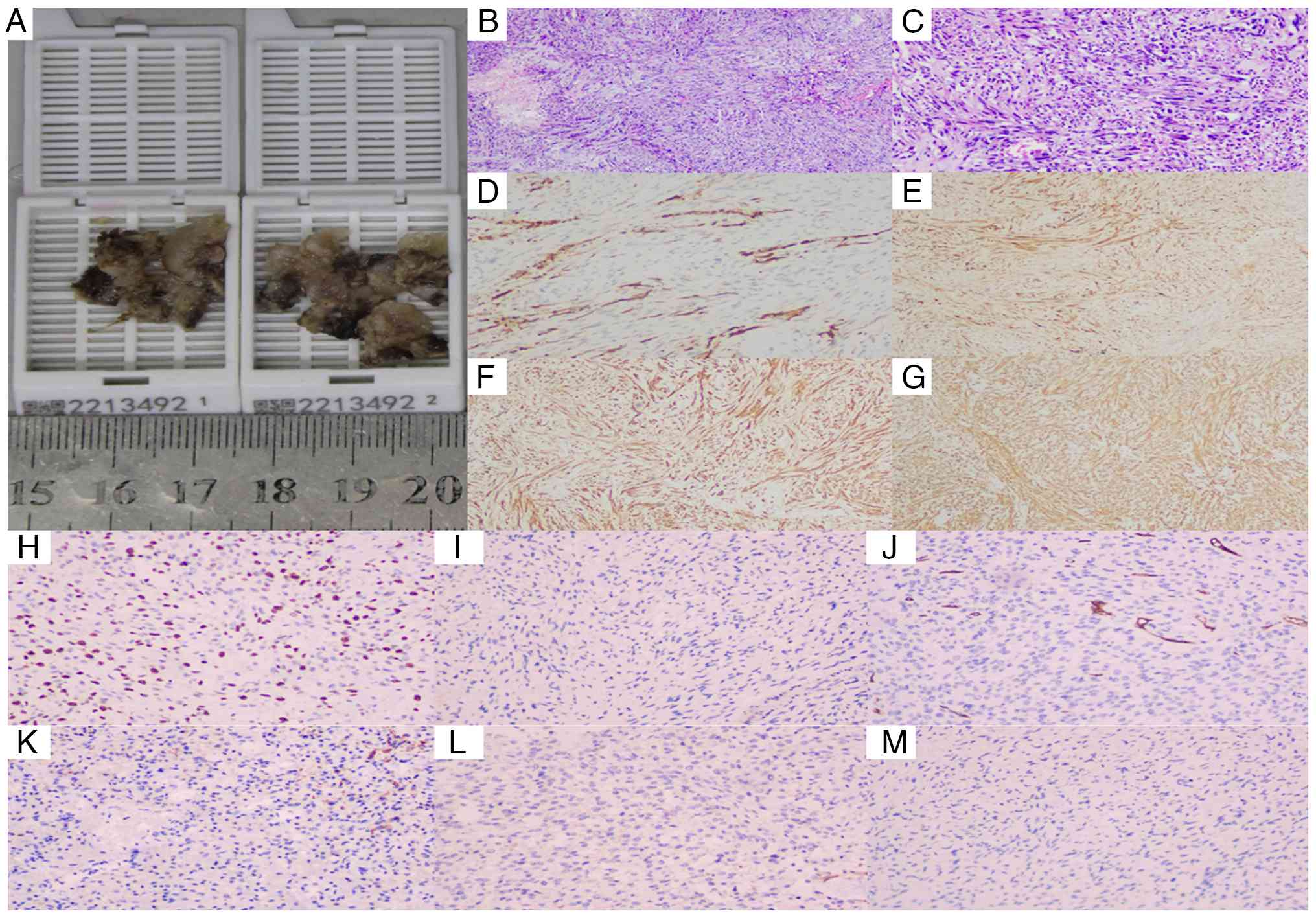
Jiang NN, Larrazabal R, Alsunbul W and Lu JQ: Multiple pathological components in gliosarcoma. J Biomed Res. 35:408–410. 2020. View Article : Google Scholar : PubMed/NCBI

13 

Castelli J, Feuvret L, Haoming QC, Biau J, Jouglar E, Berger A, Truc G, Gutierrez FL, Morandi X, Le Reste PJ, et al: Prognostic and therapeutic factors of gliosarcoma from a multi-institutional series. J Neurooncol. 129:85–92. 2016. View Article : Google Scholar

14 

Adeberg S, Bernhardt D, Harrabi SB, Diehl C, Koelsche C, Rieken S, Unterberg A, von Deimling A and Debus J: Radiotherapy plus concomitant temozolomide in primary gliosarcoma. J Neurooncol. 128:341–348. 2016. View Article : Google Scholar

15 

Xu L, Yang Z, Chen H, Sun C, Tu C, Gu Z and Luo M: Conditional survival and changing risk profile in patients with gliosarcoma. Front Med (Lausanne). 11:14431572024. View Article : Google Scholar : PubMed/NCBI

16 

Martinez A, Binks S, Pumarola M, Hardas A, Easton A, Campo L, Browne M, Martins S, Garosi LS, Di Dona F and Tauro A: Gliosarcoma associated with bilateral hippocampal sclerosis in a cat presenting complex partial seizures with orofacial involvement: A case report. Clin Case Rep. 12:e91232024. View Article : Google Scholar : PubMed/NCBI

17 

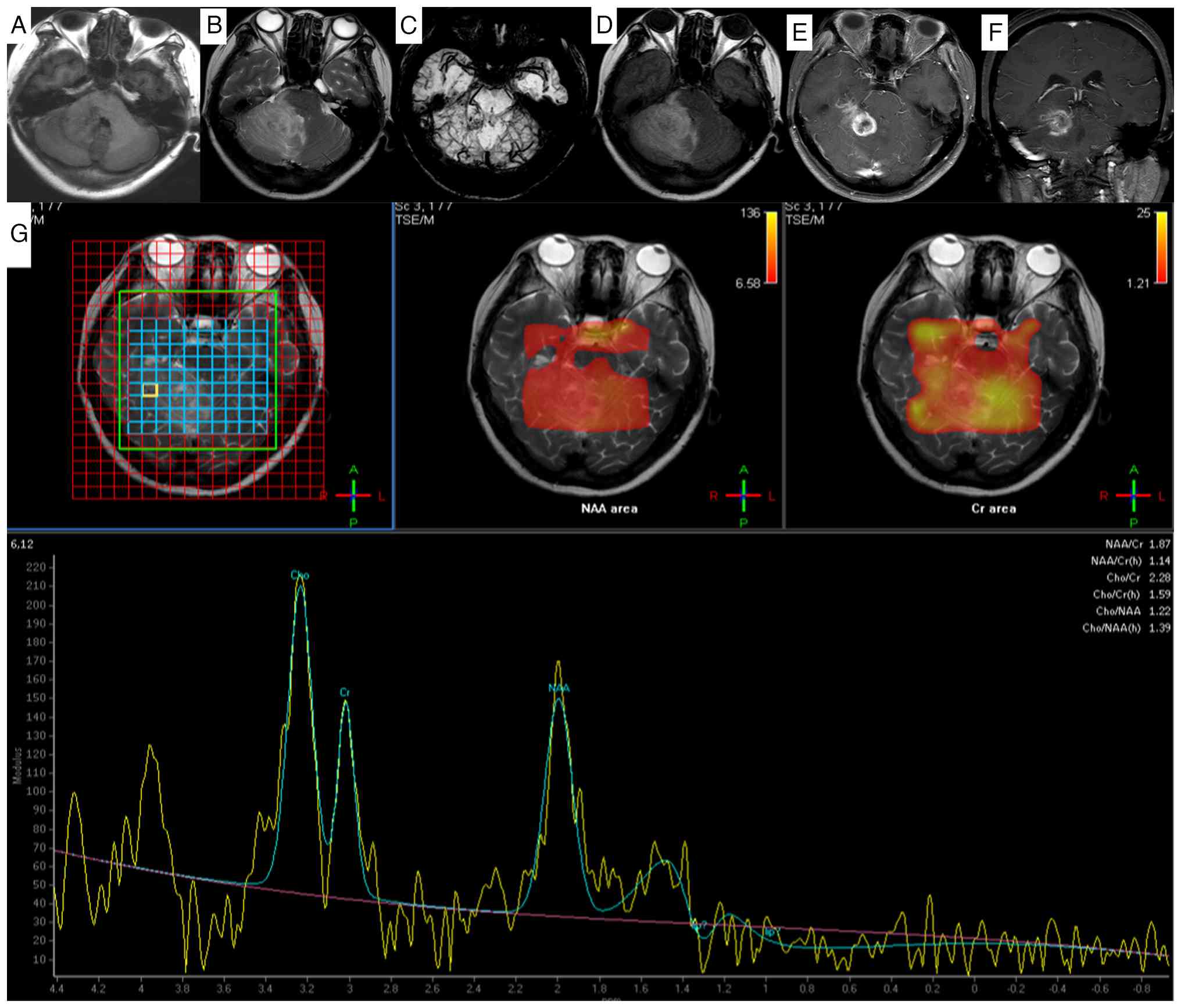
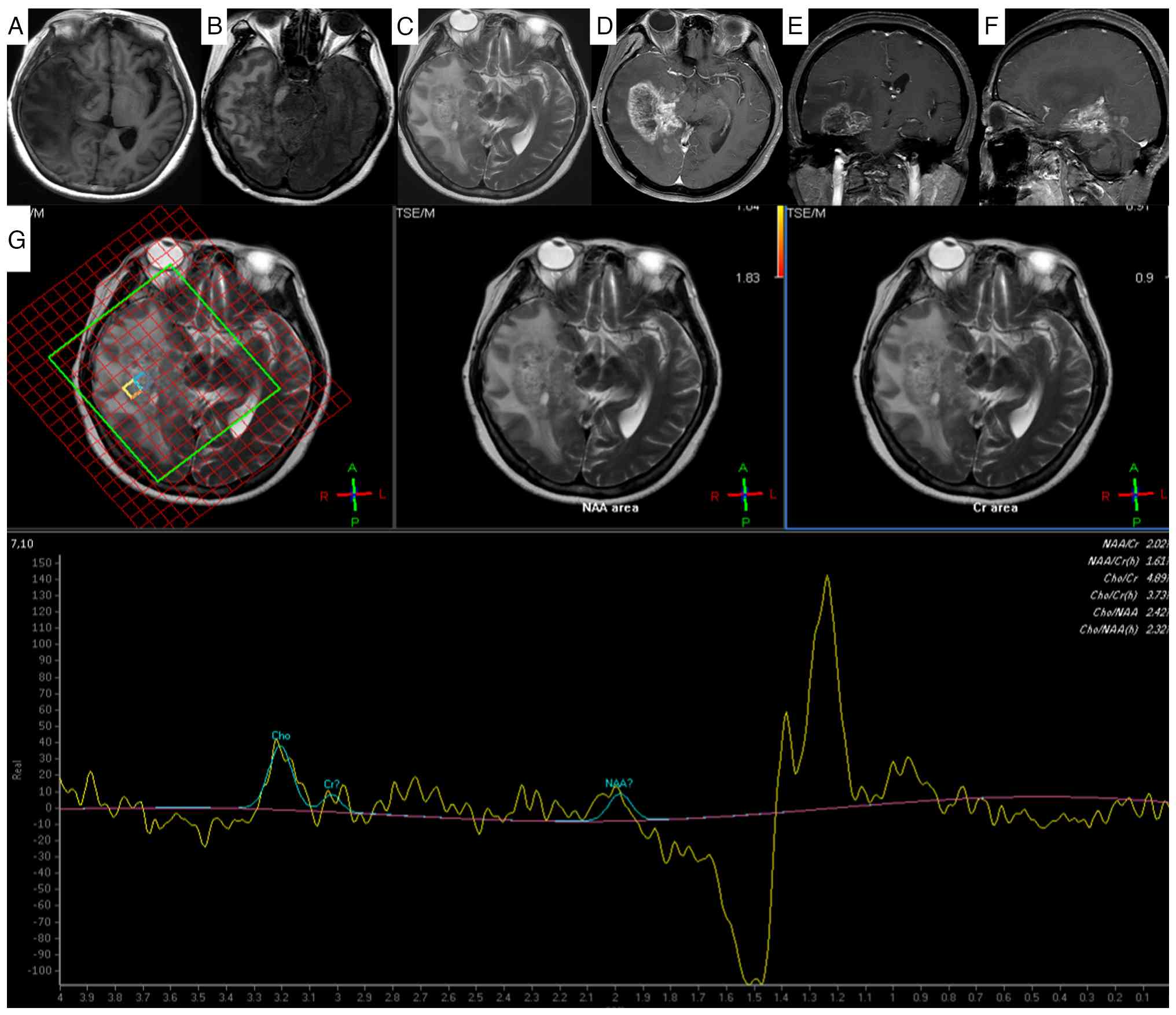
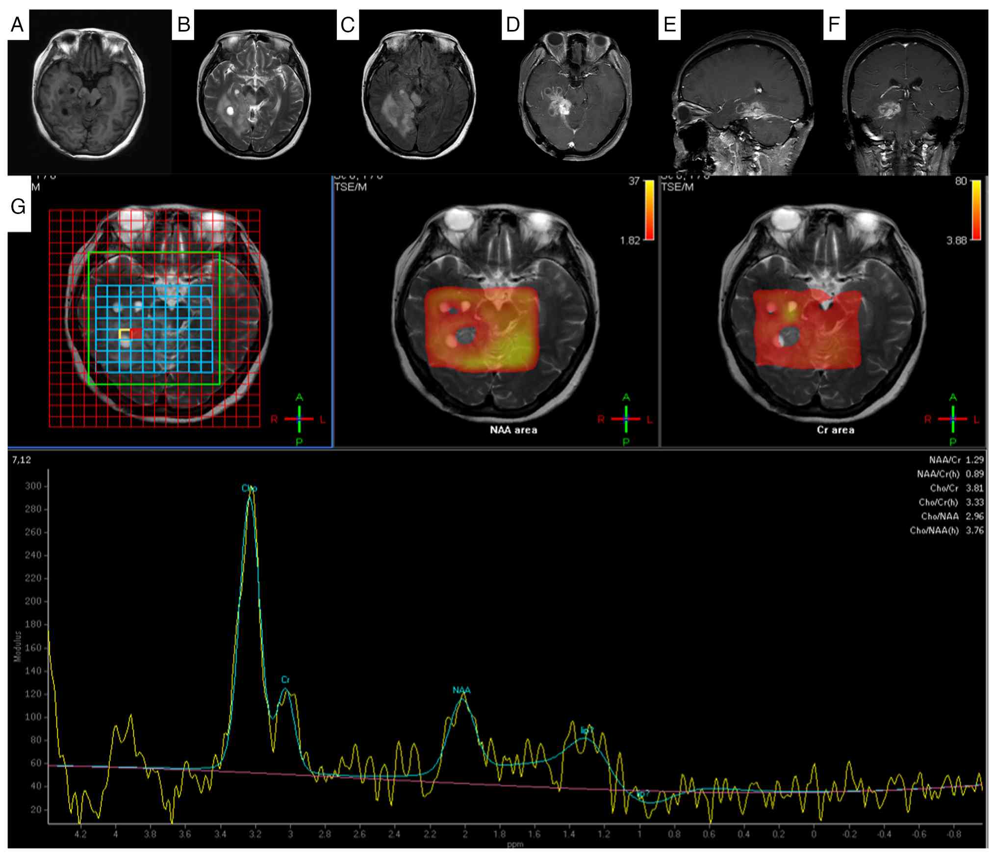
Fukada A, de Souza Queiroz L and Reis F: Gliosarcomas: Magnetic resonance imaging findings. Arq Neuropsiquiatr. 78:112–120. 2020. View Article : Google Scholar : PubMed/NCBI

18 

Gondi V, Bauman G, Bradfield L, Burri SH, Cabrera AR, Cunningham DA, Eaton BR, Hattangadi-Gluth JA, Kim MM, Kotecha R, et al: Radiation therapy for brain metastases: An ASTRO clinical practice guideline. Pract Radiat Oncol. 12:265–282. 2022. View Article : Google Scholar

Related Articles

  • Abstract
  • View
  • Download
  • Twitter
Copy and paste a formatted citation
Spandidos Publications style
Wu F, Yang Y, Wang Z, Jiang R, Wu T, Yu T and Wang P: Gliosarcoma of the right cerebellar hemisphere and parahippocampal region: A case report and literature review. Oncol Lett 31: 147, 2026.
APA
Wu, F., Yang, Y., Wang, Z., Jiang, R., Wu, T., Yu, T., & Wang, P. (2026). Gliosarcoma of the right cerebellar hemisphere and parahippocampal region: A case report and literature review. Oncology Letters, 31, 147. https://doi.org/10.3892/ol.2026.15500
MLA
Wu, F., Yang, Y., Wang, Z., Jiang, R., Wu, T., Yu, T., Wang, P."Gliosarcoma of the right cerebellar hemisphere and parahippocampal region: A case report and literature review". Oncology Letters 31.4 (2026): 147.
Chicago
Wu, F., Yang, Y., Wang, Z., Jiang, R., Wu, T., Yu, T., Wang, P."Gliosarcoma of the right cerebellar hemisphere and parahippocampal region: A case report and literature review". Oncology Letters 31, no. 4 (2026): 147. https://doi.org/10.3892/ol.2026.15500
Copy and paste a formatted citation
x
Spandidos Publications style
Wu F, Yang Y, Wang Z, Jiang R, Wu T, Yu T and Wang P: Gliosarcoma of the right cerebellar hemisphere and parahippocampal region: A case report and literature review. Oncol Lett 31: 147, 2026.
APA
Wu, F., Yang, Y., Wang, Z., Jiang, R., Wu, T., Yu, T., & Wang, P. (2026). Gliosarcoma of the right cerebellar hemisphere and parahippocampal region: A case report and literature review. Oncology Letters, 31, 147. https://doi.org/10.3892/ol.2026.15500
MLA
Wu, F., Yang, Y., Wang, Z., Jiang, R., Wu, T., Yu, T., Wang, P."Gliosarcoma of the right cerebellar hemisphere and parahippocampal region: A case report and literature review". Oncology Letters 31.4 (2026): 147.
Chicago
Wu, F., Yang, Y., Wang, Z., Jiang, R., Wu, T., Yu, T., Wang, P."Gliosarcoma of the right cerebellar hemisphere and parahippocampal region: A case report and literature review". Oncology Letters 31, no. 4 (2026): 147. https://doi.org/10.3892/ol.2026.15500
Follow us
  • Twitter
  • LinkedIn
  • Facebook
About
  • Spandidos Publications
  • Careers
  • Cookie Policy
  • Privacy Policy
How can we help?
  • Help
  • Live Chat
  • Contact
  • Email to our Support Team