Lung cancer is one of the most common causes of
cancer-related mortality worldwide, accounting for 18.7% of all
cancer fatalities (1). The
incidence of lung cancer in China has shown a persistent upward
trend since 2016 (2). Despite
advancements in early detection and standardized treatment
protocols for patients with lung cancer, the majority of cases are
still diagnosed at advanced stages with poor prognosis (3,4).
Histologically, lung cancer is categorized into two main types:
Non-small cell lung cancer (NSCLC; 85%) and small cell lung cancer
(15%) (1). One study found that
among patients with epidermal growth factor receptor (EGFR)-mutated
NSCLC treated with EGFR tyrosine kinase inhibitors, after
stratifying patients into risk groups based on a scoring system
incorporating age, performance status, metastatic sites (liver,
brain, bone, pleura), and disease control rate, the 5-year survival
rate in the low-risk group was 23.4% (5). NSCLC is divided into two principal
histological subtypes: Lung squamous cell carcinoma and lung
adenocarcinoma (LUAD) (6).
Currently, the mechanisms of drug action against NSCLC include
promoting apoptosis (7) and
inducing cellular autophagy in tumor cells (8). However, early-stage NSCLC is typically
asymptomatic and often diagnosed at advanced stages, resulting in
more challenging treatment processes with a median survival period
of <1 year (9). Therefore,
establishing in vitro biological models that are rapid and
efficient, relatively low-cost, operationally simple, and capable
of accurately replicating both the genetic characteristics of in
vivo tumors and their responses to therapeutic agents holds notable
importance for oncology research.
Three-dimensional (3D) culture is recognized as the
most promising in vitro model for oncological drug testing, as it
can recapitulate key in vivo tumor features, including the cellular
heterogeneity, intercellular signaling, structural architecture,
extracellular microenvironment, growth kinetics, gene expression
profiles and drug resistance mechanisms (10,11).
Spheroids represent one of the simplest and most effective 3D
culture models, and are formed through the self-assembly of
multiple cell types (e.g., tumor cells, stromal fibroblasts and
immune cells) (12). The complex
niche environment surrounding neoplastic cells, referred to as the
tumor microenvironment (TME), comprises diverse cellular components
(such as tumor endothelial cells, cancer-associated fibroblasts and
immune cells) and extracellular constituents (such as cytokines,
growth factors, hormones and exosomes) (13). Tumor-stroma interactions dynamically
regulate multiple biological characteristics of tumor cells,
including proliferation, migration and drug resistance (14). The TME governs fundamental processes
of tumorigenesis, progression and metastasis, and profoundly
impacts pharmacological therapeutic efficacy (15). Due to their enhanced controllability
of microenvironmental variables, 3D cell culture systems are
increasingly being employed in biomedical research domains such as
developmental biology, regenerative medicine, tissue engineering
and oncology (16,17). Therefore, the present review aims to
summarize the different 3D culture methods and the research
progress on their use for drug screening in lung cancer.
The present review is a narrative overview of
advances in 3D culture systems for lung cancer drug screening. The
literature search was performed in three electronic databases,
including PubMed (https://pubmed.ncbi.nlm.nih.gov), Web of Science
(https://www.webofscience.com) and
ScienceDirect (https://www.sciencedirect.com). The search period
covered publications from June 1997 to April 2026. The main key
words used were ‘3D culture’, ‘lung cancer’, ‘MCTS’, ‘LCO’ and
‘drug screening’. Articles were initially screened by reviewing the
titles and abstracts, after which 115 relevant publications were
selected for detailed full-text examination. All of these were
ultimately included in the present review.
The current mainstream method for in vitro cell
culture is 2D culture. Cells grown in 2D monolayers have equal
access to nutrients and growth factors present in the culture
medium, resulting in uniform growth and proliferation (18). Although the preferred in vitro 2D
culture model is standardized and simple, it is a monolayer. The
cells cultured using this method exhibit notable differences in
both morphology and architecture compared with their in vivo
counterparts under physiological conditions. The 2D culture model
cannot reproduce the TME to mimic the real cellular environment of
tumors in the human body (19). In
addition, it cannot mimic cell-cell and cell-extracellular matrix
(ECM) interactions, nor be used to study the complex spatial
organization of cells and cell interactions (20). Cells cultured in 2D systems
experience homogeneous access to nutrients, oxygen and
pharmaceutical agents within the culture medium (21). Consequently, when utilizing
monolayer-cultured tumor cells as in vitro models for anticancer
drug testing, substantial discrepancies emerge compared with in
vivo models. These differences compromise the efficiency of drug
screening processes, potentially leading to miscalculations of
therapeutic dosages and subsequent impairment of clinical efficacy
(19).
Animal models provide a more physiologically
relevant system, since the tumor develops within a complex and
dynamic microenvironment (22).
Animal models recapitulate the in vivo context, including the
complex TME. Common examples include genetically engineered mice
(23), zebrafish (24), xenografts (25) and allografts (26). Animal models are indispensable for
lung cancer research, providing critical insights into the biology
and progression of the disease, and potential therapeutic
strategies. By utilizing these models, researchers can shed light
on the intricate interactions between cancer cells and the TME,
which is vital for understanding tumor dynamics and treatment
efficacy (27). While animal models
can simulate physiological complexity at the whole-organism level,
they present insurmountable challenges in terms of external
validity due to poor applicability to clinical settings and
inherent interspecies differences between animals and humans
(28). Systematic studies on the
predictive value of animal models have revealed weak associations
between animal-derived data and human clinical outcomes (29,30).
The reliability of animal models for drug screening is further
compromised by variables such as animal sex, sample size, age and
stress levels in experimental animals (30). Furthermore, clinical trial failures
often stem from patient heterogeneity, a critical factor that
conventional methodologies and animal models fail to assess
adequately (31). These limitations
have driven the development of precision medicine and corresponding
advanced drug screening approaches (32).
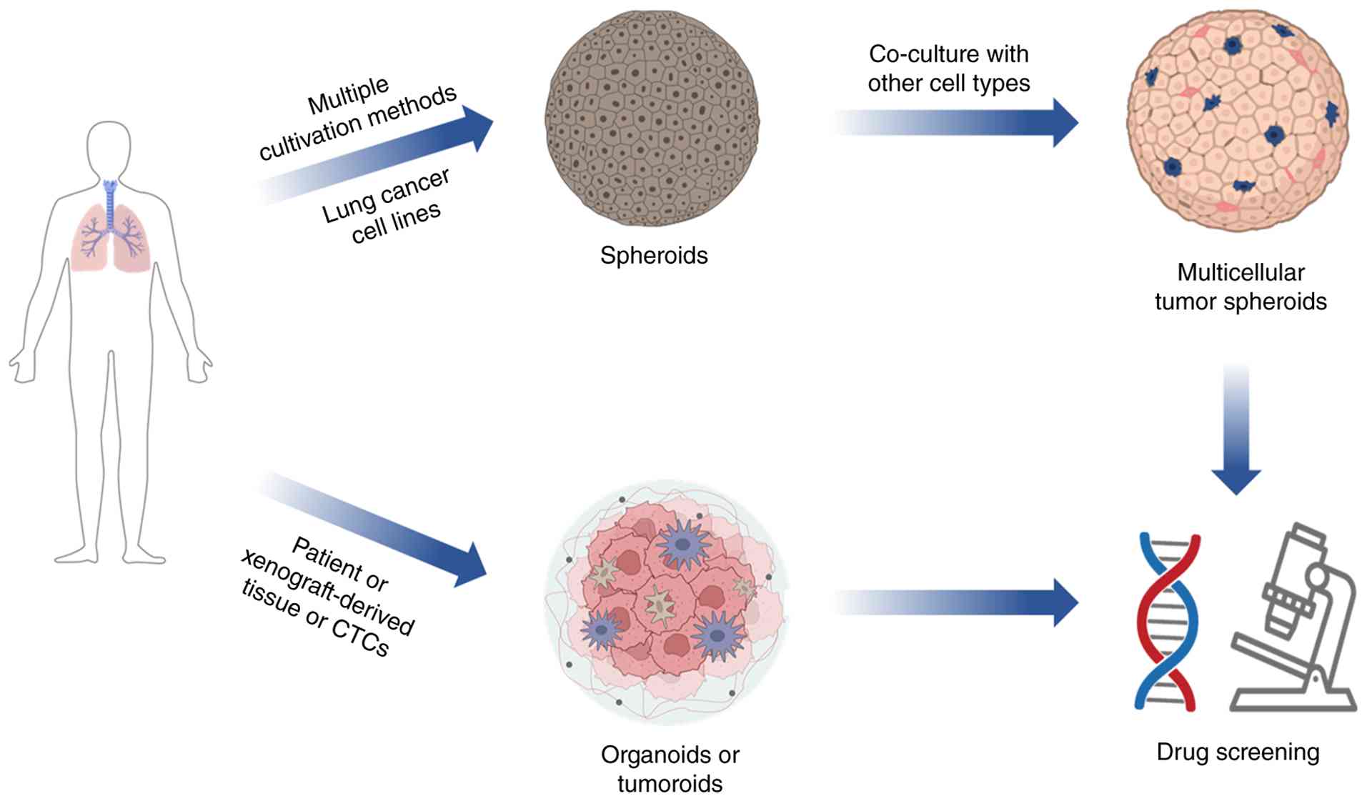
Current mainstream 3D cancer models primarily
include multicellular tumor spheroids (MCTSs), organotypic
multicellular spheroids, tumor-derived organoids and tumor
spheroids (33). As illustrated in
Fig. 1, these lung cancer models
can be generated from different cellular sources-such as cancer
cell lines, patient tissues or circulating tumor cells - and can be
further co-cultured with other cell types to increase complexity,
ultimately serving as powerful platforms for biomedical
applications including drug screening and personalized medicine.
Organoids and tumor spheroids are 3D culture models that provide
more physiologically relevant insights into tumor biology than
traditional 2D cultures (34).
However, organoids and tumor spheroids differ notably in their
culture methods, structural characteristics and applications.
Spheroids generally form through the self-assembly of single-cell
suspensions and do not require exogenous scaffolding. By contrast,
organoids represent more complex 3D structures that typically rely
on scaffold-based culture systems (35). Organoids closely mimic the
architecture and function of native tumors and often incorporate
multiple cell types along with elements of the TME (36). Organotypic culture is a culture
method in which undamaged organs in the living body are isolated
and cultured in vitro, so that they can maintain 3D growth in vitro
and maintain the original tissue structure. Organotypic culture can
successfully preserve the intact TME (37). However, organ culture has inherent
limitations: First, obtaining organ tissues is severely restricted
by their limited availability. Second, once the organ is isolated
and cultured, the proliferative activity of its cells decreases
notably and the proliferative vigor is challenging to maintain
(38). To solve the aforementioned
complications, a 3D culture model in which tumor cells grow and
proliferate in vitro to form tumor spheroids has emerged. The
method is usually applied to cell lines from solid tumor spheroids.
Notably, such cell lines from solid tumor spheroids survive well in
avascular conditions (39). In
vitro-constructed MCTSs are of great value in cancer research, with
applications including, but not limited to, radiobiological studies
(40) and novel chemotherapeutic
drug development (41).
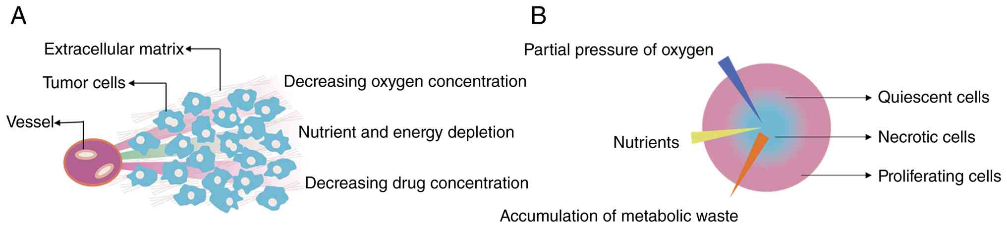
3D culture technology represents an advancement
beyond conventional 2D culture systems. Compared with 2D systems,
3D culture establishes more physiologically relevant gradients of
oxygen, nutrients, metabolic wastes and signaling molecules. 3D
culture facilitates comprehensive cell adhesion, signal
transduction and differentiation, while promoting cell-cell and
cell-matrix interactions, creating a microenvironment that is an
improved simulation of in vivo conditions. This enables cultured
tumor cells to exhibit characteristics closely resembling those of
solid tumors in vivo (16). As
shown in Fig. 2, these
characteristics include spatial gradients of oxygen, nutrients and
drugs from the periphery to the core, as well as the coexistence of
proliferating, quiescent and necrotic cell populations (42). Consequently, in vitro 3D cell
culture models are being progressively developed and implemented
for chemotherapeutic drug screening applications. Compared with
animal studies, 3D cell culture systems offer distinct advantages.
Firstly, they circumvent ethical concerns associated with human
experimentation while addressing the critical discordance observed
between animal models and clinical outcomes (with ~90% of in vivo
animal experiment results failing to align with clinical trial
findings) (43). Furthermore, 3D
cultures substantially reduce drug screening costs, shorten the
screening timeline, and enable the incorporation of diverse cell
types and enhanced physiological complexity. Additionally, this
approach overcomes the limitations inherent to animal models,
including prolonged experimental cycles, high costs and
incompatibility with high-throughput screening approaches (44).
MCTSs are commonly referred to as 3D in vitro models
that can mimic the tissue microenvironment. MCTSs are spherical
structures of a specific diameter with a dense core, composed of a
population of tumor cells. The central region of an MCTS typically
becomes hypoxic due to the limited access of cells to oxygen and
nutrients. By contrast, the cells at the edge of the spheroid can
grow normally (45). By combining
advantages such as ease of construction, high reproducibility and
compatibility with high-throughput drug screening, MCTSs have
become a commonly used model for preclinical antitumor drug
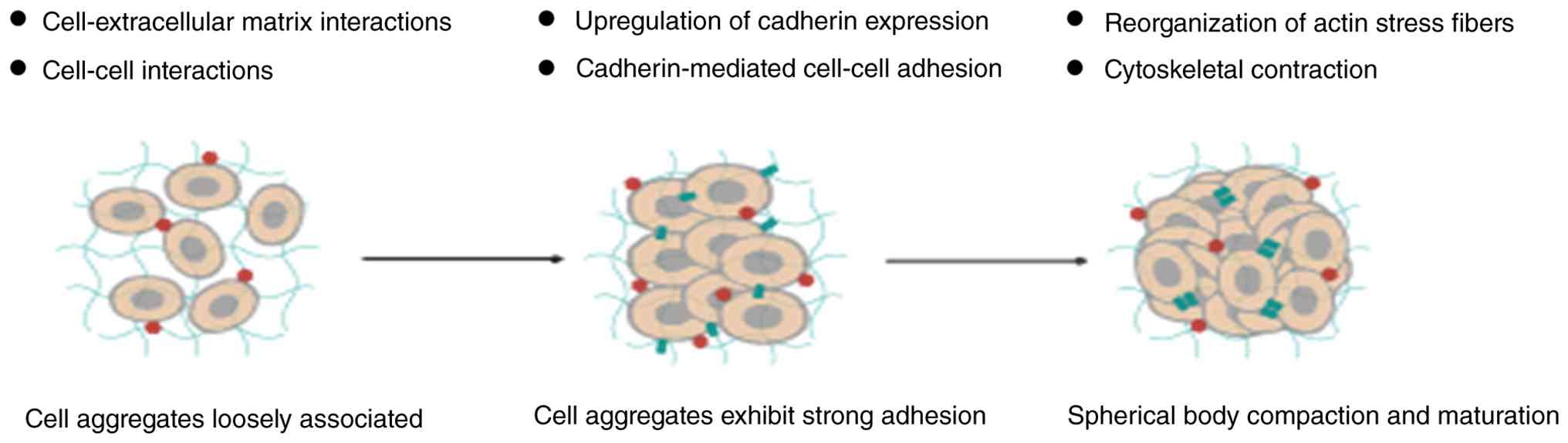
evaluation (46–49). The formation of an MCTS is roughly
divided into the following three steps (Fig. 3) (50,51).
First, cells form loose aggregates through integrin-ECM
interactions. Then, cadherin expression is upregulated, initiating
cell compaction. Finally, homophilic cadherin-cadherin interactions
mediate the formation of discrete, dense spheroids that continue to
grow. Current construction methods include the spinner culture,
pellet culture and hanging-drop method. The advantages and
disadvantages of different MCTS construction methods are summarized
in Table I (52–59).
Lung cancer organoids (LCOs) are generated by
culturing lung cancer stem cells derived from patient tissue
samples in an ECM such as Matrigel, with the addition of growth
factors to promote their proliferation and differentiation
(60). As the same malignant tumor
shows different genetic characteristics and phenotypic
heterogeneity among different patients, and tumor tissues in
different parts of the body of the same patient, personalized
precision medicine has become a novel trend in tumor treatment, and
the emergence of tumor organoids provides an opportunity for
precision medicine (61).
Individualized therapy is the latest concept in lung cancer
treatment, which refers to the development of an individualized
treatment plan with the best efficacy and the least toxicity
according to the biological characteristics of the tumor, in order
to maximize the clinical benefit to the patient (62,63).
As early as 2009, small intestinal organoids with
intestinal structure were successfully cultured using adult stem
cells derived from the mouse intestine, which led to an increase in
organoid research (64). Novel 3D
cell models have been used for drug screening, predicting patient
response to therapy and providing guidance for personalized
medication, which has been considered a breakthrough in tumor stem
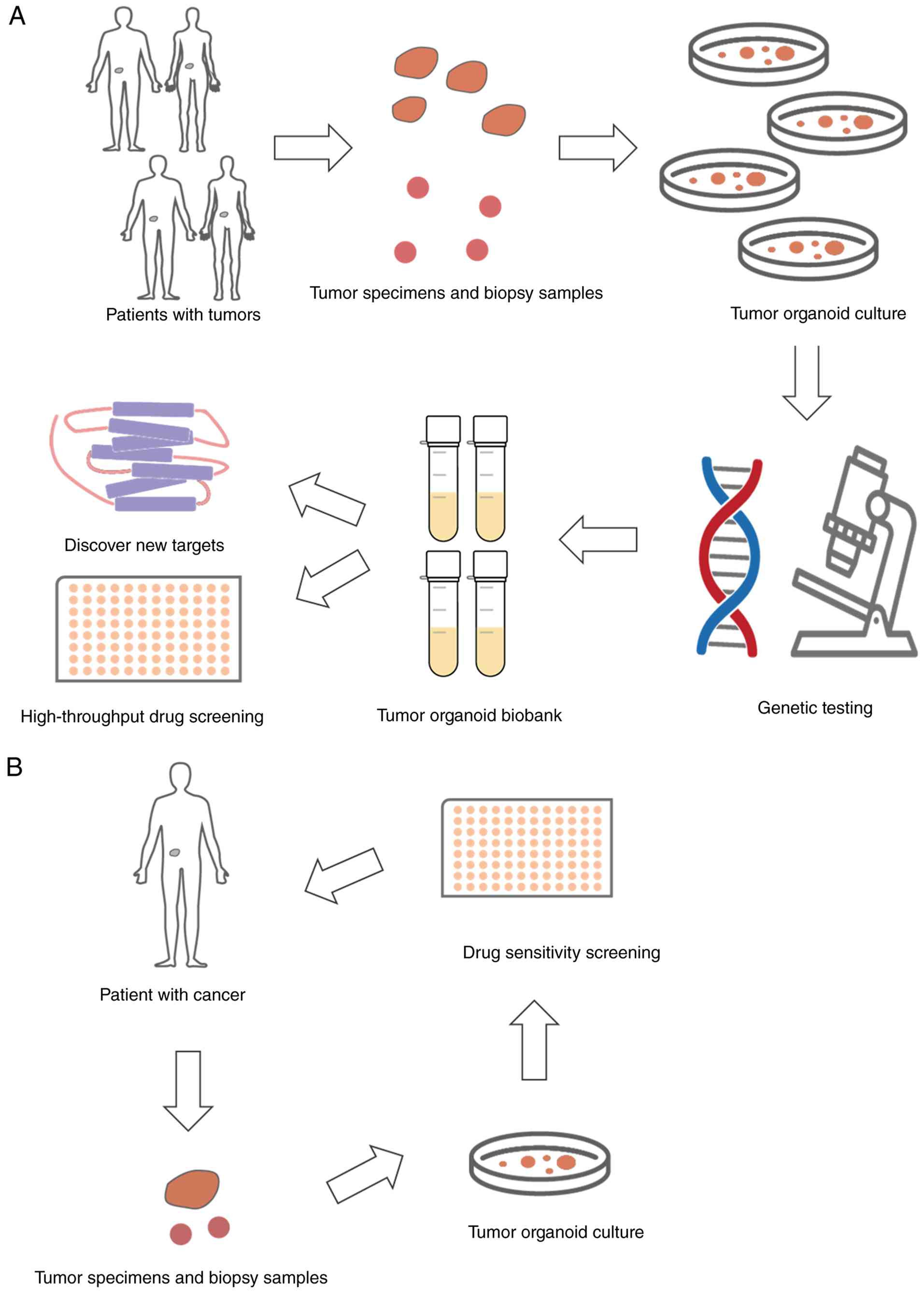
cell technology in the last decade (65). Organoid models constructed from
tissue samples from patients with lung cancer can reproduce the
histomorphology and biomarker characterization of tumors in
patients, maintain tumor heterogeneity and exhibit drug sensitivity
similar to that of their lung cancer tissues of origin. As
illustrated in Fig. 4A, tumor
organoids derived from patient tumor tissues or biopsy samples can
be used for high-throughput drug screening, genetic testing,
discovery of new therapeutic targets and the establishment of a
tumor organoid biobank, thereby supporting basic research and drug
development (66). In parallel,
Fig. 4B shows the clinical
application pathway: Patient-derived tumor organoids are subjected
to drug sensitivity screening to guide individualized treatment
decisions (67). Together, these
organoid models serve a key role in individualized drug
administration and the prognosis of lung cancer, and have the
potential to be used as reliable preclinical models for lung cancer
research (68). Organoid production
methods are generally divided into three categories: Conventional
3D culture, organoid-on-a-chip and 3D bioprinting (69).
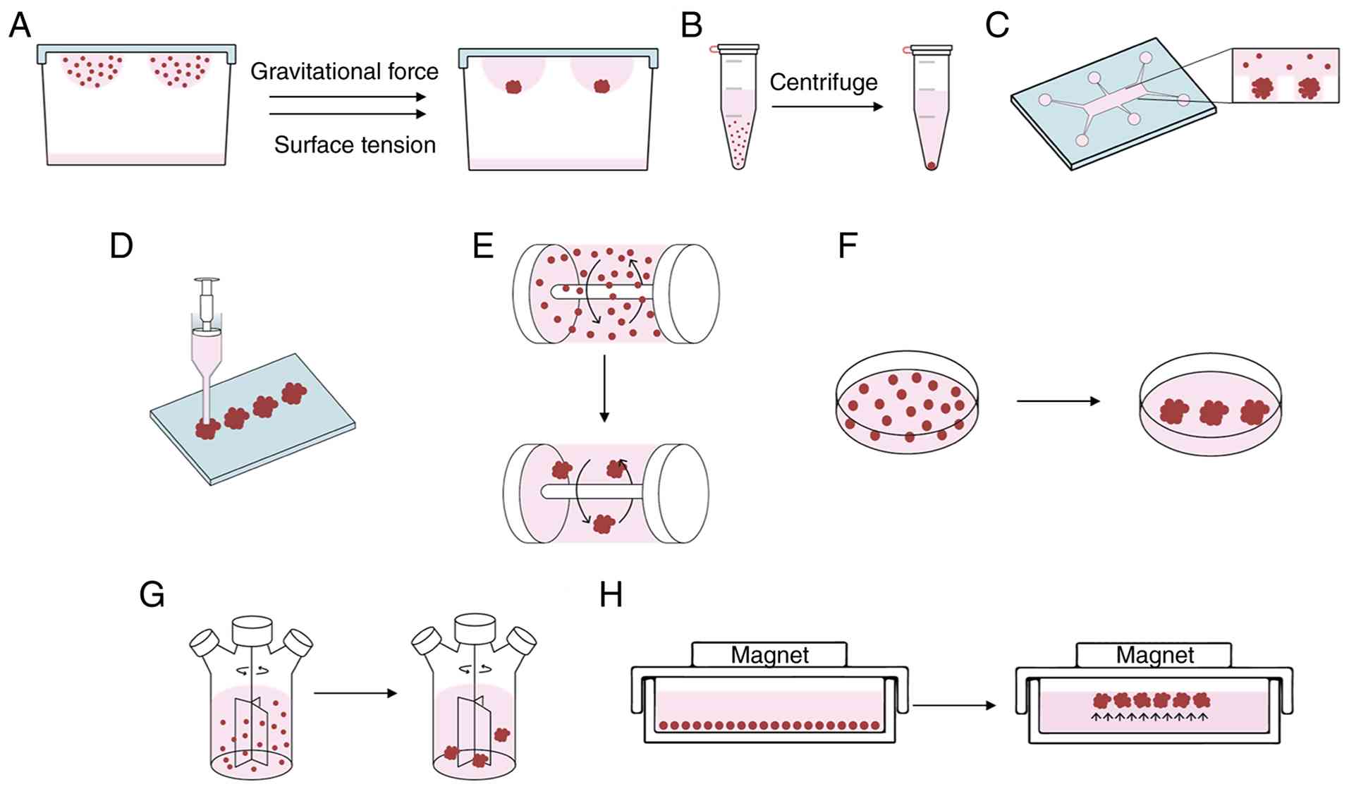
The hanging-drop method is a commonly used method
for 3D culture of tumor spheroids (70,71).
After flipping the lid of a Petri dish with attached cell
suspension droplets, the cell suspension will hang on the lid due
to surface tension. The tumor cells within the droplets will
spontaneously gather at the tip of the droplets due to gravity,
which promotes the coalescence of the cell droplets at the
air-liquid interface to form a spheroid (72). An appropriate amount of PBS must be
added to the petri dish to prevent evaporation of the liquid within
the droplets. As only one tumor spheroid is formed within each
suspension droplet, the application of this method allows for the
accurate control of the size and homogeneity of the tumor spheroids
(Fig. 5A) (73). In addition to the adjustable size of
the spheroids, the suspension droplet culture system also has other
advantages. Since it is a small-scale experiment, it does not
require expensive or specialized equipment to form spheroids; a
multichannel pipette can easily generate numerous spheroids
(74,75). Notably, mesenchymal stem cells
(MSCs) cultured using the hanging-drop method can secrete large
amounts of anti-inflammatory and antitumor factors, which are
important in disease treatment (76). However, the traditional hanging-drop
method also has notable limitations. These include the inability to
change the medium level frequently and the likely evaporation of
the culture solution within the suspension droplet, which causes
inconvenience in the subsequent experimental work, and thus, this
method is only suitable for short-term culture (77).
The 96-well spheroid plate minimizes manual handling
and transfer of spheroids, and prevents their fusion. This shift in
cultivation methodology enhances the spheroid formation throughput,
improves spheroid uniformity and reduces the skill required to
generate consistent spheroids (78). For applications demanding a large
number of spheroids (ranging from 100 to 1,000) or substantial
biomaterial, alternative methods utilizing agarose micromolds or
agarose stamps may be preferable. These methods offer several key
advantages: i) High throughput and scalability; ii) production of
highly uniform spheroids; iii) cost-effectiveness and reusability
of the molds; and iv) easy integration with standard multi-well
plates for downstream analyses (78,79).
Conventional 3D culture methods often require tedious liquid
handling steps to separate chimeric antigen receptor (CAR)-T cells
and tumor cells killed by CAR-T cells from the spheroids when
performing cytotoxicity assays (80). The advent of the 3D hanging spheroid
plate has addressed this challenge by facilitating both spheroid
formation and the subsequent separation of unbound and dead cells
from the spheroids during the assay (81). The 3D hanging spheroid plate
features a unique design where each well comprises a hanging
dripper, spheroid wells and waste wells (82). The process begins in the
hanging-drop reservoir, where tumor spheroids are formed and
subsequently co-cultured with CAR-T cells. For the cytotoxicity
readout, the droplet containing the spheroid is deposited into the
spheroid separation well (81). A
key innovation of this system is that by tilting the plate beyond
an angle of 20°, unbound CAR-T cells and dead tumor cells are
efficiently transferred into the adjacent waste well through a
connecting channel, leaving the intact spheroid isolated for
further analysis (82). The
traditional 3D culture method, whilst simple and easy to use,
offers limited precise control and low reproducibility (83).
The pellet culture method is a simpler approach for
tumor spheroid formation compared with the hanging-drop method. In
this method, cells are incubated on a shaker for 1 h to maximize
cell-cell contact and interaction, promoting tumor spheroid
formation. Subsequently, the cell suspension is centrifuged at a
low speed (28 × g for 5 min), concentrating the cells at the bottom
of a conical-shaped test tube via centrifugal force (Fig. 5B). Afterwards, the supernatant is
removed to harvest the cell pellet, and the pellet is resuspended
in a spheroid-forming culture medium. After estimating the cell
count, the cells in the medium are dispensed into 96-well U-bottom
plates with a cell-rejecting surface (74,84).
Pellet culture can be used to induce MSC differentiation. This
system is particularly suitable for stem cell differentiation
through chondrogenesis, as the cell-cell interactions within the
pellet microenvironment mimic pre-chondrogenic cell cohesion during
embryogenesis. MSCs can change their morphology in pellet culture
from fibroblasts to polygonal shapes similar to chondrocytes. Thus,
pellet culture can be used to study cartilage formation signaling
pathways and assess stem cell chondrogenic potential (85,86).
Although the pellet culture method has the advantage of generating
uniformly sized and shaped spheroids in a short period, it requires
a large number of cell culture flasks to fill the porous plate,
making it difficult to generate spheroids on a large scale and
unsuitable for high-throughput screening (53).
Microfluidic chip technology, as a novel culture
method, has attracted increasing attention for its prospects in the
pharmaceutical industry (87,88).
Integrating the 3D culture of tumor cells onto a microfluidic chip
provides a novel platform for high-throughput screening of
antitumor drugs (89). To
facilitate cell aggregation, the microfluidic microwells are first
functionalized with a layer of cell-ECM material. Subsequently, a
controlled flow rate is applied to generate hydrodynamic forces
within the device. This induces a rotational motion that keeps the
cells in suspension, thereby promoting their organization into
aggregates within the coated microwells. The appropriate
concentration of the cell suspension is infused through the
microchannels into microwells, and then the cells are cultured on
the chip to form the tumor spheroids (Fig. 5C) (90). The advantages of this method
compared with conventional spheroid culture methods (e.g., the
hanging-drop or liquid overlay method) are that more homogeneous
tumor spheroids can be formed quickly, the cell culture environment
can be precisely controlled, and the tumor spheroids can be
observed and imaged in real time by biosensors (91). The disadvantage is that the design
and processing of the chip are more complicated, and it is
difficult to remove the formed tumor spheroids for further
experimental analysis (92,93).
Organ-on-a-chip models refer to microfluidic-based
platforms that mimic the microarchitecture and functionality of
human organs, recreating various physiological milieus (94,95).
These platforms are also termed ‘microphysiological systems’
(96). The cells utilized in these
models can be derived from primary cells, cell lines or stem cells.
Specifically, ‘organoid-on-a-chip’ denotes the integration of
organoids into microfluidic devices. This hybrid approach not only
preserves the biological complexity inherent to organoids but also
leverages the controlled microenvironments of the chip to enhance
the maturity and functionality of the organoids (97). Therefore, microfluidics, a
technology characterized by sub-millimeter fluid engineering
operations, is important as an ideal technological platform for in
vitro cellular research, and has future applications in disease
diagnosis and treatment and clinical biology research (98).
3D bioprinting, an additive manufacturing technique,
is an emerging technology that converts computer aided designs into
physical objects (Fig. 5D)
(99). The advancement of 3D
bioprinting technology and its integration with biomaterials have
provided unprecedented opportunities for constructing in vitro
tumor models (100). 3D
bioprinting uses bioinks (mainly consisting of biomaterials, cells
and growth factors) to construct 3D tumor models layer by layer and
obtain functional organoids through subsequent in vitro culture
(101,102). Inkjet-based 3D printing is
suitable for encapsulating organoids within bioinks to form stable,
isolated niches that facilitate the self-assembly of organoids.
This technique enables high-precision droplet deposition, making it
applicable in scenarios demanding meticulous placement of cells and
biomaterials (103). The
advantages of this method are the ability to form tumor spheroids
of the desired size and shape that can be easily controlled in a
short period of time, and that the technology can be used for
high-throughput rapid screening due to its precision,
controllability, versatility, flexibility and rapidity (104). Furthermore, the method shows great
potential for geometrical and spatial control because 3D
bioprinting allows the precise layer-by-layer deposition of
bioinks, enabling the fabrication of tumor spheroids with
customized sizes, shapes and internal architectures that mimic the
complex spatial organization of native tumors. The disadvantages
are the complexity of the technique and the high cost of the
instrumentation. 3D bioprinting technology has led to the emergence
of a novel era of biomedical engineering, and its multiple
advantages have become a major driver for the rapid development of
drug screening methods and drug delivery systems (105).
In the rotating wall vessel method, the culture
system is rotated horizontally around the x-axis to create a
microgravity environment, which promotes the aggregation of cells
into spheroids (Fig. 5E) (106). Studies have shown that this
microgravity affects gene expression in MSCs (107,108). Under microgravity conditions,
chondrogenic and osteogenic gene expression is reduced in stem
cells, while adipogenic gene expression is elevated (109). The literature suggests that
initially the rotational speed is ~15 rpm, and as the tumor
spheroids gather into progressively larger clusters, the rotational
speed is increased to 25 rpm to keep the cell clusters in
suspension (110). Compared with
conventional static culture, the culture environment of the
rotating microgravity bioreactor can enhance the promotion of cell
proliferation and differentiation, providing an effective platform
for studying disease pathogenesis and exploring novel therapeutic
strategies (111). The method can
be applied to a variety of cell lines and is capable of generating
a large number of usable tumor spheroids in an environment
characterized by low shear stress, high dissolved oxygen levels and
low nutrient concentration, with the disadvantage that the
self-assembly of tumor spheroids cannot be observed in real time
(112).
The liquid overlay method, also referred to as
static suspension culture, involves culturing cells on specially
designed plates coated with polyhydroxyethyl methacrylate (113) or agarose (114), which results in the formation of
spheroids by reducing cell adhesion to the non-adherent culture
plate (Fig. 5F). As cell adhesion
is inhibited, the liquid overlay method enables the rapid
cultivation of reproducible, morphologically uniformly sized and
well-defined spheroids in an automated manner (115). This method is more convenient for
changing the cell culture solution and drug administration without
interfering with tumor spheroid formation compared with the
hanging-drop method. In addition, the liquid overlay method can be
performed using 96-well ultra-low adsorbent plates, making it ideal
for inoculating cells at the desired density. However, the
preparation of culture plates used in the liquid overlay method is
labor-intensive, requires skilled personnel, is relatively costly
and the plates (or their non-adherent coating) are not reusable
(112). Despite the non-adhesive
properties of agarose, this biomaterial still has drawbacks in
culturing cancer cells; agarose has difficulty interacting with
tumor cells and is unable to activate specific signaling pathways
associated with the response of tumor cells to therapeutic
processes because agarose lacks the bioactive adhesive motifs that
engage cell surface receptors (e.g., integrins) (116).
The spinner culture method generates 3D aggregates
by using a magnetic stir bar to create a turbulent flow field that
mixes air and nutrients-a process driven by magnetic force, which
was initially used to study the effects of microgravity on cells
and tissues (117). The spinner
culture method has been successfully applied for the large-scale
cultivation of bacteria and yeast to produce vaccines, recombinant
proteins and other metabolites, and in tissue engineering for
generating cartilage constructs as well as for expanding
mesenchymal stem cells on microcarriers (Fig. 5G) (118,119). During system operation, a constant
rotational speed regulation serves as the critical parameter
(120). The advantage of this
method is that the fluid environment helps exchange substances
between the tumor spheroids and allows for the formation of
multiple tumor spheroids (121).
In addition to reducing the stirring speed, the tumor spheroids
were studied by changing the medium to add drugs or growth factors
to the spheroids (48). A notable
drawback of this method is that the mechanical stirring device in
the culture system imposes substantial fluid shear stress on the
cells, thereby causing a certain degree of cellular damage.
Consequently, this approach is likely unsuitable for cells with low
adhesion strength or high sensitivity to shear forces, or those
that are anchorage-dependent and prone to anoikis when in
suspension (122).
The external force method uses external forces such
as magnetic fields, electric fields and ultrasound to concentrate
dispersed cell suspensions into high-density solutions, thereby
promoting cell aggregation into spheroids. This method relies on
magnetic forces termed magnetic levitation, which improves upon
traditional suspension techniques by enabling more rapid and
uniform spheroid formation without the need for mechanical
agitation, generating higher yields with better reproducibility and
allowing co-culture of multiple cell types (Fig. 5H) (123,124). For example, cells are incubated
with magnetic cationic liposomes containing Fe3O4 magnetic nuclei,
and when the magnetic nanoparticles are endocytosed by the cells,
the cells become magnetic under the influence of the magnetic
nanoparticles. The magnetized cells are then attracted to the
center to form a tumor spheroid by the force of an external
magnetic field (59). This method
allows for high yields and applications for long-term culture of
tumor spheroids, but the resulting tumor spheroids are usually not
uniform in size (125). The use of
magnetic forces to induce the aggregation of cells into spheroids
can be used to elucidate the interactions between cells. However,
the introduction of foreign materials (e.g., magnetic
nanoparticles) may raise safety concerns, which limits their
potential for future clinical applications (e.g., in vivo cell
therapy or tissue implantation), although this method remains
valuable for in vitro 3D culture (120).
A previous study has analyzed and compared the
morphological characteristics, protein expression, ECM distribution
and drug resistance of scaffold-free and scaffold-based A549
spheroids, and found that compared with monolayer A549 cell
cultures, scaffold-free and scaffold-based A549 spheroids exhibited
elevated levels of epithelial-mesenchymal transition (EMT) markers
and protein expression levels associated with drug resistance
(126). Drug resistance is a
leading cause of treatment failure and mortality in patients with
cancer. Among patients who succumb to cancer due to metastasis or
recurrence, >90% of cases are closely associated with multidrug
resistance, a phenomenon commonly observed in both traditional
chemotherapy and novel targeted therapies (127).
Notably, 3D culture systems generally exhibit more
potent drug resistance properties, with most compounds showing
notably reduced efficacy in a 3D environment compared with 2D
culture (128,129). Huang and Hsu (130) successfully accelerated the
formation of tumor spheroids of NSCLC cells using an
easy-to-prepare chitosan-hyaluronic acid composite membrane as a 3D
culture platform. By comparing the biological properties of 3D
tumor spheroids formed by this composite membrane with those of
2D-cultured cells, it was found that the expression levels of tumor
stem cell-related markers (such as CD133 and CD44) and EMT markers
were notably upregulated in the 3D culture system, and that cell
viability and invasive capacity were enhanced 2- to 4-fold compared
with those in 2D culture. Notably, 3D-cultured cells showed a
5–6-fold enhancement of cisplatin resistance and an even greater
16-56-fold increase in methotrexate resistance, confirming that the
composite membrane could provide an efficient 3D culture platform
for tumor stem cell research and anticancer drug screening.
A similar effect on the growth of NSCLC was
demonstrated using a multicellular spheroid chip. H1650 NSCLC cell
suspensions were incubated in the chip under a pressure range of
145–155 Pa. The chip, which was equipped with a removable cell
capture barrier, was capable of forming and extracting 3D tumor
spheroids (92). The study provided
a simple and effective method for obtaining uniform and small-sized
3D tumor spheroids for the next steps in cell-based biomedical
research, including gene expression analysis and tumor spheroid
inoculation in animal models. Ganguli et al (137) designed a modular and versatile
microchip 3D droplet culture platform that allows for reproducible
and high-content drug screening. The droplets were designed to form
a predetermined geometry, thus enabling manipulation of the
geometry of the cultured cell clusters, which can be performed
directly on the chip in real time using high-resolution confocal
microscopy. In addition, the cells in the micro-platform aggregated
into MCTSs notably faster (forming tumor spheroids in 1 day)
compared with the conventional hanging-drop culture. Fully
automated 3D screening of MCTSs has been demonstrated using a novel
angle plate adapter technology that facilitates efficient liquid
handling in a 1,536-well format. This approach identified active
natural products from the microbial Natural Products Library at UF
Scripps (138). Because this
method enables high-throughput, automated screening of 3D tumor
models in a physiologically relevant microenvironment, thereby
improving the predictive accuracy of drug responses and reducing
reliance on animal models, it may continue to serve an important
role in drug discovery and development for the foreseeable future
(139). 3D tumor spheroids are
critical for understanding the mechanisms by which the TME
contributes to cancer onset and progression, which can lead to
metastasis, and can help identify novel molecular therapeutic
approaches (140). 3D tumor
spheroid models have led to a renewed appreciation of MCTSs due to
their ability to mimic the complexity of in vivo tumors and serve
as a potential bridge between traditional 2D culture and in vivo
studies (141).
3D in vitro co-culture models improve the study of
cell-cell and cell-matrix interactions, as well as the role of the
microenvironment in cell differentiation, proliferation, apoptosis,
gene expression, and tumor cell drug resistance (142). In tumor cell culture for drug
screening, co-culture of normal cells with tumor cells could be a
potential technique to reconstitute the heterogeneous multicellular
environment of solid tumors and to promote tumor migration
(53). Due to the multi-component
and multi-target action characteristics of Traditional Chinese
Medicine, the traditional 2D monolayer cell model is too
oversimplified to comprehensively reflect its overall regulatory
effects. By contrast, the 3D tumor spheroids model with multi-cell
co-culture can more accurately mimic the in vivo microenvironment
(143). Lamichhane et al (144) used the hanging-drop method to
establish a tumor spheroid model consisting of epithelial cells,
endothelial cells and bone marrow MSCs in lung cancer, and to study
the different cell distributions and interactions within the tumor
spheroids to investigate the effects of therapeutic drugs on
different cells. Tumor spheroids constructed with a mixture of
different cells were a good model to study the interactions between
cells within the tumor, and it was found that the composition of
real tumor tissues could be further simulated by adjusting the
ratio of different cells.
MCTSs are an inevitable choice for simulating the
TME. However, relatively few cell types and compositions are
currently used to construct MCTSs, partly due to the difficulty of
co-culturing multiple cells in vitro (145). One study found that using
fibroblasts, endothelial cells and immune cells in a
feeder-layer-like manner as the external microenvironment of tumor
spheroids effectively explores stromal cell effects and overcomes
co-culture challenges (146). Han
and Hsu (147) constructed a
co-culture system of MSCs and A549 lung cancer cells on ultra-thin
(2 µm) hyaluronan-grafted chitosan, and the cells successfully
self-assembled to form 3D tumor co-spheroids with a core-shell
structure. To verify the different degrees of tumorigenicity, A549
cells or cells co-cultured with MSCs were transplanted into
zebrafish embryos for in vivo evaluation, and the tumorigenicity
obtained in the zebrafish xenograft model was consistent with that
observed in vitro. This evidence suggests that a 3D co-culture
platform of cancer cells and MSCs based on hyaluronan-grafted
chitosan substrates may be a convenient tool for studying cell-cell
interactions in tumor-like microenvironments and could potentially
be used for in vitro cancer drug testing. With the continuous
development of precision medicine, MCTSs may be further developed
into a reliable platform that can simulate solid tumors to a
greater extent than conventional 2D cultures. This platform opens
up a valuable pathway for subsequent high-throughput and
high-resolution analyses. Compared with traditional preclinical
models, it can more accurately predict clinical outcomes and become
an effective tool for real-world drug-based research and
individualized tumor treatment.
Currently, a variety of tumor organoid models have
been successfully constructed, including colorectal cancer organoid
models (148), gastric cancer
organoid models (149),
hepatocellular carcinoma organoid models (150), pancreatic cancer organoid models
(151), bladder cancer organoid
models (152), ovarian cancer
organoid models (153), prostate
cancer organoid models (154),
lung cancer organoid models (155)
and breast cancer organoid models (156). These 3D culture systems
successfully mimic the biological characteristics of the source
tissues in terms of gene expression profiles, tissue structural
features and physiological functions, and provide a promising
technological platform for developmental biology research, disease
modeling, drug screening and cell therapy (157). Several laboratories have
successfully established LCO biosample libraries for lung cancer
research, including organoid models derived from the tissues of
patients with LUAD (158,159). Systematic analyses have shown that
such models completely retained the histological architecture
(verified by H&E staining), genomic features (confirmed by
whole-exome sequencing) and gene expression profiles (analyzed by
RNA-sequencing) of the parental tumors (160,161). The models can be used for
high-throughput drug screening to further understand the
pathophysiology of lung cancer and to facilitate personalized
medicine by predicting individual patient responses to anticancer
drugs (162).
Lung cancer exhibits notable genetic and phenotypic
heterogeneity among individuals, which drives the need for
personalized medicine (108). LCOs
can be generated from tumor cells isolated from patient tissues
(163). The response of LCOs to
drugs is based on their genomic alterations. Specifically, olaparib
is effective against breast cancer gene 2-mutant-like organoids,
erlotinib is effective against EGFR-mutant organoids and crizotinib
is effective against EGFR mutation/mesenchymal-epithelial
transition factor amplification organoids. LCOs exhibit the
functional heterogeneity of tumors that is difficult to capture
with traditional techniques (108). One study identified Wnt-dependent
and Wnt-independent subtypes in LUAD, regulated by the alveolar
factor NK2 homeobox 1 (NKX2-1). Loss of NKX2-1 triggers lineage
reprogramming, leading to Wnt dependency and sensitivity to
porcupine inhibitors (159). This
organoid-based classification model links specific molecular
phenotypes (Wnt-dependent vs. Wnt-independent LUAD subtypes defined
by NKX2-1 status) to targeted therapies (e.g., porcupine inhibitors
for Wnt-dependent tumors), thereby advancing a novel paradigm for
precision medicine in lung cancer. Additionally, EGFR-mutated LCOs
have been constructed, revealing the mechanisms of resistance to
EGFR-tyrosine kinase inhibitors (164). Kim et al (165) established LCOs using five cancer
tissue subtypes, and surgically resected lung cancer tissues were
separated into individual cells or clusters of cells, embedded in
Matrigel, and cultured in Wnt3a- and Noggin-free medium. The
organoids of different lung cancer subtypes were obtained in ~4
weeks. H&E staining and immunohistochemical analysis showed
that the organoids of the five lung cancer subtypes retained LUAD
markers (e.g., napsin-A), as well as the histological features of
the cancerous tissues. The lung cancer tissues retained the genetic
features of cancer tissues as demonstrated by gene-targeted
sequencing, single nucleotide polymorphisms genotyping and mutation
concordance analysis. Meanwhile, some related studies have
confirmed that patient-derived organoids retained the histological
and genetic features of primary tumors, and were reliable models
for evaluating drug sensitivity and predicting therapeutic
responses in NSCLC, thereby guiding personalized precision medicine
(166,167).
With the advent of precision therapy, in vitro drug
sensitivity prediction of tumors is an important research direction
for individualized treatment (168). Tumor organoid technology has
advanced significantly, enabling the development of in vitro models
that can predict patient responses to anticancer drugs across
various cancer types (169).
However, in lung cancer, the construction and application of LCO
models are more difficult than those in other tumor organoid models
due to efficiency and time consumption limitations (170). Hu et al (171) used a novel integrated
superhydrophobic microtiter array chip, instead of the traditional
96-well plate, for high-throughput 3D cultivation and analysis of
LCOs. A week-long drug sensitivity test on LCO microarrays
demonstrated great potential in predicting patient treatment
response and drug screening. Importantly, the one-week drug test
results were cross-validated against three independent benchmarks:
i) The drug responses of LCOs correlated with the in vivo
anti-tumor efficacy observed in patient-derived xenografts; ii) the
sensitivity patterns aligned with the specific genetic mutation
profiles of the tumors (e.g., EGFR-TKI sensitivity in EGFR-mutant
cases); and iii) the test predictions effectively reflected the
actual clinical outcomes (e.g., progression-free survival and
objective response rates) of the matched patients, establishing the
InSMAR-chip platform as a technically feasible means for rapid
patient-specific drug response prediction in clinical settings. The
cutting-edge technology of combining LCO models with microfluidic
chips provides an effective and reliable technological tool for
predicting patient-specific drug responses in a clinical setting. A
recent study detailed a novel tumor-on-a-chip platform
incorporating organoids, which was designed to model the
physiological processes of tumor growth and metastasis in vivo.
This platform enabled practical evaluation of the invasive and
proliferative capacities of patient-derived tumor cells, thereby
serving as a valuable tool for investigating metastatic mechanisms,
and advancing targeted cancer therapies and drug discovery
(172).
Although the development of organoids has
progressed to some extent, in vitro cancer models that can
simultaneously reproduce the complexity of the TME and its diverse
ECM and genetic properties remain challenging to establish. The
emergence of 3D bioprinting technology offers a promising approach
to address this limitation, although its full potential has not yet
been realized. Dong et al (173)
utilized 3D bioprinting technology to establish a 3D bioprinting
strategy based on a bioink system (sodium alginate, hyaluronic acid
and arginine-glycine-aspartic acid peptide) in vitro, and
successfully constructed an array of LCOs for drug evaluation.
Hydrogel, as a support material for cells in 3D models, is
essential for achieving important cell-cell and cell-matrix
interactions that induce cells to form tissues and organoids
(174). 3D bioprinting allows
precise control of the spatial deposition of biomaterials and cells
based on predefined positions to create personalized tumor models,
enabling the establishment of animal-free and personalized
drug-screening platforms (175).
Despite progress in the establishment of LCOs, a
large proportion of patients remain unresponsive to current
immunotherapies (176). To better
recapitulate the living TME, a cell co-culture system can be
established by adding stromal cells, adipocytes or lymphocytes to
the culture system (177).
Dijkstra et al (178) demonstrated
that the co-culture of autologous tumor-like organs and peripheral
blood lymphocytes could be used to enrich tumor-reactive T cells.
In addition, the killing efficiency of these T cells against
matched tumor organoids could be assessed. A recent study has
demonstrated that a gel-liquid interface co-culture model,
established between LCOs and matched peripheral blood mononuclear
cells, could enhance the interaction between immune cells and tumor
organoids (155). This model
thereby allows for an improved recapitulation of the systemic
antitumor immune response observed in vivo.
The present review systematically examines the
advantages and research progress of 3D culture systems, including
MCTSs and organoids, for lung cancer drug screening, providing a
theoretical basis for their application in preclinical drug
development. Nevertheless, translating these findings into routine
clinical practice remains challenging. To advance this field and
enable clinical translation, future research should prioritize the
standardization of culture protocols and the integration of
emerging analytical technologies. Currently, considerable
heterogeneity exists across studies in terms of cell sources, ECM
components, spheroid formation methods and culture durations, which
limits the comparability and reproducibility of drug response data
(179). Furthermore, although 3D
models improve the recapitulation of the TME compared with
traditional 2D cultures, they still lack key physiological features
such as functional vasculature, dynamic immune cell infiltration
and complex stromal interactions (180). The majority of existing studies
rely on bulk analyses of spheroids or organoids, which may obscure
the intrinsic heterogeneity of tumor cell populations and their
differential drug responses (181). Integrating patient-derived
organoid biobanks with single-cell and spatial multi-omics
technologies holds considerable potential for dissecting
intratumoral heterogeneity at high resolution and mapping the
spatial distribution of drug-resistant subclones (182). Furthermore, combining 3D culture
systems with microfluidic organ-on-a-chip platforms and 3D
bioprinting will enable the construction of more physiologically
relevant models that incorporate multiple cell types and
recapitulate tissue perfusion. Addressing these limitations through
technological refinement and standardization will accelerate the
translation of 3D culture systems from bench to bedside.
The application of spheroids and organoids
represents a notable advancement in lung cancer research. They are
crucial for studying cancer biology, drug responses and resistance
mechanisms. Despite these advancements, challenges remain,
including the variability in organoid cultures, the need for
enhanced standardization and the difficulty in fully recapitulating
the TME. Nevertheless, the continuous refinement of these models
holds promise for improving their clinical relevance, facilitating
drug development and enhancing the present understanding of cancer
progression. These models provide a promising pathway for
personalized cancer therapy, reducing reliance on animal models and
improving the prediction of human-specific drug toxicity and
efficacy, thereby driving innovation in lung cancer research and
treatment.
Although both MCTSs and LCOs are superior to 2D
culture techniques, they are not mutually exclusive but rather
coexist complementarily. MCTSs provide a robust and scalable system
for investigating fundamental mechanisms and large-scale compound
screening. By contrast, LCOs, although often more
resource-intensive and time-consuming to establish, offer
high-fidelity patient-specific models for validating personalized
treatment strategies. Recently, it has been demonstrated that
malignant pleural effusion can be used as a cell source or medium
supplement for LCO model establishment; MPE not only improves the
success rate of LCO culture by enabling extended passaging and
facilitating the initial formation from difficult-to-culture
samples, but also supports the rapid generation of LCOs for
personalized drug testing (183).
Integrating both models through a tiered screening strategy,
employing MCTSs for initial high-throughput screening followed by
validation with LCOs in a patient-specific context, can effectively
optimize the drug development pipeline. In addition, the
complementary strengths of tumor spheroids in terms of modeling the
TME and organoids in terms of mirroring human tissue specificity,
can be leveraged through their integration (184). This synergistic approach bridges
the gap between model systems and deepens research insights.
Despite progress, both MCTSs and LCOs face
challenges. For MCTSs, these include standardizing size and
cellular composition, as well as incorporating complex TME
components. Ongoing advances regarding scaffold materials, imaging
technologies and culture methodologies may help overcome these
limitations in the future. For LCOs, limitations include low
culture success rates, difficulty in reconstituting a complete TME
(including functional vasculature and immune cells), and reducing
the associated time and costs of establishment. One recent study
successfully integrated multiple organoid modules on a single chip,
enabling high-throughput drug testing using this platform (185). Future directions may involve
converging these models with advanced technologies such as
microfluidic organ-on-a-chip systems and 3D bioprinting. This
integrative strategy will help construct a more physiologically
relevant TME, including stromal and immune components, thereby
enhancing the predictive power of 3D in vitro models.
In summary, the synergistic application of MCTSs
and LCOs is driving a paradigm shift in lung cancer research. MCTSs
serve as versatile tools for fundamental and applied screening,
while LCOs provide a powerful means for personalized oncology.
Together, they form a comprehensive and complementary toolkit that
enhances the capacity to develop novel anticancer strategies and
predicts clinical outcomes more accurately than conventional 2D
cultures or animal models, ultimately advancing the era of
precision medicine for patients with lung cancer.
Not applicable.
The present study was supported by The National Natural Science
Foundation of China (grant no. 82260985), The Natural Science
Foundation of Jiangxi Province (grant no. 20242BAB25572), The
Science and Technology Project of Ganzhou (grant no. 2022DSYS9969),
The Science and Technology Research Project of Education Department
of Jiangxi Province (grant nos. GJJ2201459 and GJJ2201415), and The
Science and Technology Project of Jiangxi Provincial Health
Commission (grant nos. 202310052 and 202210949).
Not applicable.
JY and MZ conceived the idea of the study and
provided overall supervision for the project. YT and HL collected
materials and wrote the manuscript. AC, LS, JZ and HC helped with
literature screening and manuscript writing. BW, HH and ML provided
constructive guidance and revised the manuscript. Data
authentication is not applicable. All authors read and approved the
final version of the manuscript.
Not applicable.
Not applicable.
The authors declare that they have no competing
interests.
|
1
|
Smolarz B, Łukasiewicz H, Samulak D,
Piekarska E, Kołaciński R and Romanowicz H: Lung
Cancer-Epidemiology, pathogenesis, treatment and molecular aspect
(Review of Literature). Int J Mol Sci. 26:20492025. View Article : Google Scholar : PubMed/NCBI
|
|
2
|
Ferrari Ardicini G, Matetic A,
Kontopantelis E, Chieffo A, Moldovan CM, Raisi-Estabragh Z, Asher
E, Mallen C and Mamas MA: Trends and future projections of cancer
prevalence in patients with cardiovascular admissions. Eur Heart J.
6:oeag0302026. View Article : Google Scholar : PubMed/NCBI
|
|
3
|
Guo LW, Lyu ZY, Meng QC, Zheng LY, Chen Q,
Liu Y, Xu HF, Kang RH, Zhang LY, Cao XQ, et al: A risk prediction
model for selecting high-risk population for computed tomography
lung cancer screening in China. Lung Cancer. 163:27–34. 2022.
View Article : Google Scholar : PubMed/NCBI
|
|
4
|
Shen M, Sun Q, Zhang J, Zhang T and Zhang
L: KAT2A accelerates lung cancer progression through succinylation
of TGFβR2. Tissue Cell. 99:1032542026. View Article : Google Scholar : PubMed/NCBI
|
|
5
|
Wang QA, Tsai IL, Lin CY, Su PL, Lin CC,
Chang JW, Huang CY, Fang YF, Chang CF, Kuo CS, et al: Multivariable
model for predicting 5-year survival in patients with EGFR-mutated
non-small cell lung cancer treated with EGFR tyrosine kinase
inhibitors: A retrospective study. Ther Adv Med Oncol.
17:175883592513219012025. View Article : Google Scholar : PubMed/NCBI
|
|
6
|
Song KJ, Choi S, Kim K, Hwang HS, Chang E,
Park JS, Shim SB, Choi S, Heo YJ, An WJ, et al: Proteogenomic
analysis reveals non-small cell lung cancer subtypes predicting
chromosome instability, and tumor microenvironment. Nat Commun.
15:101642024. View Article : Google Scholar : PubMed/NCBI
|
|
7
|
Yang J, Cao L, Li Y, Liu H, Zhang M, Ma H,
Wang B, Yuan X and Liu Q: Gracillin Isolated from Reineckia carnea
induces apoptosis of A549 cells via the mitochondrial pathway. Drug
Des Devel Ther. 15:233–243. 2021. View Article : Google Scholar : PubMed/NCBI
|
|
8
|
Li Y, Liu H, Liu X, Xiao B, Zhang M, Luo
Y, Li M and Yang J: Gracillin shows potential efficacy against
Non-small cell lung cancer through inhibiting the mTOR pathway.
Front Oncol. 12:8513002022. View Article : Google Scholar : PubMed/NCBI
|
|
9
|
Rosell R, Moran T, Viteri S, Carcereny E,
Gasco A, Quiroga V, Wei J, Camps C and Massuti B: Optimization of
genetics to create therapies for metastatic (stage IV)
non-small-cell lung cancer. Expert Opin Pharmacother. 11:1683–1693.
2010. View Article : Google Scholar : PubMed/NCBI
|
|
10
|
Breslin S and O'Driscoll L: The relevance
of using 3D cell cultures, in addition to 2D monolayer cultures,
when evaluating breast cancer drug sensitivity and resistance.
Oncotarget. 7:45745–45756. 2016. View Article : Google Scholar : PubMed/NCBI
|
|
11
|
Arnaldi P, Zotti VD, Bellese G, Gagliani
MC, Orecchia P, Castagnola P and Cortese K: 3D breast cancer
spheroids reveal Architecture-dependent HER2 expression and
signaling. Biology (Basel). 14:16542025.PubMed/NCBI
|
|
12
|
An HJ, Kim HS, Kwon JA, Song J and Choi I:
Adjustable and versatile 3D tumor spheroid culture platform with
interfacial elastomeric wells. ACS Appl Mater Interfaces.
12:6924–6932. 2020. View Article : Google Scholar : PubMed/NCBI
|
|
13
|
Kumari S, Advani D, Sharma S, Ambasta RK
and Kumar P: Combinatorial therapy in tumor microenvironment: Where
do we stand? Biochim Biophys Acta Rev Cancer. 1876:1885852021.
View Article : Google Scholar : PubMed/NCBI
|
|
14
|
Xue X, Ji D, Xue L, Tan Y, Wang B, Wang J,
Ma F, Luo Y, Lan B, Chen S, et al: Spatially resolved
transcriptomics identifies tumor-stroma-immune networks and
therapeutic targets in endocrine-resistant advanced breast cancer
treated with Everolimus+Letrozole: Insights from the MIRACLE trial.
Cancer Lett. 645:2183272026. View Article : Google Scholar : PubMed/NCBI
|
|
15
|
Sheik A, Farani MR, Kim E, Kim S, Gupta
VK, Kumar K and Huh YS: Therapeutic targeting of the tumor
microenvironments with cannabinoids and their analogs: Update on
clinical trials. Environ Res. 231:1158622023. View Article : Google Scholar : PubMed/NCBI
|
|
16
|
Pampaloni F, Reynaud EG and Stelzer EH:
The third dimension bridges the gap between cell culture and live
tissue. Nat Rev Mol Cell Biol. 8:839–845. 2007. View Article : Google Scholar : PubMed/NCBI
|
|
17
|
Weiswald LB, Bellet D and Dangles-Marie V:
Spherical cancer models in tumor biology. Neoplasia. 17:1–15. 2015.
View Article : Google Scholar : PubMed/NCBI
|
|
18
|
Breslin S and O'Driscoll L:
Three-dimensional cell culture: The missing link in drug discovery.
Drug Discovery Today. 18:240–249. 2013. View Article : Google Scholar : PubMed/NCBI
|
|
19
|
Białkowska K, Komorowski P, Bryszewska M
and Miłowska K: Spheroids as a type of Three-dimensional cell
Cultures-examples of methods of preparation and the most important
application. Int J Mol Sci. 21:62252020. View Article : Google Scholar : PubMed/NCBI
|
|
20
|
Boinapalli Y, Shankar Pandey R, Singh
Chauhan A and Sudheesh MS: Physiological relevance of in-vitro
cell-nanoparticle interaction studies as a predictive tool in
cancer nanomedicine research. Int J Pharm. 632:1225792023.
View Article : Google Scholar : PubMed/NCBI
|
|
21
|
Moghadas H, Saidi MS, Kashaninejad N and
Nguyen NT: Challenge in particle delivery to cells in a
microfluidic device. Drug Deliv Transl Res. 8:830–842. 2017.
View Article : Google Scholar : PubMed/NCBI
|
|
22
|
Jubelin C, Muñoz-Garcia J, Griscom L,
Cochonneau D, Ollivier E, Heymann MF, Vallette FM, Oliver L and
Heymann D: Three-dimensional in vitro culture models in oncology
research. Cell Biosci. 12:1552022. View Article : Google Scholar : PubMed/NCBI
|
|
23
|
Kadariya Y, Sementino E, Hua X, Kappes DJ
and Testa JR: Modeling malignant mesothelioma in genetically
engineered mice. Curr Protoc. 5:e700862025. View Article : Google Scholar : PubMed/NCBI
|
|
24
|
Szychlinska MA and Marino Gammazza A: The
zebrafish model in animal and human health research. Int J Mol Sci.
26:19452025. View Article : Google Scholar : PubMed/NCBI
|
|
25
|
Jiao X, Liu J, Wu Y, Zhong Q, Zhu L, Wang
L, Li H, Xiang M, Zhao X, Zhao G, et al: Sequential treatment with
PARPi and WEE1i enhances antitumor immune responses in preclinical
models of ovarian cancer. Sci Transl Med. 17:eadu69892025.
View Article : Google Scholar : PubMed/NCBI
|
|
26
|
Jebran AF, Seidler T, Tiburcy M, Daskalaki
M, Kutschka I, Fujita B, Ensminger S, Bremmer F, Moussavi A, Yang
H, et al: Engineered heart muscle allografts for heart repair in
primates and humans. Nature. 639:503–511. 2025. View Article : Google Scholar : PubMed/NCBI
|
|
27
|
Xiao B, Zhang M, Liu H, Cui A, Tian Y,
Tang F, Liao S, Li M, Huang H, Peng W and Yang J: Study on the
mechanism of gracillin inhibiting the proliferation of lung cancer
NCI-H1299 cells based on MAPK signaling pathway. J Cancer.
16:3048–3064. 2025. View Article : Google Scholar : PubMed/NCBI
|
|
28
|
Pound P and Ritskes-Hoitinga M: Is it
possible to overcome issues of external validity in preclinical
animal research? Why most animal models are bound to fail. J Transl
Med. 16:3042018. View Article : Google Scholar : PubMed/NCBI
|
|
29
|
Namdari R, Jones K, Chuang SS, Van
Cruchten S, Dincer Z, Downes N, Mikkelsen LF, Harding J, Jäckel S,
Jacobsen B, et al: Species selection for nonclinical safety
assessment of drug candidates: Examples of current industry
practice. Regul Toxicol Pharmacol. 126:1050292021. View Article : Google Scholar : PubMed/NCBI
|
|
30
|
Malaney P, Nicosia SV and Davé V: One
mouse, one patient paradigm: New avatars of personalized cancer
therapy. Cancer Lett. 344:1–12. 2014. View Article : Google Scholar : PubMed/NCBI
|
|
31
|
Jiang S, Zhao H, Zhang W, Wang J, Liu Y,
Cao Y, Zheng H, Hu Z, Wang S, Zhu Y, et al: An automated organoid
platform with Inter-organoid Homogeneity and Inter-patient
Heterogeneity. Cell Rep Med. 1:1001612020. View Article : Google Scholar : PubMed/NCBI
|
|
32
|
Lensink MA, Boers SN, Jongsma KR, Carter
SE, van der Ent CK and Bredenoord AL: Organoids for personalized
treatment of Cystic Fibrosis: Professional perspectives on the
ethics and governance of organoid biobanking. J Cyst Fibros.
20:443–451. 2021. View Article : Google Scholar : PubMed/NCBI
|
|
33
|
Ishiguro T, Ohata H, Sato A, Yamawaki K,
Enomoto T and Okamoto K: Tumor-derived spheroids: Relevance to
cancer stem cells and clinical applications. Cancer Sci.
108:283–289. 2017. View Article : Google Scholar : PubMed/NCBI
|
|
34
|
Yousafzai NA, El Khalki L, Wang W,
Szpendyk J and Sossey-Alaoui K: Advances in 3D culture models to
study exosomes in Triple-negative breast cancer. Cancers (Basel).
16:8832024. View Article : Google Scholar : PubMed/NCBI
|
|
35
|
Wheeler NH, Dada KA and Minnaar CA: An
overview of the developments in 3D cancer cell models, assay
techniques, and imaging modalities. Human Cell. 39:592026.
View Article : Google Scholar : PubMed/NCBI
|
|
36
|
El Harane S, Zidi B, El Harane N, Krause
KH, Matthes T and Preynat-Seauve O: Cancer spheroids and organoids
as novel tools for research and therapy: State of the art and
challenges to guide precision medicine. Cells. 12:10012023.
View Article : Google Scholar : PubMed/NCBI
|
|
37
|
Haga K, Kamimura Y, Yamazaki M, Funayama
A, Saito Y, Kida M, Tanuma JI and Izumi K: Quantitative and
longitudinal monitoring of cancer cell invasion in a
three-dimensional in vitro model of oral cancer using optical
coherence tomography. Sci Rep. 15:454492025. View Article : Google Scholar : PubMed/NCBI
|
|
38
|
Page H, Flood P and Reynaud EG:
Three-dimensional tissue cultures: Current trends and beyond. Cell
Tissue Res. 352:123–131. 2012. View Article : Google Scholar : PubMed/NCBI
|
|
39
|
Shannon AE, Boos CE, Searle BC and Hummon
AB: Gas-phase fractionation Data-Independent acquisition analysis
of 3D cocultured spheroid tumor model reveals altered translational
processes and signaling using proteomics. J Proteome Res.
23:3188–3199. 2024. View Article : Google Scholar : PubMed/NCBI
|
|
40
|
Raitanen J, Barta B, Fuchs H, Hacker M,
Balber T, Georg D and Mitterhauser M: Radiobiological assessment of
targeted radionuclide therapy with [177Lu]Lu-PSMA-I&T in 2D vs.
3D cell culture models. Int J Mol Scie. 24:170152023. View Article : Google Scholar
|
|
41
|
Domingues M, Leite Pereira C, Sarmento B
and Castro F: Mimicking 3D breast tumor-stromal interactions to
screen novel cancer therapeutics. Eur J Pharm Sci. 190:1065602023.
View Article : Google Scholar : PubMed/NCBI
|
|
42
|
Canha-Borges A, Nunes B, Estevão D,
Fernandes R, Mondini M, Cruz T, Jerónimo C, Lencart J, Paredes J,
Deutsch E, et al: Radiotherapy induces layer-specific molecular and
ultrastructural alterations in triple-negative breast cancer
spheroids. Radiother Oncol. 219:1115182026. View Article : Google Scholar : PubMed/NCBI
|
|
43
|
Tipple E, Slay E, Tsigkou O, Choudhury A
and Gough J: Using biomaterial-based 3D in vitro cancer models to
solve current clinical problems. Br J Cancer. Apr 9–2026.(Epub
ahead of print). doi: 10.1038/s41416-026-03392-3. View Article : Google Scholar : PubMed/NCBI
|
|
44
|
Mirahmad M, Sabourian R, Mahdavi M,
Larijani B and Safavi M: In vitro cell-based models of drug-induced
hepatotoxicity screening: Progress and limitation. Drug Metab Rev.
54:161–193. 2022. View Article : Google Scholar : PubMed/NCBI
|
|
45
|
Shah S and D'Souza GGM: Modeling tumor
microenvironment complexity in vitro: Spheroids as physiologically
relevant tumor models and strategies for their analysis. Cells.
14:7322025. View Article : Google Scholar : PubMed/NCBI
|
|
46
|
Li Y and Kumacheva E: Hydrogel
microenvironments for cancer spheroid growth and drug screening.
Sci Adv. 4:eaas89982018. View Article : Google Scholar : PubMed/NCBI
|
|
47
|
Ferreira LP, Gaspar VM and Mano JF: Design
of spherically structured 3D in vitro tumor models-Advances and
prospects. Acta Biomater. 75:11–34. 2018. View Article : Google Scholar : PubMed/NCBI
|
|
48
|
Sant S and Johnston PA: The production of
3D tumor spheroids for cancer drug discovery. Drug Discov Today
Technol. 23:27–36. 2017. View Article : Google Scholar : PubMed/NCBI
|
|
49
|
Viegas J, Costa S, Dias S, Pereira CL and
Sarmento B: Patient-derived melanoma Immune-Tumoroids as a platform
for precise high throughput drug screening. Adv Sci (Weinh).
11:e24087072024. View Article : Google Scholar : PubMed/NCBI
|
|
50
|
Tsai AC, Liu Y, Yuan X and Ma T:
Compaction, fusion, and functional activation of Three-dimensional
human mesenchymal stem cell aggregate. Tissue Eng Part A.
21:1705–1719. 2015. View Article : Google Scholar : PubMed/NCBI
|
|
51
|
Lin RZ, Chou LF, Chien CCM and Chang HY:
Dynamic analysis of hepatoma spheroid formation: Roles of
E-cadherin and beta1-integrin. Cell Tissue Res. 324:411–422. 2006.
View Article : Google Scholar : PubMed/NCBI
|
|
52
|
Baral T, Kirtana R, Manna S, Mishra J,
Chakraborty S, Nandi P, Niharika Roy A, Mishra P, Pradhan B, et al:
Cholesterol in glioblastoma: Impaired Hh signaling enhances
epigenetic modifiers and decreases CAV1. Cell Struct Funct.
51:121–137. 2026. View Article : Google Scholar : PubMed/NCBI
|
|
53
|
Ryu NE, Lee SH and Park H: Spheroid
culture system methods and applications for mesenchymal stem cells.
Cells. 8:16202019. View Article : Google Scholar : PubMed/NCBI
|
|
54
|
Pang L, Tian C, Wang Q, Zhao Z, Pan B, Luo
Z, Wu S, Li X and Fan J: An integrating microfluidic system for
concentration gradient generation of exosomes and Exosome-assisted
Single-Cell-Derived tumor-sphere formation. ACS Sens. 10:678–688.
2025. View Article : Google Scholar : PubMed/NCBI
|
|
55
|
Zhang T, Sheng S, Cai W, Yang H, Li J, Niu
L, Chen W, Zhang X, Zhou Q, Gao C, et al: 3-D bioprinted
human-derived skin organoids accelerate full-thickness skin defects
repair. Bioact Mater. 42:257–269. 2024.PubMed/NCBI
|
|
56
|
Moghe AS, Nandi SS, Bhonde RR, Kamyab SS,
Sawant SA and Karandikar MN: Engineering of cell line assembled
enteric organoid for enterovirus infection. Arch Virol.
171:1182026. View Article : Google Scholar : PubMed/NCBI
|
|
57
|
Depresle M, Daumar P, Goisnard A, Roux M,
Aubel C, Penault-Llorca F, Mounetou E and Bamdad M: Development of
a liquid overlay-based three-dimensional cell culture panel for
drug screening applications. Sci Rep. 15:429892025. View Article : Google Scholar : PubMed/NCBI
|
|
58
|
Dauphin T, de Beaurepaire L, Salama A,
Pruvost Q, Claire C, Haurogné K, Sourice S, Dupont A, Bach JM,
Hervé J, et al: Scalability of spheroid-derived small extracellular
vesicles production in stirred systems. Front Bioeng Biotechnol.
13:15164822025. View Article : Google Scholar : PubMed/NCBI
|
|
59
|
Haisler WL, Timm DM, Gage JA, Tseng H,
Killian TC and Souza GR: Three-dimensional cell culturing by
magnetic levitation. Nat Protoc. 8:1940–1949. 2013. View Article : Google Scholar : PubMed/NCBI
|
|
60
|
van der Vaart J and Clevers H: Airway
organoids as models of human disease. J Intern Med. 289:604–613.
2020. View Article : Google Scholar : PubMed/NCBI
|
|
61
|
Zheng Q, Liao E, Chen D and Shan G:
Bladder cancer organoids: Bridging pathological features and drug
response for precision oncology. Int Urol Nephrol. Mar 8–2026.(Epub
ahead of print). doi: 10.1007/s11255-026-05081-9. View Article : Google Scholar
|
|
62
|
Kim HS, Mitsudomi T, Soo RA and Cho BC:
Personalized therapy on the horizon for squamous cell carcinoma of
the lung. Lung Cancer. 80:249–255. 2013. View Article : Google Scholar : PubMed/NCBI
|
|
63
|
Tahayneh K, Idkedek M and Abu Akar F:
NSCLC: Current evidence on its pathogenesis, integrated treatment,
and future perspectives. J Clin Med. 14:10252025. View Article : Google Scholar : PubMed/NCBI
|
|
64
|
Sato T, Vries RG, Snippert HJ, van de
Wetering M, Barker N, Stange DE, van Es JH, Abo A, Kujala P, Peters
PJ and Clevers H: Single Lgr5 stem cells build crypt-villus
structures in vitro without a mesenchymal niche. Nature.
459:262–265. 2009. View Article : Google Scholar : PubMed/NCBI
|
|
65
|
Zhu L, Cheng C, Liu S, Yang L, Han P, Cui
T and Zhang Y: Advancements and application prospects of
three-dimensional models for primary liver cancer: A comprehensive
review. Front Bioeng Biotechnol. 11:13431772023. View Article : Google Scholar : PubMed/NCBI
|
|
66
|
Gerhards NM and Rottenberg S: New tools
for old drugs: Functional genetic screens to optimize current
chemotherapy. Drug Resist Updat. 36:30–46. 2018. View Article : Google Scholar : PubMed/NCBI
|
|
67
|
Chen Y, Xing H, Yang X, Shen J, Jiang W,
Huang J, Qiu Y, Li X, Wu K, Yang S, Zhu L, et al: Patient-derived
malignant effusion organoids guide tailored therapy and dissect
third-generation EGFR-tyrosine kinase inhibitor resistance
mechanisms in lung cancer. Cancer Lett. 645:2183562026. View Article : Google Scholar : PubMed/NCBI
|
|
68
|
Liu L, Wu Q, Fang S, Hu Z, Yu S, Wang H,
Song H and Liu Q: Organoid-based modeling unveils Dnmt3a-driven
epigenetic regulation of phenotypic plasticity in small cell lung
cancer. Cancer Lett. 650:2185162026. View Article : Google Scholar : PubMed/NCBI
|
|
69
|
Baskar G, Palaniyandi T, Viswanathan S,
Wahab MRA, Surendran H, Ravi M, Sivaji A, Rajendran BK, Natarajan S
and Govindasamy G: Recent and advanced therapy for oral cancer.
Biotechnol Bioeng. 120:3105–3115. 2023. View Article : Google Scholar : PubMed/NCBI
|
|
70
|
Mohamedahmed SM, Kamarudin MNA, Ramdas P,
Sundralingam U and Radhakrishnan AK: Optimising the method to
develop spheroids from MDA-MB-468 human triple negative breast
cancer cells. Mol Biol Rep. 53:3222026. View Article : Google Scholar : PubMed/NCBI
|
|
71
|
Fallah A, Zgheib A, Zilinski R, Danalache
BA, Eliopoulos N and Annabi B: HIF-1α silencing downregulates
SLC37A2, SLC37A3, and G6PC3 gene expression and impacts
glioblastoma stemness features in 3D neurospheres. Biochem Biophys
Res Commun. 794:1530502026. View Article : Google Scholar : PubMed/NCBI
|
|
72
|
Huang SW, Tzeng SC, Chen JK, Sun JS and
Lin FH: A dynamic hanging-drop system for mesenchymal stem cell
culture. Int J Mol Sci. 21:42982020. View Article : Google Scholar : PubMed/NCBI
|
|
73
|
Bartosh TJ and Ylostalo JH: Preparation of
anti-inflammatory mesenchymal stem/precursor cells (MSCs) through
sphere formation using hanging-drop culture technique. Curr Protoc
Stem Cell Biol. 28:2B.6.1–2B.6.23. 2014. View Article : Google Scholar : PubMed/NCBI
|
|
74
|
Achilli TM, Meyer J and Morgan JR:
Advances in the formation, use and understanding of multi-cellular
spheroids. Expert Opini Biol Ther. 12:1347–1360. 2012. View Article : Google Scholar : PubMed/NCBI
|
|
75
|
Kelm JM, Timmins NE, Brown CJ, Fussenegger
M and Nielsen LK: Method for generation of homogeneous
multicellular tumor spheroids applicable to a wide variety of cell
types. Biotechnol Bioeng. 83:173–180. 2003. View Article : Google Scholar : PubMed/NCBI
|
|
76
|
Bartosh TJ, Ylöstalo JH, Mohammadipoor A,
Bazhanov N, Coble K, Claypool K, Lee RH, Choi H and Prockop DJ:
Aggregation of human mesenchymal stromal cells (MSCs) into 3D
spheroids enhances their antiinflammatory properties. Proc Natl
Acad Sci USA. 107:13724–13729. 2010. View Article : Google Scholar : PubMed/NCBI
|
|
77
|
Huang BW and Gao JQ: Application of 3D
cultured multicellular spheroid tumor models in tumor-targeted drug
delivery system research. J Control Release. 270:246–259. 2018.
View Article : Google Scholar : PubMed/NCBI
|
|
78
|
Liszewski J, Klingelhutz A, Sander EA and
Ankrum J: Development and analysis of scaffold-free adipose
spheroids. Adipocyte. 13:23472152024. View Article : Google Scholar : PubMed/NCBI
|
|
79
|
Basu A, Dydowiczová A, Trosko JE, Bláha L
and Babica P: Ready to go 3D? A semi-automated protocol for
microwell spheroid arrays to increase scalability and throughput of
3D cell culture testing. Toxicol Mech Methods. 30:590–604. 2020.
View Article : Google Scholar : PubMed/NCBI
|
|
80
|
Chen Z and Park S: Three-dimensional
hanging drop spheroid plates for easy chimeric antigen receptor
(CAR) T cytotoxicity assay. Methods Mol Biol. 2764:35–42. 2024.
View Article : Google Scholar : PubMed/NCBI
|
|
81
|
Chen Z, Han S, Kim S, Lee C, Sanny A, Tan
AH and Park S: A 3D hanging spheroid-filter plate for
high-throughput drug testing and CAR T cell cytotoxicity assay.
Analyst. 149:475–481. 2024. View Article : Google Scholar : PubMed/NCBI
|
|
82
|
Chen Z, Han S, Sanny A, Chan DL, van Noort
D, Lim W, Tan AH and Park S: 3D hanging spheroid plate for
high-throughput CAR T cell cytotoxicity assay. J Nanobiotechnol.
20:302022. View Article : Google Scholar
|
|
83
|
Banerjee D, Singh YP, Datta P, Ozbolat V,
O'Donnell A, Yeo M and Ozbolat IT: Strategies for 3D bioprinting of
spheroids: A comprehensive review. Biomaterials. 291:1218812022.
View Article : Google Scholar : PubMed/NCBI
|
|
84
|
Mulligan LM, Lian EY and Maritan SM: An
efficient and flexible cell aggregation method for 3D spheroid
production. J Vis Exp. Mar 27–2017.(Epub ahead of print). doi:
10.3791/55544. PubMed/NCBI
|
|
85
|
Pan X, Huang X, Zhang B, Pei F, Zhao Z and
Cen X: miR-20a-5p regulated SMAD6 to inhibit chondrogenesis of
hDPSCs. Oral Diseases. 29:3433–3446. 2022. View Article : Google Scholar : PubMed/NCBI
|
|
86
|
Zhang L, Su P, Xu C, Yang J, Yu W and
Huang D: Chondrogenic differentiation of human mesenchymal stem
cells: a comparison between micromass and pellet culture systems.
Biotechnol Lett. 32:1339–1346. 2010. View Article : Google Scholar : PubMed/NCBI
|
|
87
|
Fuchs S, Johansson S, Tjell AØ, Werr G,
Mayr T and Tenje M: In-line analysis of Organ-on-Chip systems with
sensors: Integration, fabrication, challenges, and potential. ACS
Biomat Sci Eng. 7:2926–2948. 2021. View Article : Google Scholar : PubMed/NCBI
|
|
88
|
Fernandez-Carro E, Angenent M,
Gracia-Cazaña T, Gilaberte Y, Alcaine C and Ciriza J: Modeling an
optimal 3D Skin-on-Chip within microfluidic devices for
pharmacological studies. Pharmaceutics. 14:14172022. View Article : Google Scholar : PubMed/NCBI
|
|
89
|
Akbari M: Transforming drug discovery with
miniaturized predictive tissue models. Micromachines (Basel).
16:2992025. View Article : Google Scholar : PubMed/NCBI
|
|
90
|
Lipreri MV, Totaro MT, Baldini N and Avnet
S: From spheroids to Tumor-on-a-Chip for cancer modeling and
therapeutic testing. Micromachines. 16:13432025. View Article : Google Scholar : PubMed/NCBI
|
|
91
|
Sabhachandani P, Motwani V, Cohen N,
Sarkar S, Torchilin V and Konry T: Generation and functional
assessment of 3D multicellular spheroids in droplet based
microfluidics platform. Lab Chip. 16:497–505. 2016. View Article : Google Scholar : PubMed/NCBI
|
|
92
|
Jin HJ, Cho YH, Gu JM, Kim J and Oh YS: A
multicellular spheroid formation and extraction chip using
removable cell trapping barriers. Lab Chip. 11:115–119. 2011.
View Article : Google Scholar : PubMed/NCBI
|
|
93
|
Feng Y, Wang B, Tian Y, Chen H, Liu Y, Fan
H, Wang K and Zhang C: Active fluidic chip produced using
3D-printing for combinatorial therapeutic screening on liver tumor
spheroid. Biosens Bioelectron. 151:1119662020. View Article : Google Scholar : PubMed/NCBI
|
|
94
|
Mao M, Meng Z, He J and Li D:
Microphysiological systems inspired by leaf venation. Trends
Biotechnol. 42:1331–1334. 2024. View Article : Google Scholar : PubMed/NCBI
|
|
95
|
Cao UMN, Zhang Y, Chen J, Sayson D, Pillai
S and Tran SD: Microfluidic Organ-on-A-chip: A guide to biomaterial
choice and fabrication. Int J Mol Sci. 24:32322023. View Article : Google Scholar : PubMed/NCBI
|
|
96
|
Bhatia SN and Ingber DE: Microfluidic
organs-on-chips. Nat Biotechnol. 32:760–772. 2014. View Article : Google Scholar : PubMed/NCBI
|
|
97
|
Zhong C, Wang S, Yan K, Zhao Y, Yang H and
Yu J: Integrating organoids and organ-on-a-chip in dental research.
Biomaterials. 330:1240652026. View Article : Google Scholar : PubMed/NCBI
|
|
98
|
Whitesides GM: The origins and the future
of microfluidics. Nature. 442:368–373. 2006. View Article : Google Scholar : PubMed/NCBI
|
|
99
|
Cheng JTY, Tan ECK and Kang L: Pharmacy 3D
printing. Biofabrication. 172024.doi: 10.1088/1758-5090/ad837a.
|
|
100
|
Yu J, Zhang Y, Ran R, Kong Z, Zhao D, Zhao
W, Yang Y, Gao L and Zhang Z: Research progress in the field of
tumor model construction using bioprinting: A review. Int J
Nanomedicine. 19:6547–6575. 2024. View Article : Google Scholar : PubMed/NCBI
|
|
101
|
Matai I, Kaur G, Seyedsalehi A, McClinton
A and Laurencin CT: Progress in 3D bioprinting technology for
tissue/organ regenerative engineering. Biomaterials.
226:1195362020. View Article : Google Scholar : PubMed/NCBI
|
|
102
|
Jian H, Wang M, Dong Q, Li J, Wang A, Li
X, Ren P and Bai S: Dipeptide Self-assembled hydrogels with tunable
mechanical properties and degradability for 3D bioprinting. ACS
Appl Mater Interfaces. 11:46419–46426. 2019. View Article : Google Scholar : PubMed/NCBI
|
|
103
|
Kim W, Lee Y, Kang D, Kwak T, Lee HR and
Jung S: 3D Inkjet-Bioprinted Lung-on-a-Chip. ACS Biomater Sci Eng.
9:2806–2815. 2023. View Article : Google Scholar : PubMed/NCBI
|
|
104
|
Kurzątkowska M, Frankowski J, Sobczak M
and Piotrowska U: 3D bioprinting of cancer models: A game-changer
in drug discovery and development. Int J Pharm. 682:1259152025.
View Article : Google Scholar : PubMed/NCBI
|
|
105
|
Mihaylova A, Shopova D, Parahuleva N,
Yaneva A and Bakova D: (3D) Bioprinting-next dimension of the
pharmaceutical sector. Pharmaceuticals (Basel). 17:7972024.
View Article : Google Scholar : PubMed/NCBI
|
|
106
|
Steinwerth P, Bertrand J, Sandt V, Marchal
S, Sahana J, Bollmann M, Schulz H, Kopp S, Grimm D and Wehland M:
Structural and molecular changes of human chondrocytes exposed to
the rotating wall vessel bioreactor. Biomolecules. 14:252023.
View Article : Google Scholar : PubMed/NCBI
|
|
107
|
Mayer-Wagner S, Hammerschmid F, Redeker
JI, Schmitt B, Holzapfel BM, Jansson V, Betz OB and Müller PE:
Simulated microgravity affects chondrogenesis and hypertrophy of
human mesenchymal stem cells. Int Orthop. 38:2615–2621. 2014.
View Article : Google Scholar : PubMed/NCBI
|
|
108
|
Nishi M, Matsumoto R, Dong J and Uemura T:
Engineered bone tissue associated with vascularization utilizing a
rotating wall vessel bioreactor. J Biomed Mater Res Part A.
101:421–427. 2012.PubMed/NCBI
|
|
109
|
Sheyn D, Pelled G, Netanely D, Domany E
and Gazit D: The effect of simulated microgravity on human
mesenchymal stem cells cultured in an osteogenic differentiation
system: A bioinformatics study. Tissue Eng Part A. 16:3403–3412.
2010. View Article : Google Scholar : PubMed/NCBI
|
|
110
|
Ingram M, Techy GB, Saroufeem R, Yazan O,
Narayan KS, Goodwin TJ and Spaulding GF: Three-dimensional growth
patterns of various human tumor cell lines in simulated
microgravity of a NASA bioreactor. In Vitro Cell Dev Biol Anim.
33:459–466. 1997. View Article : Google Scholar : PubMed/NCBI
|
|
111
|
Grimm D, Egli M, Krüger M, Riwaldt S,
Corydon TJ, Kopp S, Wehland M, Wise P, Infanger M, Mann V and
Sundaresan A: Tissue engineering under microgravity conditions-use
of stem cells and specialized cells. Stem Cells Dev. 27:787–804.
2018. View Article : Google Scholar : PubMed/NCBI
|
|
112
|
Phelan MA, Gianforcaro AL, Gerstenhaber JA
and Lelkes PI: An air bubble-isolating rotating wall vessel
bioreactor for improved Spheroid/Organoid formation. Tissue Eng
Part C Methods. 25:479–488. 2019. View Article : Google Scholar : PubMed/NCBI
|
|
113
|
Zare M, Bigham A, Zare M, Luo H, Rezvani
Ghomi E and Ramakrishna S: pHEMA: An1 overview for biomedical
applications. Int J Mol Sci. 22:63762021. View Article : Google Scholar : PubMed/NCBI
|
|
114
|
Khodadadi Yazdi M, Taghizadeh A,
Taghizadeh M, Taghizadeh M, Stadler FJ, Farokhi M, Mottaghitalab F,
Zarrintaj P, Ramsey JD, Seidi F, et al: Agarose-based biomaterials
for advanced drug delivery. J Control Release. 326:523–543. 2020.
View Article : Google Scholar : PubMed/NCBI
|
|
115
|
Ma R, Mandell J, Lu F, Heim T, Schoedel K,
Duensing A, Watters RJ and Weiss KR: Do Patient-derived spheroid
culture models have relevance in chondrosarcoma research? Clin
Orthop Relat Res. 479:477–490. 2021. View Article : Google Scholar : PubMed/NCBI
|
|
116
|
Piazza F, Ravaglia B, Caporale A, Svetić
A, Parisse P, Asaro F, Grassi G, Secco L, Sgarra R, Marsich E, et
al: Elucidating the unexpected cell adhesive properties of agarose
substrates. The effect of mechanics, fetal bovine serum and
specific peptide sequences. Acta Biomateri. 189:286–297. 2024.
View Article : Google Scholar
|
|
117
|
Sucosky P, Osorio DF, Brown JB and Neitzel
GP: Fluid mechanics of a spinner-flask bioreactor. Biotechnol
Bioeng. 85:34–46. 2003. View Article : Google Scholar : PubMed/NCBI
|
|
118
|
Çolak G, Uzar S and Birbir M: Production
of the Bakırköy strain of sheeppox vaccine virus in spinner flasks
using an alternative method with Cytodex 1 microcarriers on Vero
cells. BMC Biotechnol. 26:92025. View Article : Google Scholar : PubMed/NCBI
|
|
119
|
Loverdou N, Cuvelier M, Nilsson Hall G,
Christiaens AS, Decoene I, Bernaerts K, Smeets B, Ramon H, Luyten
FP, Geris L and Papantoniou I: Stirred culture of cartilaginous
microtissues promotes chondrogenic hypertrophy through exposure to
intermittent shear stress. Bioeng Transl Med. 8:e104682022.
View Article : Google Scholar : PubMed/NCBI
|
|
120
|
Lewis NS, Lewis EEL, Mullin M, Wheadon H,
Dalby MJ and Berry CC: Magnetically levitated mesenchymal stem cell
spheroids cultured with a collagen gel maintain phenotype and
quiescence. J Tissue Eng. 8:20417314177044282017. View Article : Google Scholar : PubMed/NCBI
|
|
121
|
Yeatts AB and Fisher JP: Bone tissue
engineering bioreactors: Dynamic culture and the influence of shear
stress. Bone. 48:171–181. 2011. View Article : Google Scholar : PubMed/NCBI
|
|
122
|
Chisti Y: Hydrodynamic damage to animal
cells. Crit Rev Biotechnol. 21:67–110. 2008. View Article : Google Scholar : PubMed/NCBI
|
|
123
|
Seo JY, Park SB, Kim SY, Seo GJ, Jang HK
and Lee TJ: Acoustic and magnetic Stimuli-based Three-dimensional
cell culture platform for tissue engineering. Tissue Eng Regen Med.
20:563–580. 2023. View Article : Google Scholar : PubMed/NCBI
|
|
124
|
Tepe U, Aslanbay Guler B and Imamoglu E:
Applications and sensory utilizations of magnetic levitation in 3D
cell culture for tissue Engineering. Mol Biol Rep. 50:7017–7025.
2023. View Article : Google Scholar : PubMed/NCBI
|
|
125
|
Jafari J, Han X-l, Palmer J, Tran PA and
O'Connor AJ: Remote control in formation of 3D multicellular
assemblies using magnetic forces. ACS Biomat Sci Eng. 5:2532–2542.
2019. View Article : Google Scholar : PubMed/NCBI
|
|
126
|
Qi X, Prokhorova AV, Mezentsev AV, Shen N,
Trofimenko AV, Filkov GI, Sulimanov RA, Makarov VA and Durymanov
MO: Comparison of EMT-related and multi-drug resistant gene
expression, extracellular matrix production, and drug sensitivity
in NSCLC spheroids generated by scaffold-free and scaffold-based
methods. Int J Mol Sci. 23:133062022. View Article : Google Scholar : PubMed/NCBI
|
|
127
|
Cui A, Liu H, Liu X, Zhang M, Xiao B, Wang
B and Yang J: Steroidal saponins: Natural compounds with the
potential to reverse tumor drug resistance (Review). Oncol Lett.
28:5852024. View Article : Google Scholar : PubMed/NCBI
|
|
128
|
Xu Y, Pachnikova G, Przybilla D, Schäfer
R, Cui Y, Zhou D, Chen Z, Zhao A and Keilholz U: Evaluation of JQ1
combined with docetaxel for the treatment of prostate cancer cells
in 2D- and 3D-culture systems. Front Pharmacol. 13:8396202022.
View Article : Google Scholar : PubMed/NCBI
|
|
129
|
Alwahsh M, Al-Doridee A, Jasim S, Awwad O,
Hergenröder R and Hamadneh L: Cytotoxic and molecular differences
of anticancer agents on 2D and 3D cell culture. Mol Biol Rep.
51:7212024. View Article : Google Scholar : PubMed/NCBI
|
|
130
|
Huang YJ and Hsu SH: Acquisition of
epithelial-mesenchymal transition and cancer stem-like phenotypes
within chitosan-hyaluronan membrane-derived 3D tumor spheroids.
Biomaterials. 35:10070–10079. 2014. View Article : Google Scholar : PubMed/NCBI
|
|
131
|
Huang Y, Lu Y, Vadlamudi M, Zhao S,
Felmlee M, Rahimian R and Guo X: Intrapulmonary inoculation of
multicellular spheroids to construct an orthotopic lung cancer
xenograft model that mimics four clinical stages of non-small cell
lung cancer. J Pharmacol Toxicol Methods. 104:1068852020.
View Article : Google Scholar : PubMed/NCBI
|
|
132
|
Zhang Z, Wang H, Ding Q, Xing Y, Xu Z, Lu
C, Luo D, Xu L, Xia W, Zhou C and Shi M: Establishment of
patient-derived tumor spheroids for non-small cell lung cancer.
PLoS One. 13:e01940162018. View Article : Google Scholar : PubMed/NCBI
|
|
133
|
Motta A, Daware R, Nucci A, Breuel S, von
Stillfried S, Maurer J, Boor P, Jonigk D, Kiessling F, Lammers T,
et al: Bioengineering multicellular tumor spheroids with tunable
extracellular matrix deposition. Acta Biomaterialia. 212:357–372.
2026. View Article : Google Scholar : PubMed/NCBI
|
|
134
|
Pandya N, Khan E, Jain N, Satham L, Singh
R, Makde RD, Mishra A and Kumar A: Curcumin analogs exhibit
anti-cancer activity by selectively targeting G-quadruplex forming
c-myc promoter sequence. Biochimie. 180:205–221. 2021. View Article : Google Scholar : PubMed/NCBI
|
|
135
|
Gupta SK, Torrico Guzman EA and Meenach
SA: Coadministration of a tumor-penetrating peptide improves the
therapeutic efficacy of paclitaxel in a novel air-grown lung cancer
3D spheroid model. Int J Cancer. 141:2143–2153. 2017. View Article : Google Scholar : PubMed/NCBI
|
|
136
|
Guzman EAT, Sun Q and Meenach SA:
Development and evaluation of paclitaxel-loaded aerosol
nanocomposite microparticles and their efficacy against air-grown
lung cancer tumor spheroids. ACS Biomater Sci Eng. 5:6570–6580.
2019. View Article : Google Scholar : PubMed/NCBI
|
|
137
|
Ganguli A, Mostafa A, Saavedra C, Kim Y,
Le P, Faramarzi V, Feathers RW, Berger J, Ramos-Cruz KP, Adeniba O,
et al: Three-dimensional microscale hanging drop arrays with
geometric control for drug screening and live tissue imaging. Sci
Adv. 7:eabc13232021. View Article : Google Scholar : PubMed/NCBI
|
|
138
|
Vega VF, Yang D, Jordan LO, Ye F, Conway
L, Chen LY, Shumate J, Baillargeon P, Scampavia L, Parker C, et al:
Protocol for 3D screening of lung cancer spheroids using natural
products. SLAS Discov. 28:20–28. 2023. View Article : Google Scholar : PubMed/NCBI
|
|
139
|
Shen B, Tang Y, Baltz RH and Gonzalez R:
Introduction to the special issue: ‘Natural Product Discovery and
Development in the Genomic Era: 2021’. J Ind Microbiol Biotechnol.
48:kuab0302021. View Article : Google Scholar : PubMed/NCBI
|
|
140
|
Patel T and Jain N: Multicellular tumor
spheroids: A convenient in vitro model for translational cancer
research. Life Sci. 358:1231842024. View Article : Google Scholar : PubMed/NCBI
|
|
141
|
Rodrigues DB, Reis RL and Pirraco RP:
Modelling the complex nature of the tumor microenvironment: 3D
tumor spheroids as an evolving tool. J Biomed Sci. 31:132024.
View Article : Google Scholar : PubMed/NCBI
|
|
142
|
Vella N, Fenech AG and Petroni Magri V: 3D
cell culture models in research: Applications to lung cancer
pharmacology. Front Pharmacol. 15:14380672024. View Article : Google Scholar : PubMed/NCBI
|
|
143
|
Aydemir D, Keleş B, Polat İ, Metin E,
Sokullu E and Ulusu NN: Development and evaluation of a
3D-engineered neural co-culture system: Impacts on oxidative
stress, pentose phosphate pathway, trace element and mineral
metabolisms. J Neurosci Methods. 425:1106142026. View Article : Google Scholar : PubMed/NCBI
|
|
144
|
Lamichhane SP, Arya N, Kohler E, Xiang S,
Christensen J and Shastri VP: Recapitulating epithelial tumor
microenvironment in vitro using three dimensional tri-culture of
human epithelial, endothelial, and mesenchymal cells. BMC Cancer.
16:5812016. View Article : Google Scholar : PubMed/NCBI
|
|
145
|
Lee J, Cuddihy MJ, Cater GM and Kotov NA:
Engineering liver tissue spheroids with inverted colloidal crystal
scaffolds. Biomaterials. 30:4687–4694. 2009. View Article : Google Scholar : PubMed/NCBI
|
|
146
|
Estrada MF, Rebelo SP, Davies EJ, Pinto
MT, Pereira H, Santo VE, Smalley MJ, Barry ST, Gualda EJ, Alves PM,
et al: Modelling the tumour microenvironment in long-term
microencapsulated 3D co-cultures recapitulates phenotypic features
of disease progression. Biomaterials. 78:50–61. 2016. View Article : Google Scholar : PubMed/NCBI
|
|
147
|
Han HW and Hsu SH: Chitosan-hyaluronan
based 3D co-culture platform for studying the crosstalk of lung
cancer cells and mesenchymal stem cells. Acta Biomater. 42:157–167.
2016. View Article : Google Scholar : PubMed/NCBI
|
|
148
|
Wang Y, Zhang Z, Li X, Lu X, Zhuansun X,
Li Q, Zhang J, Xu X, Liu X, Wei Y, et al: Colorectal carcinoma
organoid and cancer-associated fibroblasts co-culture system for
drug evaluation. In Vitro Models. 4:31–44. 2025. View Article : Google Scholar : PubMed/NCBI
|
|
149
|
Yue SSK, Tong Y, Siu HC, Ho SL, Law SYK,
Tsui WY, Chan D, Huang Y, Chan ASY, Yun SW, et al: Divergent
lineage trajectories and genetic landscapes in human gastric
intestinal metaplasia organoids associated with early neoplastic
progression. Gut. 74:522–538. 2025. View Article : Google Scholar : PubMed/NCBI
|
|
150
|
Close DA and Johnston PA: Miniaturization
and characterization of patient derived hepatocellular carcinoma
tumor organoid cultures for cancer drug discovery applications.
SLAS Discovery. 30:1002012025. View Article : Google Scholar : PubMed/NCBI
|
|
151
|
Ely ZA, Kulstad ZJ, Gunaydin G, Addepalli
S, Verzani EK, Casarrubios M, Clauser KR, Wang X, Lippincott IE,
Louvet C, et al: Pancreatic cancer-restricted cryptic antigens are
targets for T cell recognition. Science. 388:eadk34872025.
View Article : Google Scholar : PubMed/NCBI
|
|
152
|
Merrill NM, Kaffenberger SD, Bao L,
Vandecan N, Goo L, Apfel A, Cheng X, Qin Z, Liu CJ, Bankhead A, et
al: Integrative drug screening and multiomic characterization of
Patient-derived bladder cancer organoids reveal novel molecular
correlates of gemcitabine response. Eur Urol. 86:434–444. 2024.
View Article : Google Scholar : PubMed/NCBI
|
|
153
|
Tomas EJ, Valdes YR, Davis J, Kolendowski
B, Buensuceso A, DiMattia GE and Shepherd TG: Exploiting cancer
dormancy signaling mechanisms in epithelial ovarian cancer through
spheroid and organoid analysis. Cells. 14:1332025. View Article : Google Scholar : PubMed/NCBI
|
|
154
|
De Felice D, Alaimo A, Bressan D, Genovesi
S, Marmocchi E, Annesi N, Beccaceci G, Dalfovo D, Cutrupi F,
Medaglia S, et al: Rarγ-Foxa1 signaling promotes luminal identity
in prostate progenitors and is disrupted in prostate cancer. EMBO
Rep. 26:443–469. 2024. View Article : Google Scholar : PubMed/NCBI
|
|
155
|
Li K, Liu C, Sui X, Li C, Zhang T, Zhao T,
Zhang D, Wu H, Liu Y, Wang S, et al: An organoid co-culture model
for probing systemic anti-tumor immunity in lung cancer. Cell Stem
Cell. 32:1218–1234.e7. 2025. View Article : Google Scholar : PubMed/NCBI
|
|
156
|
Wu T, Li B, Lei H, Zhao F and Liu Z:
Evolution and hotspots in breast cancer organoid research: Insights
from a bibliometric and visual knowledge mapping study (2005–2024).
Front Oncol. 15:16043622025. View Article : Google Scholar : PubMed/NCBI
|
|
157
|
Cacciamali A, Villa R and Dotti S: 3D cell
cultures: Evolution of an ancient tool for new applications. Front
Physiol. 13:8364802022. View Article : Google Scholar : PubMed/NCBI
|
|
158
|
Li H, Li Y, Zheng X, Chen F, Zhang S, Xu
S, Mu Y, Shen W, Tong J, Chen H, et al: RBM15 facilitates
osimertinib resistance of lung adenocarcinoma through m6A-dependent
epigenetic silencing of SPOCK1. Oncogene. 44:307–321. 2024.
View Article : Google Scholar : PubMed/NCBI
|
|
159
|
Ebisudani T, Hamamoto J, Togasaki K,
Mitsuishi A, Sugihara K, Shinozaki T, Fukushima T, Kawasaki K,
Seino T, Oda M, et al: Genotype-phenotype mapping of a
patient-derived lung cancer organoid biobank identifies
NKX2-1-defined Wnt dependency in lung adenocarcinoma. Cell Rep.
42:1122122023. View Article : Google Scholar : PubMed/NCBI
|
|
160
|
Terai H, Yasuda H and Sato T:
Organoid-based precision cancer modeling: New frontier in lung
cancer research. Cell Rep. 44:1165952025. View Article : Google Scholar : PubMed/NCBI
|
|
161
|
Shi R, Radulovich N, Ng C, Liu N, Notsuda
H, Cabanero M, Martins-Filho SN, Raghavan V, Li Q, Mer AS, et al:
Organoid cultures as preclinical models of non-small cell lung
cancer. Clin Cancer Res. 26:1162–1174. 2020. View Article : Google Scholar : PubMed/NCBI
|
|
162
|
Lee E, Lee SY, Seong YJ, Ku B, Cho HJ, Kim
K, Hwang Y, Park CK, Choi JY, Kim SW, et al: Lung cancer
organoid-based drug evaluation models and new drug development
application trends. Transl Lung Cancer Res. 13:3741–3763. 2024.
View Article : Google Scholar : PubMed/NCBI
|
|
163
|
Sachs N, Papaspyropoulos A, Zomer-van
Ommen DD, Heo I, Böttinger L, Klay D, Weeber F, Huelsz-Prince G,
Iakobachvili N, Amatngalim GD, et al: Long-term expanding human
airway organoids for disease modeling. EMBO J. 38:e1003002019.
View Article : Google Scholar : PubMed/NCBI
|
|
164
|
Shinozaki T, Togasaki K, Hamamoto J,
Mitsuishi A, Fukushima T, Sugihara K, Ebisudani T, Okada M, Saito
A, Shigematsu L, et al: Basal-shift transformation leads to EGFR
therapy-resistance in human lung adenocarcinoma. Nat Commun.
16:43692025. View Article : Google Scholar : PubMed/NCBI
|
|
165
|
Kim M, Mun H, Sung CO, Cho EJ, Jeon HJ,
Chun SM, Jung DJ, Shin TH, Jeong GS, Kim DK, et al: Patient-derived
lung cancer organoids as in vitro cancer models for therapeutic
screening. Nat Commun. 10:39912019. View Article : Google Scholar : PubMed/NCBI
|
|
166
|
Chen JH, Chu XP, Zhang JT, Nie Q, Tang WF,
Su J, Yan HH, Zheng HP, Chen ZX, Chen X, et al: Genomic
characteristics and drug screening among organoids derived from
non-small cell lung cancer patients. Thorac Cancer. 11:2279–2290.
2020. View Article : Google Scholar : PubMed/NCBI
|
|
167
|
Lee SH, Kim K, Lee E, Lee K, Ahn KH, Park
H, Kim Y, Shin S, Jeon SY, Hwang Y, et al: Prediction of TKI
response in EGFR-mutant lung cancer patients-derived organoids
using malignant pleural effusion. NPJ Precis Oncol. 8:1112024.
View Article : Google Scholar : PubMed/NCBI
|
|
168
|
van Renterghem AWJ, van de Haar J and
Voest EE: Functional precision oncology using patient-derived
assays: Bridging genotype and phenotype. Nat Rev Clin Oncol.
20:305–317. 2023. View Article : Google Scholar : PubMed/NCBI
|
|
169
|
Chitrangi S, Vaity P, Jamdar A and Bhatt
S: Patient-derived organoids for precision oncology: A platform to
facilitate clinical decision making. BMC Cancer. 23:6892023.
View Article : Google Scholar : PubMed/NCBI
|
|
170
|
Zhang K, Xi J, Zhao H, Wang Y, Xue J,
Liang N and Wei Z: A dual-functional microfluidic chip for guiding
personalized lung cancer medicine: Combining EGFR mutation
detection and organoid-based drug response test. Lab Chip.
24:1762–1774. 2024. View Article : Google Scholar : PubMed/NCBI
|
|
171
|
Hu Y, Sui X, Song F, Li Y, Li K, Chen Z,
Yang F, Chen X, Zhang Y, Wang X, et al: Lung cancer organoids
analyzed on microwell arrays predict drug responses of patients
within a week. Nat Commun. 12:25812021. View Article : Google Scholar : PubMed/NCBI
|
|
172
|
Chen M, Shan H, Tao Q, Hu R, Sun Q, Zheng
M, Chen Z, Lin Q, Yin M, Zhao S, et al: Mimicking tumor metastasis
using a transwell-integrated Organoids-On-a-Chip platform. Small.
20:e23085252024. View Article : Google Scholar : PubMed/NCBI
|
|
173
|
Dong Q, Su X, Li X, Zhou H, Jian H, Bai S,
Yin J and You Q: In vitro construction of lung cancer organoids by
3D bioprinting for drug evaluation. Colloids Surf A Physicochemical
Eng Aspects. 666:1312882023. View Article : Google Scholar
|
|
174
|
Yoo S and Lee HJ:
Spheroid-hydrogel-integrated biomimetic system: A new frontier in
advanced three-dimensional cell culture technology. Cells Tissues
Organs. 214:128–147. 2025.PubMed/NCBI
|
|
175
|
Akbaba H, Homer L, Tran TBT, Bhaskar N,
Sharma N, Erel-Akbaba G, Pahal S and Nguyen TD: Gene therapy
strategies in tissue engineering: Advances and applications for
regenerative therapy. Trends Biotechnol. Nov 27–2025.(Epub ahead of
print). doi: 10.1016/j.tibtech.2025.10.025. PubMed/NCBI
|
|
176
|
Lee D, Kim Y, Kang DH and Chung C:
Modeling drug and radiation resistance with patient-derived
organoids: Recent progress, unmet needs, and future directions for
lung cancer. Cells. 14:19942025. View Article : Google Scholar : PubMed/NCBI
|
|
177
|
Shan H, Chen M, Zhao S, Wei X, Zheng M, Li
Y, Lin Q, Jiang Z, Chen Z, Fei C, et al: Acoustic virtual 3D
scaffold for direct-interacting tumor organoid-immune cell
coculture systems. Sci Adv. 10:eadr48312024. View Article : Google Scholar : PubMed/NCBI
|
|
178
|
Dijkstra KK, Cattaneo CM, Weeber F,
Chalabi M, van de Haar J, Fanchi LF, Slagter M, van der Velden DL,
Kaing S, Kelderman S, et al: Generation of Tumor-reactive T cells
by Co-culture of peripheral blood lymphocytes and tumor organoids.
Cell. 174:1586–1598.e12. 2018. View Article : Google Scholar : PubMed/NCBI
|
|
179
|
Cordeiro S, Oliveira BB, Valente R,
Ferreira D, Luz A, Baptista PV and Fernandes AR: Breaking the mold:
3D cell cultures reshaping the future of cancer research. Front
Cell Dev Biol. 12:15073882024. View Article : Google Scholar : PubMed/NCBI
|
|
180
|
Silvestri G and Chatterjee A: Rebuilding
the marrow in vitro: Translational advances in the 3D modeling of
blood cancers. Onco (Basel). 5:512025.PubMed/NCBI
|
|
181
|
Um JH, Zheng Y, Mao Q, Nam C, Zhao H, Koh
YW, Shin SJ, Park YM and Lin DC: Genomic and Single-cell analyses
characterize patient-derived tumor organoids to enable personalized
therapy for head and neck squamous cell carcinoma. Cancer Res.
85:2726–2742. 2025. View Article : Google Scholar : PubMed/NCBI
|
|
182
|
Kluiver TA, Lu Y, Schubert SA, Kraaier LJ,
Ringnalda F, Lijnzaad P, DeMartino J, Megchelenbrink WL, Amo-Addae
V, Eising S, et al: Divergent WNT signaling and drug sensitivity
profiles within hepatoblastoma tumors and organoids. Nat Commun.
15:85762024. View Article : Google Scholar : PubMed/NCBI
|
|
183
|
Wang L, Yu Y, Fang Y, Li Y, Yu W, Wang Z,
Lv J, Wang R and Liang S: Malignant pleural effusion facilitates
the establishment and maintenance of tumor organoid biobank with
multiple patient-derived lung tumor cell sources. Exp Hematol
Oncol. 13:1152024. View Article : Google Scholar : PubMed/NCBI
|
|
184
|
Benboubker V, Ramzy GM, Jacobs S and
Nowak-Sliwinska P: Challenges in validation of combination
treatment strategies for CRC using patient-derived organoids. J Exp
Clin Cancer Res. 43:2592024. View Article : Google Scholar : PubMed/NCBI
|
|
185
|
Qureshi AA, Wehrle CJ, Ferreira-Gonzalez
S, Jiao C, Hong H, Dadgar N, Arpi-Palacios J, Phong YP, Kim J, Sun
K, et al: Tumor organoids for primary liver cancers: A systematic
review of current applications in diagnostics, disease modeling,
and drug screening. JHEP Rep. 6:1011642024. View Article : Google Scholar : PubMed/NCBI
|