Introduction
Lung cancer is one of the leading causes of
cancer-related mortalities; there were nearly 2.5 million new cases
(12.4% of all cancers in the world), and with an estimated 1.8
million deaths (18.7%) in 2022 (1,2).
Non-small cell lung cancer (NSCLC) accounts for >80% of lung
cancer cases (3), in which lung
adenocarcinoma (LUAD), the most common pathological subtype,
constitutes ~40% of lung malignancies (4). Currently, immune checkpoint blockers,
including programmed cell death protein 1 (PD-1) and cytotoxic T
lymphocyte-associated protein 4, have shown marked clinical
progress in the treatment of LUAD (5). In addition, EGFR-targeted tyrosine
kinase inhibitors and immunotherapy targeting PD-1 are widely
applied in clinical practice (6).
However, the 5-year survival rate of patients with LUAD remains low
due to factors such as high metastasis rate and resistance to
radiotherapy (7). Therefore, an
in-depth understanding of the molecular mechanisms underlying the
development of LUAD is key in improving the diagnosis and treatment
of LUAD.
Immune resistance and tumor metastasis serve key
roles in promoting the development of LUAD (8). Anoikis is a programmed cell death
process resulting from the loss of cell-extracellular matrix
interactions (7). Anoikis
resistance allows cancer cells to survive in body circulation,
thereby promoting secondary tumor formation in distant organs,
which is therefore considered an important mechanism underpinning
cancer cell metastasis in NSCLC (9). Notably, there is growing interest in
developing LUAD therapeutic strategies aimed at reducing anoikis
resistance (10–12).
ATP binding cassette subfamily A member 8 (ABCA8), a
member of the ABCA subfamily of transporter proteins, exhibits
physiological functions associated with the transport of lipids and
cholesterol (13). ABCA8 is lowly
expressed in NSCLC and its high expression may improve the overall
survival time of patients with lung cancer (14). However, its specific mechanism in
lung cancer is not been fully understood. Nicotinamide adenine
dinucleotide phosphate oxidase 4 (NOX4) has been observed to
mediate anoikis resistance in lung cancer cells (7). Upregulation of NOX4 not only drives
resistance to targeted therapy in lung cancer, but also induces a
high expression of programmed death ligand 1 through activating
factors such as IL-8, thereby synergistically facilitating immune
evasion (8). In colorectal cancer,
NOX4 promotes tumor growth, stemness and distant metastasis by
activating the MAPK/ERK signaling pathway (15). In addition, ABC transporter-mediated
drug efflux and apoptosis escape (including nest-leaving apoptosis
resistance) are two core mechanisms underlying cancer treatment
resistance (16–18). Based on this, analysis of the Gene
Expression Profiling Interactive Analysis (GEPIA) database revealed
that ABCA8 was negatively associated with NOX4 in LUAD. Therefore,
the present study hypothesized that ABCA8 may be involved in the
negative regulation of NOX4 and inhibited anoikis resistance.
Forkhead box protein A2 (FOXA2) expression is decreased in the
majority of lung cancer cells (19). FOXA2 can also act as a suppressor of
tumor metastasis, inhibiting ERK phosphorylation and epithelial
mesenchymal transition (EMT) (20).
Inhibition of ERK phosphorylation can block the expression of NOX4
(21) and EMT is considered an
important factor of anoikis resistance (7). Collectively, it was hypothesized that
FOXA2 could inhibit NOX4 expression, thereby attenuating the
anoikis resistance of LUAD cells, and that ABCA8 might play a role
in this process.
Materials and methods
Attachment culture
Human lung epithelial adenocarcinoma cell line A549
(cat. no. GDC0063) and human lung epithelial normal cell line
BEAS-2B (cat. no. GDC0139) were provided by The China Center For
Type Culture Collection. All cells were subjected to short tandem
repeat analysis and tested negative for Mycoplasma. BEAS-2B
cells were cultured in keratinocyte-SFM (cat. no. 17005;
Invitrogen; Thermo Fisher Scientific, Inc.) with supplements [30
µg/ml bovine pituitary extract (cat. no. abs9119; Absin Bioscience,
Inc.) and 0.2 ng/ml rat epidermal growth factor (cat. no. CYT-556;
Ameijie Technology Co., Ltd.)]. A549 cells were maintained in
RPMI-1640 medium (cat. no. PM150110; Procell Biotechnology Co.,
Ltd.) containing 10% FBS (cat. no. C0234; Beyotime Biotechnology),
100 U/ml penicillin and 0.1 mg/ml streptomycin (cat. no. 15140122;
Gibco; Thermo Fisher Scientific, Inc.). Cell culture was performed
in a moist incubator at 37°C with 5% CO2.
Suspension culture
Cell culture plates [60 mm; cat. no. CCD06N-060;
Belamb Biotechnology (Hangzhou) Co., Ltd.] were coated with 250 µl
poly-2-hydroxyethylmethacrylate (poly-HEMA; 50 mg/ml in 95%
ethanol; cat. no. P3932; Sigma-Aldrich, Merck KGaA) and dried in a
laminar flow hood at room temperature overnight. A549 and BEAS-2B
cells were trypsinized into single cell suspensions and
8×10⁵ cells were plated on poly-HEMA-coated dishes.
After 24 h, the cells were collected by centrifugation at 300 × g
for 5 min at room temperature and processed for cell viability,
flow cytometry and protein analyses (22).
Transfection
ABCA8/NOX4/FOXA2 was inserted into the plasmid
cytomegalovirus (pCM)-V6-Entry vector (cat. no. PS100001; OriGene
Technologies, Inc.) after amplification to construct
ABCA8/NOX4/FOXA2 overexpression (oe-) plasmids
(oe-ABCA8/oe-NOX4/oe-FOXA2) and the empty vector served as the
negative control (NC). In addition, short hairpin RNA (shRNA)
targeting ABCA8 (sh-ABCA8; cat. no. C02001;
5′-AAGTCAAATAGTTAAAGCAAATT-3′) and shRNA targeting NC (sh-NC,
5′-TTCTCCGAACGTGTCACGT-3′) were purchased from Shanghai GenePharma
Co., Ltd. These shRNA constructs were cloned into the pGPU6 vector
backbone. The pGPU6 vector contains a U6 promoter for shRNA
expression and a neomycin resistance gene for selection in
mammalian cells. The plasmid sequence is listed in Supplementary Data 1. Following the
manufacturer's instructions of Lipo6000™ transfection reagent
(Lipo6000; cat. no. C052; Beyotime Biotechnology), 3×10⁵
A549 cells were seeded in a 96-well plate (Biovision, Inc.).
Simultaneously, Lipo6000 (0.2 µl) was diluted with 5 µl DMEM (cat.
no. PM150210; Procell Biotechnology Co., Ltd.) without serum. A
total of 100 ng oe-ABCA8/oe-NOX4/oe-FOXA2 or sh-ABCA8/sh-NC was
diluted with 5 µl serum-free DMEM (1:1 ratio). After 20 min,
Lipo6000 and oe-ABCA8/oe-NOX4/oe-FOXA2 and/or sh-ABCA8/sh-NC was
introduced to the A549 cell culture medium for co-culture at 37°C
for 6 h. Subsequently, the whole culture medium was replaced with a
fresh complete medium for additional 24 h of culture. Transfection
efficiency was detected through reverse transcription quantitative
PCR (RT-qPCR) and western blotting.
Cell grouping and treatment
First, A549 and BEAS-2B cells were divided into
attachment (Att) and suspension (Sus) groups and received
attachment or suspension culture as aforementioned. Second, A549
cells were assigned to Control (without treatment), oe-NC
(transfected with oe-NC) or oe-ABCA8 (transfected with oe-ABCA8)
groups. Third, A549 cells were distributed to Control, oe-NC and
oe-NOX4 groups to validate the transfection efficiency. Fourth,
A549 cell suspension was assigned to oe-NC, oe-NOX4, oe-ABCA8 and
oe-NOX4 + oe-ABCA8 groups, where transfection with NC, oe-NOX4,
oe-ABCA8 and oe-NOX4 + oe-ABCA8 was performed. Fifth, A549 cells
were divided to Control, oe-NC (transfected with oe-NC) and
oe-FOXA2 (transfected with oe-FOXA2) groups. Sixth, A549 cells were
distributed into Control, sh-NC (transfected with sh-NC) and
sh-ABCA8 (transfected with ABCA8) groups. Seventh, A549 cell
suspension was divided into Control (no treatment), sh-ABCA8,
oe-FOXA2 and sh-ABCA8 + oe-FOXA2 groups and cells in the last 3
groups were separately transfected with sh-ABCA8, oe-FOXA2 and
sh-ABCA8 + oe-FOXA2.
Bioinformatics analysis
Prediction of FOXA2 and ABCA8 expressions in LUAD
based on the sample types (normal n=59; primary tumor n=515) was
achieved by searching the The University of Alabama at Birmingham
Cancer data analysis portal (UALCAN) (23). P<0.05 was considered
statistically significant. GEPIA (http://gepia.cancer-pku.cn/; version 2.0) was used for
predicting the association between ABCA8 and NOX4 or between ABCA8
and FOXA2 in LUAD. In addition, FOXA2 was identified as an upstream
binding transcription factor of ABCA8 from the hTFtarget database
(guolab.wchscu.cn/hTFtarget/#!/; version 1.0) (24). Subsequently, the University of
California Santa Cruz (http://genome.ucsc.edu/; version 1.0) and JASPAR
database (https://jaspar.genereg.net/; 2024
update) were employed to obtain the predicted binding sites of
FOXA2 in the ABCA8 promoter region. Default parameters were used
for transcription factor binding site prediction in JASPAR, with a
relative profile score threshold of >80% for high-confidence
predictions.
Chromatin immunoprecipitation (ChIP)
assay
Enrichment of FOXA2 in ABCA8 promoter region was
evaluated with ChIP kit (cat. no. P2078; Beyotime Biotechnology).
After fixation in formaldehyde (1%; cat. no. BTN160220; Beijing
Bio-Lab Technology Co., Ltd.) at 37°C for ~10 min, the A549 cells
were treated with glycine (125 mM; cat. no. HY-Y0966;
MedChemExpress) and lysed in 1 mM PMSF (cat. no. 10837091001;
Sigma-Aldrich; Merck KGaA) added with SDS lysis buffer (cat. no.
P0013G; Beyotime Biotechnology). To obtain the genome at the size
of 200–1,000 bp, the lysate was sonicated on ice (10 sec pulse and
50 sec break, six times; 20% amplitude). A total of 20 ml sonicated
lysate was taken as input for subsequent testing and 2 ml sonicated
lysate was centrifuged at 10,000 × g (5 min; 4°C) with 70 ml
Protein A + G Agarose/Salmon Sperm DNA. The supernatant was removed
for measurement. Next, ChIP dilution buffer containing 1 mM PMSF
was prepared and added to dilute the sonically processed lysate.
The cross-linked chromatin lysates were incubated with FOXA2
antibody (1:5; cat. no. ab256493; Abcam) and HRP-goat anti-rabbit
IgG (H+L; 1:500; cat. no. ab205718; Abcam) at 4°C overnight. A
total of 60 µl Protein A + G Agarose/Salmon Sperm DNA was added and
slowly mixed for 1 h to precipitate the primary antibody-recognized
protein or corresponding complex. The mixture was then centrifuged
again as before and was washed with the following buffers: Low-salt
immune complex washing buffer (×1); high-salt immune complex
washing buffer (×1); LiCl immune complex washing buffer (×1);
Tris-EDTA buffer (×2). Endogenous DNA-protein compounds were
precipitated by protein agarose/agarose and de-crosslinked
overnight at 65°C. Finally, DNA components were extracted by the
standard phenol-chloroform method (25). RT-qPCR was performed to detect the
enrichment of FOXA2 in the promoter region of ABCA8. The primer
sequences were as follows: Forward (5′-GCTTTCTATGTCCACCGCCT-3′);
and reverse (5′-AGCCCATCCAGCATTCAACA-3′). The results were analyzed
with Image J software (version 1.53; National Institutes of
Health).
Dual-luciferase reporter assay
Wild-type (WT-pcDNA-FOXA2) and mutant FOXA2
(MUT-pcDNA-FOXA2) were cloned into the pGL3-basic plasmid (E1751;
Promega Corporation) to construct the luciferase reporter vectors,
with the empty pGL3-Basic plasmids (WT/MUT-Empty vector) serving as
the NC. The Renilla luciferase reporter vector pRL-SV40
(cat. no. E2231; Promega Corporation) were regarded as an internal
control. The recombinant plasmids were co-transfected into A549
cells for 36 h. Then, the cells were lysed in 100 µl passive lysis
buffer (cat. no. E1941; Promega Corporation). Luciferase activity
in cell lysates was detected with a Dual Luciferase®
Reporter Assay System (cat. no. E1910; Promega Corporation)
(26).
RT-qPCR
Total RNA from BEAS-2B/A549 cells were extracted
with NucleoSpin® RNA Plus kit (cat. no. 740984.50;
Machery-Nagel GmbH). To determine the quantity of the isolated RNA,
a NanoDrop™ 2000 spectrophotometer (cat. no. ND-2000; Thermo Fisher
Scientific, Inc.) was employed. RT and amplification of the RNAs
were performed utilizing the One-Step RT-qPCR Kit (cat. no. P4305;
Beijing Genenode Biotech Co., Ltd), according to the manufacturer's
instructions. The reaction was performed on a CFX384 Touch PCR
detection system (cat. no. 1855195; Bio-Rad Laboratories, Inc.)
under the following conditions: 42°C for 30 min, 94°C for 2 min,
followed by 40 cycles of 94°C for 20 sec, 60°C for 20 sec, and 72°C
for 20 sec. The expression level was calculated using
2−ΔΔCq method (27) with
GAPDH as the internal reference. The relative primers are listed in
Table I.
 | Table I.Primers for reverse
transcription-quantitative PCR. |
Table I.
Primers for reverse
transcription-quantitative PCR.
| Gene | Forward primer,
5′à3′ | Reverse primer
(5′-3′) |
|---|
| FOXA2 |
CATGCACTCGGCTTCCAGTA |
GCGAGATGTACGAGTAGGGC |
| ABCA8 |
GTGTCAACAAACTTGGGCCTT |
CAGGCCGGTGAGGAAAGATT |
| NOX4 |
CAGTCTTTGACCCTCGGTCC |
TGATCCTCGGAGGTAAGCCA |
| GAPDH |
GACAGTCAGCCGCATCTTCT |
GCGCCCAATACGACCAAATC |
Confocal microscopy imaging
A549 cell suspension was prepared, dripped onto a
microslide and covered with a coverslip. Confocal microscopy
imaging was performed on a Zeiss LSM 510 laser scanning microscope
(magnification, ×100; Zeiss GmbH).
MTT assay
A549 cells in suspension culture were seeded into
96-well plate at a density of 3×10⁵/well and were
incubated at 37°C for 24 h. The culture medium was then discarded
and the cells were incubated with 20 µl MTT reagent (5.0 mg/ml;
cat. no. ab211091, Abcam) for 4 h at 37°C. The medium solution was
removed. Next, 100 µl DMSO (cat. no. ST038; Beyotime Biotechnology
Co., Ltd.) was added to the wells. A microplate reader (cat. no.
1681130; Bio-Rad Laboratories, Inc.) was exploited to evaluate
absorbance at 590 nm (28).
Flow cytometry
A549 apoptosis in suspension culture was monitored
using the Annexin V-FITC/PI Apoptosis Detection Kit (cat. no.
40302ES50; Shanghai Yeasen Biotechnology Co., Ltd.). Following the
instructions of the kit, A549 cells were centrifuged at 300 × g for
~5 min (at 4°C) and the cell culture medium was removed. Cells were
washed with PBS and counted. Subsequently, 100 µl binding buffer
was added to the wells, followed by addition of 5 µl Annexin V-FITC
and 10 µl PI staining solution. The cells were then cultured in an
incubator at 25°C for 10 min (in the dark). Next, 400 µl binding
buffer was added to the cells, which were then placed in an ice
bath. The results were assessed using a FACSCalibur flow cytometer
(BD Biosciences) with FlowJo software (version 10; FlowJo,
LLC).
Western blotting
With lysis buffer (cat. no. 20118ES60; Shanghai
Yeasen Biotechnology Co., Ltd.), the protein was isolated from A549
cells. Then, the extracted lysates underwent 5 min centrifugation
(1,000 × g) at 4°C and the supernatant was obtained for following
detection. A BCA assay kit (cat. no. 23225; Thermo Fisher
Scientific, Inc.) was exploited to determine the protein
concentration. Then, SDS-PAGE gel electrophoresis (8–12%; cat. no.
M00659; GenScript Biotech Corporation) was applied for
electrophoresis of equal amount of protein (30 µg/lane), which was
immediately loaded onto PDVF membranes (cat. no. MSPVDF022BX,
Membrane Solutions). Next, the membranes were blocked (10 min) with
10% blocking buffer (cat. no. YT067; Beijing Biolab Technology Co.,
Ltd.) at room temperature and probed with primary antibodies for
NOX4 (cat. no. ab154244; 67 kDa; 1:500; Abcam), cleaved-caspase3
(cat. no. ab32042; 17 kDa; 1:500; Abcam), cleaved caspase-8 (cat.
no. 9496; 1:1,000; 25 kDa; Cell Signaling Technology, Inc.),
caspase3 (cat. no. ab32351; 31 kDa; 1:5,000; Abcam), caspase8 (cat.
no. ab25901; 55 kDa; 1 µg/ml; Abcam), ABCA8 (cat. no. ab230896; 179
kDa; 1:1,000; Abcam) and GAPDH (cat. no. 5174; 37 kDa; 1:1,000;
Cell Signaling Technology, Inc.) at 4°C overnight. Subsequently,
the membranes were exposed to HRP-secondary antibody goat
anti-rabbit IgG H&L (cat. no. ab6721; 1:2,000; Abcam) for 1 h
at room temperature. The membranes with proteins were then
visualized using ECL Plus (cat. no. WBKlS0100; MilliporeSigma;
Merck KGaA) and exposed using the Kodak In-Vivo Imaging System FX
Pro (Kodak). The results were analyzed with a semi-quantification
software (Image J; version 1.53; National Institutes of Health) and
GAPDH served as the internal reference.
Statistical analysis
All measurement data are presented as mean ± SD.
Comparisons between two groups were performed by independent sample
t-tests, while comparisons among multiple groups were made through
ANOVA tests. The normality of data distribution was verified using
the Shapiro-Wilk test and the homogeneity of variances was assessed
with the Brown-Forsythe test. Statistical analyses were achieved
using GraphPad software (version 8.0; Dotmatics) and P<0.05 was
considered to indicate a statistically significant difference.
Results
FOXA2 and ABCA8 expression levels are
downregulated in LUAD
Given that ABCA8 expression is downregulated in
NSCLC (13), a series of
experiments were conducted to reveal its associated pathways in
LUAD mechanisms. Through retrieval and assessment of the UALCAN
website, expression levels of FOXA2 and ABCA8 in LUAD were
predicted based on sample type (normal n=59; primary tumor n=515).
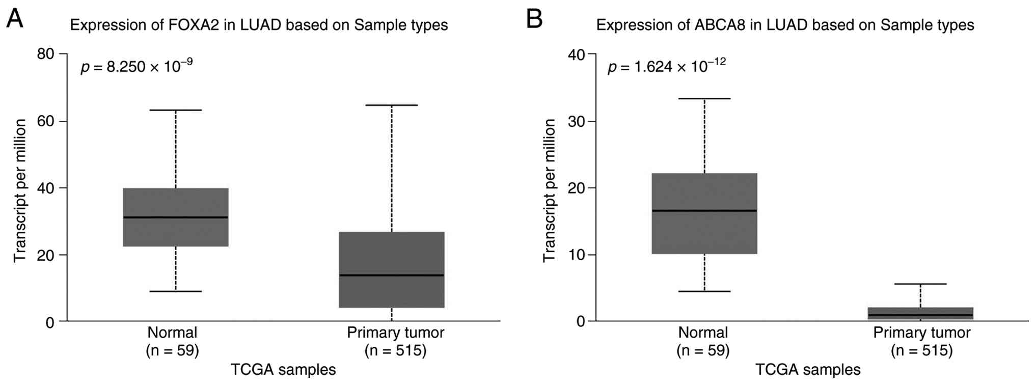
FOXA2 expression was decreased in LUAD tumor tissues, compared with
normal tissues (P=8.250×10⁻⁹; Fig. 1A) and this was also true for ABCA8
expression in LUAD tumor tissues (P=1.624×10⁻¹²;
Fig. 1B). These findings indicated
that both FOXA2 and ABCA8 expression levels were decreased in
LUAD.
ABCA8 and FOXA2 expression levels are
decreased in A549 cells, H1299 cells and H1975 cells and NOX4 is
negatively associated with ABCA8
Anoikis resistance has been established as a key
process of cancer cell metastasis in LUAD (29). NOX4 can mediate anoikis resistance
in lung cancer cells (7).
Considering the existing results and the established downregulation
of ABCA8 and FOXA2 in LUAD, the association between ABCA8 and NOX4
in the anoikis resistance of LUAD was determined. Suspension
culture was carried out for A549 cells, H1299 cells, H1975 cells
and BEAS-2B cells to induce the anoikis resistance, with an
attachment culture serving as the control. Subsequently, RT-qPCR
was conducted to detect the mRNA expression of FOXA2 and ABCA8 in
A549 cells and BEAS-2B cells undergoing attachment culture.
Compared with BEAS-2B cells, A549 cells, H1299 cells and H1975
cells presented levels of FOXA2 and ABCA8 decreased by ~50%
(P<0.001; Fig. 2A and B). Then,
mRNA levels of FOXA2, ABCA8 and NOX4 in A549 cells, H1299 cells and
H1975 cells experiencing attachment and suspension culture were
measured. The results revealed that ABCA8 and FOXA2 levels in A549
cells, H1299 cells and H1975 cells were lower in suspension culture
compared with attachment culture (P=0.0007, P=0.0003, P=0.005,
P=0.0004, P=0.001 and P=0.0008; Fig.
2C, D, F, G, I and J). Conversely, A549 cells, H1299 cells and
H1975 cells in suspension culture exhibited higher NOX4 mRNA level
compared with those in attachment culture (P=0.0011, P=0.0011 and
P=0.0013; Fig. 2E, H and K).
Subsequently, RT-qPCR data reflected that oe-ABCA8 significantly
increased ABCA8 expression by 1-fold in A549 cells (P=0.0002;
Fig. 2L), which demonstrated
successful transfection. Before experiments, a negative correlation
was predicted between ABCA8 and NOX4 in GEPIA (P=0.014; R=−0.11;
Fig. 2M), which was further
determined by western blotting results. Furthermore, oe-ABCA8
decreased NOX4 protein expression by ~70% (P=0.0001; Fig. 2N). Collectively, it was concluded
that FOXA2 and ABCA8 levels were downregulated in A549 cells and a
further reduction could be induced in A549 cells under suspension
suspension; correspondingly, NOX4 expression was upregulated and
exhibited a negative correlation with ABCA8 level.
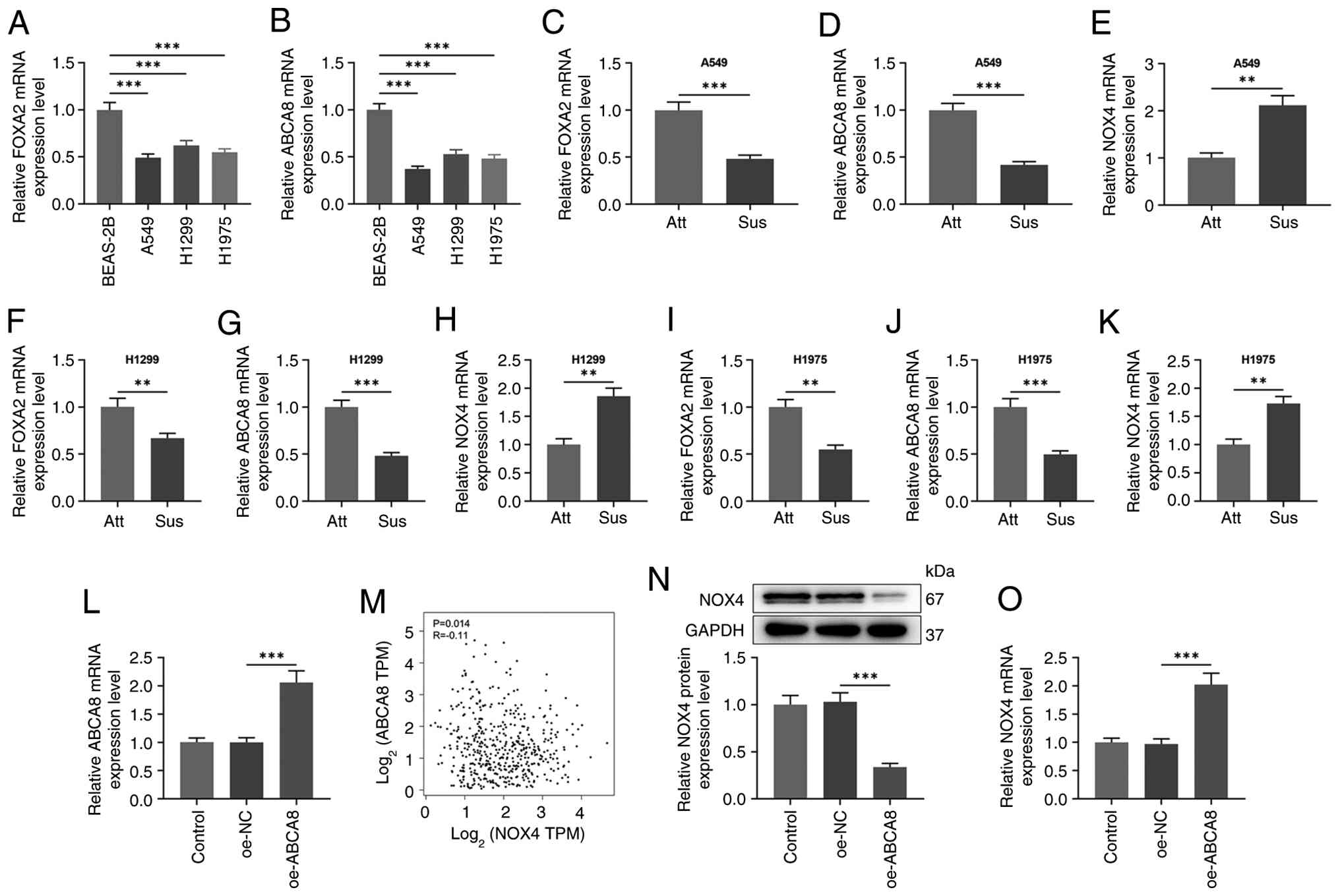 | Figure 2.ABCA8 overexpression reduces anoikis
resistance in A549 cells by inhibiting NOX4 expression. BEAS-2B
cells, A549 cells, H1299 cells and H1975 cells were divided into
Att and Sus groups and received attachment culture or suspension
culture. RT-qPCR was conducted to detect mRNA levels of (A) FOXA2
and (B) ABCA8 in BEAS-2B cells, A549 cells, H1299 cells and H1975
cells undergoing attachment culture, with GAPDH serving as the
internal reference. Relative mRNA levels of (C) FOXA2, (D) ABCA8
and (E) NOX4 in A549 cells, relative mRNA levels of (F) FOXA2, (G)
ABCA8 and (H) NOX4 in H1299 cells and relative mRNA levels of (I)
FOXA2, (J) ABCA8 and (K) NOX4 in H1975 cells were measured (in both
Att and Sus culture) by RT-qPCR, with GAPDH serving as the internal
reference. (L) A549 cells were assigned into Control (without
treatment), oe-NC (transfected with oe-NC) and oe-ABCA8
(transfected with oe-ABCA8) groups. The mRNA expression of ABCA8
was determined using RT-qPCR, with GAPDH serving as the internal
reference. (M) Gene Expression Profiling Interactive Analysis was
used for prediction of the association between ABCA8 and NOX4
(P=0.014; R=−0.11). (N) The protein expression of NOX4 was
determined using western blotting, with GAPDH serving as the
internal reference. (O) A549 cells were divided into Control
(without treatment), oe-NC (transfected with oe-NC) and oe-NOX4
(transfected with oe-NOX4) groups. The mRNA expression of NOX4 was
measured using RT-qPCR, with GAPDH serving as the internal
reference. Data are presented as the mean ± SD (n=3). There were
three biological replicates in each experimental group. **P<0.01
and ***P<0.001. FOXA2, forkhead box protein A2; ABCA8, ATP
binding cassette subfamily A member 8; NOX4, nicotinamide adenine
dinucleotide phosphate oxidase 4; Att, attachment; Sus, suspension;
oe, overexpression; NC, negative control; RT-qPCR, reverse
transcription-quantitative PCR; TPM, transcripts per million. |
Oe-ABCA8 mitigated anoikis resistance
in A549 cells by inhibiting NOX4 expression
Subsequently, oe-NOX4 plasmid was transfected into
A549 cell to ascertain the negative association between ABCA8 and
NOX4. From the RT-qPCR results, it was noted that oe-NOX4
successfully increased the NOX4 mRNA level in A549 cells (P=0.0002;
Fig. 2O). Then, confocal microscopy
images substantiated that oe-NOX4 promoted the aggregation of A549
cells, which was reversed by oe-ABCA8 (Fig. 3A). In addition, MTT assay results
indicated that oe-ABCA8 attenuated the stimulatory effect of NOX4
on A549 cell viability by ~20% (P=0.002; Fig. 3B). According to the outcomes of flow
cytometry, it was observed that oe-NOX4 reduced apoptosis rate of
A549 cells by ~63% (P<0.0001; Fig.
3C and D), which was offset by oe-ABCA8 (P<0.0001; Fig. 3C and D). In addition, the
expressions of cleaved-caspase3 and cleaved-caspase8, established
apoptosis-associated proteins (30), as well as NOX4 expression were
determined using western blotting. The relative protein expression
of cleaved-caspase3 and cleaved-caspase8 in A549 cells was reduced
by ~50% after transfection of oe-NOX4 (P=0.0028 and P<0.0001;
Fig. 3E-G), whereas oe-ABCA8
reversed this inhibitory effect of oe-NOX4 (P=0.0028 and
P<0.0001; Fig. 3E-G). NOX4
expression in A549 cells was upregulated by oe-NOX4, yet
downregulated by oe-ABCA8 (P=0.0018 and P=0.0018; Fig. 3E-H). Overall, oe-NOX4 strengthened
anoikis resistance in LUAD (A549) cells, which was counteracted by
oe-ABCA8.
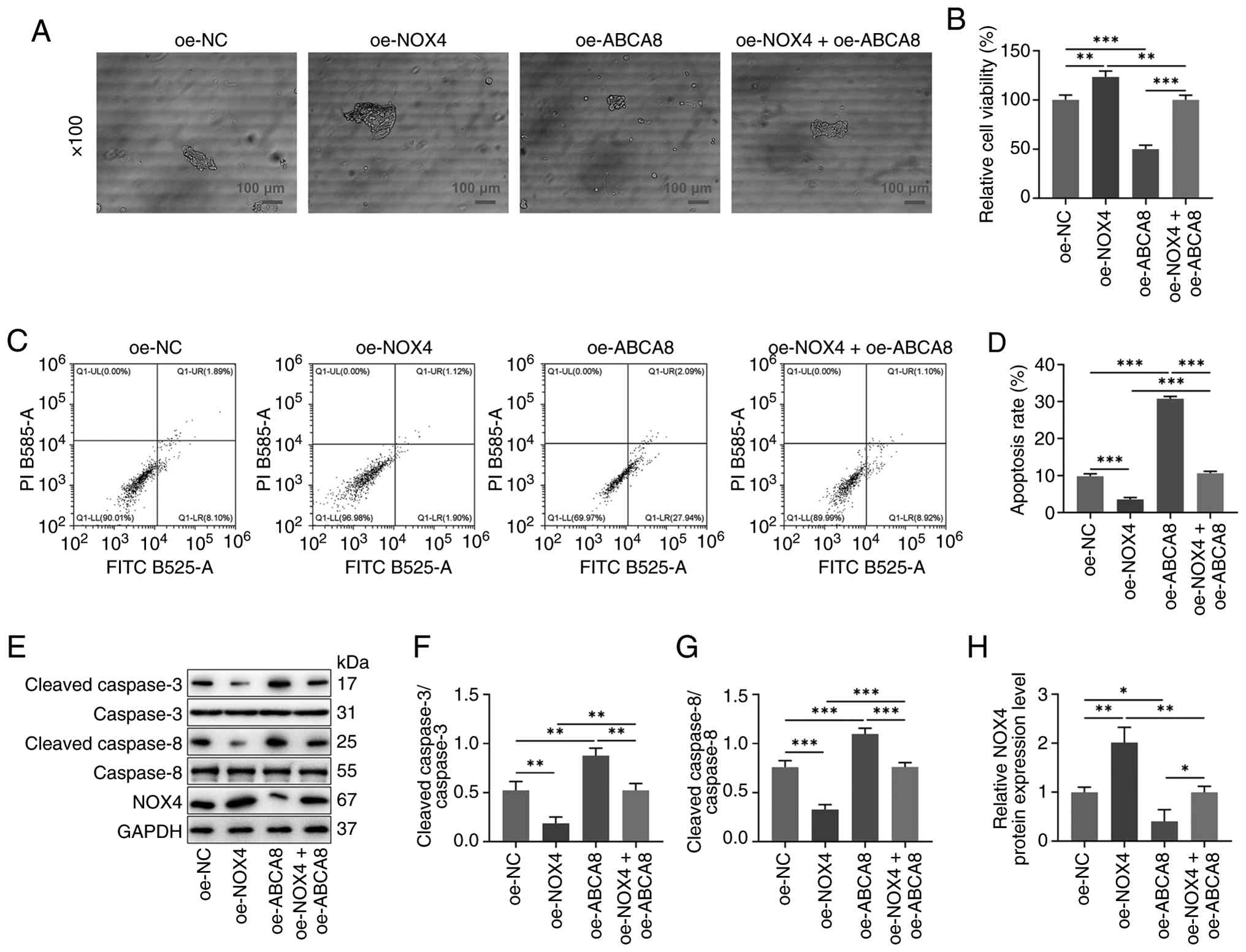 | Figure 3.Effects of NOX4 or ABCA8
overexpression on cell viability, apoptosis rate and
apoptosis-associated protein levels. (A) A549 cells in suspension
culture were assigned to oe-NC, oe-NOX4, oe-ABCA8 (treated as
before) and oe-NOX4 + oe-ABCA8 (transfected with oe-NOX4 and
oe-ABCA8) groups. Confocal microscopy images were obtained from a
microscope (magnification, ×100; scale bar, 100 µm). (B) Viability
of A549 cells (suspension culture) was monitored by MTT assay. (C)
Flow cytometry was employed to determine and (D) quantify the
apoptosis in A549 cells (suspension culture). (E) Western blotting
measured (F) cleaved-caspase3, caspase3, (G) cleaved-caspase8,
caspase8 and (H) NOX4 protein expressions in A549 cell suspension,
with GAPDH serving as the internal reference. Data are presented as
the mean ± SD (n=3). There were three biological replicates in each
experimental group. *P<0.05, **P<0.01 and ***P<0.001.
ABCA8, ATP binding cassette subfamily A member 8; NOX4,
nicotinamide adenine dinucleotide phosphate oxidase 4; oe-,
overexpression; NC, negative control. |
FOXA2 alleviates anoikis resistance of
A549 cells through promoting the expression of ABCA8
FOXA2 was considered as the upstream transcription
factor of ABCA8 after analysis and experimental validation of their
association. Transfection efficiency of oe-FOXA2 was examined by
RT-qPCR and the data revealed FOXA2 mRNA level was increased 1-fold
by oe-FOXA2 (P=0.0003; Fig. 4A). By
searching GEPIA, it was determined that ABCA8 was positively
correlated with FOXA2 (P=5.6×10⁻⁷; R=0.23, Fig. 4B). Western blotting was used for
determination of ABCA8 expression in A549 cells and the results
elucidated that relative protein expression of ABCA8 was
significantly increased by 0.8-fold after transfection with
oe-FOXA2 (P=0.0013, Fig. 4C).
Furthermore, JASPAR was employed to predict the binding sites of
FOXA2 to ABCA8 (Fig. 4D). ChIP and
dual-luciferase reporter assays were conducted to test the binding
of FOXA2 and ABCA8. It was observed that FOXA2 was enriched in the
promoter of ABCA8 (P<0.0001; Fig.
4E). In addition, the outcomes of dual-luciferase reporter
assay revealed that relative luciferase activity was ~3-fold higher
in WT-pcDNA-FOXA2 compared with that of the WT-empty vector
(P=0.0004; Fig. 4F), while no
significant difference was found in MUT groups (Fig. 4F). Sh-ABCA8 reduced ABCA8 expression
in A549 cells by ~50% (P=0.0003; Fig.
4G), indicating successful transfection. The aggregation of
A549 cells was promoted after transfection of sh-ABCA8, whereas
oe-FOXA2 weakened the effect of sh-ABCA8 (Fig. 4H). In addition, oe-FOXA2
counteracted the promoting role of sh-ABCA8 in A549 cell viability
(P=0.0023; Fig. 4I). Flow cytometry
data indicated that the apoptosis rate was decreased after
transfection with sh-ABCA8 (P<0.0001; Fig. 5A and B), which was reversed by
oe-FOXA2 (P<0.0001; Fig. 5A and
B). Western blotting results further illustrated that sh-ABCA8
inhibited cleaved-caspase3 and cleaved-caspase8 protein expressions
and promoted NOX4 expression (P=0.0029, P=0.0116 and P=0.0025;
Fig. 5C-F), which was offset by
oe-FOXA2 (P=0.0035, P<0.0001 and P=0.0026; Fig. 5C-F). Overall, these results
indicated that FOXA2 promoted anoikis of A549 cells by upregulating
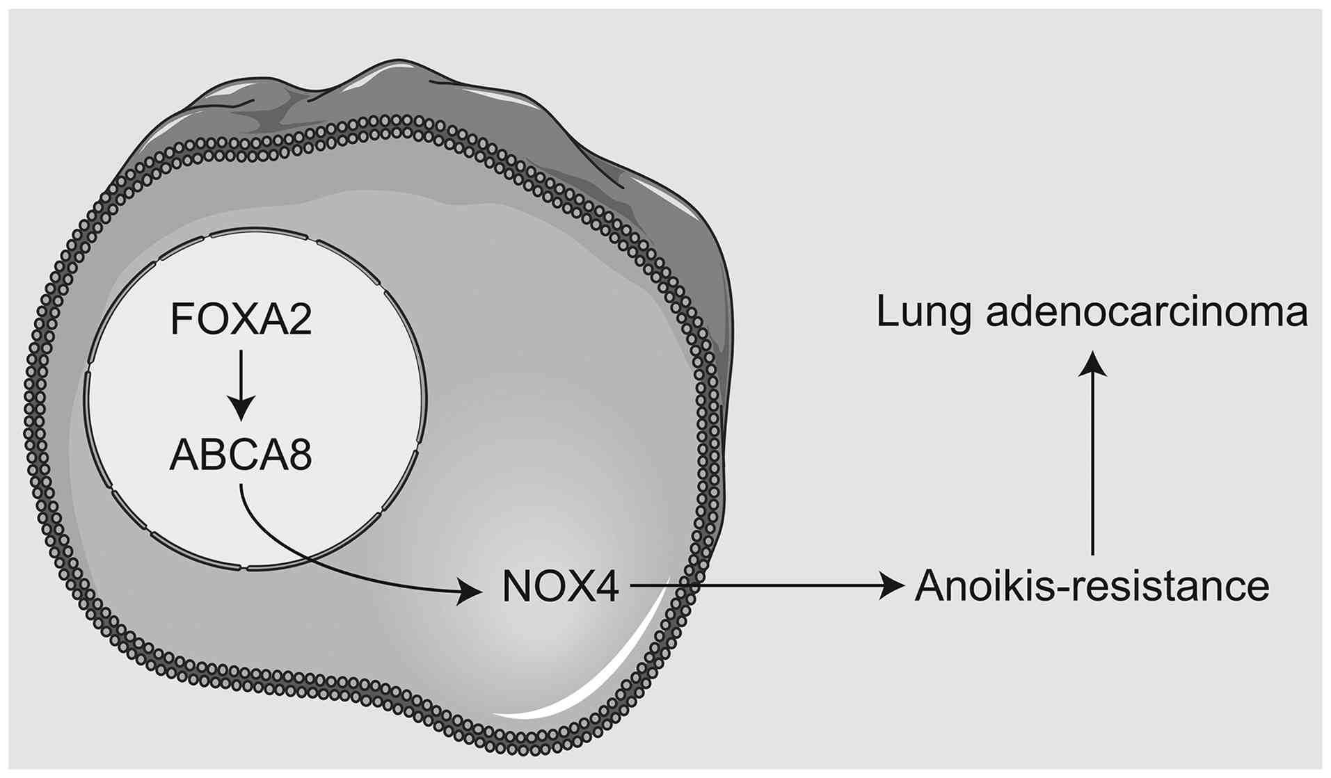
the expression of ABCA8. Collectively, these findings support a
model whereby FOXA2 upregulates ABCA8, which suppresses NOX4 and
alleviates anoikis resistance (Fig.
6).
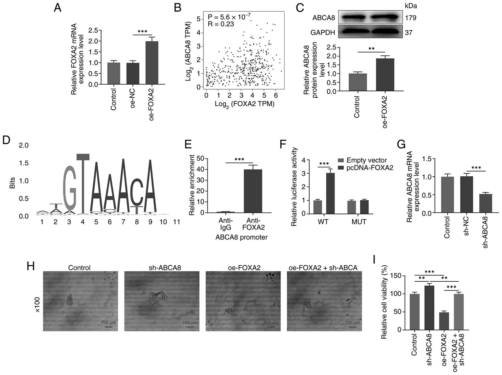 | Figure 4.FOXA2 alleviates anoikis resistance
of A549 cells through promoting the expression of ABCA8. (A) A549
cells were distributed into Control (without treatment), oe-NC and
oe-FOXA2 (transfected with oe-FOXA2) groups. The mRNA expression of
FOXA2 was detected by RT-qPCR, with GAPDH serving as the internal
reference. (B) Gene Expression Profiling Interactive Analysis was
used to predict the association between ABCA8 and FOXA2
(P=5.6×10⁻⁷; R=0.23). (C) ABCA8 protein expression in
A549 cells was determined using western blotting, with GAPDH
serving as the internal reference. (D) JASPAR was utilized to
obtain the predicted binding sites of FOXA2 in ABCA8 promoter
region. (E) Relative enrichment of FOXA2 in the promoter region of
ABCA8 was evaluated using a chromatin immunoprecipitation assay.
(F) Dual-luciferase reporter assay was performed to validate the
binding of FOXA2 and ABCA8. (G) A549 cells (suspension culture)
were divided into Control (without treatment), sh-NC (transfected
with sh-NC) and sh-ABCA8 (transfected with sh-ABCA8) groups. The
mRNA expression of ABCA8 was measured using RT-qPCR, with GAPDH
serving as the internal reference. (H) A549 cells were assigned to
Control (without treatment), sh-ABCA8 (transfected with sh-ABCA8),
oe-FOXA2 (treated as before) and oe-FOXA2 + sh-ABCA8 (transfected
with oe-FOXA2 and sh-ABCA8) groups. Confocal microscopy images were
obtained from a microscope (magnification, ×100; scale bar, 100
µm). (I) Viability of A549 cells (suspension culture) was monitored
using an MTT assay. **P<0.01 and ***P<0.001. FOXA2, forkhead
box protein A2; ABCA8, ATP binding cassette subfamily A member 8;
sh-, short hairpin; NC, negative control; oe-, overexpression;
RT-qPCR; reverse transcription-quantitative PCR. |
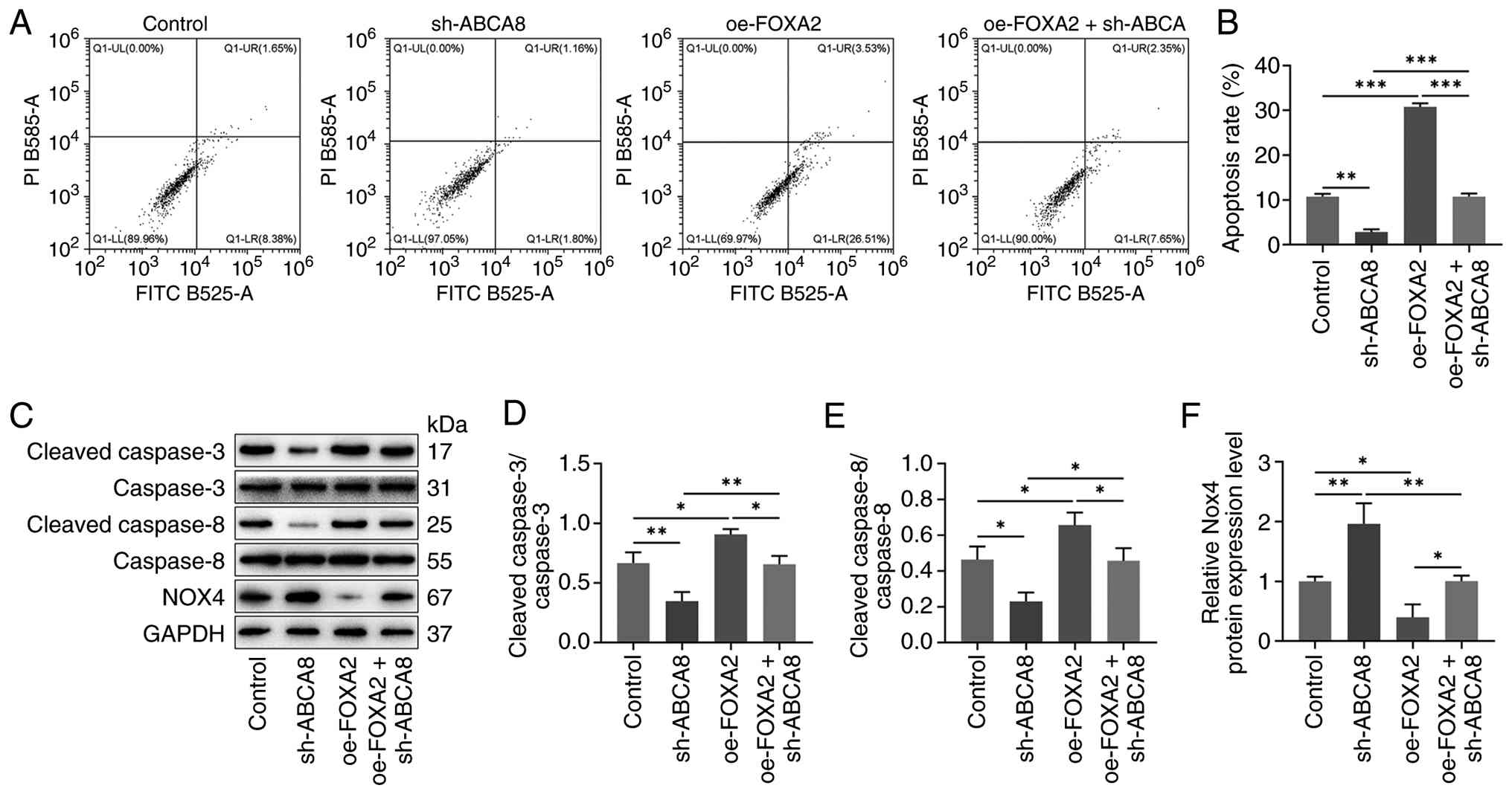 | Figure 5.Effect of knocking down ABCA8 or
overexpressing FOXA2 on the apoptosis rate and the expression of
apoptosis-associated proteins. (A) Flow cytometry was employed to
monitor and (B) quantify apoptosis in A549 cells (suspension
culture). (C) Western blotting was used to measure (D)
cleaved-caspase3, caspase3, (E) cleaved-caspase8, caspase8 and (F)
NOX4 protein expression levels in A549 cells (suspension culture),
with GAPDH serving as the internal reference. Data are presented as
the mean ± SD (n=3). There were three biological replicates in each
experimental group. *P<0.05, **P<0.01 and ***P<0.001.
FOXA2, forkhead box protein A2; ABCA8, ATP binding cassette
subfamily A member 8; NOX4, nicotinamide adenine dinucleotide
phosphate oxidase 4; sh-, short hairpin; NC, negative control; oe-,
overexpression. |
Discussion
ABCA8 belongs to the ABC transporter protein
superfamily, whose downregulation is associated with poor
prognosis, tumorigenesis and metastasis (20). In previous years, more attention has
been paid to the marked effect of ABCA8 on certain cancer types.
ABCA8 is regarded as a prognostic biomarker for gastric
adenocarcinoma, exhibiting relation to immune infiltration,
especially in M2 macrophages (9).
ABCA8 overexpression inhibits breast cancer cell proliferation by
regulating the AMPK/mTOR signaling pathway (31). ABCA8 is lowly expressed in LUAD and
associated with overall survival of patients with LUAD (14); further, ABCA8 suppresses the
proliferation of LUAD cells (5).
Based on bioinformatic analysis, FOXA2 was identified as an
upstream transcription factor of ABCA8. FOXA2 deficiency is
frequently observed in NSCLC, the main mechanism of which is
epigenetic silencing through promoter hypermethylation (32). FOXA2 also acts as a tumor suppressor
of lung cancer metastasis by inhibiting Slug gene expression and
the EMT process (33). However, the
specific role of FOXA2 as a transcription factor in LUAD remains to
be eluidates. In the present study, ABCA8 and FOXA2 mRNA levels
were significantly reduced in patients with LUAD and in A549 cells.
Normal epithelial cells are sensitive to anoikis, whereas cancer
cells are usually resistant to anoikis (34). To compare anoikis sensitivity,
immortalized normal bronchial epithelial cells (BEAS-2B) and lung
cancer cells (A549) were used.
Anoikis, a type of programmed cell death triggered
by cell detachment from the extracellular matrix, is an important
defense against the colonization of distant organs (7). Both anchorage-dependent growth and EMT
are associated with anoikis resistance and are important steps in
cancer progression and metastatic colonization (35). An increasing number of studies on
mechanisms related to anoikis resistance have attracted attention.
For example, nuclear MYH9 overexpression promotes CTNNB1 expression
and enhance anoikis resistance in gastric cancer (36). Glutamate dehydrogenase 1-mediated
reprogramming of glutaminolysis enhances anoikis resistance in
LKB1-deficient lung cancer and promotes tumor metastasis (19). Yet, additional information is still
needed regarding the genes and pathways that mitigate anoikis
resistance in LUAD. As aforementioned, NOX4 is a reactive oxygen
species-generating enzyme and silencing of NOX4 attenuated Src
expression and enhanced anoikis in lung cancer cells (7). TGF-β mediates the EMT process through
the NOX4 signaling pathway, thus promoting lung cancer metastasis
(37). The present study analyzed
and obtained a negative correlation between ABCA8 and NOX4 in LUAD
and accordingly speculated and determined in vitro that
ABCA8 overexpression could reduce anoikis resistance in LUAD by
inhibiting NOX4 expression. Suspension culture of A549 cells has
been utilized to induce anoikis, as this method blocks cell
adhesion to the extracellular matrix (38). The present results showed that NOX4
expression was elevated in suspended A549 cells and was reduced in
A549 cells after oe-ABCA8 transfection. Concurrently, oe-ABCA8
reversed the effects of oe-NOX4 on enhancing cell aggregation,
promoting cell viability, dampening apoptosis, decreasing
cleaved-caspase3 level and promoting NOX4 expression in suspended
A549 cells. This evidence suggested that ABCA8 can negatively
regulate NOX4 to ameliorate anoikis resistance in LUAD cells.
Suspended cells that form multicellular aggregates exhibit a
stronger anoikis resistance than single cells (39). In the present study, the degree of
cell aggregation served as one of the indicators to measure anoikis
resistance.
Since FOXA2 has been identified as an upstream
transcription factor of ABCA8, their association was further
determined. ChIP assay results suggested that FOXA2 was enriched in
the ABCA8 promoter region. The dual-luciferase reporter assay data
showed that FOXA2 bound to the promoter region of ABCA8. In
addition, FOXA2 was positively correlated with ABCA8 in LUAD
according to GEPIA analysis outcomes and western blotting results
demonstrated that oe-FOXA2 promoted ABCA8 protein expression. This
indicated that FOXA2 can positively regulate ABCA8. Reportedly,
oe-FOXA2 can reduce the ERK phosphorylation level, which in turn
inhibits NOX4 expression (15). The
present experimental data determined that oe-FOXA2 abrogated
sh-ABCA8-induced enhancement of cell aggregation, activation of
cell vitality, reduction of apoptosis rate, downregulation of
cleaved caspase3 and upregulation of NOX4 in suspension cultured
A549 cells.
However, the present study exhibits a number of
limitations. First, the present study primarily used cell culture
models to investigate the roles of FOXA2 and ABCA8 in tumor
resistance, which may not fully replicate the complexity of the
in vivo tumor microenvironment, systemic drug responses or
the full metastatic cascade in vivo. Future studies
employing in vivo metastasis models, such as tail vein
injection or orthotopic implantation, will be important in
validating the functional importance of this axis in suppressing
lung colonization and distant metastasis within a complex
physiological microenvironment. Furthermore, while the present
study established ABCA8 as a key downstream effector that
suppresses NOX4, the precise molecular mechanism by which this
transporter protein regulates NOX4 expression potentially through
intermediary signals (including ROS) or kinases (such as ERK)
remains to be determined and is an important focus for future
investigations. Finally, despite the established alterations in
expressions of FOXA2, ABCA8 and NOX4 in a number of LUAD cell lines
(H1299 and H1975), in-depth mechanistic studies remain
predominantly confined to the A549 model. Future research needs to
further demonstrate the regulatory network of this pathway in more
models.
In conclusion, the present findings established a
novel mechanistic association whereby FOXA2-driven ABCA8
transcription sensitizes cancer cells to anoikis by inhibiting NOX4
signaling, thereby inhibiting metastatic dissemination. The
findings provide a novel therapeutic avenue for targeting tumor
metastasis in LUAD. In therapeutic applications, restoring
FOXA2/ABCA8 function or directly inhibiting NOX4 activity could be
combined with existing immunotherapies to overcome drug resistance
and suppress metastasis. In clinical translation, low expression
levels of FOXA2 and ABCA8 may serve as prognostic biomarkers for
identifying high-risk patients with metastatic lung cancer.
Supplementary Material
Supporting Data
Acknowledgements
Not applicable.
Funding
Funding: No funding was received.
Availability of data and materials
The data generated in the present study are included
in the figures and/or tables of this article.
Authors' contributions
NNH and LL performed high-throughput sequencing
experiments and performed the bioinformatics analysis. JQH analyzed
and interpreted data and wrote the manuscript. LFL designed the
study. MMP performed the literature review. MMP and JH were
responsible for data acquisition, data analysis and interpretation,
they also assisted in the creation of charts and literature review,
and participated in the final review and proofreading of the
manuscript. LFL, MMP and JH confirm the authenticity of all the raw
data. All authors read and approved the final version of the
manuscript.
Ethics approval and consent to
participate
Not applicable.
Patient consent for publication
Not applicable.
Competing interests
The authors declare that they have no competing
interests.
References
|
1
|
Bray F, Laversanne M, Sung H, Ferlay J,
Siegel RL, Soerjomataram I and Jemal A: Global cancer statistics
2022: GLOBOCAN estimates of incidence and mortality worldwide for
36 cancers in 185 countries. CA Cancer J Clin. 74:229–263.
2024.PubMed/NCBI
|
|
2
|
Liang G, Meng W, Huang X, Zhu W, Yin C,
Wang C, Fassan M, Yu Y, Kudo M, Xiao S, et al: miR-196b-5p-mediated
downregulation of TSPAN12 and GATA6 promotes tumor progression in
non-small cell lung cancer. Proc Natl Acad Sci USA. 117:4347–4357.
2020. View Article : Google Scholar : PubMed/NCBI
|
|
3
|
Song JQ, Wang X, Zeng ZM, Liang PA, Zhong
CY and Liu AW: Efficacy of PD-1 inhibitors combined with
Anti-angiogenic therapy in driver gene mutation negative
Non-Small-cell lung cancer with brain metastases. Discov Med.
35:321–331. 2023. View Article : Google Scholar : PubMed/NCBI
|
|
4
|
Zuo Y, Shen W, Wang C, Niu N and Pu J:
Circular RNA Circ-ZNF609 promotes lung adenocarcinoma proliferation
by modulating miR-1224-3p/ETV1 signaling. Cancer Manag Res.
12:2471–2479. 2020. View Article : Google Scholar : PubMed/NCBI
|
|
5
|
Rotte A: Combination of CTLA-4 and PD-1
blockers for treatment of cancer. J Exp Clin Cancer Res.
38:2552019. View Article : Google Scholar : PubMed/NCBI
|
|
6
|
Miller KD, Siegel RL, Lin CC, Mariotto AB,
Kramer JL, Rowland JH, Stein KD, Alteri R and Jemal A: Cancer
treatment and survivorship statistics, 2016. CA Cancer J Clin.
66:271–289. 2016.PubMed/NCBI
|
|
7
|
Li Y, Yang Y, Ma Q, Cheng H, Wang H, Ma C,
Li F, Zhao S, Li X, Qi Y and Gu Z: HNRNPK/CLCN3 axis facilitates
the progression of LUAD through CAF-tumor interaction. Int J Biol
Sci. 18:6084–6101. 2022. View Article : Google Scholar : PubMed/NCBI
|
|
8
|
Liu WJ, Wang L, Zhou FM, Liu SW, Wang W,
Zhao EJ, Yao QJ, Li W, Zhao YQ, Shi Z, et al: Elevated NOX4
promotes tumorigenesis and acquired EGFR-TKIs resistance via
enhancing IL-8/PD-L1 signaling in NSCLC. Drug Resist Updat.
70:1009872023. View Article : Google Scholar : PubMed/NCBI
|
|
9
|
Guo Y, Wang ZW, Su WH, Chen J and Wang YL:
Prognostic value and immune infiltrates of ABCA8 and FABP4 in
stomach adenocarcinoma. Biomed Res Int. 2020:41451642020.
View Article : Google Scholar : PubMed/NCBI
|
|
10
|
Wang Y, Cheng S, Fleishman JS, Chen J,
Tang H, Chen ZS, Chen W and Ding M: Targeting anoikis resistance as
a strategy for cancer therapy. Drug Resist Updat. 75:1010992024.
View Article : Google Scholar : PubMed/NCBI
|
|
11
|
Dong B, Gu Y, Sun X, Wang X, Zhou Y, Rong
Z, Zhang J, Shi X, Zhang Z, He X, et al: Targeting TUBB3 suppresses
anoikis resistance and bone metastasis in prostate cancer. Adv
Healthc Mater. 13:e24006732024. View Article : Google Scholar : PubMed/NCBI
|
|
12
|
D'Amore T, Bravoco D, Di Paola G, Albano
F, Brancaccio M, Sabato C, Cesta G, Zolfanelli C, Lauciello V,
Falco G and Mazzone P: Anoikis resistance in gastric cancer: A
comprehensive review. Cell Death Dis. 16:5282025. View Article : Google Scholar : PubMed/NCBI
|
|
13
|
Wang F, Su Q and Li C: Identidication of
novel biomarkers in non-small cell lung cancer using machine
learning. Sci Rep. 12:166932022. View Article : Google Scholar : PubMed/NCBI
|
|
14
|
Yang Y, Liu X, Wang X, Zhang J, Li S and
Ma X: Comprehensive analysis of ABCA family members in lung
adenocarcinoma with prognostic values. Mol Biotechnol. 4:1441–1453.
2022. View Article : Google Scholar : PubMed/NCBI
|
|
15
|
Xu YJ, Huo YC, Zhao QT, Liu JY, Tian YJ,
Yang LL and Zhang Y: NOX4 promotes tumor progression through the
MAPK-MEK1/2-ERK1/2 axis in colorectal cancer. World J Gastrointest
Oncol. 16:1421–1436. 2024. View Article : Google Scholar : PubMed/NCBI
|
|
16
|
Hu T, Li Z, Gao CY and Cho CH: Mechanisms
of drug resistance in colon cancer and its therapeutic strategies.
World J Gastroenterol. 22:6876–6889. 2016. View Article : Google Scholar : PubMed/NCBI
|
|
17
|
Bharathiraja P, Yadav P, Sajid A, Ambudkar
SV and Prasad NR: Natural medicinal compounds target signal
transduction pathways to overcome ABC drug efflux
transporter-mediated multidrug resistance in cancer. Drug Resist
Updat. 71:1010042023. View Article : Google Scholar : PubMed/NCBI
|
|
18
|
Ji N, Li H, Zhang Y, Li Y, Wang P, Chen X,
Liu YN, Wang JQ, Yang Y, Chen ZS, et al: Lansoprazole (LPZ)
reverses multidrug resistance (MDR) in cancer through impeding
ATP-binding cassette (ABC) transporter-mediated chemotherapeutic
drug efflux and lysosomal sequestration. Drug Resist Updat.
76:1011002024. View Article : Google Scholar : PubMed/NCBI
|
|
19
|
Jin L, Chun J, Pan C, Kumar A, Zhang G, Ha
Y, Li D, Alesi GN, Kang Y, Zhou L, et al: The PLAG1-GDH1 axis
promotes anoikis resistance and tumor metastasis through
CamKK2-AMPK signaling in LKB1-deficient lung cancer. Mol Cell.
69:87–99.e7. 2018. View Article : Google Scholar : PubMed/NCBI
|
|
20
|
Cui Y, Liang S, Zhang S, Zhang C, Zhao Y,
Wu D, Wang J, Song R, Wang J, Yin D, et al: ABCA8 is regulated by
miR-374b-5p and inhibits proliferation and metastasis of
hepatocellular carcinoma through the ERK/ZEB1 pathway. J Exp Clin
Cancer Res. 39:902020. View Article : Google Scholar : PubMed/NCBI
|
|
21
|
Kim HJ, Kim D, Yoon H, Choi CS, Oh YS and
Jun HS: Prevention of oxidative Stress-induced pancreatic beta cell
damage by Broussonetia Kazinoki Siebold fruit extract via the
ERK-Nox4 pathway. Antioxidants (Basel). 9:4062020. View Article : Google Scholar : PubMed/NCBI
|
|
22
|
Kim H, Sung JY, Park EK, Kho S, Koo KH,
Park SY, Goh SH, Jeon YK, Oh S, Park BK, et al: Regulation of
anoikis resistance by NADPH oxidase 4 and epidermal growth factor
receptor. Br J Cancer. 116:370–381. 2017. View Article : Google Scholar : PubMed/NCBI
|
|
23
|
Chandrashekar DS, Karthikeyan SK, Korla
PK, Patel H, Shovon AR, Athar M, Netto GJ, Qin ZS, Kumar S, Manne
U, et al: UALCAN: An update to the integrated cancer data analysis
platform. Neoplasia. 25:18–27. 2022. View Article : Google Scholar : PubMed/NCBI
|
|
24
|
Zhang Q, Liu W, Zhang HM, Xie GY, Miao YR,
Xia M and Guo AY: hTFtarget: A comprehensive database for
regulations of human transcription factors and their targets.
Genomics Proteomics Bioinformatics. 18:120–128. 2020. View Article : Google Scholar : PubMed/NCBI
|
|
25
|
Bao J, Li D, Wang L, Wu J, Hu Y, Wang Z,
Chen Y, Cao X, Jiang C, Yan W and Xu C: MicroRNA-449 and
microRNA-34b/c function redundantly in murine testes by targeting
E2F transcription factor-retinoblastoma protein (E2F-pRb) pathway.
J Biol Chem. 287:21686–21698. 2012. View Article : Google Scholar : PubMed/NCBI
|
|
26
|
Yang Z, Wang T, Wu D, Min Z, Tan J and Yu
B: RNA N6-methyladenosine reader IGF2BP3 regulates cell cycle and
angiogenesis in colon cancer. J Exp Clin Cancer Res. 39:2032020.
View Article : Google Scholar : PubMed/NCBI
|
|
27
|
Lee CT, Li R, Zhu L, Tribble GD, Zheng WJ,
Ferguson B, Maddipati KR, Angelov N and Van Dyke TE: Subgingival
microbiome and Specialized Pro-Resolving lipid mediator pathway
profiles are correlated in periodontal inflammation. Front Immunol.
12:6912162021. View Article : Google Scholar : PubMed/NCBI
|
|
28
|
Song Z, Xiang X, Li J, Deng J, Fang Z,
Zhang L and Xiong J: Ruscogenin induces ferroptosis in pancreatic
cancer cells. Oncol Rep. 43:516–524. 2020.PubMed/NCBI
|
|
29
|
Guo X, Wang Z, Sun Q, Sun C, Hua H and
Huang Q: The inhibitory effect of microRNA-1827 on anoikis
resistance in lung adenocarcinoma A549 cells via targeting
caveolin-1. Acta Biochim Biophys Sin (Shanghai). 52:1148–1155.
2020. View Article : Google Scholar : PubMed/NCBI
|
|
30
|
Jiao Y, Li Y, Zhang J, Zhang S, Zha Y and
Wang J: RRM2 alleviates doxorubicin-induced Cardiotoxicity through
the AKT/mTOR signaling pathway. Biomolecules. 12:2992022.
View Article : Google Scholar : PubMed/NCBI
|
|
31
|
Lv C, Yang H, Yu J and Dai X: ABCA8
inhibits breast cancer cell proliferation by regulating the AMP
activated protein kinase/mammalian target of rapamycin signaling
pathway. Environ Toxicol. 37:1423–1431. 2022. View Article : Google Scholar : PubMed/NCBI
|
|
32
|
Basseres DS, D'Alò F, Yeap BY, Löwenberg
EC, Gonzalez DA, Yasuda H, Dayaram T, Kocher ON, Godleski JJ,
Richards WG, et al: Frequent downregulation of the transcription
factor Foxa2 in lung cancer through epigenetic silencing. Lung
Cancer. 77:31–37. 2012. View Article : Google Scholar : PubMed/NCBI
|
|
33
|
Jin S, He J, Zhou Y, Wu D, Li J and Gao W:
LncRNA FTX activates FOXA2 expression to inhibit non-small-cell
lung cancer proliferation and metastasis. J Cell Mol Med.
24:4839–4849. 2020. View Article : Google Scholar : PubMed/NCBI
|
|
34
|
Kim YN, Koo KH, Sung JY, Yun UJ and Kim H:
Anoikis resistance: An essential prerequisite for tumor metastasis.
Int J Cell Biol. 2012:3068792012. View Article : Google Scholar : PubMed/NCBI
|
|
35
|
Paoli P, Giannoni E and Chiarugi P:
Anoikis molecular pathways and its role in cancer progression.
Biochim Biophys Acta. 1833:3481–3498. 2013. View Article : Google Scholar : PubMed/NCBI
|
|
36
|
Ye G, Yang Q, Lei X, Zhu X, Li F, He J,
Chen H, Ling R, Zhang H, Lin T, et al: Nuclear MYH9-induced CTNNB1
transcription, targeted by staurosporin, promotes gastric cancer
cell anoikis resistance and metastasis. Theranostics. 10:7545–7560.
2020. View Article : Google Scholar : PubMed/NCBI
|
|
37
|
Ma M, Shi F, Zhai R, Wang H, Li K, Xu C,
Yao W and Zhou F: TGF-β promote epithelial-mesenchymal transition
via NF-κB/NOX4/ROS signal pathway in lung cancer cells. Mol Biol
Rep. 48:2365–2375. 2021. View Article : Google Scholar : PubMed/NCBI
|
|
38
|
Zhong X and Rescorla FJ: Cell surface
adhesion molecules and adhesion-initiated signaling: understanding
of anoikis resistance mechanisms and therapeutic opportunities.
Cell Signal. 24:393–401. 2012. View Article : Google Scholar : PubMed/NCBI
|
|
39
|
Zhang Y, Lu H, Dazin P and Kapila Y:
Squamous cell carcinoma cell aggregates escape suspension-induced,
p53-mediated anoikis: Fibronectin and integrin alphav mediate
survival signals through focal adhesion kinase. J Biol Chem.
279:48342–48349. 2004. View Article : Google Scholar : PubMed/NCBI
|