Introduction
The Cancer Genome Atlas (TCGA) defines patients with
World Health Organization (WHO) grade II or III glioma as low-grade
glioma (LGG) (1), which accounts
for 20–40% of all glioma cases (2).
The cumulative risk of progression to glioblastoma (GBM) within a
10-year follow-up period is ~70% (3), posing a severe threat to the life and
health of patients. Current treatment strategies are mainly based
on surgical resection combined with neoadjuvant therapies such as
radiotherapy and chemotherapy, yet the postoperative recurrence
rate remains considerably high (4).
With the advance of next-generation sequencing technology, a large
number of tumor-associated molecular biomarkers have been
identified and gradually applied in clinical practice, providing a
novel direction for the precise diagnosis and treatment of tumors
(5). Although GBM exhibits higher
malignancy, genomic expression disorders frequently occur during
tumorigenesis and progression, and the molecular mechanisms
underlying such disorders often differ between GBM and LGG
(6). Therefore, the present study
focused on LGG to screen molecular biomarkers associated with
disease progression, laying a theoretical foundation for subsequent
combined therapy for LGG.
Family with sequence similarity 122 (FAM122) is a
class of highly conserved proteins (7), whose members include FAM122A,
FAM122B and FAM122C. FAM122A is the most
extensively studied member of the family and has been identified as
a key negative regulator of the protein phosphatase 2A
(PP2A) holoenzyme (8).
FAM122A can specifically bind to the interaction interface
between the scaffold subunit Aα and the regulatory subunit B55α
through its conserved domain, directly impairing the assembly
stability of the PP2A/Aα-B55α-C holoenzyme, thereby inhibiting the
phosphatase activity of PP2A (9).
This inhibitory effect leads to hyperphosphorylation and stable
expression of downstream oncoproteins (such as MYC), ultimately
promoting abnormal cell cycle progression, inhibiting apoptosis and
enhancing the invasive and proliferative capacities of tumor cells
(10). For instance, FAM122A
has been confirmed to promote the progression of acute myeloid
leukemia by modulating the expression of PP2A and MYC (10). In addition, in hepatocellular
carcinoma, FAM122A can regulate PP2A-dependent downstream
pathways to facilitate cancer cell growth and increase
chemoresistance to doxorubicin (11).
In contrast to the well-characterized
FAM122A, the physiological functions and pathological
mechanisms of FAM122B remain to be fully elucidated to date.
As a homologous protein of FAM122A, FAM122B may also possess
similar oncogenic potential. In the preliminary stage of the
present study, differential analysis revealed that FAM122B
is highly expressed in LGG, suggesting its potential correlation
with the malignant phenotype of tumors. The present study aimed to
clarify whether FAM122B is involved in regulating the
proliferative capacity of LGG cells and its role in the malignant
progression of the disease, thereby providing a reference for the
subsequent development of diagnostic and therapeutic targets.
Materials and methods
Cell culture
All the cell lines used in the present study
(HA1800, SHG44 and SW1088) were purchased from TongPai (Shanghai)
Biotech Co., Ltd. The HA1800 cells (cat. no. HA1800; http://www.shtpbio.com/tongpai-Products/) used in the
present study were immortalized human normal astrocytes. HA1800 and
SHG44 cells were cultured in high-glucose DMEM medium (Cytiva; cat.
no. SH30022), while SW1088 cells were maintained in Leibovitz L-15
medium (Cytiva; cat. no. SH30525). All media were supplemented with
10% fetal bovine serum (HySigen Biosciences; cat. no. FBP-S005) and
1% penicillin-streptomycin solution (Beijing Solarbio Science &
Technology Co, Ltd.; cat. no. P1400) to sustain cell growth and
inhibit microbial contamination. The cells were incubated in a
constant-temperature cell incubator at 37°C with 5% CO2
and saturated humidity. Cell density was observed daily. When the
cell confluence reached ~90%, the cells were rinsed twice with
phosphate-buffered saline (Beijing Solarbio Science &
Technology Co, Ltd.; cat. no. P1020), followed by digestion into
single-cell suspension using 0.25% trypsin-EDTA (Beijing Solarbio
Science & Technology Co, Ltd.; cat. no. T1300). The digestion
was terminated with complete medium prior to subsequent
treatments.
Cell transfection
SHG44 and SW1088 cells were seeded at a density of
5×10⁵ cells/well into 6-well cell culture plates (Wuhan Servicebio
Technology Co., Ltd.; cat. no. CCP-6H) and cultured at 37°C
overnight until cell confluence reached 50–60%. After which, cells
were treated with the transfection reagent,
Lipofectamine® 3000 (Thermo Fisher Scientific, Inc.;
cat. no. L3000075), and the transfection medium, Opti-MEM™ I
Reduced Serum Medium (Thermo Fisher Scientific, Inc.; cat. no.
31985070). Small interfering RNA (siRNA) targeting FAM122B
was purchased from Shanghai GenePharma Co., Ltd. The sequences of
FAM122B siRNA were sense strand:
5′-CGGAGGAATAGTACAACAATTdTdT-3′, antisense strand:
5′-AATTGTTGTACTATTCCTCCGdTdT-3′ (RefSeq accession number:
NM_145284). The sequences of negative control siRNA were sense
strand: 5′-UUCUCCGAACGUGUCACGUdTdT-3′, antisense strand:
5′-ACGUGACACGUUCGGAGAAdTdT-3′. The final working concentration of
siRNA for cell transfection was 50 nM. For transfection,
FAM122B siRNA and Lipofectamine® 3000 reagent
were separately diluted in Opti-MEM Reduced Serum Medium, allowed
to stand for 5 min, gently mixed and incubated at room temperature
for 20 min to form stable transfection complexes. Subsequently, the
transfection complexes were slowly added dropwise into 6-well
plates containing reduced serum medium. After 6–8 h of
transfection, the medium was replaced with complete medium and cell
culture was continued.
Reverse transcription-quantitative
(RT-q)PCR
The cells were seeded at a confluency of 60–70%. At
24–48 h post-transfection, total RNA was extracted from LGG cells
using an RNA extraction kit (Omega Bio-Tek, Inc.; cat. no. R6834).
The concentration and purity of RNA were measured by Thermo Fisher
NanoDrop micro-spectrophotometer (Thermo Fisher Scientific, Inc.).
After which, the extracted RNA was reverse-transcribed into cDNA
using a reverse transcription kit (Novoprotein Scientific Co.,
Ltd.; cat. no. E047) under the following conditions: 25°C for 5
min, 50°C for 15 min and 85°C for 5 min, with final storage at 4°C.
qPCR was performed using a qPCR kit (Novoprotein Scientific Co.,
Ltd.; cat. no. E096) to verify transfection efficiency with the
following conditions: Pre-denaturation at 95°C for 5 min for 1
cycle; 95°C for 10 sec and 60°C for 30 sec for a total of 40
cycles. Primer sequences were as follows: FAM122B (alias
PABIR2) forward primer, AAGGGAAATGCAAACGGCAAT and reverse
primer, TCTCCGGCTTGTCAAAATCAC; 18S forward primer,
CACCAGACTTGCCCTCCAAT and reverse primer, CCTGAGAAACGGCTACCACAT. All
primers were obtained from Zhengzhou Shangzhiya Biotechnology Co.,
Ltd. RNA extraction, cDNA synthesis and qPCR were performed
strictly in accordance with the manufacturer's protocols. The
2−ΔΔCq method was used to calculate relative gene
expression levels (12), and all
aforementioned experiments were repeated in triplicate. Under the
experimental conditions used in this study, 18S was stably
expressed, with no significant differences in Cq values
between the NC and siRNA groups.
CCK-8 cell proliferation assay
Transfected SHG44 and SW1088 cells were seeded in
96-well plates at a density of 2×10³ cells/well, with 6
replicate wells set for each group to reduce experimental errors.
Assays were performed at the following five time points: 0, 24, 48,
72 and 96 h post-seeding. Specifically, 10 µl CCK-8 reagent
(Shanghai Saint-Bio Biotechnology Co., Ltd.; cat. no. ST1008) was
added to each well, and the plate was gently shaken to ensure
thorough mixing of the reagent with the medium. The plate was then
incubated at 37°C for 1 h in the dark. After incubation, a
microplate reader was used to measure the absorbance (OD value) of
each well at a wavelength of 450 nm, with the OD value serving as
an indicator of cell proliferation activity.
Colony formation assay
Transfected SHG44 and SW1088 cells were digested
into a single-cell suspension, counted and uniformly seeded in
6-well plates at a density of 1,000 cells/well, with three
replicate wells set up for each group. The cells were cultured for
10–15 days, with the medium replaced every three days. After
visible cell clones formed, the medium was discarded, and the cells
were fixed with 4% paraformaldehyde at room temperature for 30 min,
followed by staining with 0.1% crystal violet solution at room
temperature for a further 30 min. Excess dye was rinsed off with
running water, and the plates were air-dried. Cell colonies were
defined as cell clusters with a size of >100 pixel2
and analyzed using ImageJ software (version 1.54i; National
Institutes of Health).
Wound healing assay
Transfected SHG44 and SW1088 cells were seeded in
6-well plates and cultured until >90% confluence. A horizontal
line was drawn on the back of each 6-well plate as an anchor point
to ensure consistent imaging fields at all time points. A straight
scratch wound was made vertically across the horizontal line at the
center of each well using a sterile 1,000 µl pipette tip, with
effort made to maintain uniform wound width across all wells. The
cells were rinsed twice with PBS to remove floating cells, and the
medium was replaced with serum-free medium. Images of the wound
areas were captured under an inverted microscope at 0, 24 and 48 h
post-scratching, respectively. The wound healing rate was
calculated using Image software (version 1.54i, National Institutes
of Health) according to the following formula: Wound healing
rate=[(wound width at 0 h-wound width at 24/48 h)/wound width at 0
h] ×100%. This index was used to evaluate the migratory ability of
the cells.
Transwell assay
Cell migration ability was assessed using uncoated
Transwell chambers (Wuhan Servicebio Technology Co., Ltd.; cat. no.
WG3415). Transfected SHG44 and SW1088 cells were digested into a
single-cell suspension, resuspended in serum-free medium and
adjusted to a density of 1×10⁵ cells/ml. A 200 µl aliquot of the
cell suspension was added to the upper chamber, while 600 µl
complete medium was placed in the lower chamber as a
chemoattractant. After incubation at 37°C for 24 h, non-migratory
cells on the upper surface of the chamber membrane were gently
wiped off with cotton swabs. Migratory cells on the lower surface
were fixed with 4% paraformaldehyde (Wuhan Servicebio Technology
Co., Ltd.; cat. no. P1110) at room temperature for 30 min and
stained with 0.1% crystal violet solution (Wuhan Servicebio
Technology Co., Ltd.; cat. no. C8470) at room temperature for a
further 30 min. Excess dye on the membrane surface was rinsed away
slowly with running water, followed by air-drying at room
temperature. Images were captured under an inverted microscope for
cell counting, and the average number of migratory cells was
calculated to evaluate cell migration ability.
Data collection
Several public databases and datasets were employed
for bioinformatics analyses. Firstly, gene expression profiles were
retrieved from five publicly available Gene Expression Omnibus
(GEO) datasets (https://www.ncbi.nlm.nih.gov/geo/) (dataset nos.
GSE35493, GSE43378, GSE50025, GSE74187 and GSE83300), with detailed
characteristics of these datasets presented in Table SI. TCGA (https://portal.gdc.cancer.gov/) database provided mRNA
sequencing data and corresponding clinical information of 503 LGG
cases, and the Chinese Glioma Genome Atlas (CGGA) database
(http://www.cgga.org.cn/) included 403 cases of
mRNA sequencing data, 142 cases of microarray data and matched
clinical characteristics of LGG. In addition, the Human Protein
Atlas (HPA) database (https://www.proteinatlas.org/), contained
immunohistochemistry (IHC) data of FAM122B in three normal
cerebral cortex samples and five LGG tissue samples. Finally, the
TIMER database (TIMER2.0; http://cistrome.shinyapps.io/timer/) supplied immune
cell infiltration data of LGG tissues. All data were downloaded
from the official websites of the corresponding databases. The
inclusion criteria were complete mRNA expression data and available
clinical follow-up information.
Gene set enrichment analysis
(GSEA)
To explore the potential molecular pathways through
which high FAM122B expression regulates LGG progression,
GSEA was performed using GSEA software (version 4.3.3; http://www.gsea-msigdb.org/gsea/index.jsp). The gene
set employed for analysis was c2.cp.v7.5.symbols.gmt
(pathway-associated gene set), which was used to assess the
enrichment levels of pathways between the FAM122B
high-expression group and low-expression group. Pathways with a
false discovery rate (FDR) <0.25 and P<0.05 were defined as
markedly enriched pathways.
Meta-analysis
For retrospective studies, meta-analysis can
integrate and pool multiple datasets from different sources,
thereby improving the generalizability and extrapolation of
research conclusions. Previous findings of the present study
indicated that high expression of FAM122B serves as an
adverse prognostic risk factor in LGG. To further expand the sample
size and verify whether the negative effect of FAM122B
expression on the prognosis of patients with LGG is universally
applicable, the following seven independently sourced datasets were
included in the present study: i) TCGA RNA-Seq (https://portal.gdc.cancer.gov/); ii) CGGA
mRNA-array (http://www.cgga.org.cn/); iii) CGGA
RNA-Seq; iv) GSE43378 (13); v)
GSE50025 (14); vi) GSE74187
(15); and vii) GSE83300 (16) (https://www.ncbi.nlm.nih.gov/geo/). The aforementioned
datasets cover cohorts from different countries and ethnic
populations, and adopt multiple molecular detection platforms.
Prior to conducting the meta-analysis, a separate prognostic
analysis was performed for each dataset. The Cox regression model
was used to obtain the prognostic effect sizes of each cohort,
followed by meta-analysis to pool and synthesize these prognostic
effect sizes across all studies. The present study used the
I2 statistic to assess the heterogeneity among datasets.
Statistical analyses were conducted using R software (version
4.5.0; R Foundation for Statistical Computing; http://www.r-project.org/) and the ‘meta’ (https://CRAN.R-project.org/package=meta)
package. Significant statistical heterogeneity was defined as
I2>50% accompanied by P<0.05 for the heterogeneity
test. A random-effects model was used for pooled analyses to
account for potential between-study heterogeneity.
Statistical analysis
Statistical analyses and visualizations were
performed using R software (version 4.5.0) with the ‘survival’
(https://CRAN.R-project.org/package=survival),
‘survminer’ (https://CRAN.R-project.org/package=survminer),
‘corrplot’ (https://CRAN.R-project.org/package=corrplot),
‘ggplot2’ (https://CRAN.R-project.org/package=ggplot2), ‘dplyr’
(https://CRAN.R-project.org/package=dplyr), ‘pROC’
(https://CRAN.R-project.org/package=pROC) and
‘forestplot’ (https://CRAN.R-project.org/package=forestplot)
packages. A two-tailed significance level was set at α=0.05, with
P<0.05 considered to indicate a statistically significant
difference. First, data on target gene expression,
clinicopathological characteristics and survival outcomes (endpoint
events, follow-up time and censoring status) of the study samples
were collated. After quality control, samples were divided into
high and low expression groups based on the median value of gene
expression. For clinical correlation analysis, Pearson/Spearman
correlation analyses were used for continuous variables, and
χ2 test was applied for categorical variables.
Kaplan-Meier curves were plotted to visualize survival
distributions, and the Log-rank test was used to compare survival
differences between the high and low gene expression groups.
Univariate and multivariate Cox proportional hazards regression
analyses were sequentially conducted. After verifying the
proportional hazards assumption via the Schoenfeld residual method,
independent risk factors for survival outcomes were identified,
with hazard ratio (HR) and 95% confidence interval (CI) calculated,
and forest plots generated to present the results. The ‘ROC’
package was used to plot receiver operating characteristic (ROC)
curves and calculate the area under the curve (AUC) for evaluating
the predictive value of target gene expression for clinical
outcomes of the subjects. All experimental data were statistically
analyzed using GraphPad Prism 10.0 software (Dotmatics). Student's
t-test was used for comparisons between two groups, while one-way
ANOVA followed by Dunnett's multiple comparisons test was applied
for datasets with three or more groups. Quantitative data were
expressed as mean ± standard deviation. Survival data were
displayed via Kaplan-Meier survival curves. Correlation results
were presented in correlation heatmaps. HR results were visualized
by forest plots. Diagnostic efficacy was shown by ROC curves.
Enumeration data were presented as case numbers and
percentages.
Results
Expression analysis of FAM122B in
LGG
Malignant progression of gliomas is often
accompanied by disorders of genomic expression profiles (17). To clarify the expression
characteristics of FAM122B in gliomas, the present study
first performed bioinformatics analysis using the GSE35493 dataset
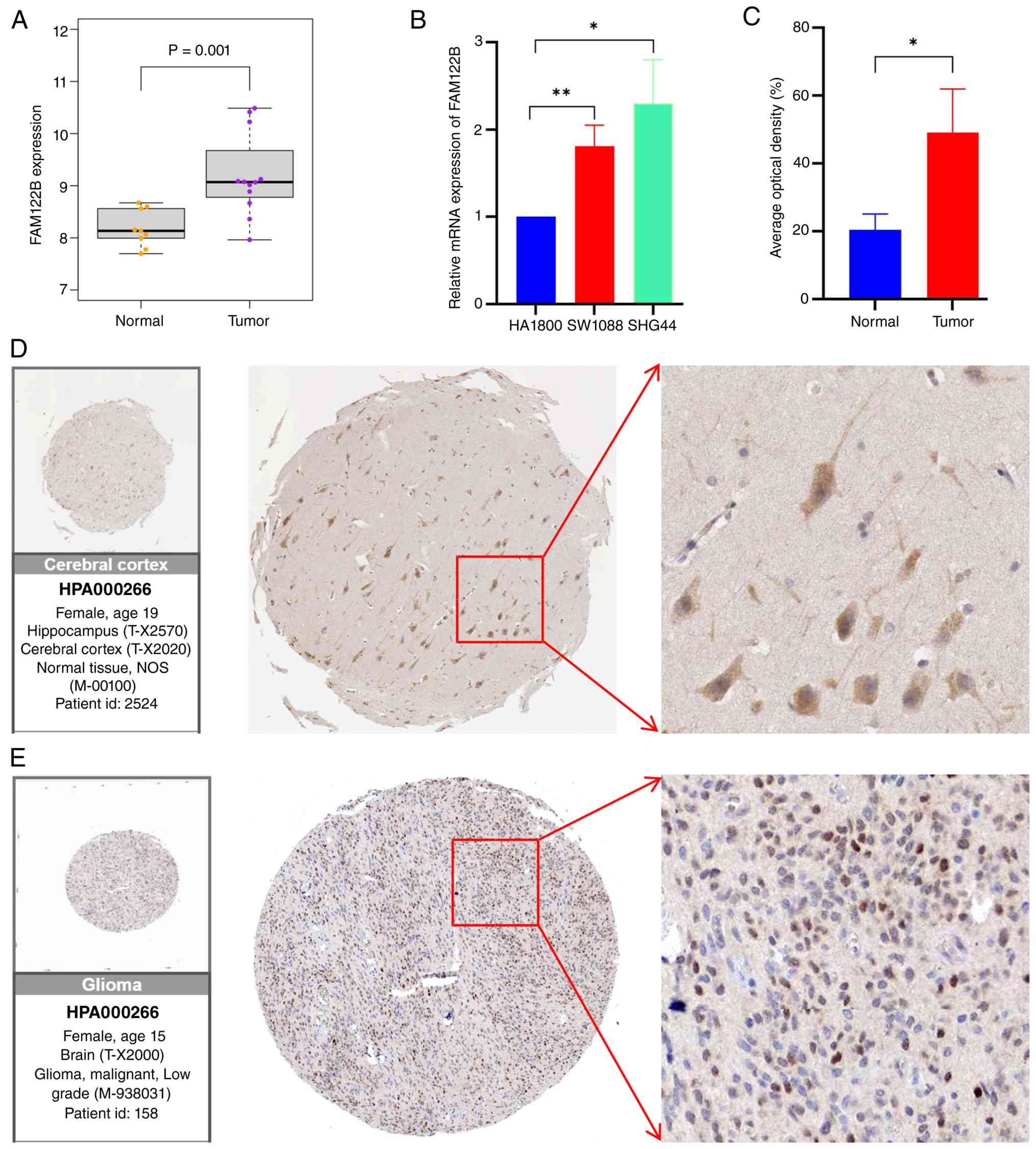
from the GEO database. The results showed that the mRNA level of
FAM122B was markedly higher in tumor tissues than in normal
brain tissues (normal group, n=9; tumor group, n=12; Fig. 1A). To verify the aforementioned
bioinformatics analysis results, the mRNA expression level of
FAM122B in LGG cell lines and normal astrocytes was detected
using the RT-qPCR assay. The human normal astrocyte cell line
HA1800 was selected as the control, and the LGG cell lines SW1088
and SHG44 were used as the research objects. The results confirmed
that the mRNA of FAM122B was markedly higher in LGG cell
lines than in normal HA1800 glial cells (Fig. 1B), which was consistent with the
database analysis results. Furthermore, the expression difference
of FAM122B was verified at the protein level. The IHC data
for FAM122B were retrieved from the HPA database, and
downloaded and analyzed the IHC results of three cases of normal
cerebral cortex tissues and five cases of LGG tumor tissues
(Figs. 1D and E; S1 and S2). The results showed that compared with
normal cerebral cortex tissues, the protein expression level of
FAM122B in LGG tumor tissues was markedly increased
(Fig. 1C). The results of the
present study demonstrated that FAM122B is highly expressed
at both the mRNA and protein levels in LGG tissues and cell lines,
which lays an experimental foundation for the subsequent in-depth
exploration of the correlation between the high expression of
FAM122B and the poor prognosis of patients with LGG.
Analysis of the correlation between
FAM122B expression and clinical characteristics of patients with
LGG
To explore the correlation between FAM122B
expression and the clinical characteristics of patients with LGG,
mRNA expression data and corresponding clinical characteristic
information from patients with LGG were downloaded from three
databases, namely TCGA-seq, CGGA-seq and CGGA microarray, and an
integrated analysis was performed. Firstly, in terms of clinical
grading, the prognosis of patients with WHO grade III LGG was
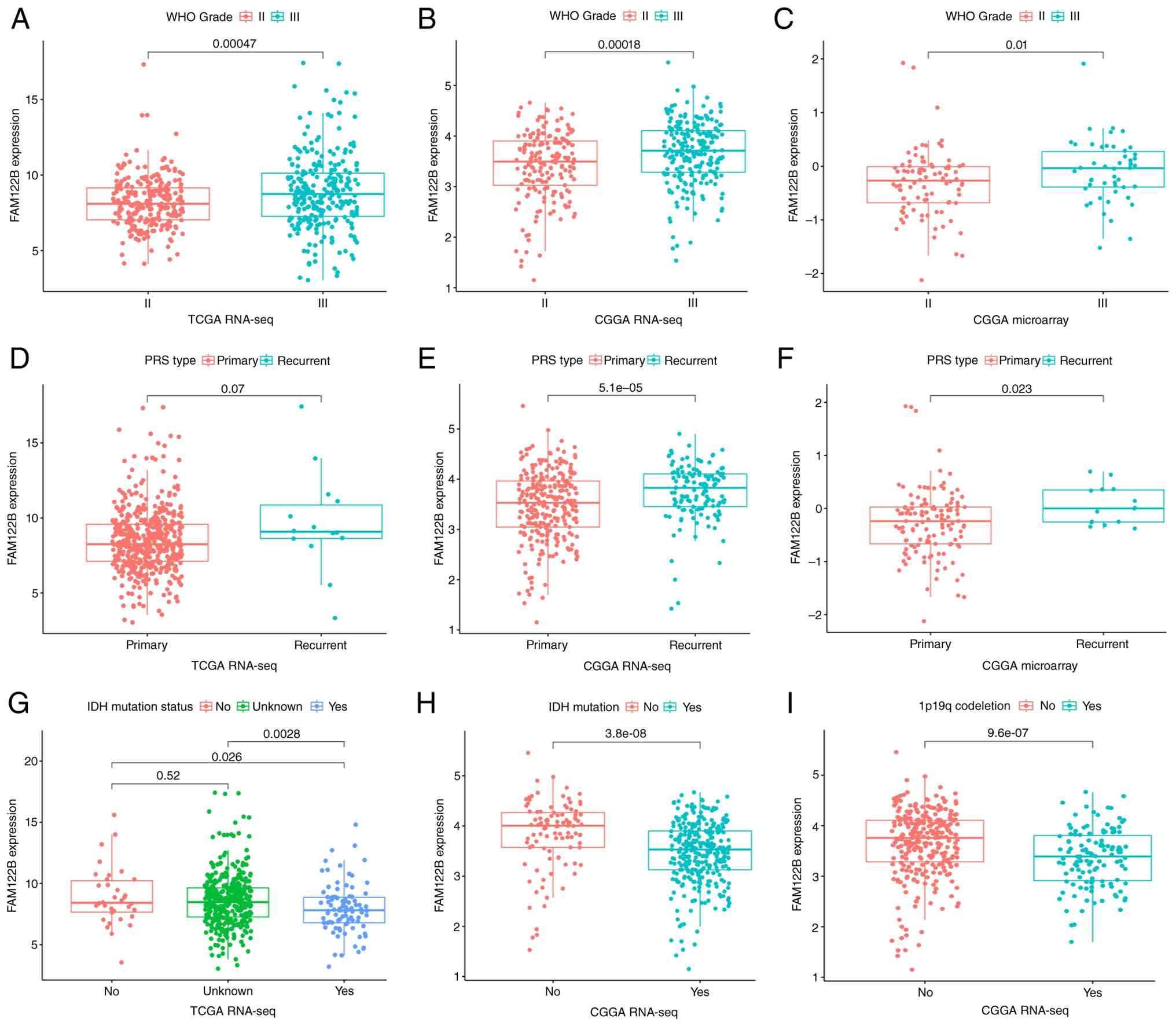
markedly worse than that of patients with WHO grade II (18). Meanwhile, in all three
aforementioned databases, it was observed that the mRNA expression
level of FAM122B was markedly upregulated with increasing
WHO grade of gliomas (Fig. 2A-C),
suggesting a positive correlation between its expression and the
malignant degree of tumors. Second, compared with primary LGG,
recurrent LGG tumor cells possess stronger invasiveness and lead to
worse prognosis (19). Data from
three databases indicated that the mRNA expression level of
FAM122B was higher in samples from recurrent patients than
in those from primary patients (Fig.
2D-F). Finally, analysis of key molecular markers based on
glioma grading showed that IDH mutation and 1p19q chromosome
co-deletion are important indicators of favorable prognosis in
patients with LGG (20). Meanwhile,
the mRNA expression level of FAM122B in tumor tissues was
correspondingly decreased in patients with LGG with IDH mutation
(Fig. 2G and H) and 1p19q
co-deletion (Fig. 2I) phenotypes,
which further confirmed that FAM122B expression is
correlated with the prognostic characteristics of patients. In
summary, the results of multi-database and multi-clinical dimension
analyses all indicate that high FAM122B expression is
closely correlated with malignant clinical phenotypes of patients
with LGG (high grade, recurrence, absence of IDH mutation and
non-co-deletion of 1p19q), suggesting that its high expression may
drive the deterioration of clinical prognosis in patients with LGG.
Based on these results, the direct correlation between
FAM122B expression level and the prognosis of patients with
LGG will be further explored in subsequent studies.
FAM122B is a potential prognostic
marker for LGG
Survival time is one of the core direct indicators
for evaluating the prognosis of patients with tumors. To clarify
the direct correlation between FAM122B expression level and
the prognosis of patients with LGG, the survival time and survival
status information of all patients with LGG were extracted from the
three aforementioned databases, the patients were divided into
high-expression and low-expression groups according to the mRNA
expression level of FAM122B, and survival prognosis analysis
was conducted. First, KM survival curve analysis was performed. The
results showed that in the three databases of TCGA-seq, CGGA-seq
and CGGA microarray, with the extension of follow-up time, the
survival rate of patients in the FAM122B high-expression
group was consistently lower than that in the low-expression group
(Fig. 3A-C), and the differences
between the groups were statistically significant (all P<0.05).
Subsequently, the predictive value of FAM122B for the
survival outcomes of patients with LGG was evaluated by ROC curve
analysis. The results showed that in the three databases, the AUC
of FAM122B for predicting the 1-, 3- and 5-year survival
outcomes of patients was stably ~0.7 (Fig. 3D-F), suggesting that the short-term
and medium-term survival prognosis of patients with LGG can be
moderately predicted based solely on the expression level of
FAM122B. Combined with the previous results on the
correlation between high FAM122B expression and malignant
clinical phenotypes of LGG, the survival analysis in this section
further confirms that FAM122B is expected to serve as a
molecular marker for evaluating the prognosis of patients with
LGG.
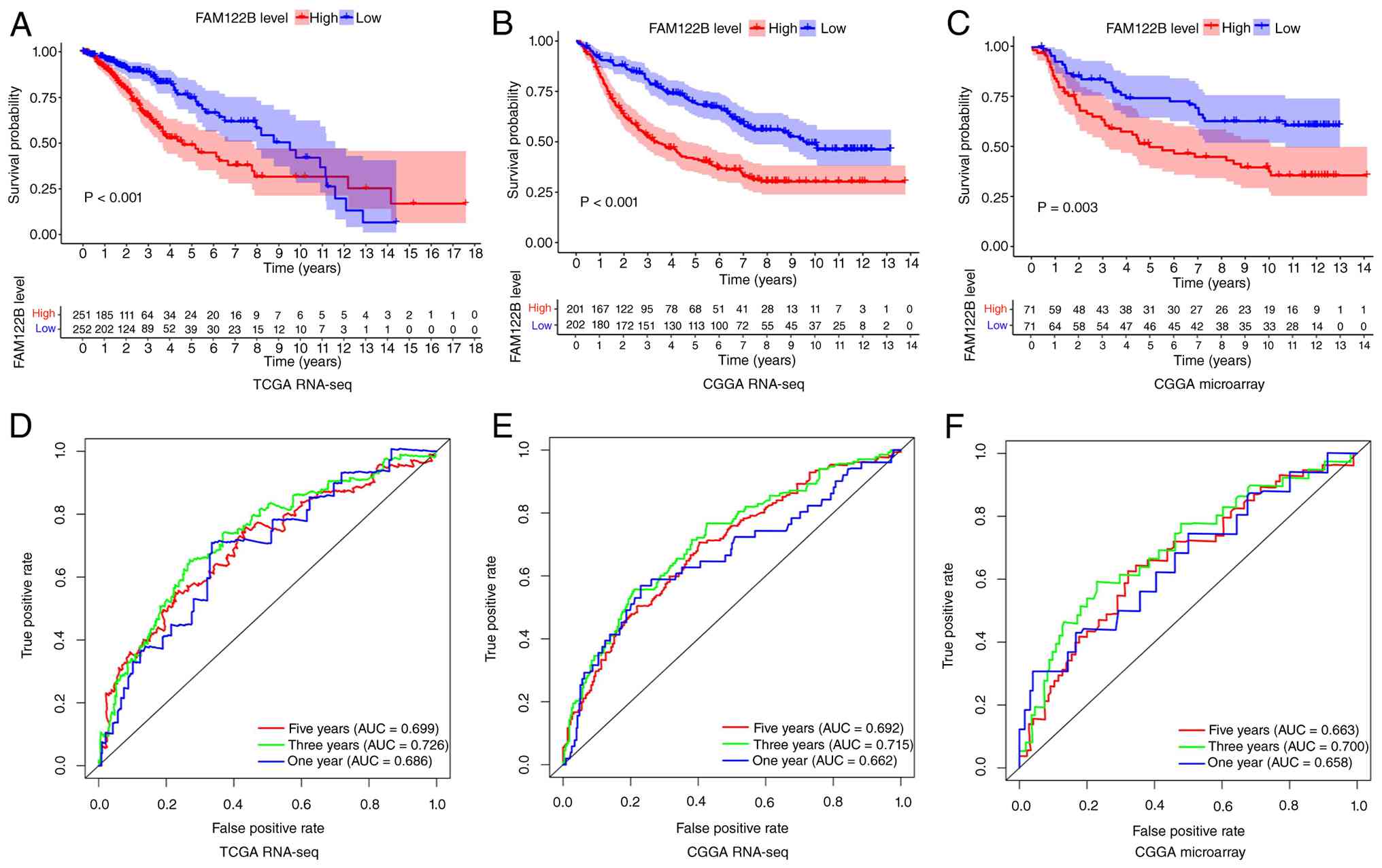 | Figure 3.Prognostic value of FAM122B
expression level in patients with LGG. (A) Kaplan-Meier survival
curves of patients with LGG stratified by FAM122B expression
levels based on TCGA RNA-seq dataset. (B) Kaplan-Meier survival
curves of patients with LGG stratified by FAM122B expression
levels based on CGGA RNA-seq dataset. (C) Kaplan-Meier survival
curves of patients with LGG stratified by FAM122B expression
levels based on CGGA microarray dataset. (D) ROC curves and AUC
values of FAM122B for predicting 1-, 3- and 5-year survival
outcomes of patients with LGG based on the TCGA RNA-seq dataset.
(E) ROC curves and AUC values of FAM122B for predicting 1-,
3- and 5-year survival outcomes of patients with LGG based on the
CGGA RNA-seq dataset. (F) ROC curves and AUC values of
FAM122B for predicting 1-, 3- and 5-year survival outcomes
of patients with LGG based on the CGGA microarray dataset.
FAM122B, Family with sequence similarity 122, member B; LGG,
low-grade glioma; TCGA, The Cancer Genome Atlas; CGGA, Chinese
Glioma Genome Atlas; ROC, plot receiver operating characteristic;
AUC, area under the curve. |
FAM122B is an independent poor
prognostic factor for patients with LGG
Although the aforementioned ROC curve analysis
clarified the predictive ability of FAM122B expression for
the survival outcomes of patients with LGG, this analysis failed to
exclude the potential confounding interference of clinical
indicators such as age, pathological features and tumor stage,
making it difficult to confirm its independent prognostic value.
Therefore, the present study further adopted univariate and
multivariate Cox proportional hazards regression models to clarify
whether high FAM122B expression remains an independent risk
factor for poor prognosis in patients with LGG after adjusting for
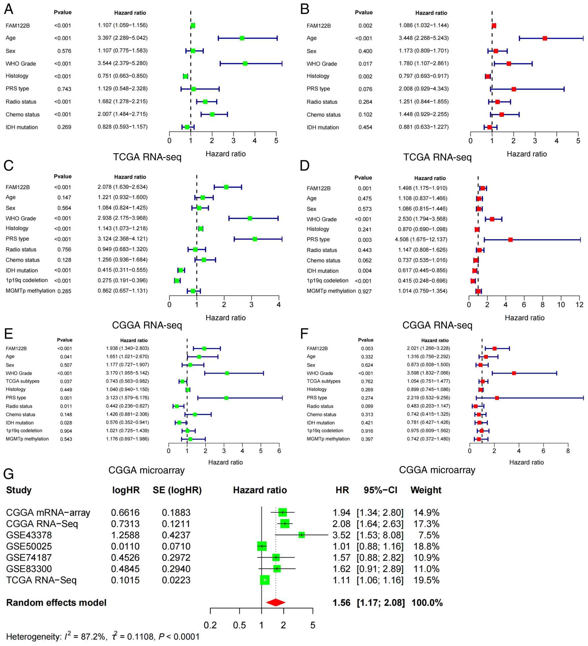
the aforementioned confounding factors. Firstly, univariate Cox
proportional hazards regression analysis was performed on samples
from the three databases separately to rapidly screen for
indicators with significant impacts on patient survival. The
results showed that FAM122B expression level, age, WHO grade
and tumor recurrence status were all significant factors affecting
the prognosis of patients with LGG (all HR>1; P<0.05;
Fig. 4A, C and E). Subsequently,
these factors were incorporated into a multivariate Cox
proportional hazards regression analysis to adjust for the mutual
interference among the various indicators. The results showed that
across all three databases, only high FAM122B expression and
elevated WHO grade were independent prognostic factors for poor
prognosis in patients with LGG (Fig.
4B, D and F). Finally, the results of the meta-analysis across
multiple datasets (HR, 1.56; 95% CI, 1.17–2.08) also indicated that
FAM122B is a risk factor for poor prognosis in patients with
LGG (Fig. 4G). In conclusion, the
independent prognostic value of FAM122B in LGG was
clarified, which lays an important theoretical foundation for
subsequent in-depth exploration of the molecular regulatory
mechanisms of FAM122B, and also provides a potential target
for LGG prognosis evaluation and targeted therapy.
Co-expression analysis and GSEA of
FAM122B
The occurrence and development of tumors typically
involve multi-gene synergistic regulatory networks (21). To further clarify the mechanism
underlying the role of FAM122B in the pathological
progression of LGG, the present study performed co-expressed gene
screening and analysis of FAM122B based on TCGA database.
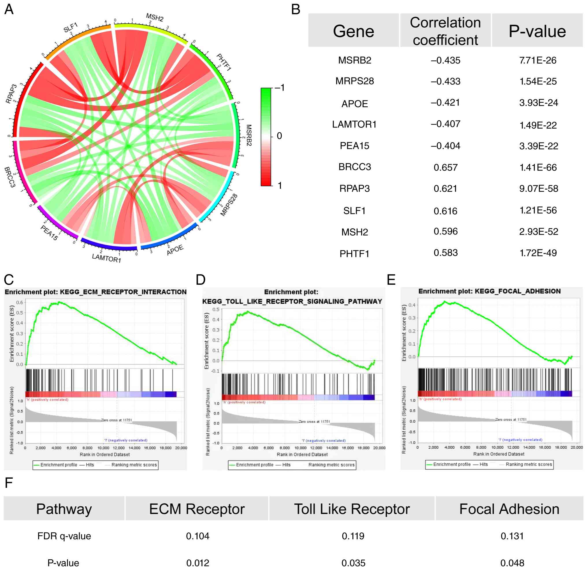
The top five genes with the strongest positive correlation with
FAM122B were identified as BRCC3, RPAP3, SLF1, MSH2
and PHTF1, while the top five genes with the strongest
negative correlation were MSRB2, MRPS28, APOE, LAMTOR1 and
PEA15 (Fig. 5A and B).
Combined with information from the literature review, it was found
that among the positively correlated genes, BRCC3, SLF1 and
MSH2 have all been reported as oncogenes involved in
tumorigenesis and progression (22–24).
By contrast, LAMTOR1 and PEA15 are among the
negatively correlated genes which have been confirmed to exert
tumor-suppressive functions (25,26).
The correlation between the aforementioned co-expression analysis
results and genes with established functions further corroborates
the potential oncogenic properties of FAM122B in LGG.
To clarify the specific molecular pathways through
which FAM122B regulates the malignant progression of LGG,
GSEA was performed. The results showed that the samples in the
FAM122B high-expression group were markedly enriched in the
extracellular matrix (ECM)-receptor interaction pathway, Toll-like
receptor pathway and focal adhesion (Fig. 5C-E). Moreover, FDR <0.25 and
P<0.05 was noted for all enriched pathways (Fig. 5F). Previous studies have confirmed
that the aforementioned pathways are closely associated with tumor
cell proliferation, invasion and metastasis, as well as the
remodeling of the tumor immune microenvironment (27–29).
Accordingly, it was suggested that FAM122B may regulate the
activity of these key pathways either positively or negatively,
thereby affecting the malignant biological behaviors of LGG, which
provides an important direction for further elucidating its
molecular regulatory mechanisms.
Regulatory effect of FAM122B high
expression on the immune microenvironment of LGG
To elucidate the specific mechanism through which
high FAM122B expression modulates the malignant biological
behaviors of LGG, first, the ESTIMATE algorithm was employed to
calculate the TME scores of patients with LGG stratified into high
and low FAM122B expression groups. The results demonstrated
that a statistically significant difference existed exclusively in
the immune score between the two groups (Fig. 6A). This finding suggested that
FAM122B may participate in the remodeling of the LGG immune
microenvironment by regulating immune cell infiltration. Second,
analysis of immune cell subset scatter plots revealed that the
expression level of FAM122B may correlate with the
infiltration levels of memory CD4+ T cells, M2-type macrophages,
activated natural killer cells and monocytes (Fig. 6B). It was therefore hypothesized
that FAM122B may reshape the immune microenvironment by
regulating changes in the distribution of these immune cell
subsets, thereby promoting the malignant progression of LGG.
Subsequently, the correlation between FAM122B expression and
immune cell infiltration was explored using the TIMER database. The
results showed that only CD8+ T cells were positively correlated
with FAM122B expression (Fig.
S3A). Moreover, survival analysis indicated that high
expression of both CD8+ T cells and FAM122B markedly
shortened the overall survival of patients with LGG (Fig. S3B), suggesting that the two may
synergistically affect patient prognosis. Immune checkpoints play a
pivotal role in tumor immune escape and immunotherapy (30). Based on TCGA database, Spearman
correlation analysis was performed to investigate the expression
correlation between FAM122B and classical immune checkpoint
molecules (CD274, CTLA4, HAVCR2, CD96, KLRB1 and IDO1). The results
showed that FAM122B was positively correlated with most of
these immune checkpoint molecules, among which the correlation
coefficient with CD274 was the highest (Table SII). As the primary ligand for
programmed cell death protein 1 (PD-1), CD274 can mediate tumor
cells to escape immune system attacks (31). Collectively, these bioinformatics
analyses suggest that FAM122B may regulate the progression
of LGG via participating in the remodeling of tumor immune
microenvironment, which indicates that it may serve as a promising
candidate target for immunotherapy of LGG.
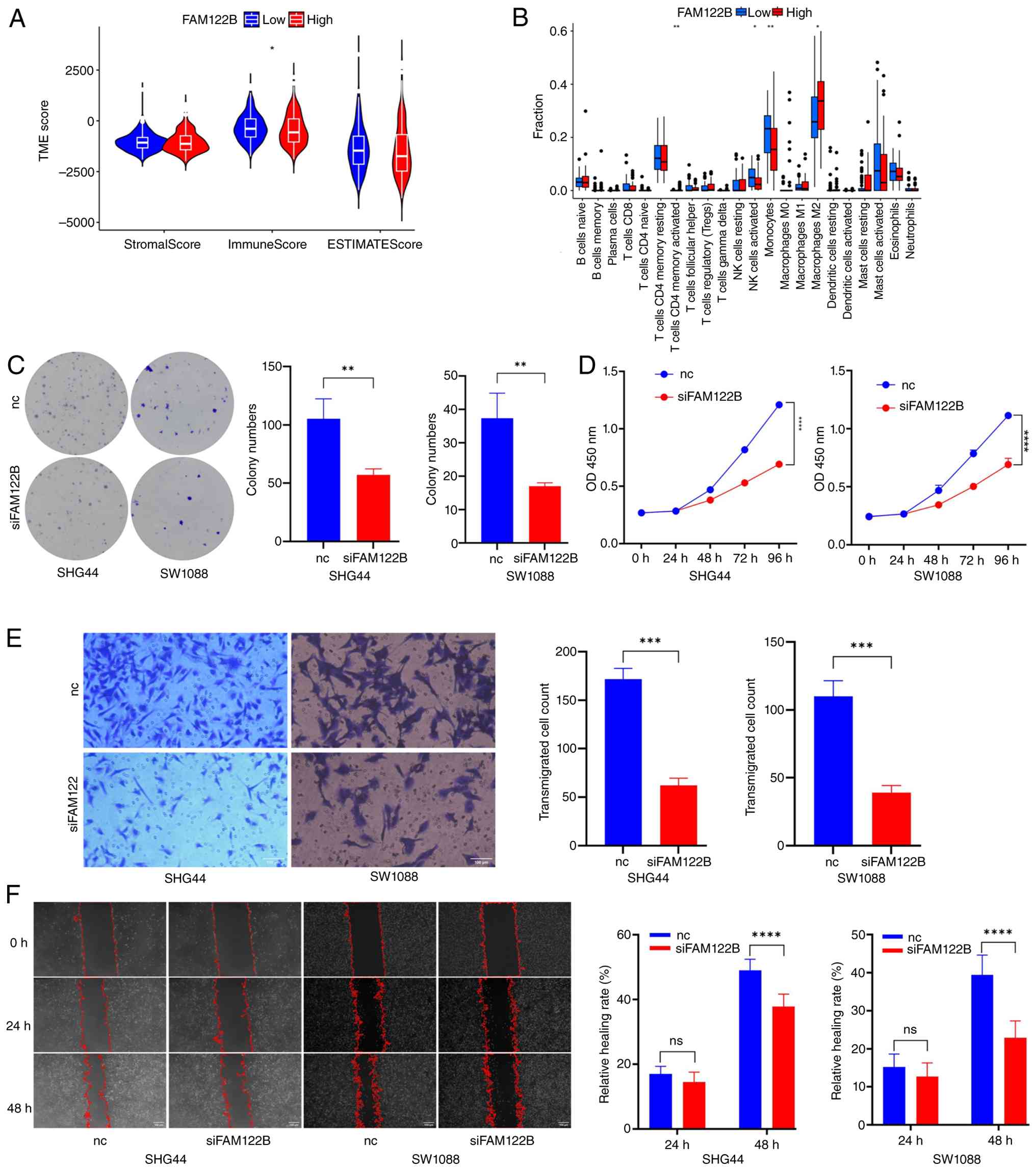 | Figure 6.FAM122B affects LGG immune
microenvironment and malignant cell behaviors. (A) TME scores of
patients with LGG in FAM122B high- and low-expression
groups, calculated by the ESTIMATE algorithm. (B) Correlation
analysis between FAM122B expression level and infiltration
levels. (C) Colony formation assay to detect the proliferation
capacity of SHG44 and SW1088 cells after FAM122B knockdown.
(D) CCK-8 assay to detect the proliferation viability of SHG44 and
SW1088 cells after FAM122B knockdown. (E) Transwell assay to
detect the invasion capacity of SHG44 and SW1088 cells after
FAM122B knockdown. Scale bar, 100 µm. (F) Wound healing
assay to detect the migration capacity of SHG44 and SW1088 cells
after FAM122B knockdown. Scale bar, 100 µm. *P<0.05,
**P<0.01, ***P<0.001, ****P<0.0001. FAM122B, Family
with sequence similarity 122, member B; LGG, low-grade glioma; TME,
tumor microenvironment; ns, no significance; si, short interfering;
LGG, low-grade glioma. |
In vitro experimental verification of
the biological effects of FAM122B on LGG cells
Following the aforementioned comprehensive analyses,
it was found that FAM122B can affect multiple biological
processes of glioma. However, bioinformatics analysis alone cannot
completely avoid potential false-positive results. To ensure the
rigor of the present research, in vitro cell experiments
were performed to explore the effects of FAM122B expression
on the proliferation and migration abilities of the LGG cell lines,
SHG44 and SW1088. siRNA was used to specifically knockdown
FAM122B expression in cell lines SHG44 and SW1088 (Fig. S3C). As shown in Fig. 6C, silencing FAM122B markedly
inhibited the colony-forming ability of LGG cell lines SHG44 and
SW1088. In addition, the CCK-8 assay results showed that knocking
down FAM122B markedly reduced the cell proliferation rate,
with the difference gradually widening over time (Fig. 6D). Furthermore, the Transwell
migration assay demonstrated that silencing FAM122B markedly
decreased the number of cells migrating through the chamber
(Fig. 6E). Finally, the wound
healing assay further confirmed that the relative healing rate in
the siFAM122B group was markedly lower than that in the
control group at 48 h (Fig. 6F).
Collectively, these results suggest that silencing FAM122B
markedly inhibits the proliferation and migration abilities of LGG
cells, further corroborating the database analysis indicating that
FAM122B functions as an oncogene.
Discussion
Glioma is the most common malignant tumor in the
central nervous system (32). As an
important subtype of glioma, LGG carries a lower degree of
malignancy than GBM, yet it exhibits a high risk of malignant
progression and recurrence (33),
posing a notable threat to the lives, health and quality of life of
patients. Exploring tumor-specific biomarkers and their regulatory
mechanisms is the key to breaking through the therapeutic
bottleneck of LGG. In the present study, the expression pattern,
clinical importance and biological functions of FAM122B in
LGG were systematically investigated through a combination of
bioinformatics analysis and in vitro experiments.
The FAM122 is a group of highly conserved endogenous
inhibitors of PP2A, whose members are involved in diverse
physiological and pathological processes by regulating PP2A
activity (9). As a core member of
this family, the oncogenic role of FAM122A has been
validated by numerous studies (10,11,34).
FAM122A can regulate biological behaviors of tumor cells
including proliferation, apoptosis and invasion in various
malignant tumors such as acute myeloid leukemia and hepatocellular
carcinoma by inhibiting PP2A activity (9), thus exerting a key oncogenic role.
However, the biological function and regulatory mechanisms of
FAM122B, another member of the FAM122 family, in tumors
remain unclear, and its role in glioma in particular has not been
systematically investigated.
In the present study, the expression characteristics
of FAM122B in LGG were first clarified through
multi-dimensional verification. Based on the GEO database and LGG
cell lines, it was verified that the mRNA level of FAM122B
was markedly upregulated. In addition, IHC data of cerebral cortex
and LGG tissues from the HPA database demonstrated that
FAM122B protein expression was also markedly increased,
indicating that high expression of FAM122B in LGG is a
consistent feature at both transcriptional and translational
levels, laying a foundation for the subsequent investigation of its
biological functions.
To clarify the correlation between high
FAM122B expression and the clinical characteristics as well
as prognosis of LGG, the present study integrated mRNA expression
data and clinical information from three databases (TCGA-seq,
CGGA-seq and CGGA microarray) for comprehensive analysis. The
results revealed that FAM122B expression was closely
associated with multiple clinical phenotypes of LGG. Specifically,
the expression of FAM122B was markedly upregulated with the
elevation of WHO grade and the expression level of FAM122B
was higher in recurrent LGG than in primary LGG. At the level of
key molecular markers, FAM122B expression was markedly
decreased in patients with LGG with an IDH mutation and 1p19q
chromosomal codeletion, both of which are well-recognized favorable
prognostic indicators for LGG (35). Therefore, high expression of
FAM122B is associated with a poor prognostic phenotype,
preliminarily suggesting that it may be involved in driving the
malignant progression of LGG.
The ability to serve as a tumor prognostic marker
largely depends on its prognostic predictive power. In the present
study, Kaplan-Meier survival analysis showed that patients with
high FAM122B expression exhibited markedly shorter survival
across three independent databases, confirming the negative
correlation between FAM122B expression and patient
prognosis. ROC curve analysis further demonstrated that
FAM122B exhibited moderate predictive value for 1-, 3- and
5-year survival in patients with LGG. The AUC remained at ~0.7,
suggesting that the expression level of FAM122B alone has
fair predictive potential for the short- to medium-term prognosis
of patients with LGG. However, univariate survival analysis alone
cannot exclude the interference of confounding factors such as age,
pathological grade and recurrence status. Therefore, the present
study further performed univariate and multivariate Cox regression
analyses. The results demonstrated that after adjusting for other
clinical factors, high expression of FAM122B and elevated
WHO grade were independent adverse risk factors for patients with
LGG. Based on the aforementioned analyses, it was hypothesized that
FAM122B may mediate the malignant biological behaviors of
glioma.
In the present study, three independent datasets
were analyzed separately rather than being merged and integrated,
which effectively avoided the difficulty of normalization among
multiple datasets. This approach not only maintained the
independence of each analytical dataset, but also enabled mutual
verification among the three datasets, fully demonstrating the
reliability of the findings. On this basis, four additional
datasets from different sources were further incorporated to
perform a meta-analysis together with the original core data,
thereby improving the reliability and general applicability of the
research conclusions.
Although multiple independent datasets and
analytical methods were included in the present study, and the
corresponding results were well mutually validated, the overall
analysis remains based on retrospective bioinformatic data.
Retrospective research inevitably has inherent limitations and
relevant biases cannot be completely avoided (36). Therefore, in vitro
experiments were performed by selecting two LGG cell lines to
explore the effects of FAM122B knockdown on the biological
behaviors of tumor cells. The results showed that knocking down
FAM122B markedly inhibited the proliferation and migration
of both LGG cell lines, which further indicated that FAM122B
is involved in regulating tumor malignant phenotypes and verified
the reliability of the conclusions.
As overexpression experiments were not carried out
in the present study, follow-up studies should add such experiments
to further validate the functional role of FAM122B.
Malignant tumor progression is a complex process involving the
coordinated regulation of multiple genes and pathway disorders
(37). To further explore the
potential oncogenic role of FAM122B in LGG, the present
study first performed co-expressed gene screening and analysis for
FAM122B based on TCGA database. The results showed that the
top five genes with the strongest positive correlation with
FAM122B were BRCC3, RPAP3, SLF1, MSH2 and
PHTF1. According to the literature review, several of these
genes have been confirmed to possess distinct pro-tumor functions.
Specifically, BRCC3 markedly promotes the proliferation,
migration and invasion of cancer cells in various tumors, including
bladder cancer (22), breast cancer
(38), cervical cancer (39), pancreatic cancer (40) and colon cancer (41). In addition, as a mismatch repair
protein, MSH2 plays a crucial role in the drug resistance
mechanisms of multiple cancers such as bladder cancer (24) and lung cancer (42). Furthermore, serving as a predictive
indicator, SLF1 can reflect the sensitivity of patients with
colon cancer to oxaliplatin chemotherapy (23). By contrast, among the genes
negatively correlated with FAM122B, deficiency of
LAMTOR1 can markedly suppress the malignant progression of
bladder cancer (43) and colon
cancer (44). Finally,
PEA15, another negatively correlated gene, inhibits the
epithelial-mesenchymal transition process in triple-negative breast
cancer and exerts a tumor-suppressive effect. The consistent
association of FAM122B with these established oncogenic or
tumor-suppressive genes further corroborates its oncogenic
potential in LGG, suggesting that FAM122B may form a
synergistic regulatory network with these genes to jointly drive
the progression of LGG.
The present study adopted GSEA to explore the
potential regulatory mechanisms of FAM122B in LGG. This
method is particularly suitable for investigating gene functional
mechanisms, as it does not rely on artificially set thresholds for
screening differentially expressed genes and thus avoids missing
key regulatory genes. Moreover, it can accurately identify
signaling pathways enriched with FAM122B at the pathway
level, thereby clarifying the core molecular pathways by which
FAM122B regulates the occurrence and progression of LGG. The
results revealed that FAM122B was markedly enriched in three
cellular signaling pathways in LGG, including ECM-receptor
interaction, Toll-like receptor signaling pathway and focal
adhesion. Activation of these cellular signaling pathways plays a
pivotal role in tumor progression and regulates malignant
phenotypes through multiple pathways and molecular mechanisms
(45–47).
In the present study, indirect exploration of the
biological function of FAM122B was attempted via the
aforementioned analytical strategies. Given that the three
signaling pathways exert essential regulatory effects on the
formation of the immunosuppressive tumor microenvironment, the role
of FAM122B in the immune microenvironment of LGG was further
investigated. The results showed that the enrichment of tumor
immune cells was markedly higher in the FAM122B
high-expression group, particularly for M2-type tumor-associated
macrophages and CD4+ T cells. Meanwhile, high
FAM122B expression was positively correlated with the levels
of multiple immune checkpoint molecules.
Collectively, these findings suggest that
FAM122B may be associated with the formation of the
immunosuppressive microenvironment in LGG via the three
aforementioned signaling pathways, which potentially contributes to
the malignant progression of glioma. For example, glioma cells
secrete abundant ECM components, such as collagen and fibronectin,
which bind to surface receptors on macrophages (such as integrins,
CD36 or CD44). This interaction activates downstream signaling
pathways, recruiting monocytes to the tumor microenvironment and
inducing their polarization into M2-type macrophages, which exhibit
immunosuppressive and pro-angiogenic functions (48–50).
In glioma, abnormal activation of the Toll-like
receptor signaling pathway can upregulate the expression of immune
checkpoint molecules such as PD-L1, thereby enabling tumor cells to
evade immune surveillance and elimination. Meanwhile, glioma
further activates the TLR signaling cascade to enhance the
PD-1/PD-L1 immune checkpoint pathway, which reduces the
infiltration of CD4+ T cells and suppresses Th1-mediated
cytotoxicity within the tumor microenvironment, ultimately
maintaining an overall immunosuppressive state. Furthermore,
targeted intervention of the Toll-like receptor signaling pathway
can effectively regulate macrophage polarization and may help
reverse resistance to immune checkpoint therapy (51).
Studies have indicated that the activation of
CD4+ T cells is regulated by multiple factors during
tumor progression (52–54). Alterations in the activity of the
focal adhesion signaling pathway can modulate the function of
CD4+ T cells within the tumor immune microenvironment,
thereby affecting the malignant progression of tumors (52). Previous literature reports also
provide indirect support for these analytical findings. The present
study only preliminarily explored the biological regulatory role of
FAM122B in the pathological progression of LGG, and its
specific molecular mechanisms remain to be further verified by
experiments.
The major strengths of the present study lie in the
full utilization of data from multiple databases to conduct highly
logical and scientifically rigorous analyses. A variety of
analytical tools were adopted, enabling mutual verification among
the research findings. Nevertheless, there are several
shortcomings. The suggested roles of FAM122B in signaling
pathways and the immune microenvironment are based on correlational
bioinformatic analyses and were not directly validated through
mechanistic experiments in the present study. The present study
only provided correlational analytical outcomes rather than direct
conclusive results. Further attention and in-depth improvement by
subsequent researchers are required in this regard and the present
work merely serves as a reference basis. The present study did not
perform analyses on clinical tissue samples from patients, which
constitutes a limitation of the present research. In addition,
significant heterogeneity was observed in this meta-analysis. The
data were collected from populations across different countries and
ethnic groups and the inconsistent detection methods also
contributed to heterogeneity, which may affect the reliability and
interpretation of the pooled results.
In summary, the results of the present study
suggest that FAM122B is relatively highly expressed in LGG
and may act as a potential oncogene in LGG. The present study may
partially fill the research gap related to the FAM122 family in LGG
and help enrich the pathophysiological regulatory network of the
FAM122 protein family. The present study provided preliminary
evidence supporting a potential role of FAM122B in LGG and
that further experimental and clinical studies are required to
validate its clinical significance.
Supplementary Material
Supporting Data
Supporting Data
Acknowledgements
Not applicable.
Funding
The present study was supported by Henan Provincial Key Research
and Development Program (grant no. 251111312600) and Henan Young
and Middle-aged Health Science and Technology Innovation Talent
Project (grant no. LJRC2023007).
Availability of data and materials
The data generated in the present study may be
requested from the corresponding author.
Authors' contributions
HW was responsible for data collection and
manuscript drafting. YZ participated in data collection. TG and YG
were responsible for data analysis. RQ participated in study
conception and experimental design, critical revision of the
manuscript, project review and supervision, and funding
acquisition. HW and RQ confirm the authenticity of all the raw
data. All authors have read and approved the final version of the
manuscript.
Ethics approval and consent to
participate
Not applicable.
Patient consent for publication
Not applicable.
Competing interests
The authors declare that they have no competing
interests.
References
|
1
|
Patel SH, Poisson LM, Brat DJ, Zhou Y,
Cooper L, Snuderl M, Thomas C, Franceschi AM, Griffith B, Flanders
AE, et al: T2-FLAIR mismatch, an imaging biomarker for IDH and
1p/19q status in lower-grade gliomas: A TCGA/TCIA project. Clin
Cancer Res. 23:6078–6085. 2017. View Article : Google Scholar : PubMed/NCBI
|
|
2
|
Louis DN, Perry A, Wesseling P, Brat DJ,
Cree IA, Figarella-Branger D, Hawkins C, Ng HK, Pfister SM,
Reifenberger G, et al: The 2021 WHO classification of tumors of the
central nervous system: A summary. Neuro Oncol. 23:1231–1251. 2021.
View Article : Google Scholar : PubMed/NCBI
|
|
3
|
Jooma R, Waqas M and Khan I: Diffuse
Low-grade glioma-changing concepts in diagnosis and management: A
review. Asian J Neurosurg. 14:356–363. 2019. View Article : Google Scholar : PubMed/NCBI
|
|
4
|
Nahed BV, Redjal N, Brat DJ, Chi AS, Oh K,
Batchelor TT, Ryken TC, Kalkanis SN and Olson JJ: Management of
patients with recurrence of diffuse low grade glioma: A systematic
review and evidence-based clinical practice guideline. J
Neurooncol. 125:609–630. 2015. View Article : Google Scholar : PubMed/NCBI
|
|
5
|
Fournier C, Mercey-Ressejac M, Derangère
V, Al Kadi A, Rageot D, Charrat C, Leroy A, Vollaire J, Josserand
V, Escudé M, et al: Nanostructured lipid carriers based mRNA
vaccine leads to a T cell-inflamed tumour microenvironment
favourable for improving PD-1/PD-L1 blocking therapy and long-term
immunity in a cold tumour model. EBioMedicine. 112:1055432025.
View Article : Google Scholar : PubMed/NCBI
|
|
6
|
Sun X, Jia Q, Li K, Tian C, Yi L, Yan L,
Zheng J, Jia X and Gu M: Comparative genomic landscape of
lower-grade glioma and glioblastoma. PLoS One. 19:e03095362024.
View Article : Google Scholar : PubMed/NCBI
|
|
7
|
Yang YS, Liu MH, Yan ZW, Chen GQ and Huang
Y: FAM122A is required for mesendodermal and cardiac
differentiation of embryonic stem cells. Stem Cells. 41:354–367.
2023. View Article : Google Scholar : PubMed/NCBI
|
|
8
|
Fan F, Zhao J, Liu Y, Zhao H, Weng L, Li
Q, Chen G and Xu Y: Identifying the SUMO1 modification of FAM122A
leading to the degradation of PP2A-Cα by ubiquitin-proteasome
system. Biochem Biophys Res Commun. 500:676–681. 2018. View Article : Google Scholar : PubMed/NCBI
|
|
9
|
Wasserman JS, Faezov B, Patel KR,
Kurimchak AM, Palacio SM, Glass DJ, Fowle H, McEwan BC, Xu Q, Zhao
Z, et al: FAM122A ensures cell cycle interphase progression and
checkpoint control by inhibiting B55α/PP2A through helical motifs.
Nat Commun. 15:57762024. View Article : Google Scholar : PubMed/NCBI
|
|
10
|
Liu MH, Chen J, Yang YS, Wang YQ, Chen GQ,
Zhang Y and Huang Y: FAM122A promotes acute myeloid leukemia cell
growth through inhibiting PP2A activity and sustaining MYC
expression. Haematologica. 106:903–907. 2021.PubMed/NCBI
|
|
11
|
Zhou Y, Shi WY, He W, Yan ZW, Liu MH, Chen
J, Yang YS, Wang YQ, Chen GQ and Huang Y: FAM122A supports the
growth of hepatocellular carcinoma cells and its deletion enhances
Doxorubicin-induced cytotoxicity. Exp Cell Res. 387:1117142020.
View Article : Google Scholar : PubMed/NCBI
|
|
12
|
Livak KJ and Schmittgen TD: Analysis of
relative gene expression data using real-time quantitative PCR and
the 2(−Delta Delta C(T)) method. Methods. 25:402–408. 2001.
View Article : Google Scholar : PubMed/NCBI
|
|
13
|
Kawaguchi A, Yajima N, Tsuchiya N, Homma
J, Sano M, Natsumeda M, Takahashi H, Fujii Y, Kakuma T and Yamanaka
R: Gene expression signature-based prognostic risk score in
patients with glioblastoma. Cancer Sci. 104:1205–1210. 2013.
View Article : Google Scholar : PubMed/NCBI
|
|
14
|
Buczkowicz P, Hoeman C, Rakopoulos P,
Pajovic S, Letourneau L, Dzamba M, Morrison A, Lewis P, Bouffet E,
Bartels U, et al: Genomic analysis of diffuse intrinsic pontine
gliomas identifies three molecular subgroups and recurrent
activating ACVR1 mutations. Nat Genet. 46:451–456. 2014. View Article : Google Scholar : PubMed/NCBI
|
|
15
|
Yu X, Feng L, Liu D, Zhang L, Wu B, Jiang
W, Han Z and Cheng S: Quantitative proteomics reveals the novel
co-expression signatures in early brain development for prognosis
of glioblastoma multiforme. Oncotarget. 7:14161–14171. 2016.
View Article : Google Scholar : PubMed/NCBI
|
|
16
|
Feng L, Qian H, Yu X, Liu K, Xiao T, Zhang
C, Kuang M, Cheng S, Li X, Wan J and Zhang K: Heterogeneity of
tumor-infiltrating lymphocytes ascribed to local immune status
rather than neoantigens by multi-omics analysis of glioblastoma
multiforme. Sci Rep. 7:69682017. View Article : Google Scholar : PubMed/NCBI
|
|
17
|
Park CK, Park I, Lee S, Sun CH, Koh Y,
Park SH, Kim JE, Yun H and Lee SH: Genomic dynamics associated with
malignant transformation in IDH1 mutated gliomas. Oncotarget.
6:43653–43666. 2015. View Article : Google Scholar : PubMed/NCBI
|
|
18
|
Björkblom B, Wibom C, Eriksson M,
Bergenheim AT, Sjöberg RL, Jonsson P, Brännström T, Antti H,
Sandström M and Melin B: Distinct metabolic hallmarks of WHO
classified adult glioma subtypes. Neuro Oncol. 24:1454–1468. 2022.
View Article : Google Scholar : PubMed/NCBI
|
|
19
|
Chen W, Lei C, Liu P, Liu Y, Guo X, Kong Z
and Wang Y, Dai C and Wang Y, Ma W and Wang Y: Progress and
prospects of recurrent glioma: A recent scientometric analysis of
the web of science in 2019. World Neurosurg. 134:e387–e399. 2020.
View Article : Google Scholar : PubMed/NCBI
|
|
20
|
Decuyper M, Bonte S, Deblaere K and Van
Holen R: Automated MRI based pipeline for segmentation and
prediction of grade, IDH mutation and 1p19q co-deletion in glioma.
Comput Med Imaging Graph. 88:1018312021. View Article : Google Scholar : PubMed/NCBI
|
|
21
|
Huang S, Ernberg I and Kauffman S: Cancer
attractors: A systems view of tumors from a gene network dynamics
and developmental perspective. Semin Cell Dev Biol. 20:869–876.
2009. View Article : Google Scholar : PubMed/NCBI
|
|
22
|
Tao H, Liao Y, Yan Y, He Z, Zhou J, Wang
X, Peng J, Li S and Liu T: BRCC3 promotes tumorigenesis of bladder
cancer by activating the NF-κB signaling pathway through targeting
TRAF2. Front Cell Dev Biol. 9:7203492021. View Article : Google Scholar : PubMed/NCBI
|
|
23
|
Han X, Wang Z, Zhang L, Shen Y, Tan Q, Sun
Y, Wang J, Qian X, Yang H and Shi Y: SLF1 polymorphism predicts
response to oxaliplatin-based adjuvant chemotherapy in patients
with colon cancer. Am J Cancer Res. 11:1522–1539. 2021.PubMed/NCBI
|
|
24
|
Zhang H, Xiao X, Wei W, Huang C, Wang M,
Wang L, He Y, Sun J, Jiang Y, Jiang G and Zhang X: CircLIFR
synergizes with MSH2 to attenuate chemoresistance via MutSα/ATM-p73
axis in bladder cancer. Mol Cancer. 20:702021. View Article : Google Scholar : PubMed/NCBI
|
|
25
|
Wu B, Huang X, Shi X, Jiang M, Liu H and
Zhao L: LAMTOR1 decreased exosomal PD-L1 to enhance immunotherapy
efficacy in non-small cell lung cancer. Mol Cancer. 23:1842024.
View Article : Google Scholar : PubMed/NCBI
|
|
26
|
Mohammed HN, Pickard MR and
Mourtada-Maarabouni M: The protein phosphatase 4-PEA15 axis
regulates the survival of breast cancer cells. Cell Signal.
28:1389–1400. 2016. View Article : Google Scholar : PubMed/NCBI
|
|
27
|
Neophytou CM, Panagi M, Stylianopoulos T
and Papageorgis P: The role of tumor microenvironment in cancer
metastasis: Molecular mechanisms and therapeutic opportunities.
Cancers. 13:20532021. View Article : Google Scholar : PubMed/NCBI
|
|
28
|
Zhang P, Cao X, Guan M, Li D, Xiang H,
Peng Q, Zhou Y, Weng C, Fang X, Liu X, et al: CPNE8 promotes
gastric cancer metastasis by modulating focal adhesion pathway and
tumor microenvironment. Int J Biol Sci. 18:4932–4949. 2022.
View Article : Google Scholar : PubMed/NCBI
|
|
29
|
Benencia F, Alaniz LD and McCall KD:
Editorial: Toll-like receptor expression in transformed cells: Role
in tumor development and cancer therapies. Front Immunol.
15:14784312024. View Article : Google Scholar : PubMed/NCBI
|
|
30
|
Mortezaee K: Immune escape: A critical
hallmark in solid tumors. Life Sci. 258:1181102020. View Article : Google Scholar : PubMed/NCBI
|
|
31
|
Pardoll DM: The blockade of immune
checkpoints in cancer immunotherapy. Nat Rev Cancer. 12:252–264.
2012. View Article : Google Scholar : PubMed/NCBI
|
|
32
|
Ostrom QT, Price M, Neff C, Cioffi G,
Waite KA, Kruchko C and Barnholtz-Sloan JS: CBTRUS statistical
report: Primary brain and other central nervous system tumors
diagnosed in the United States in 2015–2019. Neuro Oncol. 24 (Suppl
5):v1–v95. 2022. View Article : Google Scholar : PubMed/NCBI
|
|
33
|
Nakasu S and Nakasu Y: Malignant
progression of diffuse Low-grade Gliomas: A systematic review and
Meta-analysis on incidence and related factors. Neurol Med Chir
(Tokyo). 62:177–185. 2022. View Article : Google Scholar : PubMed/NCBI
|
|
34
|
Wang YQ, Yang YS, Chen J, Liu MH, Chen GQ
and Huang Y: FAM122A maintains DNA stability possibly through the
regulation of topoisomerase IIα expression. Exp Cell Res.
396:1122422020. View Article : Google Scholar : PubMed/NCBI
|
|
35
|
Familiari P, Lapolla P, Picotti V,
Palmieri M, Pesce A, Carosi G, Relucenti M, Nottola S, Gianno F,
Minasi S, et al: Role of 1p/19q codeletion in diffuse Low-grade
glioma tumour prognosis. Anticancer Res. 43:2659–2670. 2023.
View Article : Google Scholar : PubMed/NCBI
|
|
36
|
Talari K and Goyal M: Retrospective
studies-utility and caveats. J R Coll Physicians Edinb. 50:398–402.
2020. View Article : Google Scholar : PubMed/NCBI
|
|
37
|
Charmpi K, Guo T, Zhong Q, Wagner U, Sun
R, Toussaint NC, Fritz CE, Yuan C, Chen H, Rupp NJ, et al:
Convergent network effects along the axis of gene expression during
prostate cancer progression. Genome Biol. 21:3022020. View Article : Google Scholar : PubMed/NCBI
|
|
38
|
Huang Q, Zheng S, Gu H, Yang Z, Lu Y, Yu X
and Guo G: The deubiquitinase BRCC3 increases the stability of ZEB1
and promotes the proliferation and metastasis of triple-negative
breast cancer cells. Acta Biochim Biophys Sin (Shanghai).
56:564–575. 2024.PubMed/NCBI
|
|
39
|
Zhang F and Zhou Q: Knockdown of BRCC3
exerts an anti-tumor effect on cervical cancer in vitro. Mol
Med Rep. 18:4886–4894. 2018.PubMed/NCBI
|
|
40
|
Hu X, Wu J and Xu J: UCA1 executes an
oncogenic role in pancreatic cancer by regulating miR-582-5p/BRCC3.
Front Oncol. 13:11332002023. View Article : Google Scholar : PubMed/NCBI
|
|
41
|
Feng X, He S, Chen Y and Zhang L:
Deubiquitinase BRCC3 promotes the migration, invasion and EMT
progression of colon adenocarcinoma by stabilizing MET expression.
Genes Genomics. 46:637–646. 2024. View Article : Google Scholar : PubMed/NCBI
|
|
42
|
Ko JC, Chiu HC, Wo TY, Huang YJ, Tseng SC,
Huang YC, Chen HJ, Syu JJ, Chen CY, Jian YT, et al: Inhibition of
p38 MAPK-dependent MutS homologue-2 (MSH2) expression by metformin
enhances gefitinib-induced cytotoxicity in human squamous lung
cancer cells. Lung Cancer. 82:397–406. 2013. View Article : Google Scholar : PubMed/NCBI
|
|
43
|
Sun Y, Guan Z, Sheng Q, Duan W, Zhao H,
Zhou J, Deng Q and Pei X: N-myristoyltransferase-1 deficiency
blocks myristoylation of LAMTOR1 and inhibits bladder cancer
progression. Cancer Lett. 529:126–138. 2022. View Article : Google Scholar : PubMed/NCBI
|
|
44
|
Zhao L, Gao N, Peng X, Chen L, Meng T,
Jiang C, Jin J, Zhang J, Duan Q, Tian H, et al: TRAF4-mediated
LAMTOR1 ubiquitination promotes mTORC1 activation and inhibits the
inflammation-induced colorectal cancer progression. Adv Sci
(Weinh). 11:e23011642024. View Article : Google Scholar : PubMed/NCBI
|
|
45
|
Kidd LC, Rogers EN, Yeyeodu ST, Jones DZ
and Kimbro KS: Contribution of toll-like receptor signaling
pathways to breast tumorigenesis and treatment. Br Cancer (Dove Med
Press). 5:43–51. 2013.PubMed/NCBI
|
|
46
|
Liu Z, Zhang X, Ben T, Li M, Jin Y, Wang T
and Song Y: Focal adhesion in the tumour metastasis: From molecular
mechanisms to therapeutic targets. Biomark Res. 13:382025.
View Article : Google Scholar : PubMed/NCBI
|
|
47
|
Lu P, Weaver VM and Werb Z: The
extracellular matrix: A dynamic niche in cancer progression. J Cell
Biol. 196:395–406. 2012. View Article : Google Scholar : PubMed/NCBI
|
|
48
|
Cui X, Morales RT, Qian W, Wang H, Gagner
JP, Dolgalev I, Placantonakis D, Zagzag D, Cimmino L, Snuderl M, et
al: Hacking macrophage-associated immunosuppression for regulating
glioblastoma angiogenesis. Biomaterials. 161:164–178. 2018.
View Article : Google Scholar : PubMed/NCBI
|
|
49
|
Xiong J, Xiao R, Zhao J, Zhao Q, Luo M, Li
F, Zhang W and Wu M: Matrix stiffness affects tumor-associated
macrophage functional polarization and its potential in tumor
therapy. J Transl Med. 22:852024. View Article : Google Scholar : PubMed/NCBI
|
|
50
|
Ellert-Miklaszewska A, Poleszak K,
Pasierbinska M and Kaminska B: Integrin signaling in glioma
pathogenesis: From biology to therapy. Int J Mol Sci. 21:8882020.
View Article : Google Scholar : PubMed/NCBI
|
|
51
|
Norollahi SE, Babaei K, Rashidy-Pour A,
Yousefi B, Baharlou R, Far BF, Jalali A and Samadani AA: Role of
the TLR signaling pathway in the pathogenesis of glioblastoma
multiforme with an emphasis on immunotherapy. Biochem Biophys Rep.
43:1021492025.PubMed/NCBI
|
|
52
|
Wiemer AJ, Wernimont SA, Cung TD, Bennin
DA, Beggs HE and Huttenlocher A: The focal adhesion kinase
inhibitor PF-562,271 impairs primary CD4+ T cell activation.
Biochem Pharmacol. 86:770–781. 2013. View Article : Google Scholar : PubMed/NCBI
|
|
53
|
Guo M, Liu MYR and Brooks DG: Regulation
and impact of tumor-specific CD4(+) T cells in cancer and
immunotherapy. Trends Immunol. 45:303–313. 2024. View Article : Google Scholar : PubMed/NCBI
|
|
54
|
Accogli T, Bruchard M and Végran F:
Modulation of CD4 T cell response according to tumor cytokine
microenvironment. Cancers. 13:3732021. View Article : Google Scholar : PubMed/NCBI
|