Introduction
Osteosarcoma (OS) remains the most common primary
malignant bone tumor in children, adolescents and young adults
(1). Standard treatment consists of
neoadjuvant chemotherapy, complete surgical resection and
postoperative chemotherapy (2).
However, outcomes remain poor in recurrent, metastatic or
chemotherapy-refractory disease (2). Pulmonary metastasis and recurrence
remain the principal causes of OS-related mortality (1,2). OS
has shown a limited response to immunotherapies that have
revolutionized the treatment of other solid tumors (for example,
anti-programmed cell death protein 1 immune checkpoint inhibitors
have revolutionized the treatment of melanoma and non-small cell
lung cancer) suggesting that therapeutic failure is multifactorial
rather than attributable to a single biological defect (3,4).
Traditional explanations have emphasized tumor
cell-intrinsic mechanisms, such as genomic instability and clonal
evolution (5). These mechanisms are
interconnected, but they do not fully account for several
long-standing clinical observations (6). Immune-cell infiltration is evident in
a subset of OS tumors; however, its presence does not necessarily
translate into durable tumor clearance (7,8).
Similarly, technically successful local control does not assure
freedom from pulmonary recurrence (1,2).
Despite employing different mechanisms, immune checkpoint
blockades, engineered cellular therapies and combination strategies
often yield modest and inconsistent clinical benefits (3,6,9,10).
Thus, tumor cell-intrinsic biology explains an important component
of OS aggressiveness but has limited explanatory scope when
considered in isolation (9).
Recent advances in single-cell, spatial and
multi-omics studies support a more tissue-specific interpretation
of OS biology (11,12). OS arises within a complex skeletal
microenvironment rather than at a generic tumor site (13). Mineralized matrix, marrow-derived
cells, abnormal vasculature, stromal populations and immune signals
may interact to regulate immune-cell trafficking, local immune
suppression and pulmonary niche remodeling (14). This interpretation is supported by
recent cellular atlases, tumor microenvironment studies and
analyses of pulmonary metastases (11–15).
In the present review, layered antitumor immune
failure refers to the progressive accumulation of defects in immune
cell trafficking, antigen presentation, effector activation,
cytotoxic elimination of tumor cells and pulmonary immune
surveillance. This term is used as an organizational concept rather
than as a replacement for established concepts such as
immunosuppression, immune exclusion or immunologically cold tumors
(8,16).
Layered antitumor immune failure differs from immune
exclusion or an immunologically cold phenotype in that it
emphasizes the spatial and temporal accumulation of defects across
the primary bone niche, treatment-induced tissue repair programs,
systemic niche communication and pulmonary immune surveillance. The
term therefore describes not only insufficient immune activation,
but also the misdirection of immune activity toward
non-eradicating, repair-associated or metastasis-permissive
programs. This concept explains the presence of measurable immune
activity in OS, yet the failure of this cancer to succeed across
the downstream checkpoints needed to achieve durable tumor
elimination and prevent relapse.
The term ‘bone niche-driven’ is used in the present
review in a qualified sense. This does not mean that the bone niche
has been established as a universal or exclusive causal driver of
immune failure in OS. Rather, it is an evidence-based framework
through which the structural, cellular, stromal and metabolic
features of the skeletal niche may orchestrate multiple levels of
antitumor immune dysfunction.
Therefore, the present review tracks the biological
progression of OS, rather than simply cataloguing immune
abnormalities. The suggested sequence involves bone niche
remodeling, myeloid cell predominance, tolerogenic processing of
therapy-induced cell death, extracellular vesicle (EV)-mediated
niche communication, physical and metabolic adaptation, and
pulmonary niche conditioning. These modules are synthesized from
OS-specific evidence and contextual tumor-biology literature
(8,11–14,16–21),
with relatively mature evidence distinguished from
hypothesis-generating or extrapolated mechanisms.
First, the present review recharacterizes OS immune
resistance as a bone niche-organized process rather than a simple
catalogue of immune cell abnormalities or therapeutic targets.
Second, the theory of layered antitumor immune failure explains the
coexistence of immune infiltration, local tumor control and immune
activation with the failure of cytotoxic clearance and the delayed
recurrence of pulmonary tumors. Third, it clearly separates direct
evidence of OS from indirect evidence inferred from other bone
tumors or from general solid tumor biology, thus minimizing the
risk of overinterpreting causality in poorly validated modules.
Fourth, the proposed framework is operationalized through claim
evidence gap mapping, a minimal validation package, and trial
design scenarios that prioritize longitudinal sampling, spatial
functional readouts and pulmonary recurrence endpoints.
An OS-specific rationale supports a bone
niche-centered framework of immune failure (14–17).
Unlike most soft-tissue malignancies, OS develops within a
mineralized matrix, osteoid-producing tissue and an actively
remodeling marrow compartment (14,16,17).
These anatomical and biological features impose several
disease-specific constraints. First, the mineralized matrix and
osteoid-rich architecture may restrict immune-cell access and
impair effective T cell-tumor cell contact (14,16,21).
Second, the marrow compartment is naturally enriched with myeloid
progenitors, macrophages, osteoclast-lineage cells and mesenchymal
stromal cells (MSCs), thereby favoring myeloid-dominant immune
regulation (14,16–18).
Third, bone remodeling, tissue repair and inflammatory resolution
are tightly coupled, allowing therapy-induced cell death to be
processed through repair-associated or tolerogenic programs
(18). Fourth, bone-derived
stromal, vascular and EV signals may communicate with the
circulation and pulmonary microenvironment (15,19,20).
Finally, pulmonary recurrence requires not only disseminated tumor
cells but also permissive immune and stromal conditions in the lung
(15,19–21).
Together, these features support the view that the bone niche
functions as an organizing variable in OS immune failure, rather
than merely serving as a passive site of tumor growth.
Search strategy and evidence
classification
The present review is a mechanistic,
problem-oriented narrative review; its reporting and organization
were informed by established guidance for narrative reviews
(22) and the SANRA quality
framework (23). PubMed (https://pubmed.ncbi.nlm.nih.gov/), Web of Science
(https://www.webofscience.com/), and
Scopus (https://www.scopus.com/). were searched
using combinations of ‘osteosarcoma’, ‘bone niche’, ‘tumor immune
microenvironment’, ‘macrophage’, ‘efferocytosis’, ‘extracellular
vesicles’, ‘premetastatic niche’, ‘metabolic reprogramming’ and
‘immunotherapy resistance’. The last search was updated to April
2026. As the review aimed to develop and refine a mechanistic
framework rather than to quantify pooled effects, no quantitative
search protocol was preregistered. Mechanistic studies in English,
and single-cell and spatial-omics studies, animal models, clinical
specimens, translational therapy studies and high-quality reviews
were prioritized. Detailed search strings and key inclusion
criteria are provided in Table
SI.
Since the aim was framework construction rather than
quantitative synthesis, a meta-analysis or a formal risk-of-bias
assessment was not performed. Candidate sources were first screened
for topic relevance, and then re-evaluated for OS specificity,
mechanistic support, functional or spatial validation, and
translational value. At least two authors reviewed the key evidence
domains, and evidence classification was finalized by consensus of
the authors; disagreements were resolved by discussion. The
reference list was restricted to sources that directly supported a
mechanistic or translational claim within the text, tables or
figure legends; broad background articles were used only to clarify
mechanistic context not yet directly validated in OS.
Evidence from other bone tumors or broader solid
tumor literature was treated as contextual rather than as
OS-specific proof. Given that the objective was conceptual
synthesis rather than systematic evidence aggregation, the
resulting framework should be interpreted as an author-derived,
evidence-weighted organizational tool rather than as a validated
grading system.
For the purposes of the present review, mechanistic
evidence was separated from grading and translational maturity
staging. Mechanistic evidence was graded according to OS
specificity, functional validation, spatial or longitudinal
support, and translational relevance (Table I). Tier 1 comprised the relatively
better-supported OS-specific mechanisms of bone niche remodeling
(11–14,17,24–26)
and myeloid-cell enrichment (8,11,13,27–31).
Tier 2 comprised OS-supported but longitudinally or functionally
incomplete mechanisms, including pulmonary premetastatic niche
conditioning (15,19,20,32–34),
efferocytosis-associated immune tolerance (18,35–37)
and EV-mediated bone-lung communication (19,32,33,38–43).
Tier 3 comprised mechanisms that remain largely associative,
computational or extrapolated from broader tumor biology, including
physical and metabolic immune adaptation (21,34,44–47).
Translational maturity was staged separately as clinical, early
translational, preclinical or conceptual (Table II).
 | Table I.Mechanistic evidence grading matrix
of the bone niche-driven framework. |
Table I.
Mechanistic evidence grading matrix
of the bone niche-driven framework.
| Module | Mechanistic
evidence tier | Evidence
source | Main
limitation | Validation
priority | (Refs.) |
|---|
| Bone niche
remodeling | Tier 1: Relatively
better-supported OS-specific mechanism | Bone TME,
osteoimmune remodeling, bone-mimetic models and single-cell/spatial
studies | Limited
longitudinal causality across treatment stages | Paired
pre-/post-treatment sampling and spatial functional validation | (11–14,17,24–27) |
| Myeloid cell
enrichment/TAM remodeling | Tier 1: Relatively
better-supported OS-specific mechanism | Macrophage-enriched
OS lesions, TAM-related risk models and spatial immune-exclusion
studies | State, compartment
and patient-context dependence | Lineage-resolved
perturbation and macrophage-state functional assays | (8,11,13,27–31) |
| Pulmonary niche
conditioning | Tier 2:
OS-supported but longitudinally uneven | Metastatic lung
microenvironment studies and OS-derived EV/chemokine findings | Few longitudinal
datasets pairing primary tumors with lung specimens | Paired primary
tumor-lung cohorts, EV/chemokine monitoring and recurrence
endpoints | (15,19,20,32–34) |
| Efferocytosis | Tier 2: Emerging
and incompletely validated |
MerTK/efferocytosis-associated OS studies
and transcriptomic/spatial inference | Limited direct
blockade and causality testing in OS | MERTK/AXL
perturbation, dying-cell-state mapping and immune readouts | (18,35–37) |
| EV-mediated
bone-lung communication | Tier 2: Emerging
and incompletely validated | OS-derived small
EVs, EV-packaged S100A11 and lung niche-related EV studies | Cargo enrichment or
recipient-cell inference does not prove causality | Functional EV cargo
experiments and recipient cell validation in OS models | (19,32,33,41–43) |
| Physical/metabolic
immune adaptation | Tier 3:
Hypothesis-generating or contextual evidence | Multi-omics,
mitophagy, UPR, pyruvate metabolism, hypoxia and immune-signature
studies | Mostly prognostic
modeling or computational inference | Metabolic
perturbation with antigen-presentation, T-cell, NK-cell and
ICI-sensitivity readouts | (21,34,44–47) |
| Table II.Candidate niche-reprogramming
strategies, translational maturity, safety considerations and trial
windows. |
Table II.
Candidate niche-reprogramming
strategies, translational maturity, safety considerations and trial
windows.
| Therapeutic
step | Candidate
intervention | Evidence
stagea | Preferred trial
window | Major
feasibility/safety concern | Key readout or
candidate context | (Refs.) |
|---|
| Niche
reprogramming | TAM
blockade/repolarization or TAM recruitment inhibition | Preclinical to
early translational | Neoadjuvant or
perioperative window | Myelosuppression,
infection risk, impaired tissue repair, macrophage-state
plasticity | Macrophage-state
shift, T-cell infiltration, cytokine profile; myeloid-enriched or
immune-excluded tumors | (28,31,49,50) |
| Niche
reprogramming | Gas6-MERTK/AXL or
efferocytosis-related blockade |
Preclinical/conceptual in OS | Post-therapy cell
death window or perioperative window | Interference with
physiological clearance, inflammation control and wound
healing | Antigen
presentation, PD-L1 change, T-cell activation after therapy; tumors
with tolerogenic processing of therapy-induced cell death | (18,35–37,58) |
| Niche
reprogramming | TGF-β mitigation,
ECM modulation or vascular normalization | Conceptual to
preclinical | Neoadjuvant or
pre-cell therapy window | Stromal toxicity,
impaired bone or wound repair, uncertain dosing window | Cell trafficking,
persistence, exhaustion, stromal barrier reduction; stromal-rich
lesions with poor homing | (14,21,53,65) |
| Immune
activation | B7-H3-directed
therapy, CAR-T/CAR-NK or checkpoint-based combinations | Early
translational; variable by modality | After partial niche
relief or in biomarker-selected disease | Antigen
heterogeneity, on-target/off-tumor toxicity, poor homing and
limited persistence | Response
durability, cytotoxic function and antigen-positive tumors after
partial niche relief | (3,64,67) |
| Pulmonary niche
maintenance | EV/chemokine
monitoring, lung-directed NK cell-supportive approaches or
postoperative immuno-modulation |
Preclinical/conceptual | Postoperative
surveillance or high-risk maintenance | Pulmonary delivery
toxicity, immune overactivation and biomarker instability | Pulmonary
recurrence-free survival, circulating EV/chemokine biomarkers, lung
niche activity | (19,33,63,68,69) |
| Implementation
framework | Biomarker-guided
perioperative or window-of-opportunity testing | Conceptual
trial-design framework | Neoadjuvant,
perioperative or early postoperative window | Causality, safety,
timing and validation of predictive biomarkers | Patients selected
by myeloid, EV/chemokine, spatial or metabolic profiles | (8,11,19,33,44–47,61,62) |
The mechanistic synthesis was organized as a
partially overlapping biological progression while preserving the
evidence hierarchy. The sequence moves from the limitations of
tumor cell-intrinsic explanations and the structural constraints of
the primary bone niche to myeloid-cell remodeling, emerging
efferocytosis and EV-mediated modules, metabolic adaptation,
pulmonary niche conditioning and therapeutic implications. These
modules should not be interpreted as a fixed unidirectional
cascade; they may occur in parallel, overlap across disease stages
or form feedback loops depending on treatment exposure and patient
phenotype. For each module, the current strength of evidence, major
uncertainties, priority validation strategies and falsifiable tests
are indicated (Tables I and
II).
Bone-niche constraints and myeloid
remodeling
Limitations of tumor cell-intrinsic
explanations
Tumor cell-intrinsic mechanisms are important but
insufficient to explain the full pattern of therapeutic failure.
Genomic instability contributes to tumor progression and treatment
resistance (5), yet several
persistent clinical observations remain incompletely explained
(1,2,6).
Immune infiltration may occur without durable tumor clearance
(7,8), technically achievable local control
may not prevent pulmonary recurrence (1,2), and
immunotherapies directed at distinct proximal targets have
generally produced limited and inconsistent clinical benefits
(3,6,9,10).
This interpretation does not diminish the importance
of tumor cell biology. Rather, it suggests that assigning
explanatory primacy to tumor cell-intrinsic mechanisms alone may be
insufficient to account for immune dysfunction and delayed
pulmonary recurrence (4).
The skeletal niche may contribute earlier and more
actively than implied by purely cancer cell-centric models
(17,24). Rather than providing a detailed
structural account in this section, the niche is used as an
explanatory bridge: Immune infiltration can coexist with
ineffective tumor clearance, local control can be followed by
pulmonary recurrence and different immune-based therapies can
encounter convergent microenvironmental constraints. These
observations suggest that the relevant failure may involve not only
malignant cell programs but also the spatial and cellular context
in which antitumor immunity is organized. The structural, myeloid,
efferocytic, EV-related, metabolic and pulmonary modules of this
context are therefore considered in the following sections
(Fig. 1).
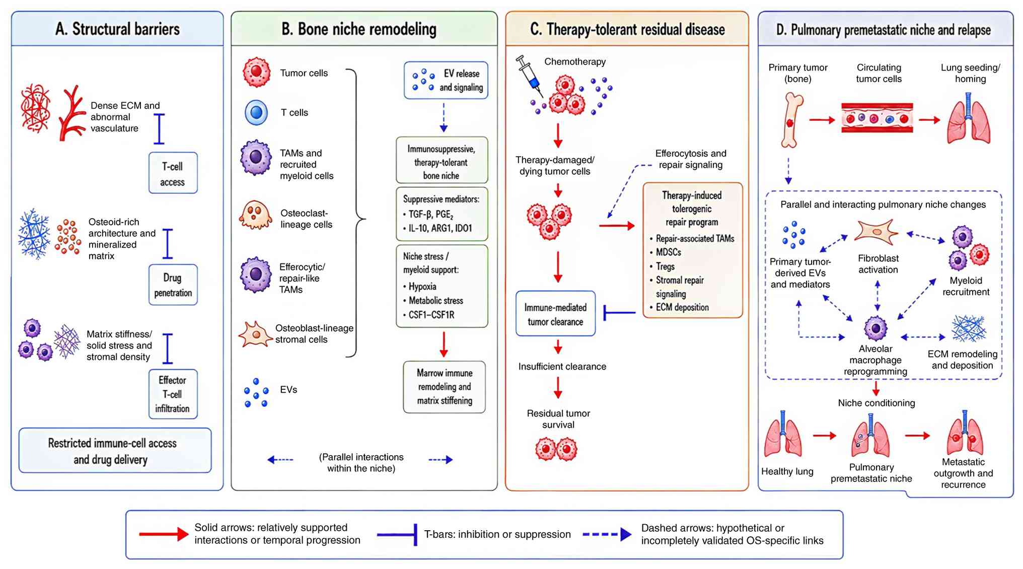 | Figure 1.Bone niche-driven layered antitumor
immune failure in OS. The schematic summarizes four partially
overlapping layers: Structural restriction within mineralized or
osteoid-rich bone; myeloid cell-predominant remodeling; tolerogenic
processing of therapy-stressed or dying tumor cells; and systemic
communication that may support pulmonary niche conditioning. Solid
arrows indicate relatively better-supported OS-specific links,
dashed arrows indicate candidate or hypothesis-generating links and
the T-bar indicates inhibition or suppression. Figure created using
BioRender. EV, extracellular vesicle; TAM, tumor-associated
macrophage; IDO1, indoleamine 2,3-dioxygenase 1; ARG1, arginase 1;
CSF1R, colony-stimulating factor 1 receptor; TGF-β, transforming
growth factor-β; PGE2, prostaglandin E2; LOX-1,
lectin-like oxidised low-density lipoprotein receptor-1; MDSC,
myeloid-derived suppressor cell; Treg, regulatory T cell; ECM,
extracellular matrix; T cell, T lymphocyte; OS, osteosarcoma. |
Structural basis of immune failure in
the bone niche
The bone niche not only provides the anatomical
setting for OS growth and dissemination but also actively shapes
the tumor microenvironment (TME), which may progressively acquire
an immune-restricted and tumor-permissive state (4,24).
Mineralized matrix and osteoid-rich architecture may restrict the
spatial access of immune cells and affect drug distribution in
certain contexts, whereas osteolytic remodeling may release
matrix-bound growth factors that support malignant adaptation
(4,24,25).
Abnormal vasculature and mechanical stress may further promote
invasion, repair-associated remodeling and immune tolerance
(14,25). Thus, normal skeletal architecture
may be converted into a structural barrier that limits early
therapeutic efficacy.
Beyond these structural constraints, the bone marrow
compartment adds an additional regulatory layer (26). Myeloid progenitors, macrophages,
osteoclast precursors and immunomodulatory MSCs normally maintain
skeletal homeostasis, tissue repair and regenerative remodeling
(24). However, within the
microenvironment of OS, these homeostatic programs may be co-opted
to promote tumor persistence, immune evasion and stromal
maintenance. Therefore, OS might exploit the intrinsic bone
remodeling capacity rather than simply appear in defective
tissue.
Such an interpretation is consistent with
bone-mimetic experimental models (17). In co-culture systems comprising
osteoid-like scaffolds and MSCs, the bone-like environment can
promote OS cell proliferation, activate stemness-associated
programs, stimulate matrix production and remodel the extracellular
matrix (ECM) (17). These results
suggest that the niche is not biologically passive. Instead,
osteoid-like structures and stromal interactions may exert
selective microenvironmental pressures that reinforce malignant
adaptation and promote an invasive, immune-evasive tissue state
(17).
Osteogenic and osteoclast-lineage cells coordinate
bone modeling and remodeling via the receptor activator of nuclear
factor κB ligand (RANKL)/RANK/osteoprotegerin system (48) under physiological conditions. This
homeostatic program may be reprogrammed to support tumor signaling,
immune regulation and metastatic progression in OS (24,25).
This implies that biological processes that, under normal
conditions, preserve skeletal integrity may, under malignant
stress, contribute to tumor maintenance.
Bone niche remodeling is one of the more
mechanistically well-supported anchors of the model (4,11–14,17,24,25)
among the modules considered within the present review. It proposes
a plausible link among structural constraints, myeloid
accumulation, altered immunological handling of dying tumor cells,
EV release, metabolic stress and conditioning of the pulmonary
niche.
This integrative sequence is a synthesis of evidence
across distinct modules rather than a single experimentally
established cascade (17–19,21,33).
Beyond a local anatomical context, the remodeled bone niche may
connect tissue architecture to systemic relapse mechanisms, thereby
linking skeletal turnover, immunosuppressive remodeling and
metastatic preparation within a sustained pattern of treatment
failure.
Myeloid cell-predominant local immune
remodeling
OS does not arise within an immunologically inert
tissue bed (49). Compared with
several soft-tissue malignancies, it develops in an actively
remodeling bone marrow compartment enriched in myeloid progenitors,
bone marrow macrophages, osteoclast precursors and immunomodulatory
MSCs (28). Myeloid cell enrichment
may therefore be embedded within the baseline cellular architecture
of the OS niche rather than merely representing a late consequence
of tumor progression (49).
Within this bone marrow niche, tumor-associated
macrophages (TAMs) occupy a potentially consequential regulatory
position (29). OS lesions are
frequently macrophage-enriched, and TAMs participate in antigen
processing, inflammatory regulation, matrix remodeling, angiogenic
remodeling and adaptive immune regulation (30). The function of TAMs cannot,
therefore, be reduced to the conventional label of
‘immunosuppressive cells’. More precisely, TAMs constitute a
multifunctional regulatory population through which skeletal
remodeling, impaired antitumor immunity, stromal adaptation and
therapeutic failure may become biologically interconnected
(29).
The binary M1/M2 dichotomy is also likely to be
overly restrictive in this setting (50). Macrophages occupy a functional
continuum, an observation that is particularly relevant in OS,
where macrophage-rich and immune-excluded niches have been
identified (8,11,13,27,31).
TAM-fibroblast crosstalk, antigen-presenting macrophage states, and
perivascular macrophage or endothelial-associated niches may alter
T-cell localization, activation thresholds and exhaustion-like
phenotypes (11,13,28,51,52).
Macrophages may therefore reshape the spatial and functional
organization of antitumor immunity rather than uniformly suppress
it.
This interpretation has therapeutic implications
(53). In OS, T-cell dysfunction
may arise less from an autonomous defect in T cells than from a
surrounding microenvironment dominated by suppressive myeloid,
stromal, vascular and metabolic constraints (8). Even when effector T cells are present,
their spatial access to tumor cells and their capacity to form
productive cytotoxic contacts may remain limited (8). Impaired antigen presentation,
myeloid-derived inhibitory signals, stromal immune exclusion,
aberrant vasculature and metabolic deprivation may collectively
reduce cytotoxic efficacy. Failure of T-cell-directed therapy may
therefore reflect persistent myeloid-dominated tissue regulation
and insufficient downstream immune activation (50).
TAMs may consequently represent a rational
therapeutic entry point, particularly in myeloid-rich tumors
(28,29,31,49).
This rationale is supported by OS-specific datasets and by broader
immuno-oncology evidence linking macrophage states to immune
exclusion, treatment resistance and metastatic progression
(50,51). The importance of the TAM axis lies
in its ability to integrate local immunosuppression, dying cell
clearance, extracellular communication, matrix remodeling and
mechanisms of systemic recurrence (18,28,54).
Targeting TAM recruitment or state may therefore provide a
mechanistically coherent strategy when downstream T-cell activation
remains constrained by a myeloid-dominated suppressive environment
(49,50).
One candidate process through which myeloid
remodeling may convert therapy-induced tumor cell death into immune
tolerance is efferocytosis. Efferocytosis is the phagocytic
clearance of apoptotic cells by macrophages and other phagocytes
(55). Given that direct causal
evidence in OS remains limited, this process is treated here as an
emerging extension of the myeloid-remodeling framework rather than
as an established mechanism.
Efferocytosis as a potential mediator of
therapy-induced immune tolerance
In OS, efferocytosis may be particularly relevant
since treatment-induced tumor cell death occurs within a bone
tissue environment already characterized by repair, remodeling and
resolution of inflammation (18).
Physiologically, efferocytosis limits excessive inflammation and
restores tissue integrity (55).
Within the OS niche, however, this program may have different
consequences. Clearance of dying tumor cells may trigger
repair-associated signaling, reprogram macrophage activity,
attenuate pro-inflammatory activation and weaken effective
antitumor immune responses in selected contexts (18).
The central issue is not just whether M2-like
polarization occurs after therapy, but why tumor cell death from
chemotherapy, radiotherapy or thermal damage does not consistently
result in long-lasting immune-mediated tumor clearance (18,56).
In healthy tissues, efferocytosis terminates inflammation and
maintains tissue architecture (55). In OS, such a clearance program could
also promote immune tolerance at a time when an immunogenic
activation would be therapeutically desirable. Thus,
therapy-induced cell death may provide a substrate for
repair-associated immunosuppression in specific conditions of the
bone niche (18).
Direct OS data link MER proto-oncogene tyrosine
kinase (MerTK)-mediated efferocytosis with M2-like macrophage
polarization, programmed death-ligand 1 (PD-L1) induction and
immune-tolerant progression (18),
and an MAGEA11-growth arrest-specific 6 (Gas6)-MERTK/AXL receptor
tyrosine kinase (AXL) transcriptomic signature has been associated
with prognostic and immune landscape features (35). The broader efferocytosis and
TAM-receptor literature supports biological plausibility, but it
remains contextual (36,37,57,58).
The priority is therefore not further correlation, but treatment
stage-specific perturbation that tests whether this pathway affects
antigen presentation and effector activation.
At present, efferocytosis is well positioned as a
testable link between therapy-induced tumor cell death and myeloid
tolerance. Enhanced framework integration will require paired pre-
and post-treatment specimens, dying cell state mapping, and direct
evaluation of whether Gas6-MERTK/AXL modulations improve T-cell
infiltration, cytotoxic contact or antigen presentation beyond
tumor burden reduction.
Extracellular vesicles as a candidate
bone-lung communication axis
EVs are increasingly recognized as active mediators
rather than inert tumor-derived secretions (38); they may transmit signals originating
from the primary bone TME beyond the local lesion (39). Experimental observations suggest
that EV cargo can influence skeletal remodeling, stromal
activation, myeloid suppression, vascular remodeling and metabolic
reprogramming (40). Through this
vesicle-mediated exchange, primary OS may communicate with
circulating and pulmonary cell populations (32).
Across sarcoma and broader tumor literature, EVs may
modulate macrophage polarization, alter dendritic cell maturation,
affect the cytotoxic function of natural killer (NK) cells and
CD8+ T cells, activate cancer-associated fibroblasts,
remodel vasculature and reorganize the ECM (38–40).
These effects likely reflect population-level vesicle signaling
rather than the action of individual vesicles. Collectively,
EV-mediated communication may contribute to a matrix-rich,
immunosuppressive and invasion-permissive microenvironment;
however, not all these effects have been functionally validated in
OS.
A particularly important but incompletely validated
question concerns the pulmonary activity of OS-derived EVs
(19,32,33,41).
These vesicles may target pulmonary fibroblasts, alveolar
macrophages, endothelial cells and bone marrow-derived myeloid
populations, thus favoring stromal activation, ECM remodeling and
the recruitment or reprogramming of immunosuppressive myeloid
cells. EV-packaged S100A11 provides direct, OS-specific evidence of
pulmonary granulocytic myeloid-derived suppressor cell (gMDSC)
recruitment and premetastatic niche formation (33). In some settings, EV-mediated
conditioning may precede, rather than simply accompany,
radiologically detectable metastatic colonization (19,32,33).
Current OS data support the role of EVs in pulmonary
niche remodeling, gMDSC recruitment and inflammatory stromal
conditioning (19,32,33,41).
The stronger assertion that EVs mediate a lasting bone-lung program
driving relapse still requires paired primary blood-lung sampling
and perturbation of defined cargo, as cargo enrichment and inferred
recipient cell interactions alone do not establish causality
(33,34).
Key validation studies should include EV depletion
or cargo perturbation, pulmonary recipient cell mapping, alveolar
macrophage and fibroblast functional endpoints, and pulmonary
recurrence endpoints. Until then, EVs provide a feasible bone-lung
communication module, but not a stand-alone explanation for
pulmonary metastasis.
Physical and metabolic stress adaptation in
the osteosarcoma niche
Metabolic reprogramming should not be interpreted
solely as an autonomous consequence of accelerated tumor
proliferation (21). Instead, it
should be considered within the physical, vascular and metabolic
constraints imposed by the OS bone niche. Mineralized matrix,
aberrant ECM deposition, increased tissue stiffness, hypoxia,
heterogeneous perfusion, nutrient competition and acidic metabolic
waste may jointly impose selective pressures on both malignant and
immune cells. These constraints can limit oxygen and substrate
availability, impair perfusion, and reshape tumor cell adaptation
and immune cell function (21).
OS studies have associated GABA type A
receptor-associated protein-mediated mitophagy and
pyruvate-metabolism programs with disease progression (44), endoplasmic reticulum (ER) stress and
unfolded protein response-related signatures with prognosis and
therapy resistance (45,47), BCL2 interacting protein 3
(BNIP3)-related hypoxic and mitophagy programs with immune features
(46), and tumor-microenvironmental
cell communication with metastatic behavior (34). These studies provide useful
stratification signals but do not by themselves establish direct
metabolic suppression of antitumor immunity; they may instead
reflect proliferation, regional hypoxia, necrotic burden,
therapy-induced clonal selection or general aggressiveness rather
than immune escape itself (21,34,44–47,59).
Causal testing should ask whether metabolic
perturbation restores major histocompatibility complex (MHC)
expression, antigen presentation, T-cell infiltration, NK cell
activity, immune checkpoint sensitivity or control of pulmonary
recurrence, independently of tumor burden reduction (21,47).
Physical and metabolic stress adaptation is
therefore retained as a lower-tier extension of the framework; it
should be upgraded only when perturbation studies link metabolic or
mechanical constraints to immune recognition, effector function and
recurrence-related endpoints (21,34).
Pulmonary premetastatic niche formation and
recurrence
The pulmonary premetastatic niche should not be seen
as merely a result of metastatic disease; it might also be a
distant indication of systemic conditioning from the initial bone
lesion (60). Myeloid-mediated
immunosuppression, EV-mediated communication, chemokine network
remodeling, ECM reorganization, mechanical cues and
hypoxia-associated stress may converge prior to radiographically
visible metastatic expansion. Such activities may therefore
generate a permissive pulmonary milieu that enables survival,
lodging and subsequent proliferation of disseminated OS cells
(15,60).
Pulmonary metastasis is the most common cause of
death due to OS (1,2), although the processes dictating
whether disseminated cells survive and proliferate are still not
well understood. The lung can serve as the physical site of tumor
cell spread, but also as a conditioned milieu comprising
inflammatory, stromal, vascular and myeloid changes preceding
detectable metastatic development (15,60).
Therefore, biological relapses may precede clinical or radiographic
recurrence.
Potential mediators include chemokines, EV cargo,
macrophage-derived signals and other soluble components. C-C motif
chemokine ligand 5 (CCL5) has been associated with OS development
and immune-stromal interactions, suggesting a potential, rather
than established, role in promoting a metastasis-permissive milieu
(61). C-X-C motif chemokine ligand
10 (CXCL10)/C-X-C chemokine receptor type 3 signaling has been
identified as a potential axis for regulating immune cell
trafficking and metastatic behavior; however, its precise role in
pulmonary niche conditioning remains unclear (62). Another way tumor-lung communication
occurs is via OS-derived EVs, with direct data implicating EV
cargo, pulmonary macrophages and gMDSC recruitment in stromal and
immunological remodeling (19,32,33).
The core of this approach is temporal dynamics.
Pulmonary niche conditioning may begin before radiographic
metastases become detectable, and recurrence may reflect not only
tumor-cell dissemination but also systemic conditioning of the lung
by signals released from the primary tumor (60). Monitoring circulating biomarkers of
pulmonary niche activity may therefore complement conventional
assessment of the primary lesion, particularly during the
perioperative period and post-chemotherapy surveillance.
Treatment-induced inflammatory and stromal responses during these
intervals may further modify metastatic permissiveness (60,63).
Pulmonary recurrence should nevertheless not be
attributed exclusively to ongoing bone-lung communication. Occult
micrometastatic dissemination, tumor cell dormancy and
reactivation, therapy-driven clonal selection and tumor
cell-intrinsic pulmonary tropism are alternative or complementary
explanations. Studies of metastasis-initiating OS subpopulations
and organotropic EV biology are conceptually consistent with some
of these processes (20,43), but they do not establish the
relative contribution of each mechanism. The proposed framework,
therefore, distinguishes pulmonary premetastatic conditioning from
micrometastatic maintenance and metastatic outgrowth. Longitudinal
cohorts with paired primary tumors and lung specimens will be
required to resolve these stages (Fig.
2).
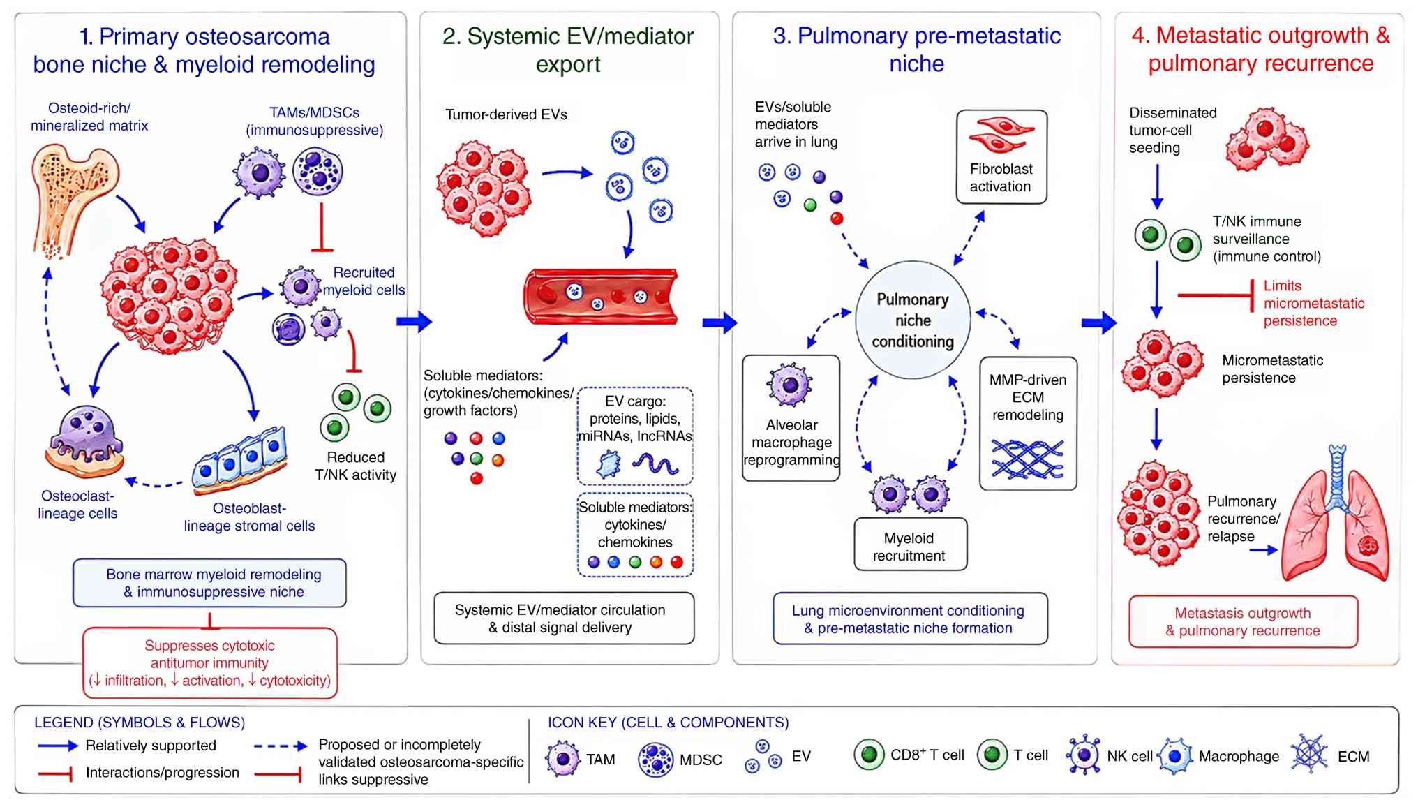 | Figure 2.Proposed bone-circulation-lung axis
linking primary OS niche remodeling to pulmonary premetastatic
conditioning and recurrence progression. The schematic depicts
primary bone niche remodeling, export of circulating
EVs/chemokines, lung-niche conditioning, micrometastatic
maintenance and metastatic outgrowth. Solid links indicate
relatively better-supported OS evidence, dashed links indicate
OS-supported candidate mechanisms, and dotted links indicate
contextual extrapolation requiring functional validation. Figure
created using BioRender. MDSC, myeloid-derived suppressor cell; EV,
extracellular vesicle; CCL5, C-C motif chemokine ligand 5; CXCL10,
C-X-C motif chemokine ligand 10; lncRNAs, long non-coding RNAs;
ECM, extracellular matrix; Mφ, macrophage; MMP, matrix
metalloproteinase; DTC, disseminated tumor cell; MDSC,
myeloid-derived suppressor cell; OS, osteosarcoma; TAM,
tumor-associated macrophage; NK, natural killer; miRNA,
microRNA. |
Therapeutic implications, validation
strategies and future perspectives
Therapeutic bottlenecks and sequential
treatment logic
Chemotherapy, checkpoint blockade and engineered
cellular therapies encounter distinct but partly convergent
microenvironmental limitations (3).
Chemotherapy can reduce tumor burden while leaving regenerative and
immunosuppressive networks intact. Checkpoint blockade can
attenuate inhibitory signaling but may act too late in
immune-excluded or myeloid cell-predominant lesions (8). Chimeric antigen receptor (CAR)-T and
CAR-NK cells provide antigen-specific cytotoxicity, yet their
homing, persistence and effector function remain constrained by
transforming growth factor-β (TGF-β) signaling, suppressive myeloid
circuits, structural barriers, and physical or metabolic stress
within the TME (64). These shared
barriers suggest that therapeutic failure may occur upstream of
final cytotoxic engagement.
These observations can be organized into a staged
translational logic rather than a catalogue of experimental
strategies. The first stage, niche reprogramming, aims to relieve
dominant myeloid, stromal, EV-related or metabolic constraints
before or alongside immune activation. Representative approaches
include TAM recruitment blockade or state reprogramming,
Gas6-MerTK/AXL-axis inhibition, TGF-β or stromal modulation,
metabolic immunomodulation, and exploratory EV-directed strategies
(18,19,21,28,31,35–37,49,58,65).
These interventions should be interpreted as candidate approaches
for testing whether niche-directed modulation can improve immune
trafficking, antigen presentation and effector-cell function,
rather than as established therapeutic requirements.
The second stage, immune activation, would deploy
checkpoint blockade, B7 homolog 3 (B7-H3)-directed therapy,
CAR-T/CAR-NK platforms, organoid-guided T-cell reconstruction or
other engineered cellular approaches after at least partial relief
of upstream constraints (3,64,66,67).
This timing is intended to reduce the mismatch between downstream
cytotoxic activation and an upstream immune-restrictive niche,
although antigen heterogeneity, inefficient homing, limited
persistence, stromal exclusion, metabolic stress and suppressive
myeloid populations remain major barriers (64,67,68).
The third stage, pulmonary niche maintenance,
focuses on high-risk relapse settings after local treatment or
during minimal residual disease surveillance. EV/chemokine
monitoring, lung-directed immune support, CAR macrophages generated
in situ using EV-delivered mRNA, inhaled NK cell-supportive
platforms and postoperative immunomodulatory strategies may be
considered as investigational approaches, but this stage remains
predominantly preclinical or conceptual (19,33,63,68,69).
The preferred endpoint should shift from short-term tumor shrinkage
alone to suppression of micrometastatic persistence and improvement
in pulmonary recurrence-free survival.
Clinical implementation would require careful
patient selection, explicit timing and predefined biological
readouts. Candidate populations may include patients at high risk
of perioperative pulmonary recurrence, patients with primary
resistance to T-cell-directed immunotherapy, and patients whose
tumors show myeloid enrichment, abnormal EV or chemokine profiles,
or spatially organized suppressive niches (8,11,19,33,61,62,70).
EV cargo, CCL5/CXCL10 profiles, macrophage-distribution patterns
and spatial immune features may help identify candidates for niche
reprogramming or pulmonary niche maintenance, but these biomarkers
require prospective validation.
Accordingly, the proposed sequence should be
regarded as a trial-design framework rather than a clinically
established algorithm. Future studies should map candidate
interventions to the phase in which they are most biologically
plausible: Niche reprogramming before or during cytotoxic injury;
immune activation after partial relief of upstream constraints; and
pulmonary niche maintenance during postoperative surveillance or in
high-risk minimal residual disease. Myeloid-enrichment biomarkers,
spatial immune profiling, EV/chemokine monitoring, pulmonary
recurrence endpoints and safety outcomes should be incorporated
rather than relying solely on tumor-size response (Fig. 3; Table
II).
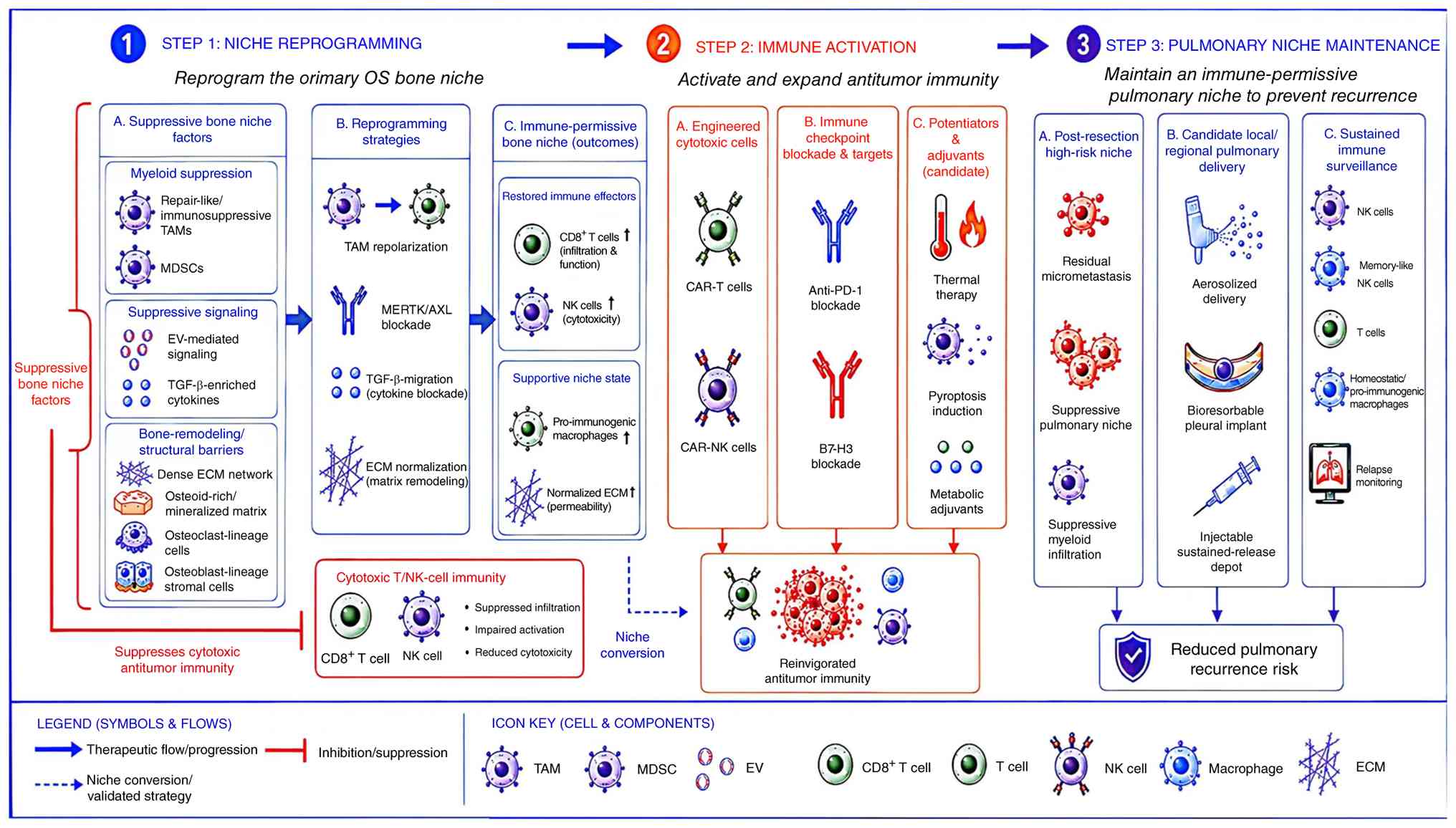 | Figure 3.Conceptual three-step therapeutic
sequence aligned with the proposed bone-lung immune axis in OS.
Niche reprogramming during the neoadjuvant/perioperative window
aims to relieve structural, myeloid, stromal, EV-related, or
metabolic constraints; immune activation follows partial niche
relief through checkpoint, B7-H3, CAR-T/CAR-NK or other engineered
approaches; and pulmonary niche maintenance during postoperative
minimal-residual-disease surveillance focuses on lung-directed
immune support, EV/chemokine monitoring, NK cell-supportive
platforms and relapse monitoring. The sequence is evidence-staged
and should not be interpreted as an established clinical algorithm.
Figure created using BioRender. TAM, tumor-associated macrophage;
MDSC, myeloid-derived suppressor cell; EV, extracellular vesicle;
ECM, extracellular matrix; TME, tumor microenvironment; Gas6,
growth arrest-specific 6; MERTK, MER proto-oncogene tyrosine
kinase; AXL, AXL receptor tyrosine kinase; TGF-β, transforming
growth factor-β; CAR, chimeric antigen receptor; ICB, immune
checkpoint blockade; PD-1, programmed cell death protein 1; B7-H3,
B7 homolog 3; NK, natural killer; OS, osteosarcoma. |
Future directions and testable
hypotheses
Efferocytosis should be explored as a potential
proximal cause of immune suppression in specific OS cases (18,35).
The impact of blocking Gas6-MERTK/AXL or related pathways on
macrophage status, antigen presentation, PD-L1 expression and
T-cell activation should be studied. Additionally,
metabolism-associated decreases in immune recognition may exist in
certain types of tumors; however, this is one of the least
established sections of the model. Studies investigating mitophagy,
BNIP3-mediated adaptability, ER stress or metabolic buffering
should directly assess immune recognition, rather than merely tumor
proliferation (44–47).
Third, the risk of pulmonary recurrence may
potentially be classified prior to radiographic development. EV
cargo, CCL5, CXCL10, spatial macrophage patterns and other
niche-related traits are possible biomarkers. Prospective
validation in cohorts with specific pulmonary outcomes is needed to
evaluate their predictive value (19,33,61,62).
Fourth, spatial organization may be more
informative than mean expression levels. 3D spatial transcriptomics
and multimodal imaging can uncover compact suppressive niches of
macrophages, fibroblasts, endothelial cells and defective
lymphocytes (11,52,71).
Such microenvironments may be better predictors of the response to
therapy than bulk transcriptome averages; however, prospective
predictive validation is yet to be shown. This approach can be
operationalized using a claim evidence gap validation matrix
(Table III) (72–77). A
minimal validation strategy for a bone niche-driven framework
should include paired pre- and post-treatment primary tumor
specimens, circulating EV and chemokine profiling during
neoadjuvant therapy and postoperative surveillance, matched
pulmonary metastatic or recurrent specimens when available, and
spatial mapping of myeloid, stromal, endothelial and T-cell
compartments. In parallel, candidate pathways such as TAM
recruitment, MerTK/AXL signaling, EV cargo transfer and metabolic
stress adaptation should be tested through cell type-resolved
perturbation models. The clinical relevance of this framework would
be strengthened by predefined pulmonary recurrence endpoints and
external validation in an independent cohort. Evidence would be
particularly compelling if niche-directed disruption altered immune
trafficking, antigen presentation, effector-cell function and
pulmonary recurrence risk beyond changes in tumor burden alone.
| Table III.Claim evidence gap validation matrix
for the bone niche-driven framework. |
Table III.
Claim evidence gap validation matrix
for the bone niche-driven framework.
| Claim | Direct OS
evidence | Contextual
evidence | Current gap | Falsifiable
validation test | (Refs.) |
|---|
| Bone niche
remodeling restricts effective antitumor immunity | Single-cell/spatial
atlases, bone TME studies and bone-mimetic models | Osteoimmunology and
bone malignancy literature | Limited
longitudinal evidence across neoadjuvant therapy, surgery and
recurrence | Paired
pre-/post-treatment spatial mapping showing that niche remodeling
changes immune trafficking, tumor-cell contact, antigen
presentation and recurrence risk | (11–14,17,24–27) |
| Myeloid-rich niches
impair downstream cytotoxic function | Macrophage-enriched
lesions, immune-exclusion studies and TAM-related OS models | Pan-cancer TAM and
macrophage-ecosystem literature | Unclear
state-specific causality and patient-context dependence | Lineage-resolved
TAM perturbation showing restored CD8+ T-cell or NK cell
function without merely reducing tumor burden | (8,11,13,27–31,50,51,53) |
| Efferocytosis
converts therapy-induced cell death into tolerance | MerTK/efferocytosis
studies and OS transcriptomic patterns | TAM receptor and
cancer efferocytosis literature | Few OS-specific
blockade studies and little longitudinal dying cell state
mapping | Gas6-MERTK/AXL
perturbation after chemotherapy-induced cell death with
antigen-presentation, PD-L1, TAM-state and T-cell-activation
readouts | (18,35–37,57,58) |
| EV-mediated
bone-lung communication conditions pulmonary relapse | OS-derived EV
studies, EV-packaged S100A11 and lung premetastatic-niche
findings | Organotropic EV and
premetastatic-niche literature | Cargo enrichment
and recipient-cell inference do not prove causality; paired
primary-lung data are scarce | EV depletion or
cargo perturbation with recipient-cell validation in lung models
and pulmonary recurrence-free survival endpoints | (19,32,33,41–43,63) |
| Physical/metabolic
stress suppresses immune recognition | OS multi-omics,
mitophagy, UPR, pyruvate, BNIP3, hypoxia and immune-signature
studies | Tumor ecology,
hypoxia and metabolic immune-suppression literature | Mostly prognostic
modeling or computational inference rather than direct immune
causality | Metabolic
perturbation coupled to MHC expression, antigen presentation,
CD8+ T-cell and NK cell assays, ICI sensitivity and lung
recurrence endpoints | (21,34,44–47,59) |
| Timed niche
reprogramming improves immunotherapy efficacy |
Macrophage-directed, B7-H3, organoid,
biomaterial, hyperthermia, NK and engineered cell OS studies |
Microenvironment-modulation and
immunotherapy-combination literature | Optimal timing,
safety and biomarker selection remain undefined | Biomarker-guided
window or perioperative trials testing niche reprogramming before
immune activation with safety and pulmonary recurrence
endpoints | (49,53,56,64–69,72–77) |
Evidence hierarchy and model
architecture
The present review argues that therapeutic
resistance in OS and pulmonary relapse requires a tissue-specific
explanation that extends beyond tumor cell intrinsic mechanisms.
The bone niche provides a testable architecture in which structural
barriers, marrow-derived myeloid enrichment, treatment-induced
repair, extracellular communication, metabolic stress and pulmonary
surveillance converge.
The evidence hierarchy is deliberately separated
from therapeutic maturity. Bone niche remodeling and myeloid cell
enrichment are the most mature OS-supported anchors; pulmonary
niche conditioning, efferocytosis and EV-mediated bone-lung
communication have growing but incomplete longitudinal or
functional support, and physical/metabolic adaptation remains a
lower-tier extension.
Accordingly, the framework should be read as a
falsifiable model, not a fixed cascade; its modules may operate in
parallel or only in selected molecular, immune, age-related or
metastatic contexts, with Table
III defining the claim evidence validation boundaries.
Heterogeneity, scope and patient
context
OS is biologically and clinically heterogeneous;
therefore, the proposed bone niche-driven framework should not be
assumed to operate uniformly across all patients. Molecular
subtypes, transcriptional states, immune infiltration patterns,
age-related skeletal and immune contexts, chemotherapy response and
metastatic trajectory may all influence the contribution of the
bone niche to antitumor immune failure (11,13,27,70,78).
Tumors with myeloid-enriched, immune-excluded or stromal-activated
phenotypes may depend more on macrophage-centered regulation and
niche-mediated immune restraint, whereas other tumors may be driven
predominantly by tumor cell intrinsic genomic instability or
therapy-resistant clonal evolution.
Pediatric, adolescent and adult patients may also
differ in marrow composition, bone-remodeling activity, immune
competence and treatment tolerance, which may modify the strength
and timing of niche-mediated immune regulation. Similarly,
chemotherapy-sensitive and -resistant tumors may differ in the
immunological consequences of therapy-induced cell death, with
dying tumor cells being processed through more immunogenic pathways
in some contexts and through tolerogenic repair or
efferocytosis-associated programs in others. Finally, localized
disease, synchronous pulmonary metastasis and delayed pulmonary
recurrence may reflect different degrees of primary bone
lesion-lung communication. Therefore, the present framework should
be viewed as a context-dependent and testable model that helps
organize heterogeneous immune-failure patterns rather than as a
universal mechanism shared by all patients with OS.
Bone niche reprogramming and
integration with mainstream immunotherapy
Bone niche reprogramming can be viewed as a
translational extension of the proposed framework. In the present
review, bone niche reprogramming refers to the attempt to shift the
OS microenvironment from a structurally constrained,
myeloid-enriched, stromal-protective and metabolically suppressive
state toward a more immune-permissive and therapeutically
responsive state. This concept does not imply that niche-directed
intervention should replace established or emerging
immunotherapies. Rather, it suggests that the efficacy of immune
activation strategies may depend, at least in part, on whether
upstream bone niche constraints have been relieved.
This concept is particularly relevant to current
immunotherapy approaches in OS. Immune checkpoint blockade may be
insufficient when effector T cells remain spatially excluded,
functionally exhausted or exposed to persistent myeloid and stromal
inhibitory signals (3,6,8).
B7-H3-targeted therapy and CAR-T or CAR-NK strategies may also be
limited by poor homing, reduced persistence, antigen heterogeneity,
TGF-β-rich stromal niches, metabolic stress and suppressive
macrophage or MDSC populations (64,67).
Bone niche reprogramming may therefore provide a rational
preparatory step before immune activation. Candidate approaches
include TAM repolarization or recruitment inhibition,
Gas6-MERTK/AXL blockade, TGF-β mitigation, matrix or vascular
normalization, EV-directed intervention and metabolic
monosensitization (18,21,35–37,49,58,65).
These strategies may help convert an immune-restricted bone lesion
into a state in which immune-activating treatments can act more
effectively, but most remain preclinical or early
translational.
The same logic may apply in preventing pulmonary
relapses. If the primary bone lesion releases EVs, chemokines and
myeloid-recruiting signals that contribute to pulmonary niche
conditioning, bone niche reprogramming may need to be considered
together with pulmonary niche maintenance in selected high-risk
patients (19,33). Local or regional lung-directed
immune strategies, including CAR macrophages generated through
EV-delivered mRNA and inhaled NK cell-supportive platforms, warrant
investigation as relapse-prevention strategies (68,69).
However, these approaches remain preclinical or early translational
and should be evaluated in biomarker-guided perioperative studies,
window-of-opportunity trials and longitudinal cohorts incorporating
spatial immune readouts, EV/chemokine monitoring and pulmonary
recurrence endpoints.
Testing and falsifying the bone
niche-driven framework
Prespecified studies should test whether
niche-directed perturbation improves immune trafficking, antigen
presentation, effector-cell function or pulmonary niche quiescence
beyond tumor shrinkage alone. Failure to change these readouts
after adequate pathway perturbation would restrict the model to
specific phenotypes, windows or metastatic contexts.
A perioperative window study is the most direct
first test: Paired pre- and post-neoadjuvant specimens should be
profiled for TAM state, MerTK/AXL activity, PD-L1 induction,
antigen-presentation markers, T cell-tumor spatial contact, stromal
barriers and circulating EV/chemokine signals.
A primary-lung paired cohort should integrate
primary tumor, serial blood EV/chemokine data, and pulmonary
metastatic or recurrent tissue to determine whether primary bone
niche states predict pulmonary myeloid recruitment, stromal
activation, micrometastatic persistence and pulmonary
recurrence-free survival.
Functional perturbation models should pair TAM
modulation, Gas6-MERTK/AXL blockade, EV cargo perturbation, TGF-β
or matrix modulation, and metabolic-stress intervention with
antigen-presentation assays, CD8+ T-cell and NK cell
function, killing assays, and in vivo lung recurrence
endpoints.
Potential trial-design scenarios
Table IV (72–77)
translates the framework into three practical settings. In poor
histological responders after neoadjuvant chemotherapy,
perioperative profiling of TAM state, MerTK/AXL activation, PD-L1,
tumor cell MHC and CD8+ T-cell proximity could determine
whether niche reprogramming should precede or accompany immune
activation.
| Table IV.Practical trial-design scenarios for
testing the bone niche-driven frameworka. |
Table IV.
Practical trial-design scenarios for
testing the bone niche-driven frameworka.
| Scenario | Candidate
population | Key biological
assessment | Intervention
window | Primary
endpoint/readout |
|---|
| Poor histological
response after neoadjuvant chemotherapy | Poor necrosis,
residual viable or tumor persistent
myeloid-enriched/immune-excluded spatial niche | TAM density and
state; MerTK/AXL activity; PD-L1 induction; tumor-cell MHC
expression; CD8+ T-cell proximity to viable tumor
cells | Perioperative or
immediate post-chemotherapy window | Shift toward an
immune-permissive niche, improved antigen presentation,
effector-cell contact and recurrence-risk biomarkers |
| High-risk localized
OS after definitive surgery | No visible lung
disease but high-risk clinicopathological, spatial, EV or chemokine
features | Serial circulating
EV cargo; CCL5/CXCL10 profiles; myeloid signatures; spatial immune
features in the resected tumor | Postoperative
surveillance or minimal residual disease window | Pulmonary
recurrence-free survival, biomarker stability and safety of
maintenance immunomodulation |
| Pulmonary
micro-metastatic disease or early lung recurrence | Micrometastatic
lung disease, early recurrence or high likelihood of pulmonary
niche activation | Lung immune niche,
alveolar-macrophage state, myeloid recruitment, systemic
EV/chemokine communication and cytotoxic cell function | Lung-directed
immune-support window or early recurrence treatment window | Pulmonary control,
immune safety, pulmonary delivery toxicity, infection risk and
micrometastatic persistence |
In high-risk localized disease after definitive
surgery, serial EV cargo, CCL5/CXCL10, myeloid signatures and
spatial features from the resected tumor could support pulmonary
recurrence-prevention studies that use pulmonary recurrence-free
survival rather than short-term shrinkage as the primary
endpoint.
In pulmonary micrometastatic disease or early lung
recurrence, the lung immune state, systemic EV/chemokine
communication and cytotoxic cell function should be assessed
alongside lung-directed immune support or engineered cell
approaches, with predefined monitoring for pulmonary toxicity,
immune overactivation, infection and impaired repair.
Limitations
The present review has limitations. As a
mechanistic narrative review rather than a systematic review or
meta-analysis, it was designed to construct and stress-test a
conceptual framework, not to capture every eligible study, pool
effects or formally grade risk of bias. Selection and evidence
weighting, therefore, involve author judgment, and the proposed
hierarchy should be viewed as a practical organizational tool
rather than a validated grading system. The maturity of evidence is
also uneven: Bone niche remodeling and myeloid cell enrichment have
broader OS support, while efferocytosis, EV-mediated bone-lung
communication and metabolic immune adaptation are more reliant on
transcriptomic, computational, spatial-inference or limited
functional data. Heterogeneity between patients, models, ages,
treatment settings and illness stages further limits
generalizability. Future validation will need a longitudinal
sample, coupled with primary lung specimens, independent
replication, lineage-resolved perturbation, clinically relevant
immune readouts and predefined pulmonary recurrence endpoints.
Conclusion
The present review proposes an evidence-graded and
testable bone niche-driven framework for understanding layered
antitumor immune failure in OS. Within this framework, treatment
resistance and pulmonary relapse may partly reflect the interaction
of structural constraints within the bone niche, myeloid
cell-predominant immune remodeling, tolerogenic processing of
therapy-induced tumor cell death, systemic niche communication and
impaired pulmonary immune surveillance. Bone niche remodeling and
myeloid cell enrichment represent the relatively better-supported
components of this model, whereas pulmonary niche conditioning is
increasingly supported but remains uneven. Efferocytosis,
EV-mediated bone-lung communication, and physical or metabolic
immune adaptation remain emerging or hypothesis-generating modules
that require OS-specific functional validation.
The principal translational implication is a shift
from treatment intensification alone toward biologically timed
intervention according to niche state. The proposed strategy
follows a three-step sequence: Niche reprogramming to relieve
upstream structural, stromal and myeloid immunosuppressive
constraints; immune activation to restore effective antitumor
responses; and pulmonary niche maintenance to limit
disseminated-cell survival, micrometastatic persistence and
pulmonary recurrence. This sequence should be regarded as a
conceptual framework for biomarker-guided trial design rather than
as an established clinical algorithm.
The model is unlikely to explain treatment failure
uniformly across all patients. Its relevance may vary according to
molecular subtype, age, immune and stromal phenotype, chemotherapy
response and metastatic trajectory. Future studies should therefore
combine longitudinal paired sampling of primary tumors and
pulmonary metastases or recurrences, spatial and single-cell
profiling, lineage-resolved functional perturbation, perioperative
intervention studies and pulmonary recurrence endpoints. Such work
will be required to distinguish causal mechanisms from associative
signals and to define the patient and disease contexts in which
bone niche-directed intervention may provide clinical value.
Supplementary Material
Supporting Data
Acknowledgements
Not applicable.
Funding
This review was supported by the Yunnan Provincial Department of
Science and Technology-Kunming Medical University Special Projects
(grant no. 202301AY070001-124).
Availability of data and materials
Not applicable.
Authors' contributions
BT and YW contributed to study conception,
literature review, manuscript drafting and table organization. JL
and RP supervised the study design, revised key academic content
and approved the final manuscript. All authors contributed to
writing and revising the manuscript. All authors read and approved
the final version of the manuscript. JL is the primary
corresponding author. Data authentication is not applicable.
Ethics approval and consent to
participate
Not applicable.
Patient consent for publication
Not applicable.
Competing interests
The authors declare that they have no competing
interests.
Glossary
Abbreviations
Abbreviations:
|
OS
|
osteosarcoma
|
|
TME
|
tumor microenvironment
|
|
TAM
|
tumor-associated macrophage
|
|
EV
|
extracellular vesicle
|
|
ECM
|
extracellular matrix
|
|
MSC
|
mesenchymal stromal cell
|
|
gMDSC
|
granulocytic myeloid-derived
suppressor cell
|
|
NK
|
natural killer
|
|
CAR
|
chimeric antigen receptor
|
|
MHC
|
major histocompatibility complex
|
|
PD-L1
|
programmed death-ligand 1
|
|
B7-H3
|
B7 homolog 3
|
|
TGF-β
|
transforming growth factor-β
|
|
RANKL
|
receptor activator of nuclear factor
κB ligand
|
|
CCL5
|
C-C motif chemokine ligand 5
|
|
CXCL10
|
C-X-C motif chemokine ligand 10
|
|
Gas6
|
growth arrest-specific 6
|
|
MerTK/MERTK
|
MER proto-oncogene tyrosine
kinase
|
|
AXL
|
AXL receptor tyrosine kinase
|
References
|
1
|
Yu Y, Li K, Peng Y, Zhang Z, Pu F, Shao Z
and Wu W: Tumor microenvironment in osteosarcoma: From cellular
mechanism to clinical therapy. Genes Dis. 12:1015692025. View Article : Google Scholar : PubMed/NCBI
|
|
2
|
Zhang L, Jiang H and Ma H: Progress in
immune microenvironment, immunotherapy and prognostic biomarkers in
pediatric osteosarcoma. Front Immunol. 16:15485272025. View Article : Google Scholar : PubMed/NCBI
|
|
3
|
Yu S and Yao X: Advances on immunotherapy
for osteosarcoma. Mol Cancer. 23:1922024. View Article : Google Scholar : PubMed/NCBI
|
|
4
|
Dutour A, Pasello M, Farrow L, Amer MH,
Entz-Werlé N, Nathrath M, Scotlandi K, Mittnacht S and
Gomez-Mascard A: Microenvironment matters: Insights from the FOSTER
consortium on microenvironment-driven approaches to osteosarcoma
therapy. Cancer Metastasis Rev. 44:442025. View Article : Google Scholar : PubMed/NCBI
|
|
5
|
El Motassime A, Vitiello R, Comodo RM,
Capece G, Bocchino G, Bocchi MB, Maccauro G and Meschini C:
Osteosarcoma: A comprehensive morphological and molecular review
with prognostic implications. Biology (Basel).
14:14072025.PubMed/NCBI
|
|
6
|
Hattinger CM, Salaroglio IC, Fantoni L,
Godel M, Casotti C, Kopecka J, Scotlandi K, Ibrahim T, Riganti C
and Serra M: Strategies to overcome resistance to immune-based
therapies in osteosarcoma. Int J Mol Sci. 24:7992023. View Article : Google Scholar : PubMed/NCBI
|
|
7
|
Sun CY, Zhang Z, Tao L, Xu FF, Li HY,
Zhang HY and Liu W: T cell exhaustion drives osteosarcoma
pathogenesis. Ann Transl Med. 9:14472021. View Article : Google Scholar : PubMed/NCBI
|
|
8
|
Ligon JA, Choi W, Cojocaru G, Fu W, Hsiue
EHC, Oke TF, Siegel N, Fong MH, Ladle B, Pratilas CA, et al:
Pathways of immune exclusion in metastatic osteosarcoma are
associated with inferior patient outcomes. J Immunother Cancer.
9:e0017722021. View Article : Google Scholar : PubMed/NCBI
|
|
9
|
Wang H, Zhu T, Yang K, Xu C, Zhang Z, Liu
G and Ren H: Decoding the immune microenvironment in osteosarcoma:
New insights into checkpoints, vaccines, and CAR-T cells. Front
Oncol. 15:17022192025. View Article : Google Scholar : PubMed/NCBI
|
|
10
|
Chen C, Xie L, Ren T, Huang Y, Xu J and
Guo W: Immunotherapy for osteosarcoma: Fundamental mechanism,
rationale, and recent breakthroughs. Cancer Lett. 500:1–10. 2021.
View Article : Google Scholar : PubMed/NCBI
|
|
11
|
Zheng X, Liu X, Zhang X, Zhao Z, Wu W and
Yu S and Yu S: A single-cell and spatially resolved atlas of human
osteosarcomas. J Hematol Oncol. 17:712024. View Article : Google Scholar : PubMed/NCBI
|
|
12
|
Zhang Y, Jiang S, Lv J, Feng W, Yu Y and
Zhao H: Osteosarcoma immune microenvironment: Cellular struggle and
novel therapeutic insights. Front Immunol. 16:15844502025.
View Article : Google Scholar : PubMed/NCBI
|
|
13
|
Liu W, Hu H, Shao Z, Lv X, Zhang Z, Deng
X, Song Q, Han Y, Guo T, Xiong L, et al: Characterizing the tumor
microenvironment at the single-cell level reveals a novel immune
evasion mechanism in osteosarcoma. Bone Res. 11:42023. View Article : Google Scholar : PubMed/NCBI
|
|
14
|
Tian H, Cao J, Li B, Nice EC, Mao H, Zhang
Y and Huang C: Managing the immune microenvironment of
osteosarcoma: The outlook for osteosarcoma treatment. Bone Res.
11:112023. View Article : Google Scholar : PubMed/NCBI
|
|
15
|
McGee LE, Pereira JS, McEachron TA, Mazcko
C, LeBlanc AK and Beck JA: The tumor microenvironment of metastatic
osteosarcoma in the human and canine lung. Commun Biol. 8:7562025.
View Article : Google Scholar : PubMed/NCBI
|
|
16
|
Zhu T, Han J, Yang L, Cai Z, Sun W, Hua Y
and Xu J: Immune microenvironment in osteosarcoma: Components,
therapeutic strategies and clinical applications. Front Immunol.
13:9075502022. View Article : Google Scholar : PubMed/NCBI
|
|
17
|
Bassi G, Saqawa M, Apolloni L, Díaz-Prado
S, Ollivier E, Levergeois R, Sandri M, Campodoni E, Cochonneau D,
Panseri S, et al: Deciphering the interaction between osteosarcoma
and mesenchymal stem cells in a 3D bone-mimetic co-culture model.
Biomed Pharmacother. 195:1189562026. View Article : Google Scholar : PubMed/NCBI
|
|
18
|
Lin J, Xu A, Jin J, Zhang M, Lou J, Qian
C, Zhu J, Wang Y, Yang Z, Li X, et al: MerTK-mediated efferocytosis
promotes immune tolerance and tumor progression in osteosarcoma
through enhancing M2 polarization and PD-L1 expression.
Oncoimmunology. 11:20249412022. View Article : Google Scholar : PubMed/NCBI
|
|
19
|
Li H, Guo Z, Zou Y, Zhang J, Shi J, Fu X,
Tu J, Yao H, Lu X, Wen L and Xie X: Tumor extracellular vesicles
lncOSLMT drives lung inflammatory premetastatic niche formation in
osteosarcoma via m6A-dependent hnRNPA2B1/COX-2 axis. Adv
Sci (Weinh). 13:e194902026. View Article : Google Scholar : PubMed/NCBI
|
|
20
|
Reinecke JB, Jimenez Garcia L, Saraf AJ,
Hinckley J, Gross AC, Le Pommellet H, Gutpell KM, Cam M, Cannon MV,
Gust MJ, et al: Metastasis-initiating osteosarcoma subpopulations
establish paracrine interactions with lung and tumor cells to
create a metastatic niche. Cancer Res. 85:4341–4358. 2025.
View Article : Google Scholar : PubMed/NCBI
|
|
21
|
Neophytou CM, Panagi M, Stylianopoulos T
and Papageorgis P: The role of tumor microenvironment in cancer
metastasis: Molecular mechanisms and therapeutic opportunities.
Cancers (Basel). 13:20532021. View Article : Google Scholar : PubMed/NCBI
|
|
22
|
Green BN, Johnson CD and Adams A: Writing
narrative literature reviews for peer-reviewed journals: Secrets of
the trade. J Chiropr Med. 5:101–117. 2006. View Article : Google Scholar : PubMed/NCBI
|
|
23
|
Baethge C, Goldbeck-Wood S and Mertens S:
SANRA: A scale for the quality assessment of narrative review
articles. Res Integr Peer Rev. 4:52019. View Article : Google Scholar : PubMed/NCBI
|
|
24
|
Song C, Tong T, Dai B, Zhu Y, Chen E,
Zhang M and Zhang W: Osteoimmunology in bone malignancies: A
symphony with evil. J Natl Cancer Cent. 4:354–368. 2024.PubMed/NCBI
|
|
25
|
Taylor AM, Sheng J, Ng PKS, Harder JM,
Kumar P, Ahn JY, Cao Y, Dzis AM, Jillette NL, Goodspeed A, et al:
Immunosuppressive tumor microenvironment of osteosarcoma. Cancers
(Basel). 17:21172025. View Article : Google Scholar : PubMed/NCBI
|
|
26
|
Orrapin S, Moonmuang S, Udomruk S,
Yongpitakwattana P, Pruksakorn D and Chaiyawat P: Unlocking the
tumor-immune microenvironment in osteosarcoma: insights into the
immune landscape and mechanisms. Front Immunol. 15:13942842024.
View Article : Google Scholar : PubMed/NCBI
|
|
27
|
Liu Y, Feng W, Dai Y, Bao M, Yuan Z, He M,
Qin Z, Liao S, He J, Huang Q, et al: Single-cell transcriptomics
reveals the complexity of the tumor microenvironment of
treatment-naive osteosarcoma. Front Oncol. 11:7092102021.
View Article : Google Scholar : PubMed/NCBI
|
|
28
|
Huang Q, Liang X, Ren T, Huang Y, Zhang H,
Yu Y, Chen C, Wang W, Niu J, Lou J and Guo W: The role of
tumor-associated macrophages in osteosarcoma
progression-therapeutic implications. Cell Oncol (Dordr).
44:525–539. 2021. View Article : Google Scholar : PubMed/NCBI
|
|
29
|
Ma J, Chen Z, Li Q, Wang L, Chen J, Yang
X, Yang C and Quan Z: RARRES2 is involved in the ‘lock-and-key’
interactions between osteosarcoma stem cells and tumor-associated
macrophages. Sci Rep. 14:22672024. View Article : Google Scholar : PubMed/NCBI
|
|
30
|
Liu Z, Zhang L and Zhong Y:
Characterization of osteosarcoma subtypes mediated by
macrophage-related genes and creation and validation of a risk
score system to quantitatively assess the prognosis of osteosarcoma
and reflect the tumor microenvironment. Ann Transl Med.
10:13182022. View Article : Google Scholar : PubMed/NCBI
|
|
31
|
Pan F, Pan R, Hu R, Zhang H, Lei S, Zhang
L, Zhou C, Zeng Z, Tian X and Xie Q: Analysis of the effects of M2
macrophage-derived PDE4C on the prognosis, metastasis and
immunotherapy benefit of osteosarcoma. J Cell Mol Med.
28:e183952024. View Article : Google Scholar : PubMed/NCBI
|
|
32
|
Lin S, Shu L, Guo Y, Yuan J, Zhang J, Wang
Y, Yang Y and Yuan T: Cargo-eliminated osteosarcoma-derived small
extracellular vesicles mediating competitive cellular uptake for
inhibiting pulmonary metastasis of osteosarcoma. J
Nanobiotechnology. 22:3602024. View Article : Google Scholar : PubMed/NCBI
|
|
33
|
Deng C, Xu Y, Chen H, Zhu X, Huang L, Chen
Z, Xu H, Song G, Lu J, Huang W, et al:
Extracellular-vesicle-packaged S100A11 from osteosarcoma cells
mediates lung premetastatic niche formation by recruiting gMDSCs.
Cell Rep. 43:1137512024. View Article : Google Scholar : PubMed/NCBI
|
|
34
|
Li J, Bai Y, Zhang H, Chen T and Shang G:
Single-cell RNA sequencing reveals the communications between tumor
microenvironment components and tumor metastasis in osteosarcoma.
Front Immunol. 15:14455552024. View Article : Google Scholar : PubMed/NCBI
|
|
35
|
Song X, Tou X, Li L, Wang F, Qian C, Wang
R and Shen J: Efferocytosis-associated transcriptomic patterns
characterize prognosis and immune landscape in osteosarcoma. J Bone
Oncol. 57:1007432026. View Article : Google Scholar : PubMed/NCBI
|
|
36
|
Zhou L and Matsushima GK: Tyro3, Axl,
Mertk receptor-mediated efferocytosis and immune regulation in the
tumor environment. Int Rev Cell Mol Biol. 361:165–210. 2021.
View Article : Google Scholar : PubMed/NCBI
|
|
37
|
Bae SH, Kim JH, Park TH, Lee K, Lee BI and
Jang H: BMS794833 inhibits macrophage efferocytosis by directly
binding to MERTK and inhibiting its activity. Exp Mol Med.
54:1450–1460. 2022. View Article : Google Scholar : PubMed/NCBI
|
|
38
|
Wang Y, Gao S, Zhang G, Xiao W, Xu B and
Li W: Extracellular vesicles in sarcoma: Implications for tumor
progression and therapy. Int J Nanomedicine. 20:13759–13779. 2025.
View Article : Google Scholar : PubMed/NCBI
|
|
39
|
Wang J and Shi K: Role of tumor-derived
exosomes and immune cells in osteosarcoma progression and targeted
therapy. Front Immunol. 16:16583582025. View Article : Google Scholar : PubMed/NCBI
|
|
40
|
Gui Y, Peng M, Huang Y, Chen Z, Chen J,
Xiao J, Liu D and Hou Y: New insights into extracellular vesicles
in metastatic cancer: From mechanisms to diagnostics and targeted
therapies. Mol Ther. 33:4731–4747. 2025. View Article : Google Scholar : PubMed/NCBI
|
|
41
|
Zhongyu X, Wei X, Hongmei Z, Xiaodong G,
Xiaojing Y, Yuanpei L, Li Z, Zhenmin F and Jianda X: Review of
pre-metastatic niches induced by osteosarcoma-derived extracellular
vesicles in lung metastasis: A potential opportunity for diagnosis
and intervention. Biomed Pharmacother. 178:1172032024. View Article : Google Scholar : PubMed/NCBI
|
|
42
|
Kumar MA, Baba SK, Sadida HQ, Marzooqi SA,
Jerobin J, Altemani FH, Algehainy N, Alanazi MA, Abou-Samra AB,
Kumar R, et al: Extracellular vesicles as tools and targets in
therapy for diseases. Signal Transduct Target Ther. 9:272024.
View Article : Google Scholar : PubMed/NCBI
|
|
43
|
Mo Z, Cheong A, Xiang L, Le MTN, Grimson A
and Zhang DX: Extracellular vesicle-associated organotropic
metastasis. Cell Prolif. 54:e129482021. View Article : Google Scholar : PubMed/NCBI
|
|
44
|
Han X, Li Y, Liu Y, Wang F, Li T, Zhang Q,
Wang G and Feng J: Integrated multi-omics reveals GABARAP-mediated
mitophagy and pyruvate metabolism as key drivers of osteosarcoma
progression. Front Immunol. 16:16805542025. View Article : Google Scholar : PubMed/NCBI
|
|
45
|
Yu Y, Yu J and Pan Z: Endoplasmic
reticulum stress-related features predict the prognosis of
osteosarcoma and reveal STC2 as a novel risk indicator for disease
progression. Front Oncol. 14:14531732024. View Article : Google Scholar : PubMed/NCBI
|
|
46
|
Li J, Wang W, Li L and Yu K: Prognostic
and immunomodulatory roles of BNIP3 in osteosarcoma revealed by
integrated single-cell and bulk transcriptomic profiling. Int J
Genomics. 2026:92722642026. View Article : Google Scholar : PubMed/NCBI
|
|
47
|
Ilieva E, Avnet S, Baldini N and Cortini
M: The unfolded protein response in sarcomas: From proteostasis to
therapy resistance. Cancers (Basel). 17:34892025. View Article : Google Scholar : PubMed/NCBI
|
|
48
|
Boyce BF and Xing L: Functions of
RANKL/RANK/OPG in bone modeling and remodeling. Arch Biochem
Biophys. 473:139–146. 2008. View Article : Google Scholar : PubMed/NCBI
|
|
49
|
Li Z, Lin D, Wu L, Xu W, Zhang H, Zhuang
Z, Zhang L, Chen J and Su Z: Macrophage-centered immunotherapy for
osteosarcoma: Mechanisms, repolarization, and translational
strategies. World J Surg Oncol. 24:1652026. View Article : Google Scholar : PubMed/NCBI
|
|
50
|
Coelho RML, Debets R and Hammerl D:
Tumor-associated macrophages: Untapped molecular targets to improve
T cell-based immunotherapy. Mol Cancer. 24:2612025. View Article : Google Scholar : PubMed/NCBI
|
|
51
|
Kloosterman DJ and Akkari L: Macrophages
at the interface of the co-evolving cancer ecosystem. Cell.
186:1627–1651. 2023. View Article : Google Scholar : PubMed/NCBI
|
|
52
|
Tang H, Cai Y, Yang M, Tang S, Huang Q, Li
H, Liu S, Teng H, Xie T, He M, et al: Single-cell and spatial
transcriptomics reveals the key role of MCAM+ tip-like
endothelial cells in osteosarcoma metastasis. NPJ Precis Oncol.
9:1042025. View Article : Google Scholar : PubMed/NCBI
|
|
53
|
Wang Q, Shao X, Zhang Y, Zhu M, Wang FXC,
Mu J, Li J, Yao H and Chen K: Role of tumor microenvironment in
cancer progression and therapeutic strategy. Cancer Med.
12:11149–11165. 2023. View Article : Google Scholar : PubMed/NCBI
|
|
54
|
Yin C, Wu Y, Qu X, Wu W, Zhang B and Chen
J: Extracellular vesicles in osteosarcoma: Mechanisms, diagnostics
and therapeutic applications. Drug Des Devel Ther. 20:5650592026.
View Article : Google Scholar : PubMed/NCBI
|
|
55
|
Li G, Xu J, Tian X, Xiao J, Long J, Chen
Y, Shen W and Zhao S: Efferocytosis: The art of cellular clearance
and novel perspectives in disease therapy. Mol Cancer. 24:2682025.
View Article : Google Scholar : PubMed/NCBI
|
|
56
|
Jin J, Yuan P, Yu W, Lin J, Xu A, Xu X,
Lou J, Yu T, Qian C, Liu B, et al: Mitochondria-targeting polymer
micelle of dichloroacetate induced pyroptosis to enhance
osteosarcoma immunotherapy. ACS Nano. 16:10327–10340. 2022.
View Article : Google Scholar : PubMed/NCBI
|
|
57
|
Qiu H, Shao Z, Wen X, Liu Z, Chen Z, Qu D,
Ding X and Zhang L: Efferocytosis: An accomplice of cancer immune
escape. Biomed Pharmacother. 167:1155402023. View Article : Google Scholar : PubMed/NCBI
|
|
58
|
Lahey KC, Gadiyar V, Hill A, Desind S,
Wang Z, Davra V, Patel R, Zaman A, Calianese D and Birge RB: Mertk:
An emerging target in cancer biology and immuno-oncology. Int Rev
Cell Mol Biol. 368:35–59. 2022. View Article : Google Scholar : PubMed/NCBI
|
|
59
|
Hanahan D: Hallmarks of cancer: New
dimensions. Cancer Discov. 12:31–46. 2022. View Article : Google Scholar : PubMed/NCBI
|
|
60
|
Wang Y, Jia J, Wang F, Fang Y, Yang Y,
Zhou Q, Yuan W, Gu X, Hu J and Yang S: Pre-metastatic niche:
Formation, characteristics and therapeutic implication. Signal
Transduct Target Ther. 9:2362024. View Article : Google Scholar : PubMed/NCBI
|
|
61
|
Chen J, Shao Y, Guo J, Hu K, Yang X, Huang
T and Zhou H: CCL5-mediated immune interactions drive osteosarcoma
progression: Insights from Mendelian randomization, single-cell
analysis, and functional validation. J Inflamm Res.
19:5591672026.PubMed/NCBI
|
|
62
|
Gyau BB and Man TK: A spatiotemporal model
of CXCL10 as a master regulator of immune evasion and metastasis in
osteosarcoma. Int J Mol Sci. 27:3192025. View Article : Google Scholar : PubMed/NCBI
|
|
63
|
Li Y, Zheng Y, Tan X, Du Y, Wei Y and Liu
S: Extracellular vesicle-mediated pre-metastatic niche formation
via altering host microenvironments. Front Immunol. 15:13673732024.
View Article : Google Scholar : PubMed/NCBI
|
|
64
|
Li S, Zhang H and Shang G: Current status
and future challenges of CAR-T cell therapy for osteosarcoma. Front
Immunol. 14:12907622023. View Article : Google Scholar : PubMed/NCBI
|
|
65
|
Li J, Yang C, Wang L and Li S:
Multifunctional nanoplatforms deciphering immune resistance in bone
tumors: Cooperative delivery, immune reprogramming and
microenvironment remodeling. J Nanobiotechnology. 24:3562026.
View Article : Google Scholar : PubMed/NCBI
|
|
66
|
Sun W, Tao Y, He X, Zhang Q, Yang X, Dong
H, Wang H, Chen W, Yao B, Zhang L, et al: Reconstruction of T cell
infiltration in an osteosarcoma PDX-organoid interactive biobank
for personalized immunotherapy. Cell Rep Med. 7:1026442026.
View Article : Google Scholar : PubMed/NCBI
|
|
67
|
Xie Y, Wang H, Zeng F, Zhang Y, Huang J,
Chen C and Wang S: Exploiting B7-H3: Molecular insights and
immunotherapeutic strategies for osteosarcoma. Bioengineering
(Basel). 12:13442025. View Article : Google Scholar : PubMed/NCBI
|
|
68
|
Hwang S, Bates PD, Valkanioti V, Chang CC,
Mecozzi S and Capitini CM: Development and evaluation of an
inhalable nanoemulsion system for enhancing NK cell function
against osteosarcoma pulmonary metastases. Front Immunol.
17:17723752026. View Article : Google Scholar : PubMed/NCBI
|
|
69
|
Xiao Y, Zhu T, Chen Z and Huang X: Lung
metastasis and recurrence is mitigated by CAR macrophages,
in-situ-generated from mRNA delivered by small extracellular
vesicles. Nat Commun. 16:71662025. View Article : Google Scholar : PubMed/NCBI
|
|
70
|
Chen J, Zhang W, Yan H, Chen J, Liu H,
Zhou X, Zhang H and Cheng D: Single-cell dissection of PTM-related
networks reveals an immunosuppressed osteosarcoma ecosystem. Front
Mol Biosci. 12:17189412025. View Article : Google Scholar : PubMed/NCBI
|
|
71
|
Qiu G, Tang Y, Zuo J, Wu H and Wan Y:
Deciphering spatially confined immune evasion niches in
osteosarcoma with 3-D spatial transcriptomics: A literature review.
Front Oncol. 15:16406452025. View Article : Google Scholar : PubMed/NCBI
|
|
72
|
Izubuchi Y, Hosoe N, Tanaka T, Watanabe Y,
Kobayashi T, Nakajima H, Kidoya H and Matsumine A: Antitumor
effects of PD-1 blockade combined with mild hyperthermia in a
murine osteosarcoma model. Biomedicines. 14:3412026. View Article : Google Scholar : PubMed/NCBI
|
|
73
|
Sun C, Li S and Ding J:
Biomaterials-boosted immunotherapy for osteosarcoma. Adv Healthc
Mater. 13:e24008642024. View Article : Google Scholar : PubMed/NCBI
|
|
74
|
Gong R, Jin XM, Cui X, Sun JH, Xie WP and
Zhang YK: Allicin inhibits PD-L1 through the IL-6/JAK2/STAT3
pathway to suppress immune evasion in osteosarcoma. Front Immunol.
17:17350902026. View Article : Google Scholar : PubMed/NCBI
|
|
75
|
Tong Z, Li Y, Zhang L, Wen S, Wang D, Li
Z, Peng Y, Wang W, Miao J, Xiao H, et al: Manganese-potentiated
cGAS-STING activation with ATM/PRMT5 inhibition remodels the
immunosuppressive microenvironment in osteosarcoma via
bone-targeted delivery. Bioact Mater. 63:117–135. 2026.PubMed/NCBI
|
|
76
|
Mizuta K, Kubota Y, Aoki Y, Morinaga S,
Asano Y, Miyashi Y, Bouvet M, Tome Y, Nishida K and Hoffman RM:
Methionine restriction alone induces T-cell-mediated immunotherapy
of osteosarcoma in a syngeneic mouse model. In Vivo. 40:813–825.
2026. View Article : Google Scholar : PubMed/NCBI
|
|
77
|
Pei YH, Wang Z, Zheng YS, Wang ZY, Cao CW,
Liang HJ, Wang BY, Jin ZJ, Lin SY, Chen LX, et al: Activation of
α2-adrenergic receptors as a therapeutic strategy for immune
rejection in post-surgery osteosarcoma recurrence treatment. J
Orthop Transl. 56:1010092026.PubMed/NCBI
|
|
78
|
Li MP, Long SP, Liu WC, Long K and Gao XH:
EMT-related gene classifications predict the prognosis, immune
infiltration, and therapeutic response of osteosarcoma. Front
Pharmacol. 15:14190402024. View Article : Google Scholar : PubMed/NCBI
|

















