Introduction
Despite significant advances in radiological and
molecular diagnostics, histological confirmation remains the most
common approach for the diagnosis of neoplastic diseases, including
both solid tumors and hematologic malignancies. In lymphoma,
histological sampling of the affected tissue is required for
comprehensive evaluation, including morphological assessment,
immunohistochemistry and genetic profiling. Excisional biopsy
remains the gold standard, allowing full evaluation of the nodal
architecture and identification of histologic features unique to
specific types (for example, grading in follicular lymphoma)
(1). Repeat biopsy is often
necessary to confirm disease recurrence or detect histologic
transformation, particularly in the context of indolent lymphoma,
where progression to high-grade histology is well recognized. By
contrast, for the diagnosis of nodal metastatic disease in solid
tumors, current guidelines recommend that the choice of biopsy
technique should depend on the disease site, with lymph node
evaluation performed using either non-invasive or invasive staging
methods at the clinician's discretion (2,3).
In deep abdominal lymph nodes, a biopsy can be
challenging due to limited accessibility, particularly in the
retroperitoneum or sites close to major vessels and vital organs.
In addition, surgical excisional biopsy of abdominal disease can be
excessively invasive, commonly requiring hospitalization, general
anesthesia, and carrying risks of infectious complications and
delayed diagnosis. However, it may not be feasible in patients with
high operative risk (4,5).
Ultrasound-guided needle biopsy (US-NB) represents a
valuable diagnostic tool employed to diagnose abdominal masses,
thus enabling preservation of tissue architecture and allowing
multiple tissue sections for immunocytochemical or histochemical
analyses (6,7). However, its application in assessing
deeply located abdominal lymph nodes remains poorly investigated,
with only a limited number of case studies reported (8–12).
Although US-NB is established for superficial or
easily accessible lymph nodes, its role in deep abdominal lymph
nodes remains poorly documented (13). It was hypothesized that US-NB could
be considered a feasible and effective approach for diagnosing
malignancies involving deep abdominal lymph nodes. Thus, the
present study aimed to report a single-center, real-world
experience with percutaneous US-NB for the diagnosis of abdominal
lymph node malignancies.
Materials and methods
Study design and patients
In the present single-center, observational,
retrospective study, adult patients who underwent a percutaneous
US-NB for suspected malignancy of the abdominal lymph nodes between
October 2013 and September 2023 were included. All procedures were
performed as part of routine clinical care, and patient data were
recorded in an institutional database. The study population
included 77 patients, of whom 33 were female (42.8%) and 44 were
male (57.1%). The median age was 65 years (range, 26–87 years).
Institutional ethics committee approval was obtained in 2021, prior
to the initiation of the retrospective data analysis. Patients were
eligible if they met predefined safety criteria to minimize the
risk of procedure-related bleeding. These included a platelet count
of >50,000/mm3 and adequate coagulation parameters,
defined as an international normalized ratio (INR) of <1.5.
Temporary discontinuation of antithrombotic therapy prior to the
procedure was also required, as follows: At least 4 days for
antiplatelet agents, 12 h for low-molecular-weight heparin, and 24
h for activated Factor Xa inhibitors. In patients receiving
warfarin, normalization of INR prior to the procedure was required.
All patients were instructed to fast for ≥12 h before the
intervention. The exclusion criteria were as follows: Platelet
count of ≤50,000/mm3, INR ≥1.5, failure to discontinue
antiplatelet or anticoagulant therapy within the specified time
intervals or inability to comply with pre-procedural fasting
requirements. Patients were enrolled consecutively to avoid
selection bias after providing written informed consent. For
patients who were lost to follow-up or deceased, authorization was
obtained from the institutional privacy guarantor. The study was
approved by the Ethical Committee AVEC of Bologna, the competent
ethics committee for the IRCCS Azienda Ospedaliero-Universitaria di
Bologna ‘Policlinico di Sant'Orsola-Malpighi’, with initial
approval granted in 2021 (approval no. 1043/2021/Oss/AOUBo) and a
subsequent extension approved in 2024 to allow continued data
collection (approval no. EM189-2024_1043/2021/Oss/AOUBo). Due to
the retrospective nature of the study, clinical data and biological
samples collected from 2013 onward were analyzed. At the time of
hospital admission and biopsy procedures, all patients had provided
written informed consent for the diagnostic procedures and for the
use of anonymized clinical data and biological samples for research
purposes.
US-NB procedure
All biopsies were performed under real-time US
guidance after selecting the most appropriate approach based on
lymph node size and anatomical disposition. A biopsy was considered
successful if sufficient tissue was obtained for a
histopathological diagnosis. A medium-frequency (6–9 MHz) linear
transducer was used for superficial lymph nodes, while a
low-frequency (1–6 MHz) convex probe was employed for deeper lymph
nodes. A needle guide with a five-angle trajectory system was
utilized to enable accurate needle placement under on-screen US
guidance. As conventional grayscale US could not reliably
differentiate necrotic lymph nodes from viable ones,
contrast-enhanced US (CEUS) using an intravenous contrast agent
(SonoVue; Bracco) was carried out to detect the vascularized
regions and avoid necrotic areas. The biopsy path was planned to
ensure the safest access (anterior, lateral, or posterior), while
avoiding bowel and major vessels. Following skin sterilization,
local anesthesia with 2% lidocaine hydrochloride was administered.
A modified Menghini 16-gauge needle was used, and one (in most
cases) to three needle passes were performed depending on sample
size. All procedures were conducted on an outpatient basis.
Following biopsy, patients were required to remain in the supine
position for 2 h to monitor for potential complications. Follow-up
US examination was performed only in the presence of symptoms, such
as pain, to assess for post-procedural complications, particularly
bleeding.
Study endpoints
The primary endpoint of the study was the detection
rate of US-NB in establishing a diagnosis of malignant disease in
the abdominal lymph nodes. This was defined as the ratio of biopsy
procedures that yielded a specific diagnosis relative to the total
number of completed procedures. Diagnostic performance was also
assessed by calculating sensitivity, specificity, positive
predictive values (PPV), negative predictive values (NPV) and
overall diagnostic accuracy. Detection rate assessment included all
biopsies, both diagnostic and non-diagnostic, while diagnostic
accuracy was calculated only among biopsies that successfully
yielded a definitive diagnosis, in order to distinguish intrinsic
diagnostic performance from overall procedural effectiveness. This
approach was chosen to avoid conflating technical or sampling
failures with the intrinsic diagnostic capability of the technique,
thereby providing a more precise estimate of its performance once
adequate tissue is obtained. Exploratory endpoints included the
association of needle size and biopsy targeting the region with the
highest standardized uptake value (SUVmax) on positron emission
tomography (PET) imaging with diagnostic outcome. True positive
patients were those diagnosed with malignancy by histology, while
true negative patients were those histologically confirmed with
non-malignant disease. Additionally, false positive and false
negative patients were those who were incorrectly diagnosed with
malignancy or with non-malignant disease, respectively. The
secondary endpoint was procedural safety, defined by recording any
adverse events occurring during or immediately after the procedure.
The adverse events were graded according to the NCI Common
Terminology Criteria for AEs v5.0
(/ctep.cancer.gov/protocoldevelopment/electronic_applications/
ctc.htm), where applicable.
Statistical analysis
Descriptive statistics were used to summarize
patient characteristics and safety outcomes. Diagnostic performance
was assessed in terms of sensitivity, specificity, PPV, NPV and
overall accuracy. Confidence interval (CI) were calculated using
the exact binomial method at 95%. The association between needle
size and biopsy targeting the region with the highest SUVmax on PET
scan, and diagnostic outcome was assessed using Pearson's
correlation coefficient (r). A multivariable logistic regression
model was used to identify predictors of diagnostic biopsy outcome.
Odds ratios (OR) with 95% confidence intervals (CI) were reported,
with P<0.05 considered to indicate a statistically significant
difference. All statistical analyses were performed using Stata 17
(StataCorp LP).
Results
Patient characteristics and
histological findings
Between 27 March 2013 and 9 January 2024, a total of
428 US-NB was performed at the Ultrasound Unit of the IRCCS
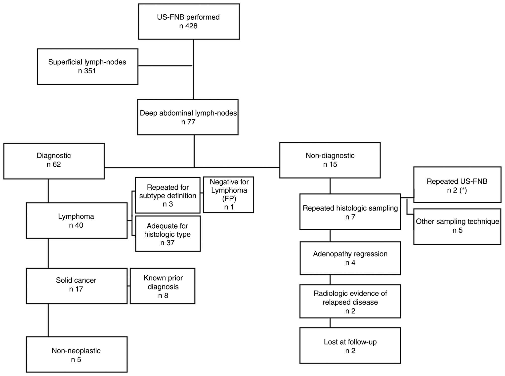
Sant'Orsola Malpighi Hospital. Among them, 77 targeted abdominal
lymph nodes (Fig. 1). Baseline
patient characteristics are listed in Table I. All patients underwent CEUS as
part of the biopsy procedure.
 | Table I.Characteristics of patients undergoing
ultrasound-guided needle biopsy (n=77). |
Table I.
Characteristics of patients undergoing
ultrasound-guided needle biopsy (n=77).
| Variable | N (%) |
|---|
| Sex, n (%) |
|
|
Female | 33 (42.8) |
| Male | 44 (57.1) |
| Site, n (%) |
|
|
Abdominal, not specified | 33 (42.8) |
|
Para-aortic | 12 (15.6) |
|
Retroperitoneal | 11 (14.3) |
|
Iliac | 6 (7.8) |
|
Epigastric | 4 (5.2) |
|
Mesogastric | 3 (3.9) |
|
Hypogastric | 1 (1.3) |
|
Mesenteric | 6 (7.8) |
| Hepatic
hilum | 1 (1.3) |
| Outcome, n (%) |
|
|
Diagnostic | 62 (80.5) |
|
Non-diagnostic | 15 (19.5) |
| Median
diameter, mm (range) |
|
| All
biopsies | 47.4 (9–200) |
|
Diagnostic | 36.5 (9–150) |
|
Non-diagnostic | 28 (15–200) |
| Calibre of needle
used for biopsy, n (%) |
|
| 14 G | 2 (2.6) |
| 16 G | 23 (29.9) |
| 18 G | 52 (67.5) |
Overall, 62 of 77 abdominal lymph node US-NBs
(80.5%) yielded a definitive histological diagnosis. Particularly,
40 patients were diagnosed with lymphoma (64.5%), 17 with
metastatic cancer (27.4%), and five with non-neoplastic disease
(8.1%; Table II). The remaining 15
procedures (19.5%) were non-diagnostic (Table III). The median lymph node
diameter, available for 70 biopsies, was 47.4 mm.
| Table II.Diagnostic biopsies. |
Table II.
Diagnostic biopsies.
| A, Lymphomas
(n=40) |
|---|
|
|---|
| Variable | Value |
|---|
| Histological type, n
(%) |
|
|
DLBCL | 9 (22.5) |
| FL | 16 (40) |
| HL | 7 (17.5) |
| HGL | 2 (5) |
| MCL | 1 (2.5) |
| CLL | 2 (5) |
| Lymphoma,
NOS | 3 (7.5) |
| Calibre of needle
used for biopsy, n (%) |
|
| 14
G | 0 (0) |
| 16
G | 14 (35) |
| 18
G | 26 (75) |
| Median age, years
(range) | 64.5 (26–87) |
|
| B, Solid cancers
(n=17) |
|
|
Variable | Value |
|
| Histological type,
n (%) |
|
| GU
tract | 5 (29.4) |
|
NET | 4 (23.5) |
| GI
tract | 3 (17.6) |
|
HCC | 1 (5.9) |
|
Breast | 1 (5.9) |
|
Lung | 1 (5.9) |
|
Melanoma | 1 (5.9) |
|
Sarcoma | 1 (5.9) |
| Calibre of needle
used for biopsy, n (%) |
|
| 14
G | 1 (5.9) |
| 16
G | 3 (17.6) |
| 18
G | 13 (76.5) |
| Median age, years
(range) | 64 (35–83) |
|
| C,
Non-neoplastic (n=5) |
|
|
|
Variable | Value |
|
| Histological type,
n (%) |
|
|
Reactive tissue | 2 (40) |
| Normal
tissue | 2 (40) |
| TB | 1 (20) |
| Calibre of needle
used for biopsy, n (%) |
|
| 14
G | 0 (0) |
| 16
G | 1 (20) |
| 18
G | 4 (80) |
| Median age, years
(range) | 52 (40–77) |
| Table III.Study outcomes and diagnostic
reliability. |
Table III.
Study outcomes and diagnostic
reliability.
|
Outcome/parameter | N (%) |
|---|
| Failure rate
(interrupted procedure)a, n (%) | 0/77 (0.0) |
| Rate of
non-diagnostic samplinga, n (%) | 15/77 (19.5) |
| Chance to repeat
any non-diagnostic procedureb, n (%) | 7/15 (46.7) |
| Chance to repeat
any diagnostic procedurec, n (%) | 3/62 (4.8) |
| Diagnostic yielda,
n (%) | 62/77 (80.5) |
| Sample adequacy for
clinical decisionsc, n (%) | 59/62 (95.2) |
Lymphoma diagnosis
Among the 40 biopsies positive for lymphoma, 32
(80.0%) were newly diagnosed, while eight (20.0%) were performed to
histologically confirm disease recurrence in patients with prior
lymphoma diagnosis. In total, three patients (7.5%) were diagnosed
with lymphoma, not otherwise specified, and required additional
sampling for further characterization. Two of these patients were
subsequently diagnosed with Hodgkin lymphoma (HL) based on bone
marrow biopsy and repeat NB of a superficial lymph node,
respectively. The third patient remained under follow-up, with
subsequent imaging being negative for pathological lymph nodes,
representing the only false-positive result. Overall, sample
adequacy for clinical decision-making in patients with lymphoma was
37/40 (92.5%).
Diagnosis of solid tumors and
non-neoplastic diseases
Among the 17 biopsies positive for solid tumors, the
majority (9 cases, 52.9%) represented histological confirmation of
metastatic lymph node in patients with a known malignancy, while
six cases (35.3%) corresponded to new diagnoses. Sample adequacy
for clinical decision-making in this setting was 100%, and no
repeat biopsies were required. Finally, five of the 62 diagnostic
biopsies did not show any neoplastic disease, including two cases
of reactive nodal tissue, two cases of normal lymph nodal tissue,
and one case of disseminated tuberculosis. No evidence of
malignancy was recorded during follow-up in patients diagnosed with
reactive or normal lymph nodes (true negative cases).
Non-diagnostic biopsies and
follow-up
A total of 15 of 77 US-NBs (19.5%) were
non-diagnostic due to missed targets or insufficient tissue
sampling. The median lymph node diameter, available for 15
biopsies, was 28 mm. Among the aforementioned patients, four
experienced spontaneous regression of lymph nodes without further
intervention; two had known aggressive lymphoma treated with CAR-T
cell therapy, and biopsies were not repeated due to radiological
and clinical disease progression; and two were lost to follow-up.
New histological sampling was performed in seven patients.
Particularly, one patient underwent repeat US-NB of abdominal
nodes, resulting in a diagnosis of follicular lymphoma; one patient
underwent US-NB of a superficial inguinal lymph node, confirming
diffuse large B-cell lymphoma (DLBCL) recurrence; three patients
were diagnosed with lymphoma following excisional biopsy of
superficial lymph nodes (two DLBCL and one HL); one patient was
diagnosed with HL following bone marrow biopsy; and two patients
underwent open abdominal surgery for resection of a suspected large
nodal mass, resulting in a final histological diagnosis of lipoma
and nodal metastasis from leiomyosarcoma, respectively.
Diagnostic performance of US-NB
Of the 77 biopsies performed, 62 provided sufficient
material for a definitive diagnosis, resulting in a diagnostic
yield of 80.5%. Among all diagnostic samples, sensitivity was 100%
(95% CI, 0.936–1.000), specificity was 83.3% (95% CI, 0.359–0.996),
PPV was 98.2% (95% CI, 0.906–0.999) and NPV was 100% (95% CI,
0.936–1.000). The overall diagnostic accuracy was 98.4%.
Needle size, PET and safety
outcomes
An 18-G, 16-G and 14-G needle was used in 69.4, 29.0
and 1.6% of diagnostic biopsies, respectively, and in 60.0, 33.3
and 6.7% of non-diagnostic biopsies. No significant association
between needle size (larger needles, 16-G and 14-G, vs. smaller
needles, 18-G) and diagnostic outcomes was recorded (P=0.53). A
total of 38 patients underwent PET scan, including 32 who had
biopsy targeting the lymph node with the highest SUVmax (range,
2.5–47). Among these, 28 of 32 biopsies yielded a diagnosis of
neoplastic disease. Among the 32 patients with available PET
imaging, 6 non-diagnostic cases were observed, 5 of which underwent
biopsy of the area with highest tracer uptake. No significant
association was found between biopsy at the SUVmax site and
diagnostic outcome. A multivariable logistic regression model was
performed to identify factors associated with a diagnostic biopsy
outcome. The model included age, needle gauge, retroperitoneal
location and lesion size, the latter categorized into tertiles
(T1-T3) based on the distribution of the study population. In the
adjusted analysis, only the largest lesion category (T3) was
significantly associated with diagnostic yield (OR 10.74, p=0.038),
whereas age, needle gauge, and retroperitoneal location were not
independently associated with the outcome. Finally, among the 77
deep lymph node biopsies performed, no procedure-related adverse
events were observed, either during or after the procedure.
Particularly, no cases of bleeding, swelling, or significant pain
were reported.
Discussion
The literature on tissue sampling of suspected
malignant abdominal lymph nodes primarily focuses on the diagnosis
of lymphoma, due to both the anatomical site involved and the
management of solid tumors, in which lymph nodes are commonly
resected for histopathological analysis during surgery or are not
biopsied in case of metastatic disease. The high diagnostic
accuracy, safety, and cost-effectiveness of NB in the diagnosis of
lymphoma have been widely reported, yielding a definitive diagnosis
in >90% of cases (14). However,
the majority of these studies mainly focus on specific
imaging-guided techniques, including computed tomography (CT)-,
PET/CT-, or US-guided NB), without taking into account whether the
disease site is superficial or deep (4,5,10,14–17).
Evidence on percutaneous US-NB in deep abdominal
lymph nodes remains limited (9,15,18–23),
thus reflecting the need for experienced operators and a preference
for CT-guided or echo-endoscopic approaches, which can provide
improved lesion visualization (16,17).
Needle selection is affected by lesion size and location.
Therefore, larger core needles (14 G) are typically used for
superficial lesions, while 16–18 G needles are preferred for deeper
lesions. Although smaller needles are associated with lower
complication rate, they can yield less informative samples. For
example, small tissue samples could fail to detect histological
transformation in lymphoma or could limit accurate assessment of
features, such as the centroblast/centrocyte ratio in follicular
lymphoma.
To the best of our knowledge, the current study
represented one of the largest series evaluating the diagnostic
yield and accuracy of percutaneous US-NB for suspected malignant
disease, including both lymphomatous and solid, in deep abdominal
lymph nodes (Table IV). The study
demonstrated a high diagnostic yield (80.5%) and excellent
diagnostic accuracy (98.4%), consistent with previous studies
(9,11,12,16,19).
Malignancy-negative results accounted for 8.1% of diagnostic
samples and were all confirmed as true negatives on follow-up.
| Table IV.Published experience with
percutaneous US-NB of abdominal lymph nodes. |
Table IV.
Published experience with
percutaneous US-NB of abdominal lymph nodes.
| First author/s,
year | US-NB, n | Abdominal deep
site, n (%) | Diagnostic
performance | Notes | (Refs.) |
|---|
| Kälkner et
al, 1994 | 129 | 62 (48) | 89% detection
rate | No abdominal biopsy
sub analysis | (24) |
| Zinzani et
al, 1998 | 55 | 55 (100) | 87% diagnostic
accuracy | Only abdominal
biopsies analyzed | (9) |
| Demharter et
al, 2001 | 128 | 12 (9.4) | 93% diagnostic
accuracy | No abdominal biopsy
sub analysis | (19) |
| De Larrinoa AF
et al, 2007 | 102 | 11 (10.8) | 88.2% diagnostic
accuracy | No abdominal biopsy
sub analysis | (22) |
| Yuan et al,
2010 | 1,119 | 197 (17.5) | 73% detection
rate | No abdominal biopsy
sub analysis | (20) |
| He et al,
2015 | 110 | 48 (43.6) | 97.8 % detection
rate | No abdominal biopsy
sub analysis | (11) |
| Pugliese et
al, 2017 | 185 | 23 (12.4) | 98.8%
sensitivity | No abdominal biopsy
sub analysis | (21) |
| Wilczynski et
al, 2020 | 793 | 256 (32) | 95% diagnostic
accuracy | No statistically
significant difference in the diagnostic accuracy between
peripheral and abdominal lymph-node biopsies | (23) |
| Zadeh et al,
2025 | 87 | 87 (100) | 94.2% diagnostic
accuracy |
| (12) |
| Present study | 428 | 77 (5.5) | 98.4% diagnostic
accuracy | - |
|
Despite the aforementioned encouraging results,
19.5% of biopsies were non-diagnostic, thus highlighting the
challenges associated with sampling deep lymph nodes, particularly
when lesions are small or necrotic. Although diagnostic biopsies
showed a NPV of 100%, sensitivity could be overestimated due to the
retrospective design and the exclusion of non-diagnostic cases from
accuracy calculations.
As expected, the majority of biopsies were positive
for lymphoma, with histological subtypes reflecting disease
epidemiology. The most common diagnoses were follicular lymphoma,
DLBCL and HL. Notably, no T-cell lymphomas were identified, which
could be explained by their relative rarity and frequent
extra-nodal involvement. Overall sample adequacy for clinical
decision-making was 92.5%. However, in three cases the histological
category could not be reliably established. Following repeat
sampling, one patient was found to be free of lymphoma, while the
remaining two were diagnosed with HL. Occasionally, distinguishing
reactive lymphoid changes from low-grade non-HL on core biopsy
could be challenging and could have contributed to the false
positive result observed. Additionally, percutaneous biopsy
yielding limited material could also lead to misclassification of
lymphoma subtypes, particularly those with low tumor cell content
or areas of sclerosis, such as HL. Notably, HL was subsequently
diagnosed in the two cases with initially uncertain subtype. The
vast majority of cases were newly diagnosed with lymphomas, while
~20% were repeat biopsies performed to confirm previous diagnoses.
In this context, repeat biopsy at relapse is clinically valuable
for excluding disease transformation and for evaluating antigen
expression to guide targeted antibody-based salvage therapy
(24,25). Notably, two non-diagnostic biopsies
were carried out in patients with rapidly progressive disease
following CAR-T cell therapy; both specimens were histologically
characterized by extensive necrosis, suggesting that alternative or
extensive tissue sampling strategies could be required in this
setting.
Solid tumor diagnoses were less frequent, accounting
for ~25% of all informative samples, with ~65% representing
metastatic disease. Importantly, six of seven patients who
underwent biopsy for suspected metastatic solid tumors were
initially assessed for possible lymphoproliferative disease,
underscoring the role of biopsy in excluding lymphoma. The most
common solid tumor diagnoses were malignancies prone to abdominal
nodal metastasis, particularly genitourinary, gastrointestinal, and
neuroendocrine tumors.
In total, ~50% of the patients had available PET
imaging to guide US biopsy, and 28 of 32 biopsies performed at
sites of maximal PET uptake were diagnostic. Although no
statistically significant association with biopsy outcome was
obtained, likely due to the limited sample size, functional imaging
could increase the likelihood of sampling representative malignant
tissue, supporting an integrated approach to optimize sampling
success (26).
Furthermore, the absence of procedure-related
complications and the feasibility of performing US-NB in an
outpatient setting further supported its safety, even among
patients with significant comorbidities who were not candidates for
more invasive approaches, such as surgery. These findings were
consistent with previous oncologic reports.
However, the present study has certain limitations,
including its retrospective design and relatively small sample
size, which could introduce selection bias and limit
generalizability. Additionally, the lack of procedural
standardization, including variability in needle size and number of
passes, could also affect outcomes. The risk of verification bias
was considered low, as all five negative biopsies were closely
monitored and no malignancies were identified. Finally, as
diagnostic accuracy was calculated conditionally on diagnostic
samples, the reported estimates may be subject to selection bias
and could appear higher than those obtained with an
intention-to-diagnose approach. The monocentric design of the
present study represented a major strength, as procedures were
performed by the same operators using consistent equipment and
standardized techniques. Nevertheless, larger prospective studies
are needed to confirm these findings.
In conclusion, our experience suggested that
US-guided percutaneous NB could be a reliable, rapid and safe
diagnostic tool for evaluating suspected malignant lymph node
disease in the deep abdomen, applicable to both initial diagnosis
and disease recurrence, with high diagnostic accuracy. However,
caution is warranted in particular scenarios. In low-grade
lymphoma, limited tissue samples could hinder differentiation
between reactive and pathological tissues. In cases of suspected
histological transformation or heterogeneous disease, NB could not
fully capture full disease complexity. Furthermore, in rapidly
proliferating lesions, extensive necrosis could reduce diagnostic
yield, thus indicating that alternative sampling approaches capable
of providing larger histological specimens should be
considered.
Acknowledgements
The authors would like to thank Mr. Massimo Agostini
(Associazione Italiana Contro Leucemia, Linfomi e Mieloma; Bologna,
Italy) for collecting patient data.
Funding
Funding: No funding was received.
Availability of data and materials
The data generated in the present study may be
requested from the corresponding author.
Authors' contributions
CM, LA, CS and PLZ designed and conceived the study
and drafted and revised the manuscript. LA and CM conducted all
data analyses. CM, DDB, SMB, LDM and MM collected the data. DDB,
SMB, BC, CP, LDM, MM and AB provided study materials and
interpreted data. All authors read and approved the final
manuscript. CM and LA confirm the authenticity of all the raw
data.
Ethics approval and consent to
participate
The present study was conducted in accordance with
The Declaration of Helsinki and was approved by the Ethical
Committee AVEC of Bologna (approval nos. 1043/2021/ Oss/AOUBo and
EM189-2024_1043/2021/Oss/AOUBo; Bologna, Italy).
Patient consent for publication
Not applicable.
Competing interests
The authors declare that they have no competing
interests.
References
|
1
|
Alaggio R, Amador C, Anagnostopoulos I,
Attygalle AD, Araujo IBO, Berti E, Bhagat G, Borges AM, Boyer D,
Calaminici M, et al: The 5th edition of the World Health
Organization classification of haematolymphoid tumours: Lymphoid
neoplasms. Leukemia. 36:1720–1748. 2022. View Article : Google Scholar : PubMed/NCBI
|
|
2
|
Planchard D, Popat S, Kerr K, Novello S,
Smit EF, Faivre-Finn C, Mok TS, Reck M, Van Schil PE, Hellmann MD,
et al: Metastatic non-small cell lung cancer: ESMO clinical
practice guidelines for diagnosis, treatment and follow-up. Ann
Oncol. 29 (Suppl 4):iv192–iv237. 2018. View Article : Google Scholar : PubMed/NCBI
|
|
3
|
Cervantes A, Adam R, Roselló S, Arnold D,
Normanno N, Taïeb J, Seligmann J, De Baere T, Osterlund P, Yoshino
T, et al: Metastatic colorectal cancer: ESMO clinical practice
guideline for diagnosis, treatment and follow-up. Ann Oncol.
34:10–32. 2023. View Article : Google Scholar : PubMed/NCBI
|
|
4
|
Chatani S, Hasegawa T, Kato S, Murata S,
Sato Y, Yamaura H, Yamamoto K, Yatabe Y and Inaba Y: Image-guided
core needle biopsy in the diagnosis of malignant lymphoma:
Comparison with surgical excision biopsy. Eur J Radiol.
127:1089902020. View Article : Google Scholar : PubMed/NCBI
|
|
5
|
Johl A, Lengfelder E, Hiddemann W and
Klapper W; German Low-grade Lymphoma Study Group (GLSG), : Core
needle biopsies and surgical excision biopsies in the diagnosis of
lymphoma-experience at the Lymph Node Registry Kiel. Ann Hematol.
95:1281–1286. 2016. View Article : Google Scholar : PubMed/NCBI
|
|
6
|
Fornari F and Buscarini L:
Ultrasonically-guided fine-needle biopsy of gastrointestinal
organs: Indications, results and complications. Dig Dis.
10:121–133. 1992.PubMed/NCBI
|
|
7
|
Di Stasi M, Lencioni R, Solmi L, Magnolfi
F, Caturelli E, De Sio I, Salmi A and Buscarini L:
Ultrasound-guided fine needle biopsy of pancreatic masses: Results
of a multicenter study. Am J Gastroenterol. 93:1329–1333. 1998.
View Article : Google Scholar : PubMed/NCBI
|
|
8
|
Pappa VI, Hussain HK, Reznek RH, Whelan J,
Norton AJ, Wilson AM, Love S, Lister TA and Rohatiner AZ: Role of
image-guided core-needle biopsy in the management of patients with
lymphoma. J Clin Oncol. 14:2427–2430. 1996. View Article : Google Scholar : PubMed/NCBI
|
|
9
|
Zinzani PL, Colecchia A, Festi D,
Magagnoli M, Larocca A, Ascani S, Bendandi M, Orcioni GF,
Gherlinzoni F, Albertini P, et al: Ultrasound-guided core-needle
biopsy is effective in the initial diagnosis of lymphoma patients.
Haematologica. 83:989–992. 1998.PubMed/NCBI
|
|
10
|
Bellisario F, Attili F, Campana F,
Borrelli de Andreis F, Bellesi S, Maiolo E, Alma E, Malafronte R,
Macis G, Larocca LM, et al: Endoscopic ultrasound-guided fine
needle biopsy in the diagnostic work-up of deep-seated
lymphadenopathies and spleen lesions: A monocentric experience.
Diagnostics (Basel). 13:28392023. View Article : Google Scholar : PubMed/NCBI
|
|
11
|
He Y, Ji X, Xie Y, He B, Xu X, Chen X and
Zhang Q: Clinical application of ultrasound-guided core needle
biopsy with multiple punches in the diagnosis of lymphoma. World J
Surg Oncol. 13:1262015. View Article : Google Scholar : PubMed/NCBI
|
|
12
|
Zadeh ES, Görg M, Görg C, Prosch H,
Trenker C, Westhoff CC, Dietrich CF, Raab N, Alhyari A, Huber K, et
al: The value of contrast-enhanced ultrasound in percutaneous
biopsy of retroperitoneal masses. Ultraschall Med. 46:66–72. 2025.
View Article : Google Scholar
|
|
13
|
Patel SA, Pierko K and Franco-Sadud R:
Ultrasound-guided bedside core needle biopsy: A hospitalist
procedure team's experience. Cureus. 11:e38172019.PubMed/NCBI
|
|
14
|
Syrykh C, Chaouat C, Poullot E, Amara N,
Fataccioli V, Parrens M, Traverse-Glehen A, Molina TJ, Xerri L,
Martin L, et al: Lymph node excisions provide more precise lymphoma
diagnoses than core biopsies: A French Lymphopath network survey.
Blood. 140:2573–2583. 2022. View Article : Google Scholar : PubMed/NCBI
|
|
15
|
Nguyen BM, Halprin C, Olimpiadi Y, Traum
P, Yeh JJ and Dauphine C: Core needle biopsy is a safe and accurate
initial diagnostic procedure for suspected lymphoma. Am J Surg.
208:1003–1008. 1007–1008. 2014. View Article : Google Scholar : PubMed/NCBI
|
|
16
|
Skelton E, Jewison A, Okpaluba C, Sallomi
J, Lowe J, Ramesar K, Grace R and Howlett DC: Image-guided core
needle biopsy in the diagnosis of malignant lymphoma. Eur J Surg
Oncol. 41:852–858. 2015. View Article : Google Scholar : PubMed/NCBI
|
|
17
|
Lachar WA, Shahab I and Saad AJ: Accuracy
and cost-effectiveness of core needle biopsy in the evaluation of
suspected lymphoma: A study of 101 cases. Arch Pathol Lab Med.
131:1033–1039. 2007. View Article : Google Scholar : PubMed/NCBI
|
|
18
|
Agid R, Sklair-Levy M, Bloom AI, Lieberman
S, Polliack A, Ben-Yehuda D, Sherman Y and Libson E: CT-guided
biopsy with cutting-edge needle for the diagnosis of malignant
lymphoma: Experience of 267 biopsies. Clin Radiol. 58:143–147.
2003. View Article : Google Scholar : PubMed/NCBI
|
|
19
|
Demharter J, Müller P, Wagner T, Schlimok
G, Haude K and Bohndorf K: Percutaneous core-needle biopsy of
enlarged lymph nodes in the diagnosis and subclassification of
malignant lymphomas. Eur Radiol. 11:276–283. 2001. View Article : Google Scholar : PubMed/NCBI
|
|
20
|
Yuan J and Li XH: Evaluation of
pathological diagnosis using ultrasonography-guided lymph node
core-needle biopsy. Chin Med J (Engl). 123:690–694. 2010.
View Article : Google Scholar : PubMed/NCBI
|
|
21
|
Pugliese N, Di Perna M, Cozzolino I,
Ciancia G, Pettinato G, Zeppa P, Varone V, Masone S, Cerchione C,
Della Pepa R, et al: Randomized comparison of power Doppler
ultrasonography-guided core-needle biopsy with open surgical biopsy
for the characterization of lymphadenopathies in patients with
suspected lymphoma. Ann Hematol. 96:627–637. 2017. View Article : Google Scholar : PubMed/NCBI
|
|
22
|
de Larrinoa AF, del Cura J, Zabala R,
Fuertes E, Bilbao F and Lopez JI: Value of ultrasound-guided core
biopsy in the diagnosis of malignant lymphoma. J Clin Ultrasound.
35:295–301. 2007. View Article : Google Scholar : PubMed/NCBI
|
|
23
|
Wilczynski A, Görg C, Timmesfeld N,
Ramaswamy A, Neubauer A, Burchert A and Trenker C: Value and
diagnostic accuracy of ultrasound-guided full core needle biopsy in
the diagnosis of lymphadenopathy: A retrospective evaluation of 793
cases. J Ultrasound Med. 39:559–567. 2020. View Article : Google Scholar : PubMed/NCBI
|
|
24
|
Kälkner M, Rehn S, Andersson T, Elvin A,
Hagberg H, Lindgren PG, Sundström C and Glimelius B: Diagnostics of
malignant lymphomas with ultrasound guided 1.2 mm biopsy-gun. Acta
Oncol. 33:33–37. 1994. View Article : Google Scholar : PubMed/NCBI
|
|
25
|
Lownik J, Boiarsky J, Birhiray R, Merchant
A and Mead M: sequencing of anti-CD19 therapies in the management
of diffuse large B-cell lymphoma. Clin Cancer Res. 30:2895–2904.
2024. View Article : Google Scholar : PubMed/NCBI
|
|
26
|
Broccoli A, Nanni C, Cappelli A, Bacci F,
Gasbarrini A, Tabacchi E, Piovani C, Argnani L, Ghermandi R,
Sabattini E, et al: Diagnostic accuracy of positron emission
tomography/computed tomography-driven biopsy for the diagnosis of
lymphoma. Eur J Nucl Med Mol Imaging. 47:3058–3065. 2020.
View Article : Google Scholar : PubMed/NCBI
|