Introduction
Pancreatic cancer (PC) exhibits one of the highest
mortality rates among malignancies of the digestive system, and
pancreatic ductal adenocarcinoma (PDAC) has been shown to account
for 90–95% of all PC cases (1). Due
to its aggressive biological behavior and late clinical
presentation, PDAC remains a major global health challenge
(1). Despite advances in cancer
diagnosis and treatment, the incidence of PDAC has continued to
increase over previous decades and this malignancy is projected to
become the second leading cause of cancer-related mortality in the
USA by 2030 (2). Early-stage PDAC
is often asymptomatic or presents with non-specific symptoms,
leading to delayed diagnosis in a large proportion of patients
(3). Consequently, only a minority
of patients with PDAC remain eligible for surgical resection
(4). The overall 5-year survival
rate for patients with PDAC is <12%, with limited improvement
observed over previous decades (5,6).
CA19-9 is a serum biomarker for PDAC (2). However, it has been shown to exhibit
limited sensitivity and specificity (2), particularly in the early stages of
PDAC development, which notably restricts its utility for PDAC
screening and early diagnosis. In addition, elevated CA19-9 levels
can also be observed in benign pancreatic diseases, while 5–10% of
individuals are unable to express CA19-9 due to Lewis antigen
negativity, further reducing the efficacy of this biomarker
(7,8). These limitations highlight the notable
necessity of novel, reliable and non-invasive biomarkers to improve
early detection and risk stratification of PDAC (7,8).
Microbiome research has revealed important
associations between microbial dysbiosis and multiple malignancies
(9,10). It is estimated that 10–20% of
cancers are attributable, at least in part, to infectious or
microbial factors, underscoring the role of host-microbe
interactions in carcinogenesis in specific cancer types (11). Advances in sequencing technologies
have further facilitated the identification of disease-associated
microbial signatures (12).
Increasing evidence suggests that patients with PDAC exhibit
distinct alterations in the gut, oral and tumor-associated
microbiota (9,13). Dysbiosis of the oral and gut
microbiota has been implicated in a number of mechanisms involved
in pancreatic carcinogenesis, including chronic inflammation,
immunomodulation and metabolic reprogramming (14). Intratumoral microbiome composition
is associated with survival in patients with PDAC (15).
In parallel, several studies have developed
microbiota-based diagnostic models of PDAC using fecal or oral
microbial profiles (16–20), some of which have demonstrated
improved performance when combined with conventional clinical
markers (16). Given the challenge
of early PDAC detection, such microbiota-based biomarkers hold
promise as potentially non-invasive tools for screening and
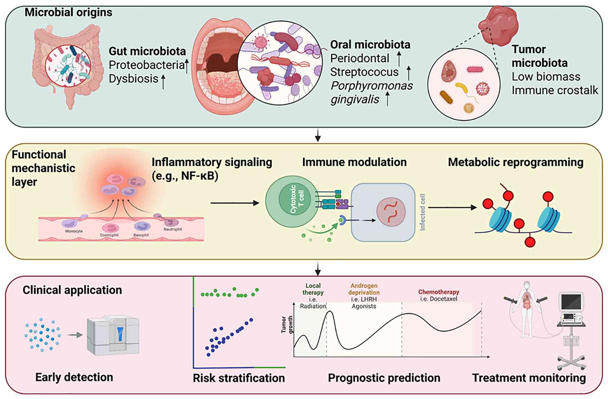
diagnosis (18,19). Fig.
1 summarizes the proposed association between microbial
dysbiosis, microbiome-based functional pathways and the potential
clinical applications of microbiome-based biomarkers in PDAC.
Additionally, accumulating evidence suggests that
microbiome-associated alterations may be detectable before the
clinical markers diagnose of PDAC, highlighting their potential as
early diagnostic biomarkers (16,20,21).
In a prospective cohort study, elevated circulating antibodies
against oral pathogens, particularly Porphyromonas
gingivalis, measured before the onset of PDAC were associated
with an increased risk of PC (22).
In a nested case-control study within the EPIC cohort, elevated
pre-diagnostic levels of antibodies targeting P. gingivalis
were markedly associated with future PC risk (22). The present review summarizes current
evidence regarding microbiota-based alterations associated with
PDAC, critically evaluates microbiota-based diagnostic approaches,
and discusses the methodological limitations and translational
challenges of such approaches that should be addressed before
clinical implementation.
Search strategy
The present review was conducted as a narrative
review. To improve transparency and reduce selection bias, a
structured literature search was performed using PubMed
(pubmed.ncbi.nlm.nih.gov), Web of Science (webofscience.com) and
Embase (embase.com) to obtain studies published from the inception
of these databases to January 31, 2026. The search terms included
combinations of key words related to PC and the microbiome, such as
‘pancreatic ductal adenocarcinoma’, ‘pancreatic cancer’,
‘microbiome’, ‘microbiota’, ‘gut microbiota’, ‘oral microbiota’,
‘tumor microbiome’, ‘diagnosis’, ‘biomarkers’, ‘early detection’
and ‘machine learning’. Only publications written in English were
included. Original studies investigating microbiome alterations or
microbiome-based diagnostic models in PDAC were included, and
relevant review articles were screened to identify additional
studies.
Studies were excluded if they: i) Lacked primary
data; ii) focused exclusively on non-human models without
translational relevance; iii) did not address diagnostic or
biomarker-related outcomes; or iv) were case reports, conference
abstracts or editorials. As the present study is a narrative
review, study selection did not follow formal systematic review
protocols. Instead, representative studies across different
populations, sample types and analytical approaches were included
in the present review to provide a balanced overview of current
evidence supporting microbiome-based biomarkers in PDAC. Where
available, studies reporting inconsistent or negative findings were
also considered.
Microbial dysbiosis signatures associated
with PDAC
Microbial dysbiosis has been increasingly reported
in patients with PDAC across multiple anatomical sites, including
the gut, tumor tissue and oral cavity (16,17,20).
Although study designs and sampling strategies differ, recurrent
alterations in microbial composition and diversity have been
identified (9,20).
Gut microbiota alterations in
PDAC
A cohort study analyzing 193 fecal samples from
patients with PDAC and healthy controls (HCs) (156 patients with
PDAC; 37 HCs) demonstrated that a number of notable alterations in
gut-microbial composition were associated with pancreatic
tumorigenesis. Notably, the inclusion of patients with
pre-malignant pancreatic lesions enabled comparisons across
different disease stages, and the results suggested that microbial
alterations may progressively emerge during pancreatic
carcinogenesis (17). A multicenter
case-control study involving Spanish and German cohorts identified
distinct fecal microbiota profiles in patients with PDAC compared
with healthy individuals and patients with chronic pancreatitis
(CP) (16,21); similarly, studies have shown that
classifiers based on 27 microbial species have achieved area under
the curve (AUC) values of 0.74–0.83 for detecting PDAC in
independent cohorts (16,21).
α-diversity findings for PDAC remain inconsistent.
Several studies have reported markedly reduced Shannon diversity, a
commonly used measure of α-diversity, in patients with PDAC
compared with HCs (23,24). Additionally, a meta-analysis
including 12 studies (535 patients with PDAC; 677 controls) also
supported an overall reduction in microbial α-diversity in patients
with PC compared with controls (25). However, other studies have failed to
detect notable differences in α-diversity between PDAC and control
groups (26,27), highlighting notable levels of
inter-study variability. By contrast, β-diversity analyses have
more consistently shown a clear separation between microbial
communities in PDAC and control groups (16,23,28),
suggesting that the overall composition of microbial communities in
patients with PDAC may be more reproducibly altered than
within-sample diversity metrics.
At the taxonomic level, several recurrent patterns
in bacterial community composition have been identified in PDAC.
Enrichment of opportunistic and facultative anaerobic taxa,
including members of the Enterobacteriaceae and Enterococcaceae
families, has been reported in PDAC cohorts (29). Furthermore, an increased abundance
of oral-associated genera, such as Streptococcus and
Veillonella, has been consistently observed in fecal samples
from patients with PDAC across multiple cohorts (30). By contrast, depletion of short-chain
fatty acid-producing commensals, including Lachnospiraceae,
Ruminococcaceae and Faecalibacterium prausnitzii, has
also been consistently reported in independent cohorts of patients
with PDAC, including multinational and Japanese cohorts (16–18,31).
Notably, a cross-population analysis indicated that microbial
signatures are influenced by ethnicity, as well as demographic,
dietary and lifestyle factors, underscoring the importance of
population-specific validation for microbial profiles (29).
In addition to indicating the presence of disease,
gut microbiota composition may also be associated with clinical
characteristics of PDAC. Compared with non-metastatic PDAC,
metastatic PDAC exhibits an overall higher relative abundance of
Gram-negative bacteria. Machine learning analysis has identified
bacterial taxa that effectively distinguished metastatic from
non-metastatic disease, such as Anaerostipes hadrus and
Porphyromonas (32).
Furthermore, an increased abundance of Klebsiella pneumoniae
in the gut is a central component of dysbiotic microbial networks
linking the gut, oral cavity and prognostic factors, and has also
been associated with poor prognosis in patients with PDAC (33). In addition, long-term survivors of
PDAC exhibit notable enrichment of Faecalibacterium
prausnitzii and Akkermansia muciniphila in the gut,
suggesting potential associations between gut microbiota
composition and patient prognosis (34). However, the aforementioned findings
require validation in larger longitudinal cohorts.
Preclinical models have provided complementary
insights into microbiome dynamics during PDAC development. In
murine models of PDAC driven by the oncogenic KrasG12D
mutation, disease progression is accompanied by time-dependent
alterations in fecal microbiota composition and reduced α-diversity
(35,36). Fecal microbiota transplantation
(FMT) experiments have further suggested that fecal microbiota from
patients with PC may transmit dysbiotic features to recipient mice
(37). In addition, dietary and
pharmacological interventions, including high-fat diet exposure and
metformin treatment, have been shown to modulate the composition of
the gut microbiota in preclinical PDAC models (38,39).
Although these murine studies have provided mechanistic insights
into microbiota-based alterations in PDAC, their direct relevance
to human clinical translation remains ambiguous. Overall, current
evidence supports the presence of gut microbiota dysbiosis in
patients with PDAC. However, heterogeneity in sampling strategies,
sequencing methods, including 16S ribosomal RNA (rRNA) sequencing
and shotgun metagenomics, bioinformatics pipelines and cohort
characteristics markedly limits direct comparisons of findings
across studies. Therefore, standardized methodologies and
multicenter validation studies are required before gut-microbial
signatures can be reliably translated into clinical diagnostic
applications (40).
Tissue-associated microbiota in
PDAC
A study analyzing 193 pancreatic tissue specimens,
including PDAC, pre-malignant lesions and benign tumors, revealed
distinct microbial compositions in tumor tissues compared with
non-malignant pancreatic samples (19). Patients with PDAC have demonstrated
markedly higher levels of α-diversity in tumor tissues compared
with adjacent normal tissues, with Proteobacteria, Firmicutes and
Actinobacteria identified as the dominant phyla found in tumor
tissues (41). Enrichment of
Pseudomonas fluorescens has also been associated with
prolonged overall survival in patients with PDAC, and in
vitro experiments have suggested that this bacterium may
inhibit PC cell proliferation (41). However, tissue-based microbiome
studies face notable methodological limitations. One study reported
notably low levels of microbial biomass in PDAC resection
specimens, which exhibited bacterial DNA levels comparable to
negative controls, raising concerns regarding contamination and
technical bias (42). In low
bacterial biomass environments such as pancreatic tissue, the
inclusion of negative controls, such as extraction blanks for
detecting reagent- or kit-derived contamination or sequencing
blanks, is key for distinguishing true microbial signals from
environmental or reagent-derived contamination (43).
Anatomical location-associated differences in
pancreatic tumor microbiota have been reported (19,36).
Distinct microbial compositions have been observed between tumors
located in the pancreatic head and those in the body or tail
(44). In addition, a spatial
analysis demonstrated associations between bacterial distribution
patterns and immune cell niches, such as T cell-rich and
T-cell-poor regions (45).
Culture-based approaches enabled the isolation of γ-proteobacteria,
including Klebsiella pneumoniae, from pancreatic cystic
lesions associated with pancreatic cancer (46). However, these approaches may capture
only a subset of the pancreatic microbiota, as culture-dependent
methods can underestimate overall microbial diversity and remain
vulnerable to contamination or selective growth bias (42,46).
Fungal components in pancreatic tissues have also
been implicated in PDAC development. For example, enrichment of
Malassezia species has been reported in PDAC tissues and
discussed as a potential contributor to pancreatic carcinogenesis
(47). A sequencing-based study
identifies site-specific microbial signatures in pancreatic and
duodenal tissues, suggesting potential microbial interactions or
translocation along the gastrointestinal tract and the pancreas
(48). Consistent with potential
microbial interactions along the gastrointestinal-pancreatic axis,
a multi-site microbiome study reported progressive microbial
alterations during the transition from CP to advanced PDAC,
including sequential enrichment of oral-associated taxa, such as
P. gingivalis and Filifactor alocis (49). Although intratumoral and
tissue-associated microbial profiles are increasingly recognized
(50,51), their diagnostic value remains
ambiguous. Low microbial biomass, spatial heterogeneity,
differences in sampling and sequencing methods, and the risk of
contamination further hinder clinical translation.
Oral and other body site microbiota in
PDAC
In a large nested case-control study analyzing
pre-diagnostic oral wash samples, the presence of P.
gingivalis and Aggregatibacter actinomycetemcomitans in
samples was associated with a markedly increased risk of PDAC
development, with adjusted odds ratio values of 1.60 and 2.20,
respectively. However, the relative abundance of Neisseria
was found to be inversely associated with PDAC risk (17). Notably, elevated levels of
antibodies against P. gingivalis have also been associated
with an ~2-fold increased risk of PC (52). A meta-analysis further supported an
association between oral microbiota alterations and PC risk
(53).
Salivary microbiome profiling has demonstrated
potential discriminatory ability between patients with PDAC and
controls (26), although fecal
microbiota-based classifiers have generally shown improved
performance compared with oral microbiota-based models (16). Notably, reduced oral-microbial
diversity and enrichment of Streptococcus have been reported
in PC cohorts (18).
Beyond the oral microbiota, an analysis of
secretin-stimulated duodenal fluid has revealed markedly reduced
α-diversity levels in patients with PDAC compared with controls and
patients with pancreatic cysts. The enrichment of specific genera,
including Bifidobacterium and Fusobacterium, has also
been observed in duodenal fluid (28). Furthermore, studies of bile
microbiota have demonstrated differences in β-diversity between
patients with malignant and benign pancreaticobiliary diseases
(54,55), although the low microbial biomass of
bile samples requires cautious interpretation of these findings
(56).
Notably, non-invasive biospecimens, including
saliva, stool and duodenal fluid, may represent promising sources
of microbial data for biomarker development (16). However, heterogeneity in study
design, limited sample sizes and the lack of standardized
predictive modeling approaches remain important limitations for the
identification of such biomarkers (57). Large prospective studies with
external validation are required to determine whether multi-site
microbial signatures can demonstrate clinically robust diagnostic
performance for PDAC. On the basis of these body site-specific
microbial alterations, several studies have attempted to develop
microbiota-based predictive models for PDAC diagnosis, as
summarized in Table I, Table II, III (16,17,30,41,58–65).
 | Table I.Representative fecal microbiome-based
diagnostic models for PDAC. |
Table I.
Representative fecal microbiome-based
diagnostic models for PDAC.
| First author/s,
year | Cohort (sample
size) | Profiling
method | Key microbial
features | Modeling
approach | Validation
strategy | Reported AUC | Diagnostic
task | (Refs.) |
|---|
| Kartal et
al, 2022 | Spanish cohort
(n=136; 57 patients with PDAC, 50 HCs and 29 patients with CP) +
German cohort (n=64; 32 patients with PDAC and 32 HCs) + 25 public
datasets (n=5,792) | Shotgun
metagenomics | 27-species
classifier ± CA19-9 biomarker | Random forest | Independent
multi-cohort validation | 0.840 (0.940 with
CA19-9) | PDAC vs. HC | (16) |
| Wang et al,
2024 | Chinese cohorts in
a multicenter study (n=193; 156 patients with pancreatic tumors and
37 HCs) | 16S rRNA
sequencing | Abundance-based
microbial features | Random forest | Internal validation
(method not reported) | NR | PDAC vs. HC | (17) |
| Wei et al,
2020 | PDAC/NPDAC cohort
(n=110; 41 patients with PDAC and 69 HCs) | 16S rRNA
sequencing | Differential
microbial taxa | Random forest | LOOCV | NR | PDAC vs. NPDAC | (65) |
| Yang et al,
2023 | Chinese cohort
(n=94; 44 patients with PDAC and 50 HCs) | 16S rRNA
sequencing |
Streptococcus enrichment | Random forest | Internal validation
(method not reported) | NR | PDAC occurrence and
liver metastasis | (30) |
| Zhang et al,
2024 | Multi-cohort
(n=183; 101 patients with PDAC and 82 HCs) | Virome
metagenomics | 219 viral OTUs | Random forest | Multi-cohort
validation | 0.879 | PDAC vs. HC | (63) |
| Li et al,
2025 | Chinese cohort
(n=187; 97 patients with PDAC and 90 HCs) | 16S rRNA
sequencing | Top 20 contributing
genera | Random forest | Internal validation
(7:3 split) | 0.890 | PDAC vs. HC | (61) |
| Gao et al,
2024 | Chinese cohort
(n=53) | 16S rRNA
sequencing | Pseudomonas
fluorescens enrichment | N/A | Internal
validation | NR |
Mechanistic/prognostic study | (41) |
| Zhou et al,
2021 | Mixed cohort (n=96;
32 patients with PDAC, 32 patients with AIP and 32 HCs) | Shotgun
metagenomics | SCFA depletion +
Proteobacteria enrichment | ML classifier | External
validation | 0.907 (PDAC vs.
HC); 0.889 (PDAC vs. AIP) | PDAC vs. HC or
AIP | (62) |
| Table II.Representative oral and salivary
microbiome-based diagnostic models for PDAC. |
Table II.
Representative oral and salivary
microbiome-based diagnostic models for PDAC.
| First author/s,
year | Cohort (sample
size) | Sample type | Profiling
method | Key microbial
features | Modeling
approach | Validation
strategy | Reported AUC | Diagnostic
task | (Refs.) |
|---|
| Li et al,
2025 | Chinese cohort
(n=187; 97 patients with PDAC and 90 HCs) | Oral | 16S rRNA
sequencing | Top 20 contributing
genera; Streptococcus enriched | Random forest | Internal validation
(7:3 split) | 0.963 | PDAC vs. HC | (61) |
| Wei et al,
2020 | Chinese cohort
(n=110; 41 patients with PDAC and 69 HCs) | Oral | 16S rRNA
sequencing |
Streptococcus and
Leptotrichia (risk); Veillonella and Neisseria
(protective) | Logistic regression
+ ML classifier | Internal
validation | 0.963 | PDAC vs. NPDAC | (65) |
| Wong et al,
2009 | UCLA cohort (n=90;
3 0 patients with PDAC, 30 patients with CP and 30 HCs) | Saliva | qPCR and
transcriptomics | Neisseria
elongata + Streptococcus mitis + ACRV1 + DMXL2 +
DPM1 | Logistic
regression | Internal
validation | 0.895 (microbial);
0.949 (combined) | PDAC vs. CP and
HC | (64) |
| Farrell et
al, 2012 | UCLA cohort (n=83;
28 patients with PDAC, 28 HCs and 27 patients with CP) | Saliva | qPCR | N. elongata
and S. mitis | Combined logistic
model | Independent
validation | 0.900 | PDAC vs. HC | (59) |
| Table III.Representative multi-modal
microbiome-based diagnostic strategies for PDAC. |
Table III.
Representative multi-modal
microbiome-based diagnostic strategies for PDAC.
| First author/s,
year | Biomarker
combination | Specimen/data
modality | Integration
method | Modeling
approach | Validation
strategy | Reported AUC | Diagnostic
task | (Refs.) |
|---|
| Wang et al,
2024 | Fecal +
tissue-associated microbiota integration | Feces + tumor
tissue | Integrated
microbial profiling | Multi-step
classifier | External validation
tumor detection | NR | Early
pancreatic | (17) |
| Kartal et
al, 2022 | Fecal microbiota +
CA19-9 | Feces + serum
biomarker | Shotgun
metagenomics + CA19-9 integration | Random forest | External
validation | 0.940 | Early PDAC
detection | (58) |
| Guo et al,
2022 | Fecal +
metabolomics integration | Feces +
metabolomics | Integrated
microbiome profiling and metabolomics | ROC analysis | Exploratory
study | NR | Resectable vs.
unresectable PDAC | (60) |
| Wong et al,
2009 | Microbiota +
salivary mRNA | Saliva +
transcriptomics i ntegration | Microbiome and
transcriptome | Combined logistic
regression model | Internal
validation | 0.949 | PDAC vs. CP and
HC | (64) |
Diagnostic modeling strategies and
performance evaluation
Previous evidence suggests that the microbiome may
represent a promising source of non-invasive biomarkers for PDAC
(16,66). Advancements in high-throughput
sequencing technologies have enabled the comprehensive profiling of
oral, fecal and tumor-associated microbial communities, and have
revealed reproducible alterations in microbiota composition that
are associated with pancreatic tumorigenesis (67,68).
As such, microbiome-based classifiers are increasingly being
developed for early detection, risk stratification and prognostic
prediction for patients with PDAC (16,69).
However, several challenges remain, including relatively small
cohort size, geographic heterogeneity and insufficient external
validation; additionally, robust clinically validated
microbiome-based diagnostic tools remain lacking (70). This section summarizes current
microbiome-derived diagnostic models, evaluates their predictive
performance and discusses emerging diagnostic strategies that
integrate microbial, metabolic and conventional clinical biomarkers
to improve diagnostic accuracy.
Fecal microbiota-based diagnostic
models
Fecal microbiota profiling remains the most
extensively studied approach for non-invasive PDAC detection.
Machine learning-based approaches have been applied to develop
microbiome-based classifiers for PDAC using shotgun metagenomics or
16S rRNA sequencing data (16).
However, microbiome datasets are typically high-dimensional,
increasing the risk of overfitting, especially in studies with
small cohorts (71). Notably, a
previous study has demonstrated that a fecal metagenomic classifier
based on 27 microbial species achieved an AUC of 0.84 for
distinguishing PDAC samples from controls, and this classifier
exhibited similar levels of accuracy for identifying both early-
and late-stage disease (16). The
aforementioned model has also been shown to maintain high disease
specificity across independent public datasets and external
validation cohorts. Additionally, the integration of this model
with serum CA19-9 levels led to further improvements in diagnostic
performance, raising the AUC to 0.94 (16).
Using a penalized logistic regression and iterative
random forest analysis, gut-microbial signatures have been able to
discriminate metastatic from non-metastatic PDAC cases (32). The integration of fecal metabolomic
profiles has been shown to further improve the discriminatory
performance of these signatures and has revealed associations
between microbial taxa and lipid metabolites, providing
complementary metabolic information for PDAC detection (60). A previous study has demonstrated
that a fecal microbiota classifier achieved an AUC of 0.856 (95%
CI, 0.740–0.972) for distinguishing PDAC from controls (26), whereas another study has reported an
AUC of 0.963 for identifying PDAC using oral microbiota profiled by
16S rRNA sequencing (61). An
additional study has shown that a classifier based on
discriminatory bacterial features exhibited for PDAC detection;
however, considerable inter-individual microbial variability in the
study cohort limited the applicability of this model for the early
identification of pre-cancerous lesions (72). Notably, numerous studies have
differed in how AUC values are reported, with inconsistent
reporting of results from training datasets, internal
cross-validation or independent external validation cohorts, which
complicates direct comparisons of model performance (16,61).
Differential diagnostic performance in
microbiota-based models has also been investigated. A previous
study showed that a random forest classifier based on the relative
abundance of the top 10 discriminatory fecal bacterial genera
distinguished PC samples from non-malignant pancreatic lesions with
an AUC of 0.894 and outperformed CA19-9-based diagnostic models in
mucinous tumor subgroups (73). In
addition, shotgun metagenomic profiling has demonstrated high
accuracy for distinguishing PDAC from autoimmune pancreatitis
(AIP), exhibiting AUC values of 0.907 for PDAC vs. HCs and 0.889
for PDAC vs. AIP (62).
Cross-population validation studies are particularly
important for clinical translation. One such study involving
Finnish and Iranian cohorts achieved an AUC of 0.88 (95% CI,
0.78–0.97) in the independent Finnish cohort (29), although microbial signatures were
partially influenced by demographic and lifestyle factors. Beyond
bacterial taxa, the diagnostic potential of the gut virome has also
been investigated. A previous study has demonstrated that a random
forest classifier based on viral operational taxonomic units
achieved an AUC of 0.879 for distinguishing patients with PDAC from
healthy controls, and exhibited notable reproducibility across
independent cohorts (63). Overall,
fecal microbiota-based diagnostic models have frequently reported
AUC values ranging from 0.80 to 0.90 (16,18);
however, the majority of studies in this field employ a
case-control design, and their performance in prospective
screenings or real-world diagnostic settings remains to be
established (57).
Oral and other body site
microbiota-based diagnostic models
Oral microbiota-based diagnostic approaches have
previously gained attention due to their convenience and high
levels of patient compliance (74).
One study has reported that a saliva-based random forest diagnostic
model demonstrated an AUC of 0.916 (95% CI, 0.832–1.000) for PDAC
detection, outperforming fecal classifiers in the same study
(26). Furthermore, another study
has demonstrated that an oral microbiota classifier achieved an AUC
of 0.963 in differentiating PC from non-malignant pancreatic
conditions, exceeding the performance of models based on CA19-9
levels (73). However, these high
AUC values should be interpreted with caution, as they are
frequently derived from analyses of small single-center cohorts and
may reflect overfitting or cohort-specific microbial patterns.
Logistic regression models based on salivary taxa
have also shown promising diagnostic performance for PDAC
detection. A model incorporating Neisseria elongata and
Streptococcus mitis has been shown to achieve an AUC value
of 0.895 (64), and subsequent
validation demonstrated that this model achieved an AUC of 0.90
(95% CI, 0.78–0.96) with a sensitivity of 96.4% and a of 82.1%
specificity (59). Additionally,
logistic regression analysis has identified Streptococcus
and Leptotrichia as risk-associated taxa, whereas
Veillonella and Neisseria have been shown to be
inversely associated with PDAC (65).
Sex-stratified classifiers integrating oral and gut
microbiota have been shown to achieve AUC values >0.90 in both
male and female patients with PDAC (75), suggesting potential sex-specific
microbial patterns. Beyond oral microbiota-based classifiers, a
previous study has shown that bile microbiome-based classifiers
exhibited notable discriminatory performance for distinguishing
between PC and cholelithiasis, with an AUC of 0.966 (76), although the low microbial biomass of
bile samples presents challenges for microbiome analysis and
requires rigorous contamination control (55). One study demonstrated compositional
differences in duodenal fluid microbiota between with PDAC compared
with HCs (28), however, predictive
modeling studies remain limited.
Overall, oral microbiota-based classifiers have
frequently reported high AUC values (>0.85). However, sample
sizes for these classifiers are typically modest, external
validation remains limited and key clinical performance indicators
are rarely reported.
Integrated multi-modal biomarker
strategies
Given the heterogeneity of microbial signatures in
PDAC, integrative approaches combining microbiota-based biomarkers
with established biomarkers or metabolomic profiles may enhance
diagnostic robustness. The integration of fecal and
tissue-associated microbial features has been shown to exhibit
improved classification performance compared with analyses based on
either fecal or tissue-associated microbial features alone
(17). Similarly, the combination
of fecal microbiota signatures with serum CA19-9 levels has been
shown to increase the AUC of the diagnostic model from 0.84 to 0.94
(16), demonstrating the potential
value of multi-modal biomarker strategies for PDAC diagnosis.
Metabolomic integration further supports
microbiome-based diagnostics. Associations between microbial taxa,
including Alistipes and Anaerostipes, and lipid
metabolites have previously been reported (60). One metabolomic analysis linked
microbe-associated metabolites to inflammatory and oncogenic
pathways, including the NF-κB and mTOR signaling pathways (77). However, integration these
complementary data types introduces additional analytical
challenges, including differences in data scale, normalization
methods and feature selection (78), which may further increase model
complexity and the risk of overfitting if not properly
controlled.
Similarly, combined salivary mRNA and microbial
biomarker models have demonstrated an AUC of 0.949 for
distinguishing patients with PDAC from healthy controls and
patients with CP (64). Beyond
diagnosis, certain microbial signatures have also been associated
with survival outcomes (36).
Although one FMT study suggested that microbiome modulation may
influence tumor growth and immune infiltration in experimental
models (36), to the best of our
knowledge, clinical validation of microbiome-based prognostic
models remains preliminary. Overall, multi-modal integration
strategies appear to exhibit improved diagnostic performance
compared with single-marker approaches. However, to the best of our
knowledge, no integrated microbiome-based diagnostic model has
achieved the level of validation necessary for routine clinical use
at present.
Critical reappraisal of reported
diagnostic performance
The aforementioned studies have reported AUC values
ranging from 0.80 to 0.96, which are often interpreted as evidence
of strong diagnostic potential. However, several notable issues
should be considered when interpreting these metrics.
Primarily, microbiome-based diagnostic models have
been developed and evaluated using retrospective case-control
designs, typically comparing established PDAC cases with HCs or
selected benign disease controls (57,74).
This design does not adequately reflect the complexity of
real-world screening or early-detection settings, where the
differential diagnosis for patients includes a broad spectrum of
pancreatic and non-pancreatic diseases, and the worldwide incidence
of PDAC was 10–15 cases per 100,000 population per year (79).
Additionally, reported AUC values alone are
insufficient to determine the clinical utility of the
aforementioned models. The positive predictive value (PPV) of a
diagnostic test is dependent on disease prevalence. In
low-prevalence screening settings, even tests with high sensitivity
and specificity may generate notably more false-positive results
than true-positive results, resulting in a low PPV (80). For example, a theoretical test with
90% sensitivity and 90% specificity applied to a population with a
PDAC prevalence of 0.01% would yield a PPV of <0.1%, indicating
that the majority of positive results would represent false
positives (81). This notable
limitation is rarely addressed in the studies discussed in the
present review, despite its relevance to assessing the potential
applicability of microbiome-based diagnostic models.
Furthermore, a number of studies have failed to
clearly distinguish between the training set performance, internal
cross-validation and independent external validation of classifier
models, despite their markedly different evidentiary strength.
Training set AUC values are inherently optimistic due to
overfitting, particularly in high-dimensional microbiome datasets
where the number of microbial variables frequently exceeds the
sample size (82). Although
internal cross-validation of these classifiers may reduce optimism
bias, it does not necessarily demonstrate robustness or
generalizability across independent populations (83). Only independent external validation
using a completely separate cohort can provide robust evidence that
a classifier will perform reliably in new populations (84). In cases where external validation
has been performed, diagnostic performance is typically decreased
compared with initial reports (85).
Collectively, although the field has generated
encouraging proof-of-principle evidence supporting the
aforementioned classifiers, these models have not yet undergone
sufficient clinical validation for routine clinical application. To
the best of our knowledge, no microbiome-based diagnostic model for
PDAC has yet undergone the rigorous prospective validation required
for clinical implementation, and none have currently been
recommended for routine clinical use in major clinical guidelines.
The high AUC values frequently reported in the aforementioned
studies should therefore be interpreted as hypothesis-generating
findings rather than clinically validated performance metrics.
Translational challenges for
microbiome-based biomarkers in PDAC
Despite the rapidly expanding evidence linking gut,
oral and tissue-associated microbiota to PDAC, the clinical
translation of microbiome-based biomarkers remains challenging.
Although multiple studies have demonstrated the promising
diagnostic performance and potential mechanistic relevance of
microbiota-based diagnostic models, notable barriers to clinical
translation persist, including methodological heterogeneity, low
microbial biomass in pancreatic tissues, confounding host-related
factors and insufficient prospective validation (57,86).
Dysbiosis has been associated with carcinogenesis through chronic
inflammation, immune evasion and microbial metabolite-mediated DNA
damage or genomic instability (87); however, whether these microbial
alterations represent causal drivers of carcinogenesis, secondary
responses to tumor progression or parallel epiphenomena remains
ambiguous (88).
A systematic review identified common microbial
alterations across pancreatic diseases, including the depletion of
beneficial microbial genera such as Bifidobacterium and
Lactobacillus and the enrichment of oral-associated genera,
including Neisseria, Streptococcus and Porphyromonas,
in PDAC (89). However, the limited
number of large-scale, prospective and independently validated
human studies, together with notable inter-study variability
highlight the need for standardized methodologies and multicenter
validation (82). This section
discusses the methodological, biological and clinical challenges
that currently hinder the implementation of microbiome-based
biomarkers in PDAC screening and risk stratification.
Methodological constraints and
technical variability
Translating microbiome research into clinically
useful diagnostic tools requires standardized and reproducible
methodologies. However, the translation of PDAC microbiome studies
is frequently limited by small sample sizes, batch effects,
inconsistent sequencing platforms and heterogeneous bioinformatics
pipelines (57,90). A Preferred Reporting Items for
Systematic Reviews and Meta-Analyses-guided systematic review
concluded that a universal 16S rRNA microbial signature for PDAC
screening has not yet been established, largely due to
heterogeneity in study design, sequencing methods and data
reporting (57).
The pancreas represents a low-biomass tissue
environment, which creates notable technical challenges for
microbiome research (56).
Investigation of tumor-associated bacteria have been hindered by
poor reproducibility and potential contamination artifacts
(91). Although modern
high-resolution approaches, such as metagenomics,
metatranscriptomics and metabolomics, have advanced PDAC-associated
microbiome research, distinguishing true tumor-resident microbes
from environmental or reagent contamination remains difficult
(91). A previous study has
demonstrated that PDAC resection specimens exhibit low microbial
biomass, with bacterial DNA levels comparable to negative controls
(42). Thus, some detected signals
may reflect contamination rather than biologically meaningful
intratumoral microbial communities (42).
Additionally, a pilot study has demonstrated that
endoscopic ultrasound-guided fine needle aspiration samples can be
used to characterize tumor-associated bacteria and fungi prior to
surgical resection under stringent contamination control conditions
(92). Therefore, similar
approaches could facilitate preoperative microbiome-based detection
of PDAC. However, standard operating procedures for sample
collection, storage, DNA extraction and sequencing, and
contamination assessment remain inconsistent across institutions
(56).
The establishment of causal relationships between
microbial alterations and tumor progression also remains
challenging. One experimental study reported that
antibiotic-mediated microbiome depletion reduce tumor growth and
delay xenograft formation despite the absence of detectable
intratumoral bacteria, suggesting that gut microbes may influence
tumor progression through indirect systemic mechanisms (93). Transcriptomic analysis in this study
further revealed changes in innate immune suppression and oncogenic
pathways (93), highlighting the
complexity of establishing direct causal relationships between gut
microbiota alterations and tumor progression. Although these
findings suggest a potential role for the microbiome in PDAC
progression, translating preclinical observations into validated
diagnostic markers requires well-designed longitudinal human
studies and standardized analytical frameworks.
Biological variability and
host-related confounders
Microbiome composition is markedly influenced by
host genetics, diet, metabolic status, medication use and
environmental exposures. This biological variability complicates
the reproducibility of microbiome-based biomarkers (94). High-fat diets have been shown to
promote microbial dysbiosis and inflammatory signaling, potentially
accelerating gastrointestinal tumorigenesis (95). Conversely, a low-protein diet has
been shown to suppress tumor development and enhance antitumor
immune activation in a microbiota-dependent manner (96). Furthermore, increased abundance of
Blautia coccoides and activation of the P2Y purinoceptor
14/STAT1 axis have been implicated in macrophage polarization
toward an immunostimulatory phenotype (96), although such microbiota-host
interactions may be influenced by inter-individual biological
variability. These observations suggest that dietary heterogeneity
across populations may markedly influence microbiome-based
biomarker profiles.
Metabolic comorbidities, such as obesity and type 2
diabetes, further complicate the interpretation of
microbiome-associated biomarkers for PDAC. Obesity and diabetes are
established risk factors for PDAC and have also been linked to
gut-microbial alterations, immune suppression, oxidative stress and
inflammatory signaling (97).
Experimental models have demonstrated that hyperglycemia enriches
drug-metabolizing bacterial populations and decreases sensitivity
to gemcitabine-based chemotherapy (98). Thus, metabolic disturbances may
confound microbial signatures identified in cross-sectional
case-control studies.
Chemotherapy itself has also been shown to alter
microbiome composition. In xenograft models, gemcitabine has been
shown to reduce Firmicutes and Bacteroidetes levels and increase
Proteobacteria and Akkermansia levels, which are changes
linked to NF-κB pathway activation (99). Similarly, gut microbiota composition
has been shown to modulate responses to immune checkpoint
inhibitors, with taxa such as Akkermansia muciniphila,
Faecalibacterium prausnitzii and Bifidobacterium spp.
being associated with improved efficacy in anti-programmed cell
death protein 1/programmed cell death 1 ligand 1 therapy (100). Although these findings are not
specific to PDAC, they suggest that therapeutic interventions can
markedly reshape microbial communities. Notably, a previous
tissue-based microbiome study included surgically resected samples
from patients who received neoadjuvant chemotherapy or
chemoradiotherapy (101). As
neoadjuvant treatment may alter microbial composition, its
potential influence should be considered when interpreting
microbiome profiling results (101,102).
Patients who use proton pump inhibitors (PPIs) have
been shown to exhibit notable enrichment of oral-associated
bacteria in fecal samples, and this microbial shift has been linked
to poorer outcomes following immune checkpoint inhibitor therapy
across solid tumors (103). These
findings suggest that medication-induced alterations in the
microbiome may produce non-specific or misleading microbial
associations.
Tobacco smoking and alcohol consumption, both
established PDAC risk factors, are key modulators of gut and oral
microbiota. Smoking alters microbial diversity and enriches
periodontal pathogens (20,53), whereas chronic alcohol consumption
disrupts intestinal barrier integrity and promotes inflammatory
dysbiosis (97). However, smoking
and alcohol consumption are potential confounding factors. These
variables have not been consistently considered or adjusted for,
which may influence the interpretation of disease-associated
microbial signatures (57).
Biliary obstruction, which is frequently present in
patients with PDAC, represents an underrecognized source of bias in
PDAC study (104). Obstructive
jaundice alters bile flow, disrupts intestinal microbial ecology
and promotes bacterial translocation (54,55).
Furthermore, biliary stent placement and endoscopic biliary
drainage procedures may independently influence the duodenal and
gut microbiome, potentially resulting in microbial signals
unrelated to the tumor microenvironment (TME) (101). However, biliary obstruction status
and stent placement are not consistently reported or incorporated
as covariates in microbiome-based PDAC studies (57,105).
The timing of biospecimen collection may also
affect microbiome profiles. In a number of PDAC microbiome studies,
the timing of stool, saliva or tissue collection relative to
diagnosis, biliary stent placement, antibiotic or PPI exposure, and
chemotherapy initiation remains poorly documented (16,28).
This lack of temporal standardization makes it difficult to
distinguish stable disease-associated microbial patterns from
transient therapy- or procedure-induced alterations, thereby
reducing the reproducibility of microbial profiles and limiting
their clinical applicability.
Mendelian randomization study identified potential
causal links between specific gut microbial taxa, such as
Collinsella and Ruminococcus torques, and PDAC risk
(106). Although these genetic
approaches strengthen causal inferences, effect sizes remain modest
and functional validation is still required. Furthermore, one
epidemiological study reported a pooled relative risk of ~1.7,
indicating an association between periodontal disease and PC risk
(107), highlighting potential
links between oral dysbiosis and PDAC. However, such associations
alone do not establish causality, and residual confounding remains
an important consideration in interpreting findings from
observational study designs.
The complexity of the PDAC TME, characterized by
dense desmoplasia and notable immunosuppression, further
complicates the clinical translation of microbiome-based biomarkers
(108). Microbiome profiles within
the TME may be influenced by stromal composition, immune
infiltration, exocrine dysfunction, biliary obstruction and prior
treatment exposure (109). In
surgical settings, the presence of Enterococcus in bile
samples has been associated with postoperative complications and
increased mortality (105),
supporting the potential clinical relevance of bile-associated
microbial profiles. However, to the best of our knowledge, there is
currently no evidence that microbiome-targeted interventions
improve survival or quality of life in patients with pancreatic
diseases (110). This may partly
reflect the complexity of the PDAC TME, in which microbial effects
are likely influenced by multiple interacting tumor, immune and
host factors. Furthermore, validated microbiome-based biomarkers
for early PDAC detection, particularly in high-risk groups such as
patients with CP and intraductal papillary mucinous neoplasms
(IPMNs), are lacking (70). Reverse
causality inherent to case-control study designs further
complicates the interpretation of microbiome-disease associations
(111).
Table IV (16,20–21,29,38,53–55,58,62,73,95–100,103,105,110), summarizes key host- and
treatment-related confounding factors that may influence the
findings of PDAC microbiome studies, including the potential
effects of these factors on microbial composition and the extent to
which current studies have controlled for them. Collectively,
microbiome-based biomarkers must be interpreted within the broader
context of metabolic, environmental and treatment-related
variability within the patient. Without careful adjustment for
confounders, reproducibility across populations may remain
limited.
| Table IV.Major host-, disease-, procedure- and
treatment-related confounding factors in PDAC microbiome
studies. |
Table IV.
Major host-, disease-, procedure- and
treatment-related confounding factors in PDAC microbiome
studies.
| Confounding
factor | Potential effects
on the microbiome and host biology | Relevance to PDAC
microbiome studies | Current status in
published PDAC microbiome studies | (Refs.) |
|---|
| Smoking | Alters oral and gut
microbial diversity; enriches periodontal pathogens; depletes
commensals; promotes chronic inflammation and oxidative
stress. | Established PDAC
risk factor; may independently affect microbial signatures in
diagnostic models. | Often incompletely
reported or inconsistently adjusted for; potential source of
bias. | (20,53) |
| Alcohol
consumption | Disrupts intestinal
barrier; alters gut microbial diversity; promotes inflammatory
dysbiosis; associated with oral microbiome shifts. | Linked to CP and
PDAC risk; may independently contribute to observed microbiota
alterations. | Inconsistently
recorded; rarely incorporated into adjustment models. | (110) |
| Diet and
nutritional status | Strongly shapes gut
composition and metabolites (SCFAs, bile acids and tryptophan
metabolites); influences inflammatory signaling. | Geographic and
dietary variability may cause inconsistent signatures across
cohorts; cachexia may influence the advanced PDAC microbiome. | Rarely standardized
or longitudinally monitored; the majority of studies lack detailed
dietary metadata. | (29,95,96) |
| Obesity and
T2D | Associated with
chronic inflammation, insulin resistance, altered gut permeability,
dysbiosis and chemotherapy responses. | Both are
established PDAC risk factors; may produce overlapping microbial
signatures independent of cancer. | Adjustment for BMI,
diabetes and metabolic parameters varies markedly; some studies
exclude diabetic patients, others include mixed populations. | (97,98) |
| PPIs and other
medications | PPIs enrich
oral-associated taxa in gut; antibiotics and metformin reshape
microbiome diversity and composition. | Medication-induced
alterations may produce false-positive associations or obscure
PDAC-specific signatures. | Medication history,
including antibiotic, PPI and metformin treatments, is often
incompletely reported. | (38,99,103) |
| Chemotherapy and
neoadjuvant therapy | Alters microbial
composition and immune-microbiome interactions;
treatment-associated dysbiosis differs from treatment-naïve
states. | Numerous
tissue-based studies use resected specimens from patients who may
have received neoadjuvant therapy, influencing microbial
signatures. | Prior therapeutic
exposure is inconsistently documented; treatment history is rarely
analyzed separately in diagnostic models. | (99,100) |
| Biliary obstruction
and stent placement | Alters bile flow
and intestinal ecology; promotes bacterial translocation; drainage
or stent placement procedures add selection pressure. | Obstructive
jaundice common in patients with PDAC; may independently influence
bile, duodenal and gut microbiota profiles. | Limited studies
explicitly report obstruction status or timing of stent placement
before sampling. | (54,55,105) |
| CP and inflammatory
pancreatic disease | Alters gut
permeability, microbial diversity and pancreatic
microenvironment. | Microbial overlap
between CP and PDAC limits diagnostic specificity and complicates
differential diagnosis. | Some studies
include CP controls, but sample sizes limited and overlap remains
notable. | (62,73,110) |
| Geographical,
ethnic and lifestyle variability | Regional diet,
sanitation, ethnicity and lifestyle influence baseline microbiome
composition. | Cross-population
variability complicates the development of universal microbial
biomarkers for PDAC. | External validation
across independent geographical cohorts remains limited;
population-specific signatures frequently observed. | (21,29,58) |
Clinical validation, regulatory
considerations and implementation
Although a previous microbiome-based diagnostic
model reported high diagnostic AUCs (16), another study relied on retrospective
case-control designs and prospective, longitudinal, multicenter
validation studies remain limited (57). A systematic review of 41 studies
noted that although multiple predictive models have shown favorable
diagnostic performance, the highest levels of diagnostic accuracy
were often observed in classifiers based on blood- and
tumor-derived microbial markers, which may be less practical for
routine screening compared with non-invasive fecal- and
oral-derived microbial markers (112). To the best of our knowledge, no
microbiome-based biomarker has currently been incorporated into
major clinical guidelines for PDAC screening or diagnosis, and
microbiome-based assays have not been established as part of
routine clinical diagnostic practice.
The clinical translation of microbiome-based
diagnostics also faces notable methodological and regulatory
challenges. Microbiome-derived disease signatures are notably
sensitive to variability in sample collection, storage, DNA
extraction, sequencing platforms and bioinformatics analysis.
Without harmonized standard operating procedures, reproducibility
across laboratories is challenging (40). For the translation of microbial
signatures into in vitro diagnostic assays, standardization
is needed throughout the entire workflow from sample acquisition to
data interpretation, including the use of quality-controlled
protocols under recognized standards, such as the International
Organization for Standardization 13485 and Good Manufacturing
Practice guidelines (113), as
well as reproducible analytical pipelines (114). Furthermore, regulatory approval
pathways represent a major obstacle for clinical translation
(115). Any diagnostic test
intended for clinical use must undergo a rigorous evaluation of
analytical validity, clinical validity and clinical utility by
agencies such as the Food and Drug Administration or the National
Medical Products Administration. However, the complexity and
dynamic nature of microbiome data create notable challenges for
establishing standardized reference ranges, ensuring
inter-laboratory reproducibility and validating machine
learning-based algorithms (116).
Overcoming the remaining challenges, including
methodological heterogeneity, technical variability, host-related
confounding factors and insufficient prospective validation,
requires large-scale, prospective, multi-ethnic cohort studies with
standardized sampling protocols, comprehensive metadata collection
and integrated multi-omics analyses. Robust validation of microbial
signatures across diverse clinical settings is necessary for the
development of clinically applicable microbiome-based screening
strategies.
Future perspectives: Toward function-based
and clinically validated biomarkers
An increasing body of evidence has indicated that
the microbiome may influence PDAC risk, progression, therapeutic
response and survival outcomes (6,117).
Although findings remain largely exploratory, advances in
mechanistic studies and multi-omics technologies have begun to
improve the translational potential of microbiome research
(118). Future clinical
application of microbiome-based biomarkers will require
methodological standardization, functional validation and targeted
implementation in high-risk populations.
Variability in specimen type, sequencing platforms
and bioinformatics pipelines has contributed to inconsistent
microbial signatures across studies. Previous multi-omics
approaches have enabled the functional characterization of
microbial communities beyond taxonomic profiling (119). Notably, one integrated
metagenomic-metabolomic study identified microbiota-derived
metabolites, such as indole-3-acetic acid, which are associated
with chemotherapy response and exert biological effects in
experimentally-induced PDAC (120). Furthermore, microbial tryptophan
metabolism and aryl hydrocarbon receptor signaling have been shown
to be involved in PDAC progression and may serve as pathway-level
biomarker candidates (77). These
observations indicate that functional microbial outputs may provide
biologically relevant diagnostic signals by reflecting microbial
activity and metabolic consequences rather than compositional
profiling alone. Fig. 2 summarizes
the interactions between microbial dysbiosis, metabolites, host
signaling pathways and TME remodeling.
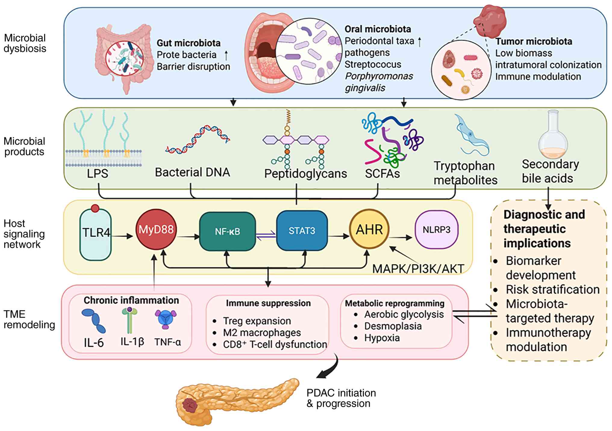 | Figure 2.Mechanistic links between
microbiota-derived products, PDAC progression and TME remodeling.
Microbial dysbiosis in the gut, oral cavity and tumor- or
tissue-associated microbial communities may contribute to PDAC
progression through microbiota-derived molecules and metabolites,
including LPS, bacterial DNA, peptidoglycans, SCFAs, tryptophan
metabolites and secondary bile acids. These microbial products may
engage or converge on host signaling pathways, including the TLR4,
MyD88, NF-κB, STAT3, AHR, NLRP3 and MAPK/PI3K/AKT signaling
pathways. Collectively, these pathways may promote chronic
inflammation via IL-6, IL-1β and TNF-α production, immune
suppression via mechanisms such as Treg expansion, M2 macrophage
polarization and CD8+ T-cell dysfunction; and iii)
metabolic reprogramming, thereby facilitating TME remodeling and
PDAC initiation and progression. Potential clinical implications of
these microbiota-based mechanistic links in PDAC include biomarker
development, microbial biomarker-based risk stratification,
microbiota-targeted therapy and immunotherapy modulation. SCFA,
short-chain fatty acid; LPS, lipopolysaccharide; TLR4, toll-like
receptor 4; MyD88, myeloid differentiation primary response 88;
AHR, aryl hydrocarbon receptor; NLRP3, nucleotide-binding
oligomerization domain-like receptor protein 3; Treg, regulatory T
cell; PDAC, pancreatic ductal adenocarcinoma; TME, tumor
microenvironment. Created with BioRender.com. |
The proposed immune-oncology-microbiome axis
highlights the interconnected roles of gut and tumor-associated
microbiota in regulating tumor metabolism and the immune
microenvironment (121). An
experimental study suggested that pancreatic tumors harbor distinct
microbial communities, and that microbiome ablation can reprogram
the TME toward an immunogenic phenotype and enhance responses to
immunotherapy (122).
Microbiota-derived metabolites, particularly short-chain fatty
acids, have been shown to promote cytotoxic T-cell activity and
modulate inflammatory signaling pathways (123,124). Furthermore, a reduction in
butyrate levels may contribute to pancreatic carcinogenesis
(125), whereas other metabolites,
including trimethylamine N-oxide, may serve as potential biomarkers
or therapeutic targets for PDAC (126).
Given the poor prognosis of PDAC and the lack of
effective population-wide screening strategies, microbiome
profiling may be particularly valuable in high-risk populations
(127). Patients with CP, IPMNs,
obesity, diabetes or a familial predisposition to PDAC may benefit
from microbiome-based risk-stratification approaches. Microbiome
analysis could also complement liquid biopsy and imaging
approaches, particularly in early-stage disease where microbial and
immune alterations may be more detectable (128). The enrichment of beneficial
microbial taxa, such as Faecalibacterium prausnitzii and
Akkermansia muciniphila, observed in long-term survivors
also supports a possible prognostic role of the microbiome in PDAC
(34).
Beyond fecal biomarkers, multi-site microbial
profiling may improve the diagnostic performance of
microbiota-based classifiers. Although fecal samples remain
attractive for non-invasive screening due to their accessibility,
oral, biliary and tissue-associated microbiota have also been
extensively investigated and may provide complementary diagnostic
information related to pancreatic carcinogenesis (119,129). Saliva, in particular, represents a
non-invasive diagnostic fluid with potential applications in
transcriptomic and metabolomic profiling (130). However, the identification of
consistent, disease-specific microbial signatures remains
challenging regardless of body site, and the functions of
individual microbial taxa in PDAC remain incompletely elucidated
(131).
Preclinical studies have also indicated that
microbiota-targeted interventions, including probiotic treatments
and metabolite modulation, may influence tumor biology and
therapeutic responses (132,133). Although the diagnostic utility of
these interventions has not yet been established, these findings
highlight the potential of precision microbiome approaches in PDAC.
Future studies should focus on large-scale, prospective,
multicenter cohorts with longitudinal sampling and integrated
multi-omics analyses. Developing reproducible, function-based
biomarker models rather than relying solely on descriptive
taxonomic associations will be important for the clinical
translation of microbiota-based biomarker models in PDAC.
Furthermore, the validation of microbiome-based strategies in
diverse clinical settings may improve early detection, risk
stratification and personalized treatment management in patients
with PDAC.
Conclusions
PDAC remains one of the most lethal malignancies,
primarily due to delayed diagnosis and the lack of effective
population-level screening strategies. Increasing evidence has
indicated that gut, oral and tissue-associated microbiota may be
potential sources of non-invasive diagnostic biomarkers.
Microbiome-based predictive models have demonstrated promising
discriminatory performance, particularly when combined with
established clinical markers of PDAC, such as CA19-9 or metabolomic
signatures.
Mechanistic studies have further suggested that
microbial dysbiosis may contribute to PDAC progression through
immune modulation, inflammatory signaling and metabolic
reprogramming. These findings provide biological support for
microbiome-informed diagnostic approaches. However, clinical
translation of these approaches remains limited by methodological
heterogeneity, host-related variability, low microbial biomass in
pancreatic tissues and a lack of large-scale prospective validation
studies.
With further methodological standardization,
functional validation and prospective clinical evaluation,
microbiome-based diagnostic strategies may eventually complement
existing diagnostic approaches and improve early detection and risk
stratification in patients with PDAC.
Acknowledgements
Not applicable.
Funding
The present review was supported by the National Natural Science
Foundation of China (grant no. 82102431), the Natural Science
Foundation of Gansu Province, China (grant no. 26JRRA817), the Key
Incubation Project Funds of The Second Hospital & Clinical
Medical School, Lanzhou University (grant no. 2025-21-zdfy-004) and
the Talent Introduction Plan of The Second Hospital & Clinical
Medical School, Lanzhou University (grant no.
yjrckyqdj-2021-03).
Availability of data and materials
Not applicable.
Authors' contributions
HJ and JY conceived the study and contributed to
writing the original draft. HJ, HS and JY reviewed and edited the
manuscript. HJ constructed figures. JY was also responsible for
supervising the study and acquiring funding. All authors read and
approved the final version of the manuscript. Data authentication
is not applicable.
Ethics approval and consent to
participate
Not applicable.
Patient consent for publication
Not applicable.
Competing interests
The authors declare that they have no competing
interests.
References
|
1
|
Mendes I and Vale N: Overcoming
microbiome-acquired gemcitabine resistance in pancreatic ductal
adenocarcinoma. Biomedicines. 12:2272024. View Article : Google Scholar : PubMed/NCBI
|
|
2
|
Chen Y, Nian F, Chen J, Jiang Q, Yuan T,
Feng H, Shen X and Dong L: Metagenomic microbial signatures for
noninvasive detection of pancreatic cancer. Biomedicines.
13:10002025. View Article : Google Scholar : PubMed/NCBI
|
|
3
|
Grossberg AJ, Chu LC, Deig CR, Fishman EK,
Hwang WL, Maitra A, Marks DL, Mehta A, Nabavizadeh N, Simeone DM,
et al: Multidisciplinary standards of care and recent progress in
pancreatic ductal adenocarcinoma. CA Cancer J Clin. 70:375–403.
2020.PubMed/NCBI
|
|
4
|
Li K, Xiao Y, Bian J, Han L, He C, El-Omar
E, Gong L and Wang M: Ameliorative effects of gut microbial
metabolite urolithin A on pancreatic diseases. Nutrients.
14:25492022. View Article : Google Scholar : PubMed/NCBI
|
|
5
|
Eid M, Martínek A, Dolina J, Uvírová M and
Dítě P: Gut microbiome and pancreatic cancer. Klin Onkol. 37:20–26.
2024. View Article : Google Scholar : PubMed/NCBI
|
|
6
|
Arsenijevic T, Nicolle R, Bouchart C,
D'Haene N, Demetter P, Puleo F and Van Laethem JL: Pancreatic
cancer meets human microbiota: Close encounters of the third kind.
Cancers (Basel). 13:12312021. View Article : Google Scholar : PubMed/NCBI
|
|
7
|
Scarà S, Bottoni P and Scatena R: CA 19-9:
Biochemical and clinical aspects. Adv Exp Med Biol. 867:247–260.
2015. View Article : Google Scholar : PubMed/NCBI
|
|
8
|
Shen MJ, Jamali A and Katona BW:
Challenges of early detection of pancreatic cancer. J Clin Invest.
135:e1919422025. View Article : Google Scholar : PubMed/NCBI
|
|
9
|
Tan X, Wang Y and Gong T: The interplay
between oral microbiota, gut microbiota and systematic diseases. J
Oral Microbiol. 15:22131122023. View Article : Google Scholar : PubMed/NCBI
|
|
10
|
Zhang W, Zhang K, Zhang P, Zheng J, Min C
and Li X: Research progress of pancreas-related microorganisms and
pancreatic cancer. Front Oncol. 10:6045312020. View Article : Google Scholar : PubMed/NCBI
|
|
11
|
Dhar AK: Human microbiome modulation: A
potential therapeutic strategy for pancreatic cancer. Synbiotics
for the Management of Cancer. Mishra N, Bhatt S, Paudel KR, Hansbro
PM and Dua K: Springer Nature Singapore; Singapore: pp. 205–242.
2023, View Article : Google Scholar
|
|
12
|
Wensel CR, Pluznick JL, Salzberg SL and
Sears CL: Next-generation sequencing: Insights to advance clinical
investigations of the microbiome. J Clin Invest. 132:e1549442022.
View Article : Google Scholar : PubMed/NCBI
|
|
13
|
Jankowski WM, Fichna J and
Tarasiuk-Zawadzka A: A systematic review of the relationship
between gut microbiota and prevalence of pancreatic diseases.
Microb Pathog. 199:1072142025. View Article : Google Scholar : PubMed/NCBI
|
|
14
|
Sexton RE, Uddin MH, Bannoura S, Khan HY,
Mzannar Y, Li Y, Aboukameel A, Al-Hallak MN, Al-Share B, Mohamed A,
et al: Connecting the human microbiome and pancreatic cancer.
Cancer Metastasis Rev. 41:317–331. 2022. View Article : Google Scholar : PubMed/NCBI
|
|
15
|
Yu Q, Jobin C and Thomas RM: Implications
of the microbiome in the development and treatment of pancreatic
cancer: Thinking outside of the box by looking inside the gut.
Neoplasia. 23:246–256. 2021. View Article : Google Scholar : PubMed/NCBI
|
|
16
|
Kartal E, Schmidt TSB, Molina-Montes E,
Rodríguez-Perales S, Wirbel J, Maistrenko OM, Akanni WA, Alashkar
Alhamwe B, Alves RJ, Carrato A, et al: A faecal microbiota
signature with high specificity for pancreatic cancer. Gut.
71:1359–1372. 2022. View Article : Google Scholar : PubMed/NCBI
|
|
17
|
Wang W, Qian C, Wang T, Jiang Y, Zhou Y,
Liu K, Ma Z, Liu P, Wu Y, Chen L, et al: A combination of faecal
and intratumour microbial community profiling reveals novel
diagnostic and prognostic biomarkers for pancreatic tumours. Clin
Transl Med. 14:e17262024. View Article : Google Scholar : PubMed/NCBI
|
|
18
|
Li P, Zhang H and Dai M: Current status
and prospect of gut and oral microbiome in pancreatic cancer:
Clinical and translational perspectives. Cancer Lett.
604:2172742024. View Article : Google Scholar : PubMed/NCBI
|
|
19
|
Michaud DS and Izard J: Microbiota, oral
microbiome, and pancreatic cancer. Cancer J. 20:203–206. 2014.
View Article : Google Scholar : PubMed/NCBI
|
|
20
|
Fan X, Alekseyenko AV, Wu J, Peters BA,
Jacobs EJ, Gapstur SM, Purdue MP, Abnet CC, Stolzenberg-Solomon R,
Miller G, et al: Human oral microbiome and prospective risk for
pancreatic cancer: A population-based nested case-control study.
Gut. 67:120–127. 2018. View Article : Google Scholar : PubMed/NCBI
|
|
21
|
Nagata N, Nishijima S, Kojima Y, Hisada Y,
Imbe K, Miyoshi-Akiyama T, Suda W, Kimura M, Aoki R, Sekine K, et
al: Metagenomic identification of microbial signatures predicting
pancreatic cancer from a multinational study. Gastroenterology.
163:222–238. 2022. View Article : Google Scholar : PubMed/NCBI
|
|
22
|
Michaud DS, Izard J, Wilhelm-Benartzi CS,
You DH, Grote VA, Tjønneland A, Dahm CC, Overvad K, Jenab M,
Fedirko V, et al: Plasma antibodies to oral bacteria and risk of
pancreatic cancer in a large European prospective cohort study.
Gut. 62:1764–1770. 2013. View Article : Google Scholar : PubMed/NCBI
|
|
23
|
Kim B, Oh S, Yang S, Ahn J, Jung K, Lee
JC, Hwang JH, Shin CM, Lee HJ, Lee HS, et al: Distinct microbiome
composition and reduced interactions in patients with pancreatic
cancer. Front Microbiol. 16:15554792025. View Article : Google Scholar : PubMed/NCBI
|
|
24
|
Sono M, Iimori K, Nagao M, Ogawa S, Maruno
T, Nakanishi Y, Anazawa T, Nagai K, Masui T, Mori H, et al:
Reduction of butyrate-producing bacteria in the gut microbiome of
Japanese patients with pancreatic cancer. Pancreatology.
24:1031–1039. 2024. View Article : Google Scholar : PubMed/NCBI
|
|
25
|
Jin D, Jin S, Zhou T, Cui Z, Guo B, Li G
and Zhang C: Quantitative evaluation of gut microbiota composition
in pancreatic cancer: A pooled study. Medicine (Baltimore).
103:e386082024.PubMed/NCBI
|
|
26
|
Chen T, Li X, Li G, Liu Y, Huang X, Ma W,
Qian C, Guo J, Wang S, Qin Q and Liu S: Alterations of commensal
microbiota are associated with pancreatic cancer. Int J Biol
Markers. 38:263–274. 2023. View Article : Google Scholar
|
|
27
|
Hashimoto S, Tochio T, Funasaka K,
Funahashi K, Hartanto T, Togashi Y, Saito M, Nishimoto Y, Yoshinori
M, Nakaoka K, et al: Changes in intestinal bacteria and imbalances
of metabolites induced in the intestines of pancreatic ductal
adenocarcinoma patients in a Japanese population: A preliminary
result. Scand J Gastroenterol. 58:193–198. 2023. View Article : Google Scholar : PubMed/NCBI
|
|
28
|
Kohi S, Macgregor-Das A, Dbouk M, Yoshida
T, Chuidian M, Abe T, Borges M, Lennon AM, Shin EJ, Canto MI and
Goggins M: Alterations in the duodenal fluid microbiome of patients
with pancreatic cancer. Clin Gastroenterol Hepatol. 20:e196–e227.
2022. View Article : Google Scholar : PubMed/NCBI
|
|
29
|
Sammallahti H, Rezasoltani S, Pekkala S,
Kokkola A, Asadzadeh Agdaei H, Azizmohammad Looha M, Ghanbari R,
Zamani F, Sadeghi A, Sarhadi VK, et al: Fecal profiling reveals a
common microbial signature for pancreatic cancer in Finnish and
Iranian cohorts. Gut Pathog. 17:242025. View Article : Google Scholar : PubMed/NCBI
|
|
30
|
Yang J, Ma Y, Tan Q, Zhou B, Yu D, Jin M,
Zhang T, Liu J and Liu H: Gut Streptococcus is a microbial marker
for the occurrence and liver metastasis of pancreatic cancer. Front
Microbiol. 14:11610682023.
|
|
31
|
Yousefi B, Mohammadlou M, Aisa B,
Falalyeyeva T, Kobyliak N and Eslami M: Immunomodulatory role of
Faecalibacterium prausnitzii in obesity and metabolic
disorders. Minerva Biotechnol Biomol Res. 33:76–85. 2021.
|
|
32
|
Villani A, Fontana A, Panebianco C, Ferro
C, Copetti M, Pavlovic R, Drago D, Fiorentini C, Terracciano F,
Bazzocchi F, et al: A powerful machine learning approach to
identify interactions of differentially abundant gut microbial
subsets in patients with metastatic and non-metastatic pancreatic
cancer. Gut Microbes. 16:23754832024. View Article : Google Scholar : PubMed/NCBI
|
|
33
|
Matsukawa H, Iida N, Kitamura K, Terashima
T, Seishima J, Makino I, Kannon T, Hosomichi K, Yamashita T, Sakai
Y, et al: Dysbiotic gut microbiota in pancreatic cancer patients
form correlation networks with the oral microbiota and prognostic
factors. Am J Cancer Res. 11:3163–3175. 2021.PubMed/NCBI
|
|
34
|
Kharofa J, Haslam D, Wilkinson R, Weiss A,
Patel S, Wang K, Esslinger H, Olowokure O, Sohal D, Wilson G, et
al: Analysis of the fecal metagenome in long-term survivors of
pancreas cancer. Cancer. 129:1986–1994. 2023. View Article : Google Scholar : PubMed/NCBI
|
|
35
|
Świdnicka-Siergiejko A, Daniluk J,
Miniewska K, Daniluk U, Guzińska-Ustymowicz K, Pryczynicz A,
Dąbrowska M, Rusak M, Ciborowski M and Dąbrowski A: Inflammatory
stimuli and fecal microbiota transplantation accelerate pancreatic
carcinogenesis in transgenic mice, accompanied by changes in the
microbiota composition. Cells. 14:3612025. View Article : Google Scholar : PubMed/NCBI
|
|
36
|
Riquelme E, Zhang Y, Zhang L, Montiel M,
Zoltan M, Dong W, Quesada P, Sahin I, Chandra V, San Lucas A, et
al: Tumor microbiome diversity and composition influence pancreatic
cancer outcomes. Cell. 178:795–806.e12. 2019. View Article : Google Scholar : PubMed/NCBI
|
|
37
|
Genton L, Lazarevic V, Stojanovic O,
Spiljar M, Djaafar S, Koessler T, Dutoit V, Gaïa N, Mareschal J,
Macpherson AJ, et al: Metataxonomic and metabolic impact of fecal
microbiota transplantation from patients with pancreatic cancer
into germ-free mice: A pilot study. Front Cell Infect Microbiol.
11:7528892021. View Article : Google Scholar : PubMed/NCBI
|
|
38
|
Dong TS, Chang HH, Hauer M, Lagishetty V,
Katzka W, Rozengurt E, Jacobs JP and Eibl G: Metformin alters the
duodenal microbiome and decreases the incidence of pancreatic
ductal adenocarcinoma promoted by diet-induced obesity. Am J
Physiol Gastrointest Liver Physiol. 317:G763–G772. 2019. View Article : Google Scholar : PubMed/NCBI
|
|
39
|
Li ZY, Chen SY, Weng MH and Yen GC:
Ursolic acid restores sensitivity to gemcitabine through the
RAGE/NF-κB/MDR1 axis in pancreatic cancer cells and in a mouse
xenograft model. J Food Drug Anal. 29:262–274. 2021.PubMed/NCBI
|
|
40
|
Knight R, Vrbanac A, Taylor BC, Aksenov A,
Callewaert C, Debelius J, Gonzalez A, Kosciolek T, McCall LI,
McDonald D, et al: Best practices for analysing microbiomes. Nat
Rev Microbiol. 16:410–422. 2018. View Article : Google Scholar : PubMed/NCBI
|
|
41
|
Gao YC, Zhou DD, Lu YB, Yang L, Gong XJ,
Chen MY, Liang S, Huang WH and Zhang W: Antitumor potentials of
onco-microbial in Chinese patients with pancreatic cancer. Heliyon.
10:e344202024. View Article : Google Scholar : PubMed/NCBI
|
|
42
|
May MS, Park H, Moallem DH, Seeram D,
Dajiang S, Hibshoosh H, Jamison JK, Uhlemann AC and Manji GA: Low
bacterial biomass in human pancreatic cancer and adjacent normal
tissue. Int J Mol Sci. 26:1402024. View Article : Google Scholar : PubMed/NCBI
|
|
43
|
Salter SJ, Cox MJ, Turek EM, Calus ST,
Cookson WO, Moffatt MF, Turner P, Parkhill J, Loman NJ and Walker
AW: Reagent and laboratory contamination can critically impact
sequence-based microbiome analyses. BMC Biol. 12:872014. View Article : Google Scholar : PubMed/NCBI
|
|
44
|
Zhu Y, Liang X, Zhang G, Li F, Xu J, Ma R,
Chen X, Ma M, Wang Y, Chen C, et al: Microbiota and metabolite
alterations in pancreatic head and body/tail cancer patients.
Cancer Sci. 115:2738–2750. 2024. View Article : Google Scholar : PubMed/NCBI
|
|
45
|
Li Y, Chang RB, Stone ML, Delman D,
Markowitz K, Xue Y, Coho H, Herrera VM, Li JH, Zhang L, et al:
Multimodal immune phenotyping reveals microbial-T cell interactions
that shape pancreatic cancer. Cell Rep Med. 5:1013972024.
View Article : Google Scholar : PubMed/NCBI
|
|
46
|
Halimi A, Gabarrini G, Sobkowiak MJ, Ateeb
Z, Davanian H, Gaiser RA, Arnelo U, Valente R, Wong AYW, Moro CF,
et al: Isolation of pancreatic microbiota from cystic precursors of
pancreatic cancer with intracellular growth and DNA damaging
properties. Gut Microbes. 13:19831012021. View Article : Google Scholar : PubMed/NCBI
|
|
47
|
Stasiewicz M, Kwaśniewski M and Karpiński
TM: Microbial associations with pancreatic cancer: A new frontier
in biomarkers. Cancers (Basel). 13:37842021. View Article : Google Scholar : PubMed/NCBI
|
|
48
|
Del Castillo E, Meier R, Chung M, Koestler
DC, Chen T, Paster BJ, Charpentier KP, Kelsey KT, Izard J and
Michaud DS: The microbiomes of pancreatic and duodenum tissue
overlap and are highly subject specific but differ between
pancreatic cancer and noncancer subjects. Cancer Epidemiol
Biomarkers Prev. 28:370–383. 2019. View Article : Google Scholar : PubMed/NCBI
|
|
49
|
Zhu Y, Liang X, Zhi M, Li L, Zhang G, Chen
C, Wang L, Wang P, Zhong N, Feng Q and Li Z: Succession of the
multi-site microbiome along pancreatic ductal adenocarcinoma
tumorigenesis. Front Immunol. 15:14872422024. View Article : Google Scholar : PubMed/NCBI
|
|
50
|
Guo X and Shao Y: Role of the oral-gut
microbiota axis in pancreatic cancer: A new perspective on tumor
pathophysiology, diagnosis, and treatment. Mol Med. 31:1032025.
View Article : Google Scholar : PubMed/NCBI
|
|
51
|
McAllister F, Khan MAW, Helmink B and
Wargo JA: The tumor microbiome in pancreatic cancer: Bacteria and
beyond. Cancer Cell. 36:577–579. 2019. View Article : Google Scholar : PubMed/NCBI
|
|
52
|
Karpiński TM: The microbiota and
pancreatic cancer. Gastroenterol Clin North Am. 48:447–464. 2019.
View Article : Google Scholar : PubMed/NCBI
|
|
53
|
Yuan M, Xu Y and Guo Z: Association of
oral microbiome and pancreatic cancer: A systematic review and
meta-analysis. Ther Adv Gastroenterol. 15:175628482211239802022.
View Article : Google Scholar : PubMed/NCBI
|
|
54
|
Merali N, Chouari T, Terroire J, Jessel
MD, Liu DSK, Smith JH, Wooldridge T, Dhillon T, Jiménez JI, Krell
J, et al: Bile microbiome signatures associated with pancreatic
ductal adenocarcinoma compared to benign disease: A UK pilot study.
Int J Mol Sci. 24:168882023. View Article : Google Scholar : PubMed/NCBI
|
|
55
|
Poudel SK, Padmanabhan R, Dave H, Guinta
K, Stevens T, Sanaka MR, Chahal P, Sohal DPS, Khorana AA and Eng C:
Microbiomic profiles of bile in patients with benign and malignant
pancreaticobiliary disease. PLoS One. 18:e02830212023. View Article : Google Scholar : PubMed/NCBI
|
|
56
|
Eisenhofer R, Minich JJ, Marotz C, Cooper
A, Knight R and Weyrich LS: Contamination in low microbial biomass
microbiome studies: Issues and recommendations. Trends Microbiol.
27:105–117. 2019. View Article : Google Scholar : PubMed/NCBI
|
|
57
|
Merali N, Chouari T, Sweeney C,
Halle-Smith J, Jessel MD, Wang B, O' Brien J, Suyama S, Jiménez JI,
Roberts KJ, et al: The microbial composition of pancreatic ductal
adenocarcinoma: A systematic review of 16S rRNA gene sequencing.
Int J Surg. 110:6771–6799. 2024. View Article : Google Scholar : PubMed/NCBI
|
|
58
|
Kartal E, Schmidt TSB, Molina-Montes E,
Rodríguez-Perales S, Wirbel J, Maistrenko OM, Akanni WA, Alashkar
Alhamwe B, Alves RJ, Carrato A, et al: A faecal microbiota
signature with high specificity for pancreatic cancer. Gut.
71:1359–1372. 2022. View Article : Google Scholar : PubMed/NCBI
|
|
59
|
Farrell JJ, Zhang L, Zhou H, Chia D,
Elashoff D, Akin D, Paster BJ, Joshipura K and Wong DT: Variations
of oral microbiota are associated with pancreatic diseases
including pancreatic cancer. Gut. 61:582–588. 2012. View Article : Google Scholar : PubMed/NCBI
|
|
60
|
Guo X, Hu Z, Rong S, Xie G, Nie G, Liu X
and Jin G: Integrative analysis of metabolome and gut microbiota in
patients with pancreatic ductal adenocarcinoma. J Cancer.
13:1555–1564. 2022. View Article : Google Scholar : PubMed/NCBI
|
|
61
|
Li P, Zhang H, Chen L, Gao X, Hu Y, Xu Q,
Liu W, Chen W, Chen H, Yuan S, et al: Oral and fecal microbiota as
accurate non-invasive tools for detection of pancreatic cancer in
the Chinese population. Cancer Lett. 612:2174562025. View Article : Google Scholar : PubMed/NCBI
|
|
62
|
Zhou W, Zhang D, Li Z, Jiang H, Li J, Ren
R, Gao X, Li J, Wang X, Wang W and Yang Y: The fecal microbiota of
patients with pancreatic ductal adenocarcinoma and autoimmune
pancreatitis characterized by metagenomic sequencing. J Transl Med.
19:2152021. View Article : Google Scholar : PubMed/NCBI
|
|
63
|
Zhang P, Shi H, Guo R, Li L, Guo X, Yang
H, Chang D, Cheng Y, Zhao G, Li S, et al: Metagenomic analysis
reveals altered gut virome and diagnostic potential in pancreatic
cancer. J Med Virol. 96:e298092024. View Article : Google Scholar : PubMed/NCBI
|
|
64
|
Wong DT, Zhang L, Farrell J, Zhou H,
Elashoff D, Gao K and Paster BJ: Salivary biomarkers for pancreatic
cancer detection. J Clin Oncol. 27 (15 Suppl):S46292009. View Article : Google Scholar
|
|
65
|
Wei AL, Li M, Li GQ, Wang X, Hu WM, Li ZL,
Yuan J, Liu HY, Zhou LL, Li K, et al: Oral microbiome and
pancreatic cancer. World J Gastroenterol. 26:7679–7692. 2020.
View Article : Google Scholar : PubMed/NCBI
|
|
66
|
Ammer-Herrmenau C, Pfisterer N, Weingarten
MF and Neesse A: The microbiome in pancreatic diseases: Recent
advances and future perspectives. United European Gastroenterol J.
8:878–885. 2020. View Article : Google Scholar : PubMed/NCBI
|
|
67
|
Kaźmierczak-Siedlecka K, Stachowska E,
Folwarski M, Przewłócka K, Makarewicz W and Bryl E: The potential
of gut microbiome as a non-invasive predictive biomarker for early
detection of pancreatic cancer and hepatocellular carcinoma. Eur
Rev Med Pharmacol Sci. 25:7275–7284. 2021.PubMed/NCBI
|
|
68
|
Wei X, Mei C, Li X and Xie Y: The unique
microbiome and immunity in pancreatic cancer. Pancreas. 50:119–129.
2021. View Article : Google Scholar : PubMed/NCBI
|
|
69
|
Li JJ, Zhu M, Kashyap PC, Chia N, Tran NH,
McWilliams RR, Bekaii-Saab TS and Ma WW: The role of microbiome in
pancreatic cancer. Cancer Metastasis Rev. 40:777–789. 2021.
View Article : Google Scholar : PubMed/NCBI
|
|
70
|
Miyabayashi K, Ijichi H and Fujishiro M:
The role of the microbiome in pancreatic cancer. Cancers (Basel).
14:44792022. View Article : Google Scholar : PubMed/NCBI
|
|
71
|
Marcos-Zambrano LJ, Karaduzovic-Hadziabdic
K, Loncar Turukalo T, Przymus P, Trajkovik V, Aasmets O, Berland M,
Gruca A, Hasic J, Hron K, et al: Applications of machine learning
in human microbiome studies: A review on feature selection,
biomarkeridentification, disease prediction and treatment. Front
Microbiol. 12:6345112021. View Article : Google Scholar : PubMed/NCBI
|
|
72
|
Cortez NE, Rodriguez Lanzi C, Hong BV, Xu
J, Wang F, Chen S, Ramsey JJ, Pontifex MG, Müller M, Vauzour D, et
al: A ketogenic diet in combination with gemcitabine increases
survival in pancreatic cancer KPC mice. Cancer Res Commun.
2:951–965. 2022. View Article : Google Scholar : PubMed/NCBI
|
|
73
|
Li P, Zhang H, Gao X, Chen L, Chen H, Yuan
S, Chen W and Dai M: Difference in fecal and oral microbiota
between pancreatic cancer and benign/low-grade malignant tumor
patients. BMC Microbiol. 24:5272024. View Article : Google Scholar : PubMed/NCBI
|
|
74
|
Pourali G, Kazemi D, Chadeganipour AS,
Arastonejad M, Kashani SN, Pourali R, Maftooh M, Akbarzade H, Fiuji
H, Hassanian SM, et al: Microbiome as a biomarker and therapeutic
target in pancreatic cancer. BMC Microbiol. 24:162024. View Article : Google Scholar : PubMed/NCBI
|
|
75
|
Khan ZA, Ghorbani M, Heffinger L,
Damdimopoulos A, Moro CF, Björnstedt M, Löhr JM, Heuchel R, Chen MS
and Sarhan D: Genderized gut and oral microbiome shifts: Uncovering
sex-specific dysbiosis in pancreatic cancer. bioRxiv. Oct
3–2024.doi: 10.1101/2024.10.02.616338.
|
|
76
|
Li Z, Chu J, Su F, Ding X, Zhang Y, Dou L,
Liu Y, Ke Y, Liu X, Liu Y, et al: Characteristics of bile
microbiota in cholelithiasis, perihilar cholangiocarcinoma, distal
cholangiocarcinoma, and pancreatic cancer. Am J Transl Res.
14:2962–2971. 2022.PubMed/NCBI
|
|
77
|
Sethi V, Tao J, Kurtom S, Vaish U, Afaq F,
Zao S, Luo X, Jain T, Sharma P, Bava E, et al: Abstract 3046: A
common gut commensal utilizes tryptophan to promote cancer via the
aryl hydrocarbon receptor. Cancer Res. 82 (12 Suppl):S30462022.
View Article : Google Scholar
|
|
78
|
Subramanian I, Verma S, Kumar S, Jere A
and Anamika K: Multi-omics data integration, interpretation, and
its application. Bioinform Biol Insights. 14:11779322198990512020.
View Article : Google Scholar : PubMed/NCBI
|
|
79
|
Zhang L, Sanagapalli S and Stoita A:
Challenges in diagnosis of pancreatic cancer. World J
Gastroenterol. 24:2047–2060. 2018. View Article : Google Scholar : PubMed/NCBI
|
|
80
|
Singhi AD, Koay EJ, Chari ST and Maitra A:
Early detection of pancreatic cancer: Opportunities and challenges.
Gastroenterology. 156:2024–2040. 2019. View Article : Google Scholar : PubMed/NCBI
|
|
81
|
Poruk KE, Firpo MA, Adler DG and Mulvihill
SJ: Screening for pancreatic cancer: Why, how, and who? Ann Surg.
257:17–26. 2013. View Article : Google Scholar : PubMed/NCBI
|
|
82
|
Wirbel J, Zych K, Essex M, Karcher N,
Kartal E, Salazar G, Bork P, Sunagawa S and Zeller G: Microbiome
meta-analysis and cross-disease comparison enabled by the SIAMCAT
machine learning toolbox. Genome Biol. 22:932021. View Article : Google Scholar : PubMed/NCBI
|
|
83
|
Steyerberg EW, Vickers AJ, Cook NR, Gerds
T, Gonen M, Obuchowski N, Pencina MJ and Kattan MW: Assessing the
performance of prediction models: A framework for traditional and
novel measures. Epidemiology. 21:128–138. 2010. View Article : Google Scholar : PubMed/NCBI
|
|
84
|
Van Smeden M, Reitsma JB, Riley RD,
Collins GS and Moons KGM: Clinical prediction models: Diagnosis
versus prognosis. J Clin Epidemiol. 132:142–145. 2021. View Article : Google Scholar : PubMed/NCBI
|
|
85
|
Asnicar F, Thomas AM, Passerini A, Waldron
L and Segata N: Machine learning for microbiologists. Nat Rev
Microbiol. 22:191–205. 2024. View Article : Google Scholar : PubMed/NCBI
|
|
86
|
Bastos AR, Pereira-Marques J, Ferreira RM
and Figueiredo C: Harnessing the microbiome to reduce pancreatic
cancer burden. Cancers (Basel). 15:26292023. View Article : Google Scholar : PubMed/NCBI
|
|
87
|
Abdul Rahman R, Lamarca A, Hubner RA,
Valle JW and McNamara MG: The microbiome as a potential target for
therapeutic manipulation in pancreatic cancer. Cancers (Basel).
13:37792021. View Article : Google Scholar : PubMed/NCBI
|
|
88
|
Li P, Shu Y and Gu Y: The potential role
of bacteria in pancreatic cancer: A systematic review.
Carcinogenesis. 41:397–404. 2020. View Article : Google Scholar : PubMed/NCBI
|
|
89
|
Memba R, Duggan SN, Ni Chonchubhair HM,
Griffin OM, Bashir Y, O'Connor DB, Murphy A, McMahon J, Volcov Y,
Ryan BM and Conlon KC: The potential role of gut microbiota in
pancreatic disease: A systematic review. Pancreatology. 17:867–874.
2017. View Article : Google Scholar : PubMed/NCBI
|
|
90
|
Meng C, Bai C, Brown TD, Hood L and Tian
Q: Human gut microbiota and gastrointestinal cancer. Genomics
Proteomics Bioinformatics. 16:33–49. 2018. View Article : Google Scholar : PubMed/NCBI
|
|
91
|
Gong Y, Huang X, Wang M and Liang X:
Intratumor microbiota: A novel tumor component. J Cancer Res Clin
Oncol. 149:6675–6691. 2023. View Article : Google Scholar : PubMed/NCBI
|
|
92
|
Wright RD, Bartelli TF, Baydogan S, White
JR, Kim MP, Bhutani MS and McAllister F: Bacterial and fungal
characterization of pancreatic adenocarcinoma from endoscopic
ultrasound-guided biopsies. Front Immunol. 14:12683762023.
View Article : Google Scholar : PubMed/NCBI
|
|
93
|
Thomas RM, Gharaibeh RZ, Gauthier J,
Beveridge M, Pope JL, Guijarro MV, Yu Q, He Z, Ohland C, Newsome R,
et al: Intestinal microbiota enhances pancreatic carcinogenesis in
preclinical models. Carcinogenesis. 39:1068–1078. 2018. View Article : Google Scholar : PubMed/NCBI
|
|
94
|
Zmora N, Suez J and Elinav E: You are what
you eat: Diet, health and the gut microbiota. Nat Rev Gastroenterol
Hepatol. 16:35–56. 2019. View Article : Google Scholar : PubMed/NCBI
|
|
95
|
Tong Y, Gao H, Qi Q, Li J, Gao J, Li P,
Wang Y, Du L and Wang C: High fat diet, gut microbiome and
gastrointestinal cancer. Theranostics. 11:5889–5910. 2021.
View Article : Google Scholar : PubMed/NCBI
|
|
96
|
Dong L, Chen Y, Nian F, et al: Low-protein
diet enhances anti-tumor immunity and immunotherapy through
microbiota-derived uridine diphosphate-galactose in pancreatic
cancer. Res Sq [Preprint]. 2025.doi:
10.21203/rs.3.rs-5804694/v1.
|
|
97
|
Ruze R, Song J, Yin X, Chen Y, Xu R, Wang
C and Zhao Y: Mechanisms of obesity- and diabetes mellitus-related
pancreatic carcinogenesis: A comprehensive and systematic review.
Signal Transduct Target Ther. 8:1392023. View Article : Google Scholar : PubMed/NCBI
|
|
98
|
Kesh K, Mendez R, Abdelrahman L and
Banerjee S and Banerjee S: Type 2 diabetes-induced microbiome
dysbiosis is associated with therapy resistance in pancreatic
adenocarcinoma. Microb Cell Fact. 19:752020. View Article : Google Scholar : PubMed/NCBI
|
|
99
|
Panebianco C, Adamberg K, Jaagura M,
Copetti M, Fontana A, Adamberg S, Kolk K, Vilu R, Andriulli A and
Pazienza V: Influence of gemcitabine chemotherapy on the microbiota
of pancreatic cancer xenografted mice. Cancer Chemother Pharmacol.
81:773–782. 2018. View Article : Google Scholar : PubMed/NCBI
|
|
100
|
Miller PL and Carson TL: Mechanisms and
microbial influences on CTLA-4- and PD-1-based immunotherapy in the
treatment of cancer: A narrative review. Gut Pathog. 12:432020.
View Article : Google Scholar : PubMed/NCBI
|
|
101
|
Nalluri H, Jensen E and Staley C: Role of
biliary stent and neoadjuvant chemotherapy in the pancreatic tumor
microbiome. BMC Microbiol. 21:2802021. View Article : Google Scholar : PubMed/NCBI
|
|
102
|
Takaori A, Hashimoto D, Ikeura T, Ito T,
Nakamaru K, Masuda M, Nakayama S, Yamaki S, Yamamoto T, Fujimoto K,
et al: Impact of neoadjuvant therapy on gut microbiome in patients
with resectable/borderline resectable pancreatic ductal
adenocarcinoma. Pancreatology. 23:367–376. 2023. View Article : Google Scholar : PubMed/NCBI
|
|
103
|
Sawada K, Yamashita R, Sakai SA, Horasawa
S, Yoshikawa A, Fujisawa T, Kadowaki S, Kato K, Ueno M, Oki E, et
al: Microbiome landscape and association with response to immune
checkpoint inhibitors in advanced solid tumors: A SCRUM-Japan
MONSTAR-SCREEN study. Cancer Res Commun. 5:857–870. 2025.
View Article : Google Scholar : PubMed/NCBI
|
|
104
|
Nalluri H, Staley C and Jensen EH:
Abstract 6097: Alterations of the pancreatic tumor microbiome: The
role of biliary stents. Cancer Res. 80 (16 Suppl):S60972020.
View Article : Google Scholar
|
|
105
|
Stein-Thoeringer CK, Renz BW, De Castilhos
J, von Ehrlich-Treuenstätt V, Wirth U, Tschaidse T, Hofmann FO,
Koch DT, Beirith I, Ormanns S, et al: Microbiome dysbiosis with
Enterococcus presence in the upper gastrointestinal tract is a risk
factor for mortality in patients undergoing surgery for pancreatic
cancer. Ann Surg. 281:615–625. 2025. View Article : Google Scholar : PubMed/NCBI
|
|
106
|
Ni P, Li L, Du K, Nov P, Wang D, Wang C,
Kou Q, Li Y, Zhang Y, Zheng C, et al: Unveiling the immunological
terrain of pancreatic ductal adenocarcinoma: Strategies to prompt
immunotherapy from Mendelian randomization. Discov Oncol.
16:6132025. View Article : Google Scholar : PubMed/NCBI
|
|
107
|
Bracci PM: Oral health and the oral
microbiome in pancreatic cancer: An overview of epidemiological
studies. Cancer J. 23:310–314. 2017. View Article : Google Scholar : PubMed/NCBI
|
|
108
|
Ho WJ, Jaffee EM and Zheng L: The tumour
microenvironment in pancreatic cancer: Clinical challenges and
opportunities. Nat Rev Clin Oncol. 17:527–540. 2020. View Article : Google Scholar : PubMed/NCBI
|
|
109
|
Langheinrich M, Wirtz S, Kneis B, Gittler
MM, Tyc O, Schierwagen R, Brunner M, Krautz C, Weber GF, Pilarsky
C, et al: Microbiome patterns in matched bile, duodenal, pancreatic
tumor tissue, drainage, and stool samples: Association with
preoperative stenting and postoperative pancreatic fistula
development. J Clin Med. 9:27852020. View Article : Google Scholar : PubMed/NCBI
|
|
110
|
Frost F, Weiss FU and Lerch MM: The role
of the microbiome in diseases of the pancreas. Internist (Berl).
63:372–378. 2022.(In German). View Article : Google Scholar : PubMed/NCBI
|
|
111
|
Daniel N, Farinella R, Belluomini F,
Fajkic A, Rizzato C, Souček P, Campa D and Hughes DJ: The
relationship of the microbiome, associated metabolites and the gut
barrier with pancreatic cancer. Semin Cancer Biol. 112:43–57. 2025.
View Article : Google Scholar : PubMed/NCBI
|
|
112
|
Hesami Z, Sabzehali F, Khorsand B, Alipour
S, Sadeghi A, Asri N, Pazienza V and Houri H: Microbiota as a
state-of-the-art approach in precision medicine for pancreatic
cancer management: A comprehensive systematic review. iScience.
28:1123142025. View Article : Google Scholar : PubMed/NCBI
|
|
113
|
World Health Organization, . WHO Expert
Committee on Specifications for Pharmaceutical Preparations:
Forty-eighth report. World Health Organization; Geneva: 2014
|
|
114
|
Acharjee A, Singh U, Choudhury SP and
Gkoutos GV: The diagnostic potential and barriers of
microbiome-based therapeutics. Diagnosis (Berl). 9:411–420. 2022.
View Article : Google Scholar : PubMed/NCBI
|
|
115
|
Adamo JE, Bienvenu RV, Fields FO, et al:
The integration of emerging omics approaches to advance precision
medicine: How can regulatory science help? J Clin Transl Sci.
2:295–300. 2018. View Article : Google Scholar : PubMed/NCBI
|
|
116
|
Porcari S, Mullish BH, Asnicar F, Ng SC,
Zhao L, Hansen R, O'Toole PW, Raes J, Hold G, Putignani L, et al:
International consensus statement on microbiome testing in clinical
practice. Lancet Gastroenterol Hepatol. 10:154–167. 2025.
View Article : Google Scholar : PubMed/NCBI
|
|
117
|
Pandya G, Kirtonia A, Singh A, Goel A,
Mohan CD, Rangappa KS, Pandey AK, Kapoor S, Tandon S, Sethi G and
Garg M: A comprehensive review of the multifaceted role of the
microbiota in human pancreatic carcinoma. Semin Cancer Biol.
86:682–692. 2022. View Article : Google Scholar : PubMed/NCBI
|
|
118
|
Fan X, Jin Y, Chen G, Ma X and Zhang L:
Gut microbiota dysbiosis drives the development of colorectal
cancer. Digestion. 102:508–515. 2020. View Article : Google Scholar : PubMed/NCBI
|
|
119
|
Wei MY, Shi S, Liang C, Meng QC, Hua J,
Zhang YY, Liu J, Zhang B, Xu J and Yu XJ: The microbiota and
microbiome in pancreatic cancer: More influential than expected.
Mol Cancer. 18:972019. View Article : Google Scholar : PubMed/NCBI
|
|
120
|
Tintelnot J, Xu Y, Lesker TR, Schönlein M,
Konczalla L, Giannou AD, Pelczar P, Kylies D, Puelles VG, Bielecka
AA, et al: Microbiota-derived 3-IAA influences chemotherapy
efficacy in pancreatic cancer. Nature. 615:168–174. 2023.
View Article : Google Scholar : PubMed/NCBI
|
|
121
|
Lin Q, Guan SW and Yu HB:
Immuno-oncology-microbiome axis of gastrointestinal malignancy.
World J Gastrointest Oncol. 15:757–775. 2023. View Article : Google Scholar : PubMed/NCBI
|
|
122
|
Pushalkar S, Hundeyin M, Daley D,
Zambirinis CP, Kurz E, Mishra A, Mohan N, Aykut B, Usyk M, Torres
LE, et al: The pancreatic cancer microbiome promotes oncogenesis by
induction of innate and adaptive immune suppression. Cancer Discov.
8:403–416. 2018. View Article : Google Scholar : PubMed/NCBI
|
|
123
|
Luu M, Riester Z, Baldrich A, Reichardt N,
Yuille S, Busetti A, Klein M, Wempe A, Leister H, Raifer H, et al:
Microbial short-chain fatty acids modulate CD8+ T cell responses
and improve adoptive immunotherapy for cancer. Nat Commun.
12:40772021. View Article : Google Scholar : PubMed/NCBI
|
|
124
|
Temel HY, Kaymak Ö, Kaplan S, Bahcivanci
B, Gkoutos GV and Acharjee A: Role of microbiota and
microbiota-derived short-chain fatty acids in PDAC. Cancer Med.
12:5661–5675. 2022. View Article : Google Scholar : PubMed/NCBI
|
|
125
|
Elango A, Nesam VD, Sukumar P, Lawrence I
and Radhakrishnan A: Postbiotic butyrate: Role and its effects for
being a potential drug and biomarker to pancreatic cancer. Arch
Microbiol. 206:1562024. View Article : Google Scholar : PubMed/NCBI
|
|
126
|
Mirji G, Worth A, Bhat SA, El Sayed M,
Kannan T, Goldman AR, Tang HY, Liu Q, Auslander N, Dang CV, et al:
The microbiome-derived metabolite TMAO drives immune activation and
boosts responses to immune checkpoint blockade in pancreatic
cancer. Sci Immunol. 7:eabn07042022. View Article : Google Scholar : PubMed/NCBI
|
|
127
|
León-Letelier RA, Dou R, Vykoukal J,
Yip-Schneider MT, Maitra A, Irajizad E, Wu R, Dennison JB, Do KA,
Zhang J, et al: Contributions of the microbiome-derived metabolome
for risk assessment and prognostication of pancreatic cancer. Clin
Chem. 70:102–115. 2024. View Article : Google Scholar : PubMed/NCBI
|
|
128
|
Vitiello GA, Cohen DJ and Miller G:
Harnessing the microbiome for pancreatic cancer immunotherapy.
Trends Cancer. 5:670–676. 2019. View Article : Google Scholar : PubMed/NCBI
|
|
129
|
Bartolini I, Nannini G, Risaliti M,
Matarazzo F, Moraldi L, Ringressi MN, Taddei A and Amedei A: Impact
of microbiota-immunity axis in pancreatic cancer management. World
J Gastroenterol. 28:4527–4539. 2022. View Article : Google Scholar : PubMed/NCBI
|
|
130
|
Sturque J, Berquet A, Loison-Robert LS,
Ahossi V and Zwetyenga N: Interest of studying the saliva
metabolome, transcriptome and microbiome in screening for
pancreatic cancer. J Stomatol Oral Maxillofac Surg. 120:554–558.
2019. View Article : Google Scholar : PubMed/NCBI
|
|
131
|
Cao C, Yue S, Lu A and Liang C: Host-Gut
microbiota metabolic interactions and their role in precision
diagnosis and treatment of gastrointestinal cancers. Pharmacol Res.
207:1073212024. View Article : Google Scholar : PubMed/NCBI
|
|
132
|
Konishi H, Isozaki S, Kashima S, Moriichi
K, Ichikawa S, Yamamoto K, Yamamura C, Ando K, Ueno N, Akutsu H, et
al: Probiotic Aspergillus oryzae produces anti-tumor mediator and
exerts anti-tumor effects in pancreatic cancer through the p38 MAPK
signaling pathway. Sci Rep. 11:110702021. View Article : Google Scholar : PubMed/NCBI
|
|
133
|
Chen SM, Chieng WW, Huang SW, Hsu LJ and
Jan MS: The synergistic tumor growth-inhibitory effect of probiotic
Lactobacillus on transgenic mouse model of pancreatic cancer
treated with gemcitabine. Sci Rep. 10:203192020. View Article : Google Scholar : PubMed/NCBI
|