Introduction
Breast cancer ranks among the most common prevalent
malignant tumors impacting the health of women, with an estimated
2.30 million new cases and 764,000 deaths globally in 2023
(1). Recently, the age of onset has
demonstrated a decreasing trend, with China accounting for 12.2% of
novel global cases and 9.69% of global mortality annually (2). Neoadjuvant chemotherapy (NAC) is a
systemic treatment administered before local interventions such as
surgery or radiation therapy, serving a key role in the management
of locally advanced breast cancer (3). The primary objectives of NAC include
shrinking tumor size, improving clinical staging, preparing for
surgical removal and accommodating the preferences of patients who
wish to conserve their breasts (4).
Furthermore, during the administration of these drugs, the
sensitivity of the tumor to chemotherapy can be evaluated, aiding
in the development of postoperative treatment strategies. This
method can increase the likelihood of breast-conserving surgeries,
with reported increases of 20–30% compared with upfront surgery
(5), and improve patient survival
rates, contributing to its growing use in recent years (4,6,7).
Previous studies indicated that patients who achieve a pathological
complete response (pCR) following NAC experience markedly improved
overall and disease-free survival rates, particularly those with
triple-negative breast cancer (TNBC) and human epidermal growth
factor receptor 2 (HER2)-positive subtypes (8–10).
Nonetheless, breast cancer displays notable molecular diversity,
and subtypes respond differently to treatments (11–13).
Thus, accurately assessing the effectiveness of NAC is key to
determining appropriate treatment paths, guiding preoperative
evaluations and informing postoperative chemotherapy, while also
offering notable prognostic insights in the future (14,15).
Ultrasonography is a quick, non-invasive imaging
method that does not involve radiation and is extensively used in
diagnosing breast conditions. Ultrasonography allows for accurate
measurement of tumor dimensions, morphology and blood flow
characteristics (16). In
comparison with mammography and physical examinations, ultrasound
(US) provides more precise measurements of the changes in breast
mass sizes. Ultrasonographic characteristics of breast cancer are
associated with pathological classifications and molecular
biological markers (17–19). However, the accuracy of US
assessment is dependent on the skill of the operator. In cases
where tumor size does not markedly decrease, the number of cancer
cells can diminish after NAC, making it inadequate in predicting
the true response to treatment. Certain studies have proposed that
integrating patient-specific factors such as clinical stage, grade
and subtype, could enhance diagnostic precision of breast cancer
(20,21).
Biomarkers such as the estrogen receptor (ER),
progesterone receptor (PR), HER2 and Ki-67 serve a notable role in
the treatment and prognosis of breast cancer. Breast cancer is
categorized into four molecular subtypes (namely, luminal A,
luminal B, triple-negative and HER2+) based on these
biomarkers. Patients with these different subtypes respond
differently to chemotherapy (21).
It is widely accepted that the effectiveness of NAC varies among
these molecular classifications (22–24).
In addition to these pathological markers, serum tumor markers (TM)
are substances identified in malignant cells or on their membranes,
resulting from abnormal expression of genes or proteins driven by
neoplastic transformation. Serum TM serve as indicators in
evaluating how tumors respond to treatment (25). Tracking TM levels provide insights
into tumor development and progression, making them key to clinical
monitoring of patients with cancer. For instance, carbohydrate
antigen 153 (CA153) is a key serum TM monitored in the early
diagnosis of breast cancer. The abnormal levels of serum TM are
often associated with clinical staging and lymph node metastasis in
patients with breast cancer (26).
This marker can aid in the initial evaluation of treatment
responses for patients with tumors (27–29).
It is possible to predict the response to NAC
(7,14,16,30).
Although there is ongoing exploration of different imaging metrics
and clinical TM to assess the effectiveness of NAC, no singular
measure has proven to be reliably predictive, to the best of our
knowledge. The present study aims to develop a clinical model that
combines US imaging with clinical characteristics to predict
response to NAC in breast cancer. Furthermore, the present study
model aims to offer a more precise, safe and effective strategy in
enhancing the accuracy of predicting pCR, identifying early
reaction to NAC and supplying dependable data for future treatment
modifications. Therefore, the present study aims to improve
survival rates and the overall quality of life of patients in the
future.
Patients and methods
Study design and patients
The present retrospective cohort study was approved
by the Ethics Committee of Hubei Cancer Hospital (approval no.
2020-KYLL-S08; Wuhan, China). The present study included a total of
883 patients diagnosed with breast cancer who underwent surgical
procedures after NAC at the Hubei Cancer Hospital (Wuhan, China)
from January 2021 to December 2022. Of these, 529 patients
initially met the inclusion criteria. All data were derived from
archived electronic medical records. The present study performed
retrospective data extraction in early 2023, after all included
patients had completed their full treatment courses and their
electronic medical records were fully archived. No prospective
patient enrollment or real-time data collection was conducted
during the clinical treatment period. The decision to administer
NAC was based on the guidelines set forth by the National
Comprehensive Cancer Network guidelines (31). The inclusion criteria were as
follows: i) Patients with a confirmed diagnosis for breast cancer
via core needle biopsy at Hubei Cancer Hospital prior to NAC; ii)
patients with complete pathological and immunohistochemical
evaluations of both pre-NAC biopsies and post-operative samples;
and iii) patients with US examinations and comprehensive reports
before and after NAC, along with serum CA153 levels measured at
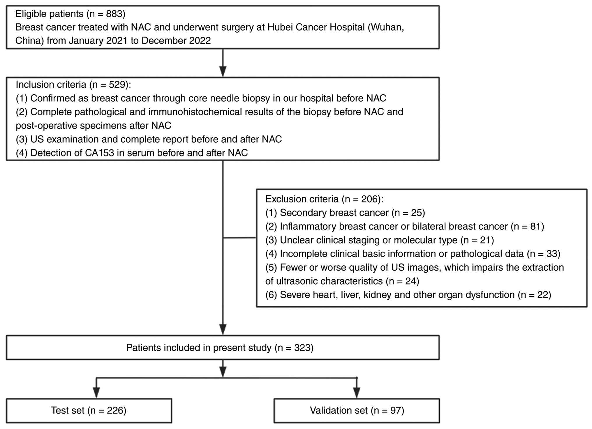
both time points. A total of 206 patients were excluded from the
analysis due to various reasons, including secondary breast cancer,
inflammatory breast cancer or bilateral breast cancer, ambiguous
clinical staging or molecular subtype, incomplete clinical and
pathological data, subpar US images that hindered the extraction of
ultrasonic characteristics and notable dysfunction of the heart,
liver, kidneys or other organs (Fig.
1). It should be noted that the initial protocol preliminarily
estimated a sample size of 200 cases based on historical admission
data. However, the expansion of the Breast Cancer Center led to a
marked rise in patient volume starting in 2021. The present study
ultimately included all consecutive eligible patients within the
predefined study period, yielding a total of 323 included
cases.
US protocol
US evaluations were performed by specialists with ≥5
years of professional experience. Each patient underwent standard
brightness-mode US at the beginning (pre-NAC) and again within 1
week before surgery after completing NAC. Patients received 6–8
cycles of NAC. The timing of the last preoperative US was
determined by the clinical team based on factors such as treatment
response, side effects and surgical scheduling, occurring after the
6th, 7th or 8th cycle. All participants in the present study were
examined using different US scanners and transducer probes,
positioned supine with their arms raised to allow complete
visibility of the breast. These assessments focused on various
parameters, including morphological characteristics, margins,
location (indicated by clock-point numbers and distance from the
skin), maximum tumor diameter, posterior echo pattern, interaction
with adjacent tissues, internal calcifications and blood flow. If
elastography or contrast-enhanced US (CEUS) examinations were
performed, those findings were documented concurrently. These
examinations were conducted at the discretion of the senior
ultrasound physician when additional assessment of tumor
vascularity (CEUS) or stiffness (elastography) was considered
necessary to aid in classification.
Histopathology
All pathological specimens obtained after surgery
were analyzed by two experienced pathologists from Hubei Cancer
Hospital, each with >5 years of experience in the field. The
evaluation utilized the Miller and Payne (MP) method, comparing
core needle biopsy samples taken before chemotherapy and those
collected post-treatment. The assessment of residual tumor cells
following NAC was categorized into five grades: i) G1, no tumor
cell loss; ii) G2, loss <30%; iii) G3, 30–90% reduction; iv) G4,
loss of >90%; and v) G5, absence of malignant cells. According
to the MP grading system (32), a
pathological incomplete response (non-pCR) is characterized by
G1-G4, where tumor cells remain in the tumor bed. A pCR is
classified as G5, indicating no invasive cancer cells are present
at the tumor bed, although ductal carcinoma in situ may
still be identified. Tumor cells were classified as
ER+/PR+ if staining was ≥1%; otherwise, the
tumor cells were considered negative. Immunohistochemistry results
were interpreted as HER2 negative for scores of (−) and (+)
(22,24), while (+++) indicated HER2
positivity. For scores of (++), additional fluorescence in
situ hybridization (FISH) test was performed, with a FISH
result of (+) considered positive and (−) negative. Tissue samples
were retrieved from archived formalin-fixed paraffin-embedded
(FFPE) blocks. Specimens were fixed in 10% neutral-buffered
formalin at room temperature for 24 h. Sections (4 µm) were mounted
on glass slides and subjected to routine hematoxylin-eosin
(H&E) staining at room temperature (hematoxylin for 5 min,
eosin for 1 min). FISH was performed on 4 µm-FFPE sections using
the HER2/CEP17 dual-color FISH probe kit (Cat. No. F.01359-01;
Guangzhou LBP Medicine Science & Technology Co., Ltd.),
according to the manufacturer's instructions. Enzymatic
permeabilization was performed with kit-supplied pepsin at 37°C for
6–10 min, with the incubation duration optimized according to
tissue conditions, followed by post-fix using 10% neutral buffered
formalin at room temperature for 10 min. Briefly, 10 µl of premixed
hybridization buffer containing HER2/CEP17 probes was applied to
tissue sections. Co-denaturation was conducted at 85°C for 5 min,
followed by hybridization at 37°C for 10–18 h in a humidified
chamber. Post-hybridization stringent washing was carried out using
kit-provided buffers. Nuclei were counterstained with DAPI at room
temperature for 5 min. Fluorescence signals were visualized and
captured using an Olympus BX51 fluorescence microscope. Hormone
receptor (HR) positivity was defined as positivity for ER and/or
PR. A Ki-67 index indicating ≥20% positive cells was classified as
high expression, while <20% was classified as low expression
(33). According to the 2023 St.
Gallen consensus (34), breast
cancer was categorized into four subtypes: i) Luminal A
(ER+ and/or PR+, HER2− and Ki-67
<14%); ii) luminal B (ER+ and/or PR+,
HER2− and Ki-67 ≥14% or ER+ and/or
PR+, HER2+ and any Ki-67); iii) HER2
upregulation (ER−/PR−, HER2+ and
any Ki-67); and iv) TNBC (ER−/PR−,
HER2− and any Ki-67).
Biopsy and NAC
Patients underwent a core needle biopsy at Hubei
Cancer Hospital, which pathology confirmed as primary invasive
breast cancer. An immunohistochemical analysis was carried out to
establish the molecular subtype. IHC was performed on 4 µm FFPE
sections. Tissue specimens were fixed in 10% neutral buffered
formalin at room temperature for 24 h and embedded in paraffin.
Antigen retrieval was performed in pre-boiled 0.01 M citrate buffer
(pH 6.0) with a pressure cooker at 121°C; slides were heated for 3
min, cooled for 20 min and rinsed under running tap water, followed
by rehydration in a descending ethanol series. Endogenous
peroxidase activity was blocked with 3% H2O2
for 10 min at room temperature. Sections were incubated overnight
at 4°C with primary antibodies against ER (cat. No. 790-4324;
1:100), PR (cat. No. 790-2223; 1:100), HER2 (all Ventana, Cat. No.
790-4493; 1:200), and Ki-67 (Dako, Cat. No. M7240; 1:100).
HRP-labelled secondary antibody from the EliVision™plus kit
(KIT-9902; Fuzhou Maxim Biotech, China) was applied for 1 h at room
temperature. Immunoreactivity was visualized with DAB, and sections
were counterstained with Mayer's hematoxylin at room temperature
for 1–3 min prior to examination under a light microscope. In cases
where there was notable enlargement of the axillary lymph nodes, a
lymph node aspiration was performed concurrently to assess if it
was due to breast cancer metastasis. All patients included in the
present study received 6–8 cycles of standard NAC, with the
specific regimen tailored by the medical team based on the
immunohistochemical molecular subtype results, following the
Chinese Society of Clinical Oncology guidelines for breast cancer
management (35). Comprehensive
information on regimens, drugs, cycles and distribution of
molecular subtype are provided in Table SI. The most frequently utilized
regimens were doxorubicin + cyclophosphamide followed by docetaxel
(AC-T; 8 cycles) and epirubicin + cyclophosphamide followed by
docetaxel (EC-T; 8 cycles), accounting for 240 patients or 74.3%.
For patients with HER2 positivity, regimens containing trastuzumab
and/or pertuzumab (such as EC-TH, AC-THP, TCHP, TCbHP and TCH/TCbH)
were administered in accordance with the Chinese Society of
Clinical Oncology Breast Cancer guidelines [2021 edition (36) and 2022 edition (35)] available at the time of
treatment.
Clinical data collection from
patients
The present study collected clinical, US and
pathological information, which included age. While the frequency
of monitoring was determined by the judgment of the physician, the
present study documented the maximum tumor diameter and serum CA153
levels during both the initial (baseline) and final (preoperative)
assessments for the present analysis. The present study also noted
US characteristics before NAC, the surgical approach adopted
post-NAC and the initial clinical T and N stages based on the 2017
American Joint Committee on Cancer 8th edition breast cancer TNM
staging criteria (37).
Furthermore, the present study evaluated biopsy pathological data
before NAC, including ER and PR status, HER2 expression, Ki-67
index and the presence of axillary lymph node metastasis.
Post-surgery, the present study recorded pathological data such as
occurrence of vascular or nerve invasion and whether a pCR was
achieved (Fig. 1).
Statistical analysis
For model development and internal validation, the
dataset was divided randomly into a test set (70%) and a validation
set (30%) using a straightforward random allocation technique
[utilizing the SPSS random number generator (version 26.0, IBM
Corp.)] (38). The percentage
reduction in maximum tumor diameter was determined using the
formula: (Initial diameter-preoperative diameter)/initial diameter
×100%. Receiver operating characteristic (ROC) curve analysis was
utilized to ascertain the ideal cut-off point in predicting pCR,
based on the Youden index (sensitivity + specificity-1).
Measurement data are presented as mean ± standard deviation, while
categorical data are indicated as frequencies (%). Normality
assessments were conducted on the measurement data from both the
test and validation set. The unpaired Student's t-test was used for
comparisons of continuous variables between groups. Categorical
data were compared between groups using either the χ2 or
Fisher's exact test. P<0.05 was considered to indicate a
statistically significant difference.
Variable selection
In the test set, the data was categorized according
to the achievement of pCR. A univariate logistic regression
analysis was conducted to investigate the factors associated with
pCR, and variables demonstrating P<0.05 were included into a
multivariate logistic regression analysis. The present analysis
used a forward stepwise selection approach, using the likelihood
ratio method, using an entry threshold of α=0.05 and a removal
threshold of α=0.10; IBM SPSS Statistics (version 26.0; IBM Corp.).
The main variables, including US characteristics, CA153 levels,
molecular subtype and pCR results, had no missing data. However,
elastography and CEUS data, which were only available for certain
subsets, were excluded from the primary model and thus, no
imputation was performed.
Interaction testing
The logistic regression model [using the ‘Enter’
method; IBM SPSS Statistics (version 26.0; IBM Corp.)] was used to
examine two predetermined interactions by incorporating product
terms: i) Molecular subtype in conjunction with US margin; and ii)
the variation in CA153 alongside the variation in tumor diameter.
An interaction of P<0.05 was considered to indicate a
statistically significant difference.
Model evaluation
An odds ratio (OR) >1 suggests an increased
probability of achieving pCR. The assessment of the model was
performed in both the test and validation sets. For the validation
process, the calibration and discrimination were evaluated using
the Hosmer-Lemeshow goodness-of-fit test and the area under the ROC
curve (AUC) (39). To examine
possible confounding effects associated with age and tumor size, a
sensitivity analysis was conducted using forced entry (‘Enter’
method) logistic regression, incorporating these two factors
alongside the initial predictors. In all evaluations, a two-sided
P<0.05 was considered to indicate a statistically significant
difference. Variance inflation factor (VIF) was calculated to
assess multicollinearity between selected variables. All
statistical analyses were carried out using the commercially
available SPSS software (version 26.0; IBM Corp.).
Results
Clinical data of patients
A total of 323 patients were included in the present
study (mean age ± standard deviation, 49.9±10.9 years; range, 21–74
years). Among the present study cohort, 226 patients were assigned
to the test set and 97 patients to the validation set.
Comprehensive clinical, US and pathological information were
collected from all participants (Table
I). The mean serum CA153 levels was recorded at 21.7±25.5 U/ml,
while the maximum tumor diameter was 3.9±2.1 cm. US analysis
revealed that a notable portion of the tumors exhibited irregular
shapes (89.8%; 290/323) and the posterior echoes indicated no
notable changes (59.1%; 191/323), with 29.1% (94/323) displaying
shadowing. The majority of patients in the present study had
axillary lymph node metastasis (81.1%; 262/323). The predominant
molecular subtype identified was luminal B (49.2%; 159/323), with
clinical stages T2 (71.5%; 231/323) and N1 (55.4%; 179/323) being
the most prevalent. Of the 323 patients, 292 (90.4%) underwent
total mastectomy as part of their radical treatment, while 31
(9.6%) opted for breast-conserving surgery after NAC, with 29.7%
(96/323) achieving a pCR. The characteristics of test and
validation sets are detailed in Table
I, demonstrating no statistically significant differences in
the distribution of all the variables between the two groups (all
P>0.05).
 | Table I.Patient and tumor characteristics in
the present study. |
Table I.
Patient and tumor characteristics in
the present study.
|
Characteristics | Total patients
(n=323) | Test set
(n=226) | Validation set
(n=97) | P-value |
|---|
| Age (years), mean ±
SD | 49.9±10.9 | 50.3±10.9 | 49.2±10.9 | 0.424 |
| CA153, U/ml, mean ±
SD | 21.7±25.5 | 22.4±25.5 | 19.9±25.6 | 0.407 |
| Tumor diameter
(cm), mean ± SD | 3.9±2.1 | 3.9±2.1 | 3.8±2.0 | 0.694 |
| Change in tumor
diameter (%), n (%) |
|
|
| 0.821 |
|
<50 | 188 (58.2) | 133 (58.8) | 55 (56.7) |
|
|
≥50 | 135 (41.8) | 93 (41.2) | 42 (43.3) |
|
| Margin, n (%) |
|
|
| 0.871 |
|
Circumscribed | 153 (47.4) | 108 (47.8) | 45 (46.4) |
|
| Not
circumscribed | 170 (52.6) | 118 (52.2) | 52 (53.6) |
|
| Morphological
characteristics, n (%) |
|
|
| 0.715 |
|
Regular | 33 (10.2) | 24 (10.6) | 9 (9.3) |
|
|
Irregular | 290 (89.8) | 202 (89.4) | 88 (90.7) |
|
| Posterior echo
pattern, n (%) |
|
|
| 0.985 |
| No
posterior features | 191 (59.1) | 133 (58.8) | 58 (59.8) |
|
|
Enhancement | 38 (11.8) | 27 (11.9) | 11 (11.3) |
|
|
Shadow | 94 (29.1) | 66 (29.3) | 28 (28.9) |
|
| Calcification, n
(%) |
|
|
| 0.780 |
|
Yes | 148 (45.8) | 104 (46.0) | 44 (44.3) |
|
| No | 175 (54.2) | 122 (54.0) | 53 (55.7) |
|
| Twisted perforating
vessels, n (%) |
|
|
| 0.742 |
|
Yes | 153 (47.4) | 105 (46.4) | 48 (49.5) |
|
| No | 170 (52.6) | 121 (53.6) | 49 (50.5) |
|
| Axillary lymph node
metastasis, n (%) |
|
|
| 0.645 |
|
Yes | 262 (81.1) | 183 (80.9) | 79 (81.4) |
|
| No | 61 (18.9) | 43 (19.1) | 18 (18.6) |
|
| Change in CA153
levels, n (%) |
|
|
| 0.913 |
|
Increase | 160 (49.5) | 111 (49.1) | 49 (50.5) |
|
|
Decrease | 163 (50.5) | 115 (50.9) | 48 (49.5) |
|
| Surgical method, n
(%) |
|
|
| 0.309 |
|
Breast-conserving | 31 (9.6) | 21 (9.3) | 10 (10.3) |
|
|
Radical | 292 (90.4) | 205 (90.7) | 87 (89.7) |
|
| Clinical T stage
(%) |
|
|
| 0.793 |
| 1 | 20 (6.2) | 12 (5.3) | 8 (8.2) |
|
| 2 | 231 (71.5) | 163 (72.1) | 68 (70.1) |
|
| 3 | 48 (14.9) | 35 (15.5) | 13 (13.5) |
|
| 4 | 24 (7.4) | 16 (7.1) | 8 (8.2) |
|
| Clinical N stage
(%) |
|
|
| 0.839 |
| 0 | 61 (18.9) | 43 (19.0) | 18 (18.6) |
|
| 1 | 179 (55.4) | 124 (54.9) | 55 (56.7) |
|
| 2 | 47 (14.6) | 33 (14.6) | 14 (14.4) |
|
| 3 | 36 (11.1) | 26 (11.5) | 10 (10.3) |
|
| Vascular/nerve
invasion (%) |
|
|
| 0.913 |
| No | 195 (60.4) | 136 (60.2) | 59 (60.8) |
|
|
Yes | 128 (39.6) | 90 (39.8) | 38 (39.2) |
|
| Hormone receptor, n
(%) |
|
|
| 0.372 |
|
Negative | 185 (57.3) | 125 (55.3) | 60 (61.9) |
|
|
Positive | 138 (42.7) | 101 (44.7) | 37 (38.1) |
|
| HER2 (%) |
|
|
| 0.101 |
|
Negative | 251 (77.7) | 175 (77.4) | 76 (78.4) |
|
|
Positive | 72 (22.3) | 51 (22.6) | 21 (21.6) |
|
| Ki-67 levels, n
(%) |
|
|
| 0.372 |
|
Low | 80 (24.8) | 58 (25.7) | 22 (22.7) |
|
|
High | 243 (75.2) | 168 (74.3) | 75 (77.3) |
|
| Molecular type, n
(%) |
|
|
| 0.272 |
| Luminal
A | 23 (7.1) | 16 (7.1) | 7 (7.2) |
|
| Luminal
B | 159 (49.2) | 109 (48.2) | 50 (51.6) |
|
|
HER2 | 72 (22.3) | 51 (22.6) | 21 (21.6) |
|
|
TNBC | 69 (21.4) | 50 (22.1) | 19 (19.6) |
|
| pCR, n (%) |
|
|
|
|
|
Yes | 96 (29.7) | 68 (30.1) | 28 (28.8) |
|
| No | 227 (70.3) | 158 (69.9) | 69 (71.2) |
|
Univariate analysis in test set
In the test set (n=226), 30.1% (68/226) of patients
achieved a pCR. Table II
illustrates the comparisons between patients in the pCR and non-pCR
groups. Tumor size reduction and the age of patients were
categorized based on the cut-off values derived from the Youden
index. The determined optimal cut-off value of tumor size reduction
was 48.5% (with sensitivity at 0.892 and specificity at 0.882,
yielding a Youden index of 0.774), which was rounded to 50% for
practical application (data not shown). Among the pCR group (n=68),
22.1% (n=15) of them were aged <46 years of age and 91.2% (n=62)
had tumors ≥2.1 cm. By contrast, in the non-pCR group, 33.5%
(n=53/158) were aged <46 years, with a notable majority (88.6%;
140/158) having tumor diameters >2.1 cm. However, these
differences were not statistically significant (all P>0.05). A
reduction in maximum diameter by ≥50% was more frequently observed
in the pCR group (73.5%; 50/68), while the non-pCR group
predominantly exhibited diameter changes <50% (72.8%; 115/158),
with these differences being statistically significant
(P<0.001). In the pCR group, the most prevalent US features in
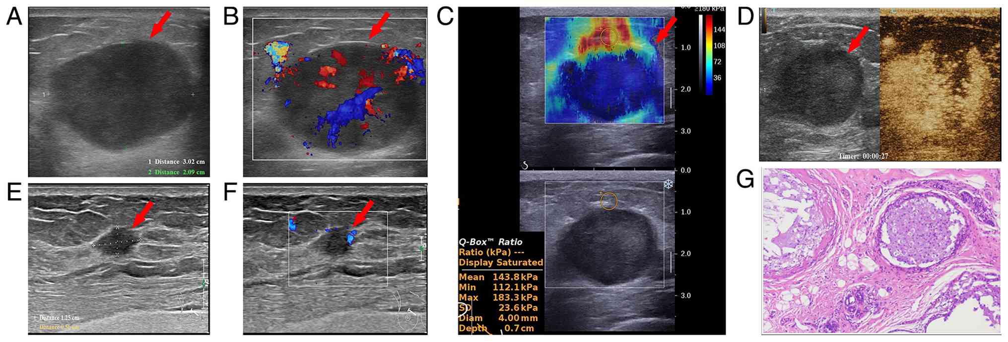
the pCR group was a circumscribed margin (82.3%; 56/68; Fig. 2), whereas only 32.9% (52/158) of the
non-PCR group displayed this characteristic (Fig. 3), indicating a statistically
significant difference (P<0.001). Both in the pCR and non-PCR
group, the absence of posterior features was common; however,
tumors with post-echo attenuation were significantly more frequent
in the non-pCR group (34.2%; 54/158) compared with the pCR group
(17.6%; 12/68) (P=0.024. No significant differences were identified
in morphological characteristics, twisted perforating vessels or
calcification between the two groups (all P>0.05). Clinical N1
status (P=0.014 and a reduction in serum CA153 levels post-NAC
(P=0.037) were significantly more prevalent in the pCR group (both
73.5%; 50/68) than in the non-pCR group (46.8%, 74/158 and 41.2%,
65/158). The pCR group also demonstrated elevated Ki-67 levels
(83.8%, 57/68) and higher proportion of negative hormone receptors
(ER− and PR−; 64.8%, 44/68) (P=0.035 and
P=0.006, respectively; Fig. S1)
compared with the non-pCR group (70.2%, 111/158 and 36.1%, 57/158).
Luminal B (55.7%, 88/158) and HER2 (21.5%, 34/158) were the most
common subtypes in the non-pCR group (Fig. S2), while TNBC (39.7%, 27/68) and
luminal B (30.8%, 21/68) subtypes were the most common subtypes in
the pCR group. The overall distribution differed significantly
(P=0.021). No significant differences were noted in clinical T
stage or HER2 expression between the two groups (all
P>0.05).
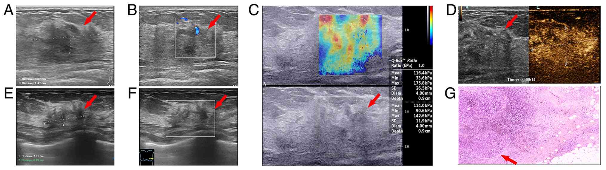 | Figure 3.A 40-year-old female patient with
invasive breast cancer, clinical T2N2M0, biopsy pathology: Estrogen
receptor (+), progesterone receptor (+), human epidermal growth
factor receptor 2 (+) and high expression of Ki-67. US examination
before treatment: Size of 3.07×1.47 cm, 3 o'clock in the left
breast, mass with dis-circumscribed margin and irregular shape. (A
and B) Color Doppler flow imaging revealed no perforator vessels.
(C) Representative SWE image of breast lesion. The mean stiffness
of the mass was 116 kPa, corresponding to the mean Young's modulus,
indicating relatively stiff tissue. (D) Contrast-enhanced US
examination with Sonovue: The tumors indicated heterogeneous
hyperenhancement, and the range did not expand significantly. (E)
After 8 cycles of TCbHP regimen (docetaxel + carboplatin +
trastuzumab + pertuzumab), the tumor size was 2.01×0.69 cm. (F) No
obvious blood flow signals were detected on CDFI. (G) After total
left mastectomy and axillary lymph node dissection, H&E
staining showed a moderate response (the original breast lesion of
the Miller-Payne grading system: grade 3) without neurovascular
invasion. Magnification: ×200). The red arrow indicates the lesion.
SWE, shear-wave elastography. |
| Table II.Univariate analysis of patients in
the test set (n=226) classified into pCR and non-pCR groups. |
Table II.
Univariate analysis of patients in
the test set (n=226) classified into pCR and non-pCR groups.
|
Characteristics | Total, n (%) | PCR (n=68), n
(%) | Non-PCR (n=158), n
(%) | B value | OR | 95% CI | P-value |
|---|
| Age, years |
|
|
|
|
|
|
|
|
≥46 | 158 (69.9) | 53 (77.9) | 105 (66.5) |
| Reference |
|
|
<46 | 68 (30.1) | 15 (22.1) | 53 (33.5) | −0.579 | 0.561 | 0.289–1.087 | 0.087 |
| Tumor diameter,
cm |
|
|
|
|
|
|
|
|
≥2.1 | 202 (89.4) | 62 (91.2) | 140 (88.6) |
| Reference |
|
|
<2.1 | 24 (10.6) | 6 (8.8) | 18 (11.4) | 0.284 | 1.329 | 0.503–3.509 | 0.566 |
| Change in tumor
diameter, % |
|
≥50 | 93 (41.2) | 50 (73.5) | 43 (27.2) |
| Reference |
|
|
<50 | 133 (58.8) | 18 (26.5) | 115 (72.8) | −2.005 | 0.135 | 0.071–0.256 |
<0.001a |
| Margin |
|
|
|
|
|
|
|
| Not
circumscribed | 118 (52.2) | 12 (17.6) | 106 (67.1) |
| Reference |
|
|
Circumscribed | 108 (47.8) | 56 (82.4) | 52 (32.9) | 2.253 | 9.513 | 4.694–19.277 |
<0.001a |
| Morphological
characteristics |
|
|
|
|
|
|
|
|
Irregular | 202 (89.4) | 62 (91.2) | 140 (88.7) |
| Reference |
|
|
Regular | 24 (10.6) | 6 (8.8) | 18 (11.3) | 0.284 | 1.329 | 0.503–3.508 | 0.566 |
| Posterior echo
pattern |
|
|
|
|
|
| 0.038a |
| No
posterior features | 133 (58.8) | 45 (66.2) | 88 (55.7) |
| Reference |
|
|
Enhancement | 27 (11.9) | 11 (16.2) | 16 (10.1) | 0.296 | 1.344 | 0.576–3.138 | 0.494 |
|
Shadow | 66 (29.3) | 12 (17.6) | 54 (34.2) | −0.833 | 0.453 | 0211-0.894 | 0.024a |
| Twisted perforating
vessels |
|
Yes | 105 (46.4) | 31 (45.5) | 74 (46.8) |
| Reference |
|
| No | 121 (53.6) | 37 (54.5) | 84 (53.2) | −0.05 | 0.951 | 0.538–1.682 | 0.863 |
| Calcification |
|
Yes | 104 (46.0) | 30 (44.1) | 74 (46.8) |
| Reference |
|
| No | 122 (54.0) | 38 (55.9) | 84 (53.2) | −0.110 | 0.896 | 0.506–1.587 | 0.707 |
| Change in CA153
levels |
|
|
|
|
|
|
|
|
Increase | 111 (49.1) | 18 (26.5) | 93 (58.8) |
| Reference |
|
|
Decrease | 115 (50.9) | 50 (73.5) | 65 (41.2) | 0.663 | 1.941 | 1.039–3.627 | 0.037a |
| Axillary lymph node
metastasis |
|
|
|
|
|
|
|
|
Yes | 183 (80.9) | 60 (88.3) | 123 (77.8) |
| Reference |
|
| No | 43 (19.1) | 8 (11.7) | 35 (22.2) | 0.05 | 1.051 | 0.509–2.174 | 0.892 |
| Clinical T
stage |
|
|
|
|
|
| 0.447 |
| 4 | 16 (7.1) | 3 (4.4) | 13 (8.2) |
| Reference |
|
| 1 | 12 (5.3) | 3 (4.4) | 9 (5.8) | 0.368 | 1.444 | 0.236–8.844 | 0.691 |
| 2 | 163 (72.1) | 54 (79.4) | 109 (68.9) | 0.764 | 2.147 | 0.587–7.854 | 0.248 |
| 3 | 35 (15.5) | 8 (11.8) | 27 (17.1) | 0.250 | 1.284 | 0.281–5.656 | 0.741 |
| Clinical N
stage |
|
|
|
|
|
| 0.016a |
| 3 | 26 (11.5) | 3 (4.4) | 23 (14.6) |
| Reference |
|
| 0 | 43 (19.0) | 8 (11.8) | 35 (22.2) | 0.966 | 2.629 | 0.668–10.346 | 0.167 |
| 1 | 124 (54.9) | 50 (73.5) | 74 (46.8) | 1.583 | 4.869 | 0.385–17.123 | 0.014a |
| 2 | 33 (14.6) | 7 (10.3) | 26 (16.4) | 0.671 | 1.769 | 0.397–7.891 | 0.455 |
| Ki-67 levels |
|
|
|
|
|
|
|
|
High | 168 (74.3) | 57 (83.8) | 111 (70.2) |
| Reference |
|
|
Low | 58 (25.7) | 11 (16.2) | 47 (29.8) | −0.786 | 0.456 | 0.220–0.946 | 0.035a |
| HER2 |
|
|
|
|
|
|
|
|
Negative | 175 (77.4) | 51 (74.9) | 124 (78.5) |
| Reference |
|
|
Positive | 51 (22.6) | 17 (25.1) | 34 (21.5) | 0.352 | 1.422 | 0.711–2.801 | 0.308 |
| Hormone
receptor |
|
|
|
|
|
|
|
|
Negative | 125 (55.3) | 44 (64.8) | 57 (36.1) |
| Reference |
|
|
Positive | 101 (44.7) | 24 (35.2) | 101 (63.9) | −0.808 | 0.446 | 0.250–0.795 | 0.006a |
| Molecular type |
|
|
|
|
|
| 0.021a |
| Luminal
A | 16 (7.1) | 3 (4.4) | 13 (8.2) |
| Reference |
|
| Luminal
B | 109 (48.2) | 21 (30.8) | 88 (55.7) | 0.285 | 1.330 | 0.353–5.014 | 0.674 |
|
HER2 | 51 (22.6) | 17 (25.1) | 34 (21.5) | 0.773 | 2.167 | 0.543–8.645 | 0.273 |
|
TNBC | 50 (22.1) | 27 (39.7) | 23 (14.6) | 1.375 | 3.957 | 0.988–15.850 | 0.052 |
Multivariate logistic regression
analysis in test set
In the present study, variables with P<0.05 from
the univariate analysis were incorporated into the multivariate
logistic regression analysis. The results of the multivariate
analysis (Table III) indicated
that, in the US evaluation, a tumor diameter reduction ≥50%
(adjusted OR, 5.197; 95%CI, 1.635–16.518) and a circumscribed
margin (adjusted OR, 9.157; 95%CI, 4.242–19.765) are significantly
associated with pCR (Figs. 2 and
3). Additional independent
predictors of pCR included molecular subtype, with the TNBC subtype
indicating a significant independent association with pCR when
compared with luminal A (adjusted OR, 4.262; 95%CI, 1.647–11.028).
A reduction in CA153 levels (adjusted OR, 2.227; 95%CI,
1.061–4.675) also demonstrated a significant relationship with pCR
(all P<0.05).
| Table III.Multivariate regression analysis in
the test set (n=226) and development of predictive model for
NAC. |
Table III.
Multivariate regression analysis in
the test set (n=226) and development of predictive model for
NAC.
|
Characteristics | B value | OR | 95% CI | P-value |
|---|
| Change in tumor
diameter, % |
|
<50 |
| Reference |
|
|
≥50 | 1.648 | 5.197 | 1.635–16.518 | 0.005a |
| Change in CA153
levels |
|
Increase |
| Reference |
|
|
Decrease | 0.801 | 2.227 | 1.061–4.675 | 0.034a |
| Margin |
| Not
circumscribed |
| Reference |
|
|
Circumscribed | 2.214 | 9.157 | 4.242–19.765 |
<0.001a |
| Molecular type |
|
|
| 0.027a |
| Luminal
A |
| Reference |
|
| Luminal
B | 0.417 | 1.518 | 0.604–3.813 | 0.374 |
|
HER2 | 0.905 | 2.472 | 0.512–11.941 | 0.260 |
|
TNBC | 1.450 | 4.262 | 1.647–11.028 | 0.003a |
Sensitivity analyses that adjusted for age and tumor
size categories did not markedly alter the ORs (Table SII) for the AUC of the present
study model (Fig. S3). To further
determine whether a circumscribed margin serves only as a
substitute for TNBC, stratified and interaction analyses were
performed (Table SIII). In the
non-TNBC subgroup (Luminal+/HER2+; n=176),
the OR for a circumscribed margin was 1.827 [OR=1.827; 95%
confidence interval (CI), 1.015–4.389; P=0.067]; for the TNBC
subgroup (luminal−/HER2−; n=50), the OR was
1.875 (95% CI, 0.482–7.372; P=0.133). Interaction tests
demonstrated no significant modification effects from molecular
subtype × margin (P=0.667) or CA153 × tumor diameter reduction
(P=0.42), suggesting that the influence of margin on pCR did not
differ significantly between patients with TNBC and without TNBC.
To evaluate the predictive value of conventional B-mode US, we
found that among patients with a ≥50% size reduction, 27% had
residual disease on pathology, corresponding to a false-positive
rate of 27%. To explain the inconsistent significance of cN stage
between univariate and multivariate analyses, collinearity testing
was performed for cN stage and the ratio of tumor diameter
reduction. Moderate multicollinearity was observed (VIF=3.713 for
cN stage; VIF=3.554 for ≥50% diameter reduction), implying
overlapping predictive information for pCR.
Development and validation of the
combined model
The integrated multivariate prediction model
utilized the adjusted ORs derived from the multivariable analysis
(Table III). The present study
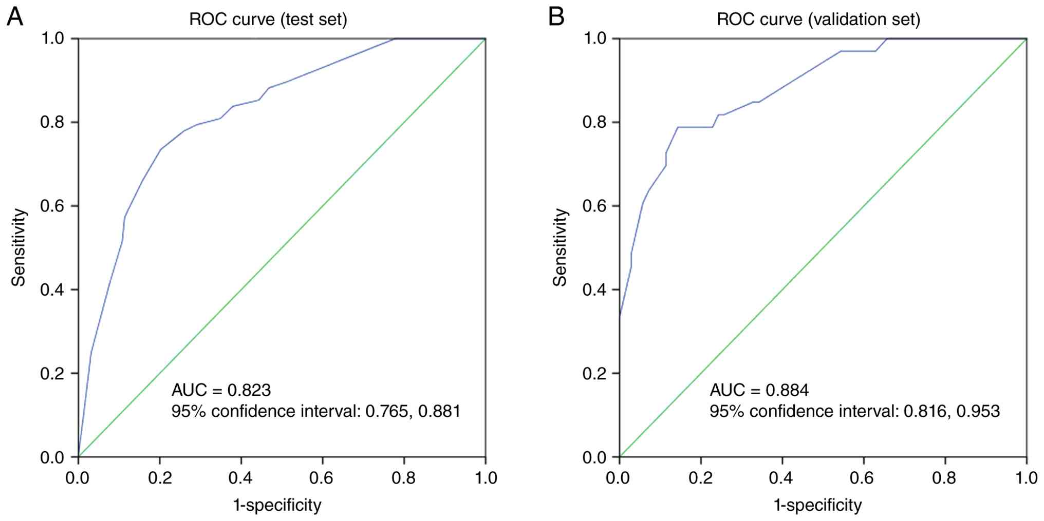
model demonstrated notable predictive capability (AUC, 0.823; 95%
CI, 0.765–0.881) and effective calibration [Hosmer-Lemeshow
goodness-of-fit test: χ2=2.779; degrees of freedom
(df)=7; P=0.905] within the test set. Furthermore, it demonstrated
robust predictive performance for pCR (AUC, 0.884; 95% CI,
0.816–0.953) and calibration ability (Hosmer-Lemeshow
goodness-of-fit test: χ2=4.298; df=7; P=0.745) in the
validation set (Fig. 4).
Discussion
As breast cancer cases continue to increase
globally, NAC has become a key part of treatment plans. Ring et
al (40) suggested that
patients who reach a pCR after NAC may be eligible for medical
management only, sparing them from the discomfort of surgery.
Furthermore, understanding the internal bioinformatics of the tumor
through treatment responses could lead to improved therapeutic
approaches that enhance patient outcomes (8,41).
Several studies indicated that achieving pCR is a key indicator of
long-term survival for patients undergoing NAC (42,43).
However, it is key to ensure the precision of pCR evaluations, as
inaccuracies could result in notable risks for patients. Thus,
establishing a reliable clinical method to assess the biological
responses to NAC before any surgical procedures is key to guiding
individualized treatment decisions.
The current progress in US diagnostic technology has
markedly enhanced the importance of US assessments in determining
the effectiveness of NAC for breast cancer (21). Several US techniques, including
two-dimensional US, elastography and CEUS, offer diverse insights
into the dimensions, shape, vascularity and stiffness of the tumor
(44). US is favored in clinical
settings due to its non-ionizing nature and patient-friendly
profile. Furthermore, two-dimensional US allows for direct
measurement and evaluation of tumor size. However, previous
research indicated that changes in tumor size alone provide limited
specificity and accuracy in assessing NAC effectiveness.
Chemotherapy may eliminate tumor cells without visibly altering the
tumor size, and residual cancerous tissue can often appear similar
to normal breast tissue on standard US scans (16,45).
By contrast, continuous monitoring of the maximum tumor diameter,
along with post-echo features and morphology, can effectively
predict NAC response (46–48). The present study indicated that a
reduction of ≥50% in maximum tumor diameter is a robust independent
predictor of pCR, aligning with previous findings (46,49).
Furthermore, the present study results indicated that tumor margins
and posterior echo characteristics associate with pCR in univariate
analysis, while circumscribed margins were a significant
independent predictor in multivariate analysis. The association
between tumor margins and pCR has not been explored in recent
literature, to the best of our knowledge. The present study
suggested that tumors with circumscribed margins are more likely to
achieve pCR (P<0.001), potentially due to the varying margins
exhibited by different breast cancer molecular subtypes. Previous
studies (46,50,51)
demonstrated that the majority of TNBC cases display circumscribed
margins on US. Furthermore, compared with other molecular subtypes,
TNBC is typically associated with a higher histological grade,
rapid cell proliferation, increased sensitivity to NAC and a
greater likelihood of achieving pCR (30,50,51).
The present study identified circumscribed margins
as a significant predictor of pCR. While this characteristic was
predominantly observed in TNBC, the interaction tests and
stratified evaluations indicated no notable variation in its impact
across different molecular subtypes, implying that the predictive
significance of circumscribed margins may not be exclusively
associated with TNBC. However, the P-values from the stratified
evaluations did not achieve statistical significance (both
P>0.05); this lack of significance is likely attributed to a
small sample size (only 11 patients without circumscribed margins
in the TNBC group) and diminished statistical power. Therefore,
further extensive prospective research is warranted to validate the
present study results.
The univariate analysis in the present study
revealed that posterior echo enhancement was a positive indicator
in achieving pCR, whereas posterior echo attenuation was associated
with a lower likelihood of pCR. The present results indicated that
tumors classified as low-grade are more prone to displaying
characteristics of posterior echo attenuation. These tumors,
characterized by their gradual growth, reduced metastatic potential
and clinical stability, tend to respond inadequately to NAC. This
finding accounts for the diminished chances of reaching pCR when
posterior echo attenuation is observed via US (52). Nonetheless, posterior echo
enhancement was excluded from the final model, indicating a need
for validation with a larger sample size in future research. The
processes of tumor growth, tumor cell invasion and metastasis in
breast cancer are associated with the formation of new blood
vessels, particularly microvascular structures (53,54).
Alterations in these micro-vessels following NAC often occur before
any morphological changes are noted (55–57).
The present study findings indicated that the presence of twisted
perforating vessels does not markedly impact the effectiveness of
NAC. However, color Doppler flow imaging has limited ability to
detect micro-vessels due to challenges in visualizing those with
slower flow rates and smaller diameters (58). This limitation may result in
underestimating the blood supply of the tumor, whereas CEUS
effectively visualizes blood flow within tumors. With advancements
in elastography technology, previous studies have observed that
drug-induced changes in the state of tumors and adjacent tissues
can lead to variations in tumor stiffness following NAC (59–61).
Furthermore, several studies have reported that higher stiffness
values in breast tumors are associated with worse prognoses
(62). In the present study, only a
limited number of patients underwent elastography and CEUS
assessments. However, with the introduction of novel technologies
in the Department of Medical Ultrasound, Hubei Cancer Hospital,
there has been a notable enhancement in diagnostic accuracy using
elastography, CEUS and superb microvascular imaging (SMI).
Therefore, data from these innovative technologies may be
integrated in future research.
TM serve a key role in indicating the presence and
development of tumors. Traditionally, TM have been utilized mainly
for the early detection of tumors and assessing prognosis (63–65).
Application of TM has expanded to include the evaluation of
pathological responses following NAC (27,29,66).
Several studies have demonstrated a positive association between
CA153 levels and cellular proliferation rates (67,68).
Furthermore, elevated serum CA153 levels are associated with
advanced stages of breast cancer, and patients exhibiting high
CA153 levels are often associated with a worse prognosis. Previous
studies reported that CA153 levels are markedly specific indicators
of breast cancer recurrence and metastasis (25,26,69).
Therefore, healthcare professionals commonly monitor CA153 levels
to assess treatment effectiveness and predict outcomes in patients
with breast cancer. In line with previous research (25,27,28,66),
the present study results indicated that a higher percentage of
patients in the pCR group demonstrated a reduction in CA153 levels
compared with those in the non-pCR group. Multivariate logistic
regression analysis suggested that a decrease in CA153 levels
serves as a positive predictor in achieving pCR.
The growth rate of breast cancer cells is a key
molecular marker for the disease, and Ki-67 levels are closely
associated with its clinicopathological features (70). Several studies indicated that
variations in Ki-67 levels before and after NAC are associated with
treatment efficacy in patients with breast cancer. Notably,
patients with elevated Ki-67 levels prior to NAC demonstrated an
improved response to the treatment, leading to enhanced outcomes
compared with patients with lower Ki-67 levels (71–73).
In the present analysis, a higher percentage of patients with
elevated Ki-67 levels were identified in the pCR group compared
with those without pCR. These findings are consistent with a
previous study conducted by Wong et al (74). However, although Ki-67 levels were
entered into the multivariate analysis, it was not retained in the
final model. This may be because its predictive information was
largely captured by molecular subtype, which are associated with
Ki-67 expression and were ultimately selected in the model. Among
the biological markers associated with breast cancer, hormone
receptors (ER and PR) and HER2 are particularly key to treatment
and prognosis assessment (75).
Patients with high HER2 levels benefit more from NAC compared with
those with lower levels (76,77).
Furthermore, Cortazar et al (41), reported that the pCR rate in
patients lacking hormone receptors was 4.2 times greater compared
with that in patients who were hormone receptor-positive,
indicating that the luminal subtype, which is hormone
receptor-positive, has a less favorable response to NAC compared
with HER2-upregulating and TNBC subtypes, making pCR harder to
achieve (78). Furthermore,
molecular subtypes respond differently to NAC, with triple-negative
and HER2+ tumors demonstrating a more robust response
(79–81). In multivariate logistic regression
analysis of the present study, molecular subtype emerged as a key
independent predictor of pCR. Specifically, TNBC was significantly
associated with a higher likelihood of pCR compared with the
luminal subtype.
The present study revealed notable variations in
tumor diameter, margin characteristics, posterior echo patterns,
CA153 levels, clinical N stage, hormone receptor status, Ki-67
expression and molecular subtypes between the pCR and non-pCR
groups. Using multivariate analysis, the present study identified
tumor diameter change, margin, CA153 fluctuations and molecular
subtypes as key components of the final predictive model, which was
subsequently validated on the validation set. These findings
suggested a promising clinical framework in predicting the response
to NAC prior to surgery. Previous research indicated that earlier N
stage is associated with increased benefit from NAC and a higher
likelihood of achieving a pCR (82). However, clinical N stage was not
retained in the final multivariate model of the present study.
Therefore, the present study performed collinearity diagnostics.
The results revealed moderate collinearity between clinical node
(cN) stage and ratio of tumor diameter reduction. These findings
suggested that cN stage and diameter reduction carry notable
overlapping predictive information for pCR. Several studies have
explored the use of radiomics in predicting pCR in patients with
breast cancer, demonstrating promising predictive capabilities
(83). Compared with MRI, US has
been reported to be more suitable for breast cancer screening
because it is simpler to perform, faster and free of ionizing
radiation (84). Certain studies
have proposed using US images from different periods after
treatment to predict pCR (85,86).
Nonetheless, accurately capturing and representing tumor images
following NAC poses notable challenges. Thus, for patients in the
present study, the tumors were marked on the skin prior to
treatment.
In summary, the present study developed an
integrated model utilizing US, serum CA153 levels and molecular
classifications, demonstrating notable potential in assessing the
effectiveness of treatment prior to surgery following NAC. However,
several limitations should be noted. Firstly, elastography and CEUS
were only conducted at the initial stage (before NAC) to evaluate
tumor characteristics and for Breast Imaging Reporting and Data
System categorization (87),
without being repeated during the pre-surgical evaluation.
Therefore, the present study cannot determine the added benefit of
these advanced US methods in lowering the false-positive rate
compared with standard B-mode US. In the present study, the
conventional B-mode US alone had a false-positive rate of 27% in
detecting notable responses (>50% reduction in size). This
indicated that with a >50% reduction in tumor size observed via
US, >25% of patients may still have residual disease. Secondly,
the present study model exclusively predicts breast pCR and does
not evaluate the status of axillary lymph nodes, thus it should not
inform decisions regarding axillary de-escalation. Several studies
have demonstrated that alterations in axillary lymph node US
characteristics, including cortical thickness, hilar integrity and
short-axis reduction rate, are predictive of axillary pCR (88–90).
Although the present study did not include this data, the findings
provide a rationale for future investigations aimed at optimizing
the model by incorporating changes in axillary US features. In the
present study, treatment regimens varied; however, no significant
pCR differences were identified among NAC regimens within each
subtype (all P>0.05). This finding suggested that pCR was driven
more by tumor biology rather than by regimen choice, consistent
with previous meta-analyses (79,91,92).
The present study model appeared robust to regimen variation.
However, subtle regimen-specific effects (for example, dose or
schedule) that affect imaging phenotypes cannot be excluded.
Group-level comparisons may still be subject to residual
confounding (for example, dose reductions and delays). Future
validation in uniformly treated cohorts is warranted. Furthermore,
the retrospective nature of the present study may lead to selection
bias, and the limited sample size, particularly within the pCR
subgroup, could affect the validity of subgroup analysis
conclusions. Future prospective studies with larger cohorts are
warranted to confirm the clinical applicability of the predictive
model in the present study. To mitigate this limitation, a
prospective study where all participants will undergo CEUS both at
the initial assessment and again after completing NAC but before
surgery is currently in progress in Hubei Cancer Hospital. Although
data collection and analysis are still ongoing, preliminary
observations suggest that CEUS may provide a precise identification
of tumor margins and residual viable tissue by evaluating
microvascular perfusion (data not published). This prospective
study is expected to quantify the advantages of CEUS in decreasing
the false-positive rate, thus offering a more dependable imaging
foundation in ensuring safe margins in breast-conserving surgeries.
With ongoing research, an increasing number of molecular markers
associated with breast cancer prognosis are being identified.
Future research may incorporate additional tumor molecular markers
and US imaging parameters, including CEUS, elastography and SMI, to
refine the present study model and enhance predictive accuracy of
NAC response, offering clinicians evidence-based guidance for
treatment decisions.
Supplementary Material
Supporting Data
Supporting Data
Acknowledgements
Not applicable.
Funding
The present study was supported by National Cancer Center
Climbing Foundation (grant no. NCC201917) and National Key Clinical
Specialty Construction Discipline (grant no. HBCHBCC-D06).
Availability of data and materials
The data generated in the present study may be
requested from the corresponding author.
Authors' contributions
The present study was conceived and designed by SZ.
MD acquired, analyzed and interpreted the data. MD drafted the
manuscript and SZ edited the manuscript. Both authors read and
approved the final manuscript. MD and SZ confirm the authenticity
of all the raw data.
Ethics approval and consent to
participate
The present retrospective study received approval
from the Ethics Committee of Hubei Cancer Hospital (approval no.
2020-KYLL-S08; Wuhan, China) prior to the initiation of data
extraction. All data were retrospectively retrieved from archived
medical records after the included patients had completed their
full treatment courses and the requirement for written informed
consent was waived by the ethics committee for the use of
de-identified clinical data, ultrasound images and pathological
staining/fluorescence images.
Patient consent for publication
Not applicable.
Competing interests
The authors declare that they have no competing
interests.
References
|
1
|
GBD 2023 Breast Cancer Collaborators, .
Global, regional, and national burden of breast cancer among
females, 1990–2023, with forecasts to 2050: A systematic analysis
for the Global Burden of Disease Study 2023. Lancet Oncol.
27:302–326. 2026. View Article : Google Scholar : PubMed/NCBI
|
|
2
|
Siegel RL, Miller KD and Jemal A: Cancer
statistics, 2017. CA Cancer J Clin. 67:7–30. 2017.PubMed/NCBI
|
|
3
|
Kaufmann M, von Minckwitz G, Bear HD,
Buzdar A, McGale P, Bonnefoi H, Colleoni M, Denkert C, Eiermann W,
Jackesz R, et al: Recommendations from an international expert
panel on the use of neoadjuvant (primary) systemic treatment of
operable breast cancer: New perspectives 2006. Ann Oncol.
18:1927–1934. 2007. View Article : Google Scholar : PubMed/NCBI
|
|
4
|
Morrow M, Jagsi R, Alderman AK, Griggs JJ,
Hawley ST, Hamilton AS, Graff JJ and Katz SJ: Surgeon
recommendations and receipt of mastectomy for treatment of breast
cancer. JAMA. 302:1551–1556. 2009. View Article : Google Scholar : PubMed/NCBI
|
|
5
|
Gulis K, Ellbrant J, Svensjö T, Skarping
I, Vallon-Christersson J, Loman N, Bendahl PO and Rydén L: A
prospective cohort study identifying radiologic and tumor related
factors of importance for breast conserving surgery after
neoadjuvant chemotherapy. Eur J Surg Oncol. 49:1189–1195. 2023.
View Article : Google Scholar : PubMed/NCBI
|
|
6
|
Buchholz TA, Mittendorf EA and Hunt KK:
Surgical considerations after neoadjuvant chemotherapy: Breast
conservation therapy. J Natl Cancer Inst Monogr. 2015:11–14. 2015.
View Article : Google Scholar : PubMed/NCBI
|
|
7
|
Gralow JR, Burstein HJ, Wood W, Hortobagyi
GN, Gianni L, von Minckwitz G, Buzdar AU, Smith IE, Symmans WF,
Singh B, et al: Preoperative therapy in invasive breast cancer:
Pathologic assessment and systemic therapy issues in operable
disease. J Clin Oncol. 26:814–819. 2008. View Article : Google Scholar : PubMed/NCBI
|
|
8
|
Masuda N, Lee SJ, Ohtani S, Im YH, Lee ES,
Yokota I, Kuroi K, Im SA, Park BW, Kim SB, et al: Adjuvant
capecitabine for breast cancer after preoperative chemotherapy. N
Engl J Med. 376:2147–2159. 2017. View Article : Google Scholar : PubMed/NCBI
|
|
9
|
Spring LM, Fell G, Arfe A, Sharma C,
Greenup R, Reynolds KL, Smith BL, Alexander B, Moy B, Isakoff SJ,
et al: Pathologic complete response after neoadjuvant chemotherapy
and impact on breast cancer recurrence and survival: A
comprehensive Meta-analysis. Clin Cancer Res. 26:2838–2848. 2020.
View Article : Google Scholar : PubMed/NCBI
|
|
10
|
Krishnan Y, Alawadhi SA, P SS, Gopal M and
Thuruthel S: Pathological responses and long-term outcome analysis
after neoadjuvant chemotheraphy in breast cancer patients from
Kuwait over a period of 15 years. Ann Saudi Med. 33:443–450. 2013.
View Article : Google Scholar : PubMed/NCBI
|
|
11
|
Rouzier R, Perou CM, Symmans WF, Ibrahim
N, Cristofanilli M, Anderson K, Hess KR, Stec J, Ayers M, Wagner P,
et al: Breast cancer molecular subtypes respond differently to
preoperative chemotherapy. Clin Cancer Res. 11:5678–5685. 2005.
View Article : Google Scholar : PubMed/NCBI
|
|
12
|
Gorzelak-Magiera A, Kabut J, Sadurska J,
Długaszek A, Domagała-Haduch M, Szot A and Gisterek-Grocholska I:
Pathological complete response after neoadjuvant chemotherapy in
breast cancer: A literature overview. Cancers (Basel). 18:17182026.
View Article : Google Scholar : PubMed/NCBI
|
|
13
|
Hashmi AA, Bukhari U, Najam J, Dowlah T,
Ali AH, Diwan MA, Anjali F, Sham S, Zia S and Irfan M: Luminal B,
human epidermal growth factor receptor 2 (HER2/neu), and
Triple-Negative breast cancers associated with a better
chemotherapy response than luminal A breast cancers in
postneoadjuvant settings. Cureus. 15:e400662023.PubMed/NCBI
|
|
14
|
Dialani V, Chadashvili T and Slanetz PJ:
Role of imaging in neoadjuvant therapy for breast cancer. Ann Surg
Oncol. 22:1416–1424. 2015. View Article : Google Scholar : PubMed/NCBI
|
|
15
|
Schott AF and Hayes DF: Defining the
benefits of neoadjuvant chemotherapy for breast cancer. J Clin
Oncol. 30:1747–1749. 2012. View Article : Google Scholar : PubMed/NCBI
|
|
16
|
Wang H and Mao X: Evaluation of the
efficacy of neoadjuvant chemotherapy for breast cancer. Drug Des
Devel Ther. 14:2423–2433. 2020. View Article : Google Scholar : PubMed/NCBI
|
|
17
|
Huang Z, Chen L, Wang Y, Fu L and Lv R:
Molecular markers, pathology, and ultrasound features of invasive
breast cancer. Clin Imaging. 79:85–93. 2021. View Article : Google Scholar : PubMed/NCBI
|
|
18
|
Zhang H, Wang L, Lin Y, Ha X, Huang C and
Han C: Classification of molecular subtypes of breast cancer using
radiomic features of preoperative ultrasound images. J Imaging
Inform Med. 38:2865–2877. 2025. View Article : Google Scholar : PubMed/NCBI
|
|
19
|
Marinovich ML, Houssami N, Macaskill P,
von Minckwitz G, Blohmer JU and Irwig L: Accuracy of ultrasound for
predicting pathologic response during neoadjuvant therapy for
breast cancer. Int J Cancer. 136:2730–2737. 2015. View Article : Google Scholar : PubMed/NCBI
|
|
20
|
Harbeck N: Neoadjuvant and adjuvant
treatment of patients with HER2-positive early breast cancer.
Breast. 62 (Suppl 1):S12–S16. 2022. View Article : Google Scholar : PubMed/NCBI
|
|
21
|
Chen Y, Qi Y and Wang K: Neoadjuvant
chemotherapy for breast cancer: An evaluation of its efficacy and
research progress. Front Oncol. 13:11690102023. View Article : Google Scholar : PubMed/NCBI
|
|
22
|
Okano M, Oshi M, Butash AL, Asaoka M,
Katsuta E, Peng X, Qi Q, Yan L and Takabe K: Estrogen receptor
positive breast cancer with high expression of androgen receptor
has less cytolytic activity and worse response to neoadjuvant
chemotherapy but better survival. Int J Mol Sci. 20:26552019.
View Article : Google Scholar : PubMed/NCBI
|
|
23
|
Schettini F and Prat A: Dissecting the
biological heterogeneity of HER2-positive breast cancer. Breast.
59:339–350. 2021. View Article : Google Scholar : PubMed/NCBI
|
|
24
|
Luen SJ, Salgado R, Dieci MV, Vingiani A,
Curigliano G, Gould RE, Castaneda C, D'Alfonso T, Sanchez J, Cheng
E, et al: Prognostic implications of residual disease
tumor-infiltrating lymphocytes and residual cancer burden in
triple-negative breast cancer patients after neoadjuvant
chemotherapy. Ann Oncol. 30:236–242. 2019. View Article : Google Scholar : PubMed/NCBI
|
|
25
|
Li X, Liu J, Wang Z, Li X, Feng K, Zhang
R, He J and Zhang H: Prediction model incorporating dynamic changes
in serum tumor markers for evaluating the efficacy of neoadjuvant
chemotherapy in patients with breast cancer: A retrospective cohort
study. Eur J Med Res. 30:12352025. View Article : Google Scholar : PubMed/NCBI
|
|
26
|
Manginstar C, Oley MH, Oley MC, Merung M,
Langi F, Kepel BJ, Rusli LV, Islam AA and Faruk M: Correlation
analysis of HIF-1α and Ca15-3 in response to neoadjuvant
chemotherapy in locally advanced breast cancer: A cohort study in
Indonesia. Breast Dis. 41:481–487. 2022. View Article : Google Scholar : PubMed/NCBI
|
|
27
|
He L, Zhang W, Huang L, Lin X and Huang W:
A retrospective analysis of a serum multi-biomarker (CA153, TSGF,
and CYFRA 21-1) logistic regression model for predicting pathologic
complete response to neoadjuvant chemotherapy in breast cancer.
Breast Cancer (Dove Med Press). 18:5673012026.PubMed/NCBI
|
|
28
|
Eniseler EB, Çetin B, Ekinci F, Erdoğan AP
and Şahbazlar M: Exploratory evaluation of CA 15-3 × NLR score for
predicting pathologic complete response in breast cancer patients
undergoing neoadjuvant chemotherapy. Clin Transl Oncol. 28:527–532.
2026. View Article : Google Scholar : PubMed/NCBI
|
|
29
|
Fujimoto Y, Higuchi T, Nishimukai A,
Miyagawa Y, Kira A, Ozawa H, Bun A, Imamura M and Miyoshi Y: High
levels of serum CA15-3 and residual invasive tumor size are
associated with poor prognosis for breast cancer patients with
non-pathological complete response after neoadjuvant chemotherapy.
J Surg Oncol. 118:228–237. 2018. View Article : Google Scholar : PubMed/NCBI
|
|
30
|
Baumgartner A, Tausch C, Hosch S,
Papassotiropoulos B, Varga Z, Rageth C and Baege A:
Ultrasound-based prediction of pathologic response to neoadjuvant
chemotherapy in breast cancer patients. Breast. 39:19–23. 2018.
View Article : Google Scholar : PubMed/NCBI
|
|
31
|
Gradishar WJ, Anderson BO, Balassanian R,
Blair SL, Burstein HJ, Cyr A, Elias AD, Farrar WB, Forero A,
Giordano SH, et al: NCCN Guidelines insights: Breast cancer,
version 1.2017. J Natl Compr Canc Netw. 15:433–451. 2017.
View Article : Google Scholar : PubMed/NCBI
|
|
32
|
Hou W, Yao Q, Niu DF and Xue WC:
Clinicopathological characteristics related to Miller/Payne grading
system of breast carcinoma after neoadjuvant therapy and
establishment of novel prediction models. Zhonghua Bing Li Xue Za
Zhi. 51:743–748. 2022.(In Chinese). PubMed/NCBI
|
|
33
|
Coates AS, Winer EP, Goldhirsch A, Gelber
RD, Gnant M, Piccart-Gebhart M, Thürlimann B and Senn HJ: Tailoring
therapies-improving the management of early breast cancer: St
Gallen international expert consensus on the primary therapy of
early breast cancer 2015. Ann Oncol. 26:1533–1546. 2015. View Article : Google Scholar : PubMed/NCBI
|
|
34
|
Thürlimann B: St. Gallen international
breast cancer conferences & Consensus. Transl Breast Cancer
Res. 5:222024. View Article : Google Scholar : PubMed/NCBI
|
|
35
|
Jiang Z, Li J, Chen J, Liu Y, Wang K, Nie
J, Wang X, Hao C, Yin Y, Wang S, et al: Chinese society of clinical
oncology (CSCO) Breast cancer guidelines 2022. Transl Breast Cancer
Res. 3:132022. View Article : Google Scholar : PubMed/NCBI
|
|
36
|
Shen B, Wang K and Jiang Z: Updated
interpretation for early breast cancer in The Chinese Society of
Clinical Oncology Breast Cancer (CSCO BC) guidelines. TBCR.
2:20212021.
|
|
37
|
Giuliano AE, Edge SB and Hortobagyi GN:
Eighth edition of the AJCC cancer staging manual: Breast cancer.
Ann Surg Oncol. 25:1783–1785. 2018. View Article : Google Scholar : PubMed/NCBI
|
|
38
|
Kim R, Chang JM, Lee HB, Lee SH, Kim SY,
Kim ES, Cho N and Moon WK: Predicting axillary response to
neoadjuvant chemotherapy: Breast MRI and US in patients with
Node-positive breast cancer. Radiology. 293:49–57. 2019. View Article : Google Scholar : PubMed/NCBI
|
|
39
|
Hosmer DW, Hosmer T, Le Cessie S and
Lemeshow S: A comparison of goodness-of-fit tests for the logistic
regression model. Stat Med. 16:965–980. 1997. View Article : Google Scholar : PubMed/NCBI
|
|
40
|
Ring A, Webb A, Ashley S, Allum WH, Ebbs
S, Gui G, Sacks NP, Walsh G and Smith IE: Is surgery necessary
after complete clinical remission following neoadjuvant
chemotherapy for early breast cancer? J Clin Oncol. 21:4540–4545.
2003. View Article : Google Scholar : PubMed/NCBI
|
|
41
|
Cortazar P, Zhang L, Untch M, Mehta K,
Costantino JP, Wolmark N, Bonnefoi H, Cameron D, Gianni L,
Valagussa P, et al: Pathological complete response and long-term
clinical benefit in breast cancer: The CTNeoBC pooled analysis.
Lancet. 384:164–172. 2014. View Article : Google Scholar : PubMed/NCBI
|
|
42
|
Fisher B, Bryant J, Wolmark N, Mamounas E,
Brown A, Fisher ER, Wickerham DL, Begovic M, DeCillis A, Robidoux
A, et al: Effect of preoperative chemotherapy on the outcome of
women with operable breast cancer. J Clin Oncol. 16:2672–2685.
1998. View Article : Google Scholar : PubMed/NCBI
|
|
43
|
Bonadonna G, Valagussa P, Brambilla C,
Ferrari L, Moliterni A, Terenziani M and Zambetti M: Primary
chemotherapy in operable breast cancer: Eight-year experience at
the Milan Cancer Institute. J Clin Oncol. 16:93–100. 1998.
View Article : Google Scholar : PubMed/NCBI
|
|
44
|
Štorkánová H, Oreská S, Špiritović M,
Heřmánková B, Bubová K, Komarc M, Pavelka K, Vencovský J, Distler
JHW, Šenolt L, et al: Plasma Hsp90 levels in patients with systemic
sclerosis and relation to lung and skin involvement: A
cross-sectional and longitudinal study. Sci Rep. 11:12021.
View Article : Google Scholar : PubMed/NCBI
|
|
45
|
Liu Y, Wang Y, Wang Y, Xie Y, Cui Y, Feng
S, Yao M, Qiu B, Shen W, Chen D, et al: Early prediction of
treatment response to neoadjuvant chemotherapy based on
longitudinal ultrasound images of HER2-positive breast cancer
patients by Siamese multi-task network: A multicentre,
retrospective cohort study. EClinicalMedicine. 52:1015622022.
View Article : Google Scholar : PubMed/NCBI
|
|
46
|
Wang X, Zhang Y, Yang M, Wu N, Wang S,
Chen H, Zhou T, Zhang Y, Wang X, Jin Z, et al: Dynamic
ultrasound-based modeling predictive of response to neoadjuvant
chemotherapy in patients with early breast cancer. Sci Rep.
14:316442024. View Article : Google Scholar : PubMed/NCBI
|
|
47
|
Zuo Y, Zhan Y, Zhou J, Xia H, Li T, Zhou
F, Luo C, Zeng H and Li Y: Prediction of pathological complete
response to neoadjuvant chemotherapy for invasive breast cancers
based on longitudinal ultrasound and superb microvascular imaging:
A single-center retrospective study. PeerJ. 13:e201712025.
View Article : Google Scholar : PubMed/NCBI
|
|
48
|
Dobruch-Sobczak KS,
Piotrzkowska-Wróblewska H, Karwat P, Klimonda Z,
Markiewicz-Grodzicka E and Litniewski J: Quantitative assessment of
the echogenicity of a breast tumor predicts the response to
neoadjuvant chemotherapy. Cancers (Basel). 13:35462021. View Article : Google Scholar : PubMed/NCBI
|
|
49
|
Xu Y, Zhang W, Wang S, Xu L, Xu H, Chen R,
Shi X, Huang X, Wang Y, He J, et al: Volume change rate before and
after neoadjuvant systemic therapy of breast cancer is an
efficacious evaluation index to predict pathological complete
response. Front Oncol. 13:9108692023. View Article : Google Scholar : PubMed/NCBI
|
|
50
|
Wojcinski S, Soliman AA, Schmidt J,
Makowski L, Degenhardt F and Hillemanns P: Sonographic features of
triple-negative and non-triple-negative breast cancer. J Ultrasound
Med. 31:1531–1541. 2012. View Article : Google Scholar : PubMed/NCBI
|
|
51
|
Dent R, Trudeau M, Pritchard KI, Hanna WM,
Kahn HK, Sawka CA, Lickley LA, Rawlinson E, Sun P and Narod SA:
Triple-negative breast cancer: Clinical features and patterns of
recurrence. Clin Cancer Res. 13:4429–4434. 2007. View Article : Google Scholar : PubMed/NCBI
|
|
52
|
Kim SY, Cho N, Choi Y, Lee SH, Ha SM, Kim
ES, Chang JM and Moon WK: Factors affecting pathologic complete
response following neoadjuvant chemotherapy in breast cancer:
Development and validation of a predictive nomogram. Radiology.
299:290–300. 2021. View Article : Google Scholar : PubMed/NCBI
|
|
53
|
Adrada BE, Candelaria R, Moulder S,
Thompson A, Wei P, Whitman GJ, Valero V, Litton JK, Santiago L,
Scoggins ME, et al: Early ultrasound evaluation identifies
excellent responders to neoadjuvant systemic therapy among patients
with triple-negative breast cancer. Cancer. 127:2880–2887. 2021.
View Article : Google Scholar : PubMed/NCBI
|
|
54
|
Ochi T, Tsunoda H, Matsuda N, Nozaki F,
Suzuki K, Takei H and Yamauchi H: Accuracy of morphologic change
measurements by ultrasound in predicting pathological response to
neoadjuvant chemotherapy in triple-negative and HER2-positive
breast cancer. Breast Cancer. 28:838–847. 2021. View Article : Google Scholar : PubMed/NCBI
|
|
55
|
Sabeti S, Larson NB, Boughey JC, Stan DL,
Solanki MH, Fazzio RT, Fatemi M and Alizad A: Ultrasound-based
quantitative microvasculature imaging for early prediction of
response to neoadjuvant chemotherapy in patients with breast
cancer. Breast Cancer Res. 27:242025. View Article : Google Scholar : PubMed/NCBI
|
|
56
|
Guo J, Wang BH, He M, Fu P, Yao M and
Jiang T: Contrast-enhanced ultrasonography for early prediction of
response of neoadjuvant chemotherapy in breast cancer. Front Oncol.
12:10266472022. View Article : Google Scholar : PubMed/NCBI
|
|
57
|
Shia WC, Chen DR, Huang YL, Wu HK and Kuo
SJ: Effectiveness of evaluating tumor vascularization using 3D
power Doppler ultrasound with high-definition flow technology in
the prediction of the response to neoadjuvant chemotherapy for T2
breast cancer: A preliminary report. Phys Med Biol. 60:7763–7778.
2015. View Article : Google Scholar : PubMed/NCBI
|
|
58
|
Zhang L, Bao LY, Tan YJ, Zhu LQ, Xu XJ,
Zhu QQ, Shan YN, Zhao J, Xie LS and Liu J: Diagnostic performance
using automated breast ultrasound system for breast cancer in
chinese women aged 40 Years or older: A comparative study.
Ultrasound Med Biol. 45:3137–3144. 2019. View Article : Google Scholar : PubMed/NCBI
|
|
59
|
Kim MJ, Eun NL, Ahn SG, Kim JH, Youk JH,
Son EJ, Jeong J, Cha YJ and Bae SJ: Elasticity values as a
predictive modality for response to neoadjuvant chemotherapy in
breast cancer. Cancers (Basel). 16:3772024. View Article : Google Scholar : PubMed/NCBI
|
|
60
|
Luo J, Lin Y, Zeng Z, Deng H and Li T:
Research of ultrasonic shear wave elastography in evaluating the
efficiency of neoadjuvant chemotherapy for breast cancer. J Clin
Ultrasound. 53:1026–1034. 2025. View Article : Google Scholar : PubMed/NCBI
|
|
61
|
Gu JH, He C, Zhao QY and Jiang TA:
Usefulness of new shear wave elastography in early predicting the
efficacy of neoadjuvant chemotherapy for patients with breast
cancer: Where and when to measure is optimal? Breast Cancer.
29:478–486. 2022. View Article : Google Scholar : PubMed/NCBI
|
|
62
|
Sigrist RMS, Liau J, Kaffas AE, Chammas MC
and Willmann JK: Ultrasound Elastography: Review of techniques and
clinical applications. Theranostics. 7:1303–1329. 2017. View Article : Google Scholar : PubMed/NCBI
|
|
63
|
Emanuelle Pereira Santos V, Luiz de França
Neto P, Eda de Oliveira Isídio B, Henrique Bezerra Fontes P,
Andrêssa de Moura I, Isabel Santos Cruz B, Máyra Gois de Sousa M,
Luana Dos Santos D, de França São Marcos B, Sousa de Pinho S, et
al: An overview about biomarkers in breast cancer: Insights into
the diagnostic and prognostic significance. Clin Chim Acta.
567:1200302025. View Article : Google Scholar : PubMed/NCBI
|
|
64
|
Krystel-Whittemore M, Tan PH and Wen HY:
Predictive and prognostic biomarkers in breast tumours. Pathology.
56:186–191. 2024. View Article : Google Scholar : PubMed/NCBI
|
|
65
|
Iwamoto N, Aruga T, Horiguchi S, Saita C,
Onishi M, Goto R, Ishiba T, Honda Y, Miyamoto H and Kuroi K:
Predictive factors of an axillary pathological complete response of
node-positive breast cancer to neoadjuvant chemotherapy. Surg
Today. 50:178–184. 2020. View Article : Google Scholar : PubMed/NCBI
|
|
66
|
Jung YJ, Lee S, Kang SK, Kim JY, Choo KS,
Nam KJ, Joo JH, Kim JJ and Kim HY: Clinicopathological factors
predicting pathological complete response to neoadjuvant Anti-HER2
therapy in HER2-positive breast cancer. Oncology. 103:351–359.
2025.PubMed/NCBI
|
|
67
|
Ruibal A, Sánchez Salmón A, Garrido M,
Bogdan Ciobotaru A and Arias JI: Preoperative CA15.3 serum levels
and cellular proliferation in patients having infiltrating ductal
carcinomas of the breast. Rev Esp Med Nucl. 26:367–371. 2007.(In
Spanish). View Article : Google Scholar : PubMed/NCBI
|
|
68
|
Nam SE, Lim W, Jeong J, Lee S, Choi J,
Park H, Jung YS, Jung SP and Bae SY: The prognostic significance of
preoperative tumor marker (CEA, CA15-3) elevation in breast cancer
patients: Data from the Korean Breast Cancer Society Registry.
Breast Cancer Res Treat. 177:669–678. 2019. View Article : Google Scholar : PubMed/NCBI
|
|
69
|
Bao H, Yu D, Wang J, Qiu T, Yang J and
Wang L: Predictive value of serum anti-p53 antibodies,
carcino-embryonic antigen, carbohydrate antigen 15-3, estrogen
receptor, progesterone receptor and human epidermal growth factor
receptor-2 in taxane-based and anthracycline-based neoadjuvant
chemotherapy in locally advanced breast cancer patients. Anticancer
Drugs. 19:317–323. 2008. View Article : Google Scholar : PubMed/NCBI
|
|
70
|
Chen R, Ye Y, Yang C, Peng Y, Zong B, Qu
F, Tang Z, Wang Y, Su X, Li H, et al: Assessment of the predictive
role of pretreatment Ki-67 and Ki-67 changes in breast cancer
patients receiving neoadjuvant chemotherapy according to the
molecular classification: A retrospective study of 1010 patients.
Breast Cancer Res Treat. 170:35–43. 2018. View Article : Google Scholar : PubMed/NCBI
|
|
71
|
Miyashita M, Sasano H, Tamaki K, Chan M,
Hirakawa H, Suzuki A, Tada H, Watanabe G, Nemoto N, Nakagawa S, et
al: Tumor-infiltrating CD8+ and FOXP3+ lymphocytes in
triple-negative breast cancer: Its correlation with pathological
complete response to neoadjuvant chemotherapy. Breast Cancer Res
Treat. 148:525–534. 2014. View Article : Google Scholar : PubMed/NCBI
|
|
72
|
Guestini F, Ono K, Miyashita M, Ishida T,
Ohuchi N, Nakagawa S, Hirakawa H, Tamaki K, Ohi Y, Rai Y, et al:
Impact of topoisomerase IIα, PTEN, ABCC1/MRP1, and KI67 on
triple-negative breast cancer patients treated with neoadjuvant
chemotherapy. Breast Cancer Res Treat. 173:275–288. 2019.
View Article : Google Scholar : PubMed/NCBI
|
|
73
|
Chen X, He C, Han D, Zhou M, Wang Q, Tian
J, Li L, Xu F, Zhou E and Yang K: The predictive value of Ki-67
before neoadjuvant chemotherapy for breast cancer: A systematic
review and meta-analysis. Future Oncol. 13:843–857. 2017.
View Article : Google Scholar : PubMed/NCBI
|
|
74
|
Wong SH, Zhao L, Zhang X, Nakatsu G, Han
J, Xu W, Xiao X, Kwong TNY, Tsoi H, Wu WKK, et al: Gavage of fecal
samples from patients with colorectal cancer promotes intestinal
carcinogenesis in germ-free and conventional mice.
Gastroenterology. 153:1621–1633.e1626. 2017. View Article : Google Scholar : PubMed/NCBI
|
|
75
|
Falo C, Azcarate J, Fernandez-Gonzalez S,
Perez X, Petit A, Perez H, Vethencourt A, Vazquez S, Laplana M,
Ales M, et al: Breast cancer Patient's outcomes after neoadjuvant
chemotherapy and surgery at 5 and 10 years for stage II–III
disease. Cancers (Basel). 16:24212024. View Article : Google Scholar : PubMed/NCBI
|
|
76
|
von Minckwitz G, Huang CS, Mano MS, Loibl
S, Mamounas EP, Untch M, Wolmark N, Rastogi P, Schneeweiss A,
Redondo A, et al: Trastuzumab emtansine for residual invasive
HER2-positive breast cancer. N Engl J Med. 380:617–628. 2019.
View Article : Google Scholar : PubMed/NCBI
|
|
77
|
Harbeck N, Penault-Llorca F, Cortes J,
Gnant M, Houssami N, Poortmans P, Ruddy K, Tsang J and Cardoso F:
Breast cancer. Nat Rev Dis Primers. 5:662019. View Article : Google Scholar : PubMed/NCBI
|
|
78
|
Feeney G, Waldron R, Miller N, Malone C,
Sweeney K, McLaughlin R, Lowery A, Barry K and Kerin M: Association
of clinical biomarkers and response to neoadjuvant therapy in
breast cancer. Irish J Med Sci. 193:605–613. 2024. View Article : Google Scholar : PubMed/NCBI
|
|
79
|
Houssami N, Macaskill P, von Minckwitz G,
Marinovich ML and Mamounas E: Meta-analysis of the association of
breast cancer subtype and pathologic complete response to
neoadjuvant chemotherapy. Eur J Cancer. 48:3342–3354. 2012.
View Article : Google Scholar : PubMed/NCBI
|
|
80
|
Castro PJTD, Janela Jacinto J, Marta Nuñez
HM, Cunha RCNB, Ladeira KM and Sousa M: 329eP Predictors of
pathological complete response following neoadjuvant chemotherapy
in breast cancer: A real-world analysis. ESMO Open. 11:1072802026.
View Article : Google Scholar
|
|
81
|
Liedtke C, Mazouni C, Hess KR, André F,
Tordai A, Mejia JA, Symmans WF, Gonzalez-Angulo AM, Hennessy B,
Green M, et al: Response to neoadjuvant therapy and long-term
survival in patients with triple-negative breast cancer. J Clin
Oncol. 26:1275–1281. 2008. View Article : Google Scholar : PubMed/NCBI
|
|
82
|
Liu YS, Li ZJ, Wang H and Zhou JQ:
Clinical value of ultrasound combined with the nutritional risk
index in predicting the efficacy of neoadjuvant chemotherapy for
breast cancer: An observational study. Medicine (Baltimore).
105:e470702026. View Article : Google Scholar : PubMed/NCBI
|
|
83
|
Gu J, Tong T, He C, Xu M, Yang X, Tian J,
Jiang T and Wang K: Deep learning radiomics of ultrasonography can
predict response to neoadjuvant chemotherapy in breast cancer at an
early stage of treatment: A prospective study. Eur Radiol.
32:2099–2109. 2022. View Article : Google Scholar : PubMed/NCBI
|
|
84
|
Jiang C, Zhang X, Qu T, Yang X, Xiu Y, Yu
X, Zhang S, Qiao K, Meng H, Li X, et al: The prediction of pCR and
chemosensitivity for breast cancer patients using DLG3, RADL and
Pathomics signatures based on machine learning and deep learning.
Transl Oncol. 46:1019852024. View Article : Google Scholar : PubMed/NCBI
|
|
85
|
Wu L, Ye W, Liu Y, Chen D, Wang Y, Cui Y,
Li Z, Li P, Li Z, Liu Z, et al: An integrated deep learning model
for the prediction of pathological complete response to neoadjuvant
chemotherapy with serial ultrasonography in breast cancer patients:
A multicentre, retrospective study. Breast Cancer Res. 24:812022.
View Article : Google Scholar : PubMed/NCBI
|
|
86
|
Jiang M, Li CL, Luo XM, Chuan ZR, Lv WZ,
Li X, Cui XW and Dietrich CF: Ultrasound-based deep learning
radiomics in the assessment of pathological complete response to
neoadjuvant chemotherapy in locally advanced breast cancer. Eur J
Cancer. 147:95–105. 2021. View Article : Google Scholar : PubMed/NCBI
|
|
87
|
Liberman L and Menell JH: Breast imaging
reporting and data system (BI-RADS). Radiol Clin North Am.
40409–430. (v)2002. View Article : Google Scholar : PubMed/NCBI
|
|
88
|
Yan Y, Jiang T, Sui L, Ou D, Qu Y, Chen C,
Lai M, Ni C, Liu Y, Wang Y, et al: Combined conventional
ultrasonography with clinicopathological features to predict
axillary status after neoadjuvant therapy for breast cancer: A
case-control study. Bri J Radiol. 96:202303702023. View Article : Google Scholar : PubMed/NCBI
|
|
89
|
Zheng Q, Yan H, He Y, Wang J, Zhang N, Huo
L, Liu Y, Wang L, Xu L and Fan Z: An ultrasound-based nomogram for
predicting axillary node pathologic complete response after
neoadjuvant chemotherapy in breast cancer: Modeling and external
validation. Cancer. 130:1513–1523. 2024. View Article : Google Scholar : PubMed/NCBI
|
|
90
|
Li Z, Tong Y, Chen X and Shen K: Accuracy
of ultrasonographic changes during neoadjuvant chemotherapy to
predict axillary lymph node response in clinical node-positive
breast cancer patients. Front Oncol. 12:8458232022. View Article : Google Scholar : PubMed/NCBI
|
|
91
|
Bahrin NWS, Matusin SNI, Mustapa A, Huat
LZ, Perera S and Hamid M: Exploring the effectiveness of molecular
subtypes, biomarkers, and genetic variations as first-line
treatment predictors in Asian breast cancer patients: A systematic
review and meta-analysis. Syst Rev. 13:1002024. View Article : Google Scholar : PubMed/NCBI
|
|
92
|
Ding Y, Ding K, He X, Mo W, Liang C, Gong
L, Huang Y and Ding X: The value of neoadjuvant anthracycline-based
regimens for HER2-positive breast cancer: A systematic review and
meta-analysis including 1366 patients. Clin Med Insights Oncol.
17:117955492311952932023. View Article : Google Scholar : PubMed/NCBI
|