Introduction
Bladder cancer is the ninth most common malignant
tumors globally, with ~614,000 new cases and 220,000 mortalities
reported in 2022, ranking second in the most common cancers of the
urinary and reproductive systems, with its prevalence increasing
year by year (1,2). Upon histological examination,
urothelial-origin bladder cancer is categorized as either
muscle-invasive bladder cancer or non-muscle-invasive bladder
cancer (3).
In early treatment, transurethral resection is the
most commonly used method for patients with bladder cancer
(4). However, patients have a high
risk of tumor progression and recurrence within 5 years after
initial treatment, accounting for 50–70% of all those treated
(5,6). It is recommended to perform lifelong
follow-up for patients with bladder cancer who have moderate or
high risk of recurrence or progression (7). Although regular cystoscopy is the most
widely used measure in the treatment of bladder cancer and is
regarded as the gold standard, it must be performed regularly. Due
to its invasiveness, it may bring inconvenience and discomfort to
patients (8). Certain patients may
present with pink urine or hematuria after cystoscopy, and may even
develop fever due to infection (9–11).
Doctors often use follow-up cytology tests which have good
specificity but low sensitivity. Moreover, the results of
cytological examination are largely influenced by the subjectivity
of the observer (12). Therefore, a
non-invasive and efficient detection method is urgently needed to
avoid follow-up cystoscopy and cytology for patients with bladder
cancer.
The development of improved diagnosis, treatment and
prevention methods for cancer is closely associated with an
improved understanding of the genetic changes in tumors (13). The Cancer Genome Atlas (TCGA) and
Gene Expression Omnibus (GEO) are two core public omics resources
that employ standardized high-throughput genomic profiling
pipelines to dissect molecular landscapes of human malignancies,
accelerating the discovery of novel diagnostic biomarkers and
therapeutic targets for patients with cancer (13). TCGA systematically collects
multi-omics and matched long-term clinical data across 33 major
cancer types, while GEO serves as the central National Center for
Biotechnology Information repository for depositing, retrieving and
reanalyzing microarray and RNA-sequencing (RNA-seq) transcriptomic
datasets worldwide (14).
Collectively, these platforms provide open-access multi-layered
data covering tumor genetics, epigenetics and transcriptomics,
enabling global researchers to systematically mine candidate
oncogenic signatures and druggable targets and translate bulk
cancer genomics findings into individualized clinical management
strategies (15). The continuous
expansion of data resources hosted by TCGA and GEO has greatly
enriched public functional genomics datasets for diverse
malignancies and has laid a solid foundation for systematic
bioinformatic screening of tumor progression-related molecular
markers. Zhang et al (16)
used bioinformatics analysis to demonstrate that follistatin-like 1
exhibits notable upregulation in bladder tumors, promotes cancer
cell movement through differentiation mechanisms and may regulate
key components of the tumor microenvironment. In addition, Aizezi
et al (17) used TCGA
database, reporting that insulin-like growth factor-like family
member 2 (IGFL2) is highly expressed in bladder cancer, which may
be associated with a poor prognosis. Therefore, IGFL2 may be a
potential biomarker and an important therapeutic target for
patients with bladder cancer (17).
However, the public microarray database of bladder cancer needs
more exploration to help to identify potential biomarkers
associated with disease progression.
In the present study, the key differentially
expressed gene keratin 14 (KRT14) in bladder cancer was screened
from the microarray dataset GSE77883. KRT14 serves an important
role in the occurrence and development of several cancers. It has
been reported to be upregulated in numerous cancers and is
considered to be a biomarker of certain malignant tumors, such as
ovarian cancer, breast cancer and renal cell carcinoma (18–20).
However, the expression and potential role of KRT14 in bladder
cancer are unknown. The present study aimed to demonstrate the role
of KRT14 in bladder cancer tissues and cells, and evaluate the
potential value of KRT14 in the development of bladder cancer.
Materials and methods
Dataset processing and hub gene
extraction
The GEO dataset GSE77883 (21), which is associated with bladder
cancer, was downloaded from the GEO database. GSE77883 is based on
the GPL17077 platform (Agilent-039494 SurePrint G3 Human GE v2
8×60K Microarray 039381; Agilent Technologies, Inc.), which
includes 3 samples from a test group and 3 samples from a control
group. This dataset profiled transcriptional differences between
parental T24 bladder cancer cells and gemcitabine-resistant T24
sublines (3 biological replicates per group). This dataset was
selected for two reasons: i) Previous studies (22–24)
have demonstrated that genes driving bladder cancer malignant
progression frequently overlap with mediators of chemotherapy
resistance; oncogenic factors overexpressed in drug-resistant
bladder cancer cells often simultaneously promote cell
proliferation, migration and invasion, which are core malignant
phenotypes of primary bladder tumors; and ii) transcriptomic
alterations in drug-resistant cell sublines can capture
low-abundance oncogenic transcripts that are easily masked in bulk
primary tumor-normal tissue comparisons, enabling the mining of
weakly expressed but functionally critical tumor-promoting genes.
The matched clinical and survival data from TCGA database
[TCGA-Bladder Urothelial Carcinoma (BLCA)] was also downloaded, and
403 patients with bladder cancer were finally included to form a
training set using TCGA data. The raw readings of the
aforementioned data were processed and normalized in R software
(version 4.2.1; Posit Software, PBC). Differentially expressed
genes between the test and control groups of bladder cancer samples
from the GEO dataset GSE77883 were screened using National Center
for Biotechnology Information GEO2R (https://www.ncbi.nlm.nih.gov/geo/geo2r/?acc=GSE77883).
GEO2R performs statistical computations through the ‘limma’ R
package (https://bioconductor.org/packages/release/bioc/html/limma.html).
Log2 fold multiples ≥1 and adjusted P<0.05 were
considered as screening criteria. The top 20 differentially
expressed genes were sorted based on logFoldChange (FC) values,
protein-protein interaction (PPI) network analysis was performed on
differentially expressed genes using the STRING website (https://cn.string-db.org/), and then hub genes were
extracted using Cytoscape visualization (version 3.10.1; Cytoscape
Consortium). KRT14 ranked within the top 3 nodes by Degree
metrics.
Bioinformatics analysis for KRT14
Based on the gene expression data of TCGA, the
expression of KRT14 in bladder cancer and normal samples was
determined. The online Kaplan Meier website (https://www.kmplot.com/analysis/) was used to evaluate
the association between KRT14 expression and the prognosis of
bladder cancer (parameters: Pan-cancer; Bladder Carcinoma; KRT14
gene; auto select best cut-off: percentiles) for the TCGA-BLCA
dataset. The receiver operating characteristic (ROC) curve was
applied to analyze the accuracy and efficacy of KRT14 in predicting
the prognosis of bladder cancer. The ROC curve can reflect the
relationship between sensitivity and specificity. The area under
curve (AUC) of the ROC curve was used to evaluate predictive
performance, with AUC typically ranging from 0.5–1; a value closer
1 indicates a greater predictive performance. Single-cell
sequencing profiles of KRT14 in bladder cancer samples was also
downloaded from the Single Cell Portal (https://singlecell.broadinstitute.org/single_cell)
database for cell type-specific expression analysis. ‘Bladder
organ’ was used to filter the results, so only one study met the
criteria. The study sample included the RNA-seq data of 2,800
IMvigor patients with bladder cancer, as well as the single cell
data of 200 patient samples and healthy donors. By exploring the
KRT14 gene, single-cell sequencing results were obtained. The
specific analysis pipeline was: Clustering selected X_umap;
annotation selected cell_type_custom; substamping selected All
Cells; continuous color scale selected Viridis; order expression
selected high. The expression of KRT14 in bladder cancer was
analyzed in a pathological tissue microarray from the Human Protein
Atlas (HPA; http://www.proteinatlas.org/). Furthermore,
immunohistochemical results were obtained from the HPA. The
upstream transcription factors of KRT14 were predicted through the
GeneCards website (https://www.genecards.org/) and the binding motif was
predicted through JASPAR (https://jaspar.elixir.no/).
Drug sensitivity and molecular docking
analysis
Query drug sensitivity correlations for KRT14 mRNA
expression were assessed using the Gene Set Cancer Analysis (GSCA)
platform (https://guolab.wchscu.cn/GSCA/#/). The top-correlated
small-molecule compounds were selected for the target gene and
protein-ligand docking was performed employing AutoDock Vina
(version 1.2.2; Scripps Institute Molecular Graphics Laboratory) to
evaluate binding affinity and interaction patterns between
candidate compounds and target genes. The full-length 3D structure
of human KRT14 was retrieved from the AlphaFold Protein Structure
Database (https://alphafold.ebi.ac.uk/search/text/krt14; entry
ID no. AF-P02533-F1). The AlphaFold model was processed using
ChimeraX (version 1.10; University of California; San Francisco,
USA) in which water molecules and low-confidence residues
(predicted Local Distance Difference Test, <70) were removed,
polar hydrogens were added and Gasteiger partial charges were
assigned before conversion to PDBQT format for docking. 3D
conformations of afatinib, erlotinib and vandetanib were downloaded
from PubChem (https://pubchem.ncbi.nlm.nih.gov/), optimized with
MMFF94 force field, protonated at physiological pH 7.4 and exported
as PDBQT files. A cubic binding pocket of 40 Å side length was
centered on the conserved globular domain of KRT14, with grid
spacing set to 0.05 nm. The exhaustiveness parameter was set to 8
to balance sampling efficiency and accuracy. A total of three
independent docking replicates with different random seeds were run
for each ligand-receptor complex. The pose with the lowest binding
free energy (kcal/mol) was selected for subsequent interaction
analysis. Protein-ligand hydrogen bonds, hydrophobic and
π-interactions were analyzed using PLIP (https://plip-tool.biotec.tu-dresden.de/plip-web/plip/index)
and LigPlus (version 2.2.9; EMBL-European Bioinformatics
Institute); binding complex structures were visualized in PyMOL 2.5
(Schrödinger, Inc.).
Tissue samples
Samples were prospectively collected for the present
study. Samples of bladder tumor tissue and adjacent healthy tissue
(3 cm away from malignant lesions) were obtained from three
patients with bladder cancer. The inclusion criteria encompassed
patients receiving total cystectomy, transurethral resection of
bladder tumor or partial cystectomy. The exclusion criteria
encompassed patients with previous or concomitant malignancies. The
cohort consisted of 2 men and 1 woman, aged 62, 83 and 77 years
old, respectively. The recruitment date was March 2026. All samples
were collected immediately before transurethral bladder tumor
resection or bladder resection in Nantong City No.1 People's
Hospital (Nantong, China). The use of patient samples was approved
by the hospital ethics committee (approval no. 2026-KT102-01).
Prior to collecting samples from the subjects, written informed
consent was obtained from each individual.
Cell culture and transfection
The cell lines used in the present study were
purchased from the cell bank of the Chinese Academy of Medical
Sciences and Zhejiang Meisen Cell Technology Co., Ltd. SV-HUC-1,
RT4 and T24 cells were cultured in RPMI 1640 (HyClone™; Cytiva)
medium containing 10% FBS (Gibco; Thermo Fisher Scientific, Inc.)
and 1% penicillin/streptomycin (Gibco; Thermo Fisher Scientific,
Inc.) solution in a moist atmosphere at 37°C and 5% CO2.
5637 and J82 cells were cultured in MEM-EBSS (Gibco; Thermo Fisher
Scientific, Inc.) containing 10% FBS and 1% penicillin/streptomycin
solution at 37°C in a moist atmosphere with 5% CO2.
The present study used small interfering (si)-RNA
(3rd generational system; Guangzhou RiboBio Co., Ltd.) for
transfection and Lipofectamine® 2000 reagent
(Invitrogen; Thermo Fisher Scientific, Inc.) according to the
manufacturer's protocol. The 293T cell line was used for lentivirus
packaging, as purchased from the cell bank of the Chinese Academy
of Medical Sciences. The lentiviral overexpression backbone vector
used in the present study was pLVX-EF1a (empty vector as control,
target gene overexpression vector). For lentivirus packaging in a
10 cm cell culture dish, the total transfection plasmid dosage was
16 µg, including 8 µg overexpression backbone plasmid, 6 µg
packaging plasmid psPAX2 and 2 µg envelope plasmid pMD2.G. The mass
ratio of backbone plasmid: psPAX2: pMD2.G was maintained at 4:3:1.
All plasmids were transfected into 293T cells using
Lipofectamine® 2000 reagent (Invitrogen; Thermo Fisher
Scientific, Inc.) following the standard manufacturer's protocol.
The cell culture and transfection environment was set at 37°C with
5% CO2. The transfection complex-containing medium was
discarded 6 h after transfection and fresh complete culture medium
was added for continuous incubation. The cell supernatant
containing lentiviral overexpression particles was separately
collected at 48 h and 72 h after plasmid transfection. The
collected supernatant was centrifuged at 4°C, 3,000 × g for 15 min
and filtered with a 0.45 µm sterile filter to eliminate cell debris
and impurities. The target cells were infected with the purified
lentivirus at a multiplicity of infection of 10 for stable
overexpression cell line construction. When the fusion degree of
5637 and RT4 cells reached 70–80% and they were in logarithmic
growth phase, siRNA against KRT14 or scrambled siRNA [si-negative
control (siNC)] at a concentration of 50 nM was transfected using
liposomes. Following transfection, the culture plate was placed in
a CO2 incubator at 37°C and incubated for 48 h prior to
subsequent experiments.
The target KRT14 sequence was
5′-GCACCAAGGTCATGGATGT-3′. The target siNC sequence was
5′-TTCCCGAACGTGTCACGTT-3′. The E2F1 overexpression vector and the
empty pENTER vector were purchased from GeneCopoeia, Inc. and
transfected using Lipofectamine® 2000 reagent
(Invitrogen; Thermo Fisher Scientific, Inc.) following the standard
manufacturer's protocol. For one well of a 6-well plate, 2.5 µg
plasmid DNA (E2F1 overexpression vector or empty pENTER vector) was
used. The DNA-transfection reagent mixture was incubated at room
temperature for 20 min to form lipoplexes. Cells were maintained at
37°C with 5% CO2 during the entire transfection process.
Culture medium was refreshed 6 h after adding complexes. Cells were
harvested or subjected to downstream experiments 48 h
post-transfection.
Western blotting
Total protein was extracted using a Protein
Isolation Kit (Beyotime Biotechnology). The protein concentration
was then measured using the Pierce™ Rapid Gold BCA Protein Assay
Kit (Beyotime Biotechnology). The protein sample was then subjected
to 10% SDS-PAGE (Beyotime Biotechnology), initially at 120 V for 20
min and then at 100 V for 1 h. Subsequently, the protein was
transferred onto a PVDF membrane (Merck KGaA) and then incubated
with the following antibodies according to standard procedures:
Anti-KRT14 (1:1,000; cat. no. ab181595; Abcam) and anti-GAPDH
(1:1,000; cat. no. sc-137179; Santa Cruz Biotechnology, Inc.),
which was loaded as a control with the same sample. The secondary
antibody used was goat anti-mouse IgG HRP (1:1,000; cat. no. A0216;
Beyotime Biotechnology). Incubation was conducted for 2 h at room
temperature. The antibody dilution buffer was purchased from
Beyotime Biotechnology. ECL reagent (Tanon Science and Technology
Co., Ltd.) was used to detect protein bands and imaged on a
ChemiDoc MP System (Bio-Rad Laboratories, Inc.).
Reverse transcription-quantitative PCR
(RT-qPCR)
Total RNA content was extracted from sample tissues
or cells (SV-HUC-1, 5637, J82, RT4 and T24) using the RNeasy Mini
assay kit (TransGen Biotech Co., Ltd.) and then reverse transcribed
to cDNA using the PrimeScript RT assay kit (TransGen Biotech Co.,
Ltd.) with a gDNA eraser according to the manufacturer's protocol.
The CFX96 instrument (Bio-Rad Laboratories, Inc.) and iTAPTM
Universal SYBR Green SuperMix (TransGen Biotech Co., Ltd.) were
used to quantify mRNA expression levels using qPCR. In qPCR, GAPDH
was used as the reference gene for mRNA. The primers used for qPCR
were designed using Premier Primer software (version 5.0; PREMIER
Biosoft) and synthesized by Sangon Biotech Co., Ltd. The relative
mRNA expression levels were calculated using the 2−ΔΔCq
method (25). The qPCR program was
as follows: Pre denaturing at 95°C for 2 min; cycle steps: 30 sec
at 60°C, 30 sec annealing at 72°C, 60 sec extension, with a total
of 35 cycles; extend to 72°C for 10 min; maintain at 4°C. The
following primers for qPCR were used: KRT14 forward:
5′-CTACTTCAAGACCATTGAG-3′ and reverse, 5′-CAACTCTGTCTCATACTTG-3′;
and GAPDH forward, 5′-GGAGCGAGATCCCTCCAAAAT-3′ and reverse,
5′-GGCTGTTGTCATACTTCTCATGG-3′.
Cell viability assay
Cells were cultured at a density of 2,000 cells per
well onto a 96-well plate. Cell proliferation was detected every 24
h using the Cell Counting Kit-8 (CCK-8) kit (Vazyme Biotech Co.,
Ltd.) for a total of 4 times (at 0, 24, 48 and 72 h). CCK-8
solution (10 µl) was added to each well of the 96 well culture
plate and incubated for 2 h, then absorbance was measured at 450 nm
using the Multiskan GO system (Thermo Fisher Scientific, Inc.).
Transwell assay
The transfected cells (5×10⁴) were added
into an upper Transwell chamber using serum-free medium with or
without Matrigel. Matrigel was stored at −20°C on ice or in a
refrigerator at 4°C in advance, before being thawed. Subsequently,
50 µl diluted Matrigel was added to the Transwell plate chamber,
before the entire polycarbonate film was covered, and incubated at
37°C for 30 min to polymerize into a gel. Culture medium containing
10% FBS was then added into the bottom chamber. After incubating at
37°C for 24 h, the cells attached to the surface of the membrane
were carefully removed with a cotton swab. Cells on the lower side
of the chamber were fixed in 4% paraformaldehyde at room
temperature for 20 min and stained with 0.1% crystal violet at room
temperature for 15 min before counting. Finally, images were
captured of the field of view under an inverted microscope (Olympus
Corporation) and the number of migrating cells was calculated
through ImageJ software (version 1.8.0; National Institutes of
Health).
Colony formation assay
Both 5637 and RT4 cell lines were cultured at a
density of 1,000 cells per well onto a 6-well plate and placed in a
moist incubator at 37°C for 2 weeks. After fixing with 4%
paraformaldehyde at room temperature for 20 min, the cells were
stained with 0.1% crystal violet at room temperature for 30 min.
Finally, images of the cells were captured and the number of
colonies (defined as a cluster containing ≥50 cells) was quantified
using ImageJ software (version 1.8.0; National Institutes of
Health).
Dual luciferase reporter assay
The 293T cell line was cultured in a 24-well plate
and transfected with E2F1 overexpression and luciferase reporter
vectors containing a KRT14 promoter region. Mutant E2F1 was used as
a negative control. After 48 h transfection using Lipofectamine
2000 reagent (Invitrogen; Thermo Fisher Scientific, Inc.),
luciferase activity was measured using the Luciferase Reporter
assay system (Promega Corporation). The firefly luciferase reporter
gene vector pGL3 Basic Vector was provided by Promega Corporation.
The ratio of the target luciferase value to the internal reference
value was used as the final result to reduce experimental
errors.
Chromatin immunoprecipitation
(ChIP)
ChIP was performing using the EZ-Magna ChIP
chromatin immunoprecipitation kit (MilliporeSigma; Merck KGaA).
5637 and RT4 cells were cultured in 10 cm plates and cross-linked
with 1% formaldehyde at room temperature for 10 min. Cells were
then collected and lysed in SDS lysis buffer, followed by chromatin
sonication on ice. The lysate was centrifuged at 12,000 × g for 10
min at 4°C to collect the supernatant. For immunoprecipitation, 100
µl anti-E2F1 antibody (1:100; cat. no. 3742; Cell Signaling
Technology, Inc.) or anti-IgG antibody (1:100; cat. no. 2729; Cell
Signaling Technology, Inc.) was added to the supernatant, followed
by incubation with protein A/G magnetic beads overnight at 4°C with
gentle rotation. In parallel, 20 µl supernatant from each sample
was retained as the input control. Finally, RT-qPCR was carried out
to detect and quantify the enrichment of target amplified DNA
products.
Statistical analysis
Western blotting, colony formation assays and
migration/invasion assays were repeated at least three times in the
present study. These experiments were performed to demonstrate
representative phenotypic changes and the data presented are
qualitative observations rather than quantitative measurements for
statistical comparison. No quantitative statistical analyses were
applied to these results. All data are presented as mean ± SD. The
results were analyzed using SPSS 25.0 (IBM Corp.) and GraphPad
Prism 8.0 (Dotmatics) analysis software. For two-group comparisons,
an unpaired Student's t-test was applied for unpaired data and a
paired Student's t-test was used for paired data. One-way ANOVA
followed by Tukey's post-hoc test was performed for multiple-group
comparisons. P<0.05 was considered to indicate a statistically
significant difference.
Results
Differentially expressed genes in the
GSE77883 dataset
GSE77883 is a data set for the study of bladder
cancer. The data set contains six samples, which are divided into a
control group (GSM2060946, GSM2060947 and GSM2060948) and an
experimental group (GSM2060949, GSM2060950 and GSM2060951).
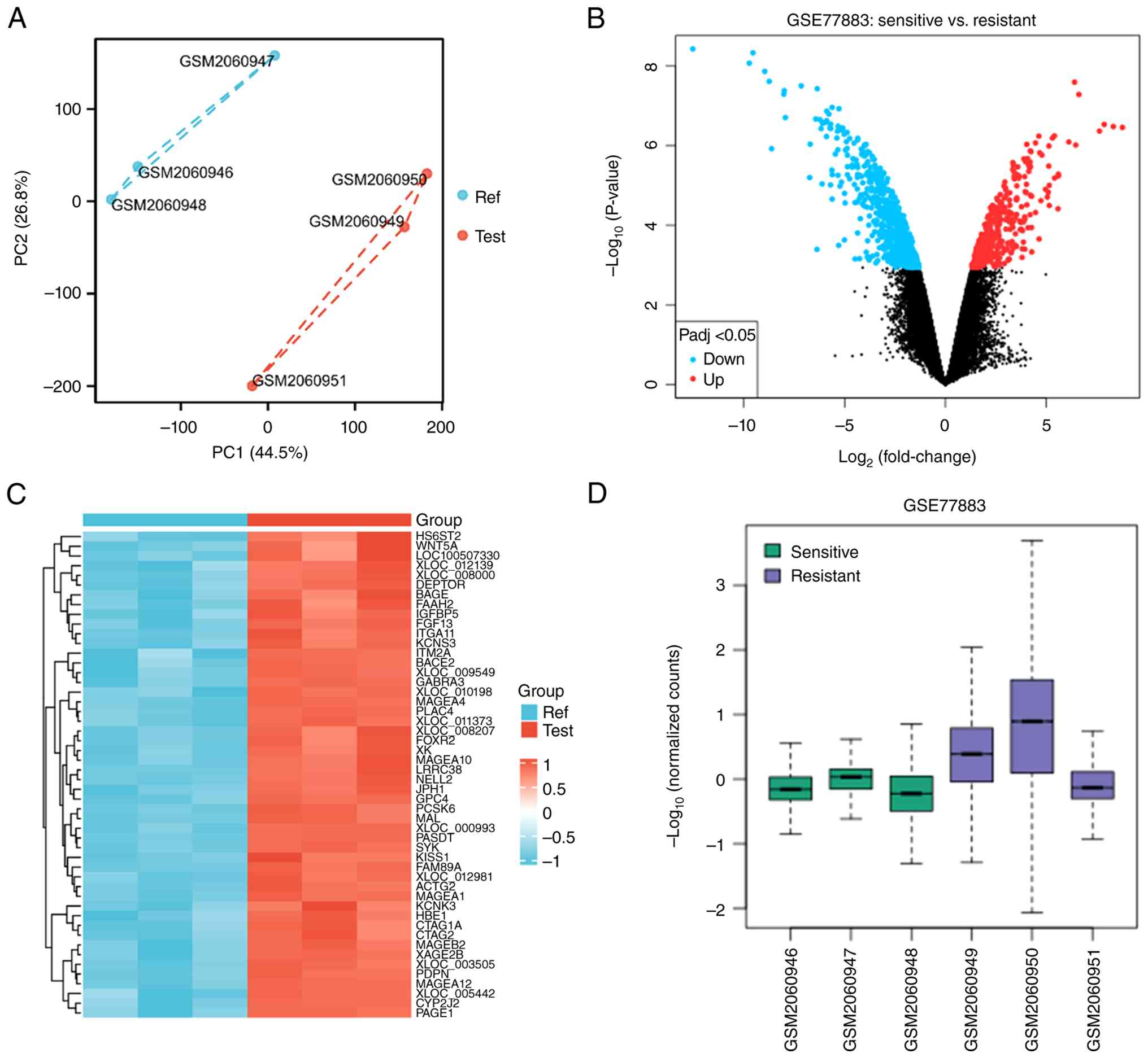
Firstly, principal component analysis was performed on the dataset
to evaluate the repeatability of the data within the group and
confirm the positive repeatability of the data in GSE77883
(Fig. 1A). Subsequently, the gene
expression data of the control group and experimental group in
GSE77883 were analyzed using the GEO2R online website. According to
the volcano map, a total of 831 differentially expressed genes were
identified, of which 495 genes were upregulated and 336 genes were
downregulated (Fig. 1B). The
upregulated and downregulated genes in the experimental and control
groups are shown in Fig. 1C. The
number of genes with abnormally high expression in the experimental
group was significantly higher than that in the control group, and
the difference was statistically significant (Fig. 1D).
Identification of hub oncogene KRT14
in bladder cancer
To identify the core functional genes from the
differentially expressed genes, the top 20 differentially expressed
genes were sorted based on logFC values and PPI network analysis
was performed using Cytoscape software. The specific ranking of
hub-gene and degree scores are shown in Table SI. The results demonstrated that
KRT14 ranked within the top 1 nodes by Degree metrics, serving an
important role in bladder cancer (Fig.
2A). Furthermore, the findings of the validation of TCGA-BLCA
dataset and paired sample analysis revealed that the expression of
KRT14 in the bladder cancer group was significantly higher than
that in the normal control group (Fig.
2B and C).
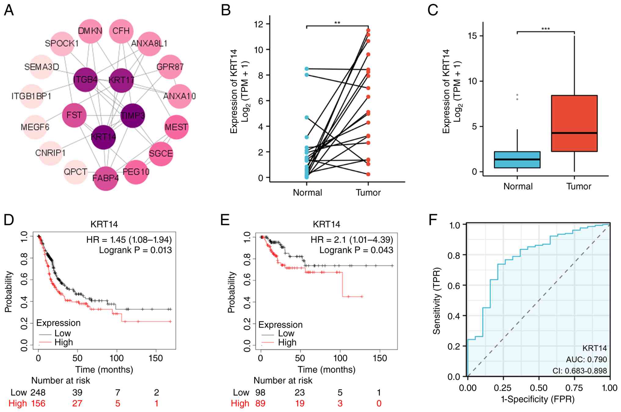 | Figure 2.Identification of the oncogene KRT14
in bladder cancer. (A) Protein-protein interaction network
screening vital differentially expressed genes in bladder cancer.
(B) Expression of KRT14 in bladder cancer in paired samples. (C)
Expression of RNA-sequencing data of KRT14 in bladder cancer. (D)
OS of KRT14 in bladder cancer (P=0.013). (E) RFS of KRT14 in
bladder cancer (P=0.043). (F) ROC curve of TCGA-BLCA according to
the expression of KRT14 (P<0.001). **P<0.01; ***P<0.001.
KRT14, keratin 14; OS, overall survival; RFS, regression-free
survival; ROC, receiver operating characteristic; TCGA-BLCA, The
Cancer Genome Atlas-Bladder Cancer; TPM, transcripts per million;
HR, hazard ratio; CI, confidence interval; AUC, area under the
curve; TPR, true positive rate; FPR, false positive rate. |
Based on the aforementioned results, we hypothesized
that the high level of KRT14 in bladder cancer is associated with
the survival and prognosis of bladder cancer. Therefore, the
overall survival (OS) and recurrence-free survival (RFS) of KRT14
in bladder cancer were assessed using the online Kaplan Meier
mapping database. The results demonstrated that the OS [hazard
ratio (HR), 1.45; 95% confidence interval (CI), 1.08–1.94; Fig. 2D] and RFS (HR, 2.1; 95% CI,
1.01–4.39; Fig. 2E) of patients
with bladder cancer with a high expression level of KRT14 were
shorter, indicating that the prognosis is worse. Moreover, the ROC
curve was used to validate the prognostic ability of KRT14 in
TCGA-BLCA cohort, demonstrating that the AUC of KRT14 was 0.79
(Fig. 2F). Therefore, combined with
high sensitivity and specificity, KRT14 can be used as a candidate
prognostic biomarker for bladder cancer.
Analysis of the expression patterns of
KRT14 in single-cell sequencing and the HPA datasets
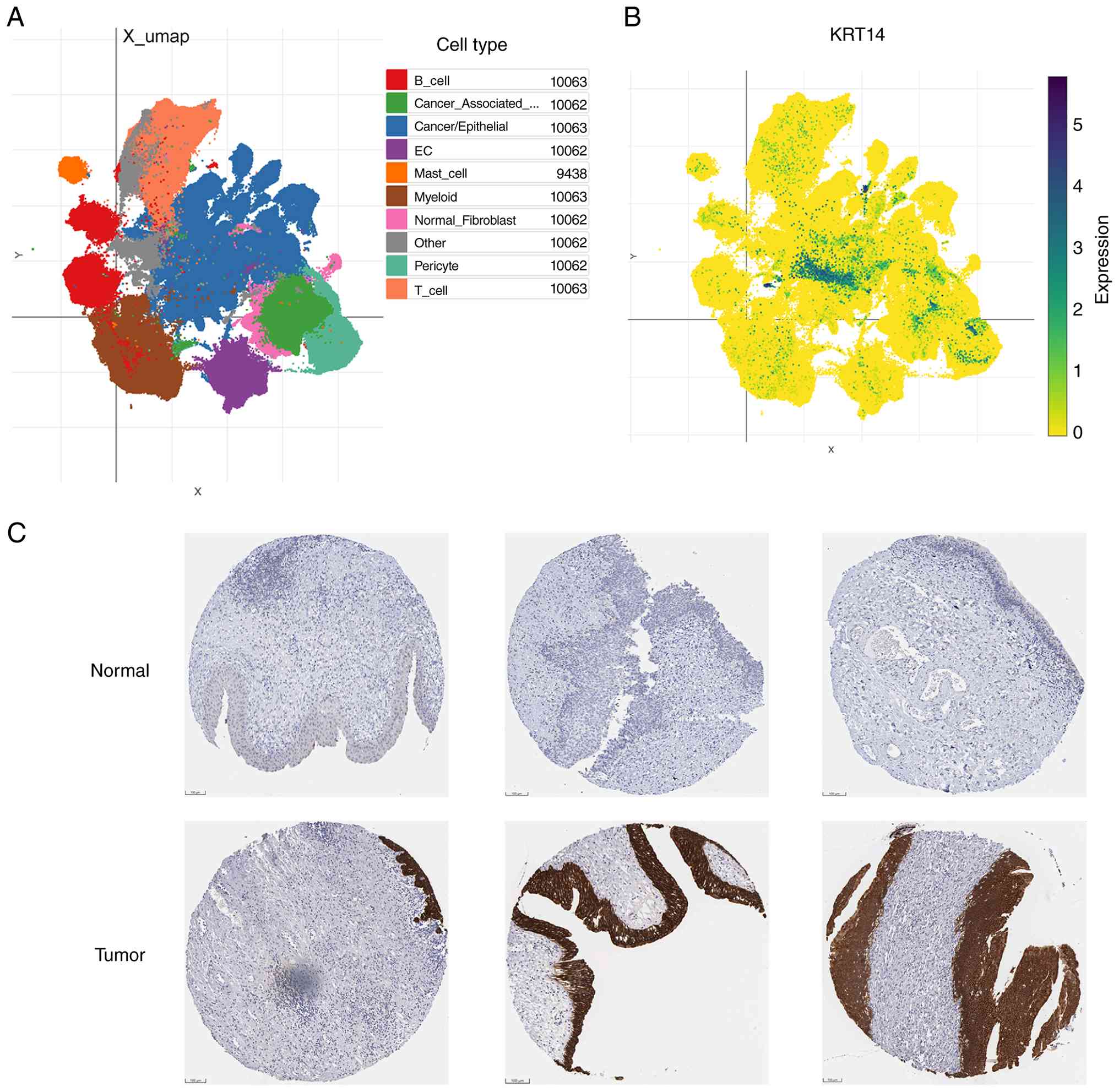
Publicly available single-cell RNA-seq data was
analyzed from 3,000 patients with BLCA (884,573 cells). In the
initial study, 10 cell types were annotated with marker genes,
including nine cell types (B cell, Cancer Associate,
Cancer/Epithelial, EC, Mast cell, Myeloid, Normal Fibroblast,
Pericyte and T cell) and one other unclassified mixed cell cluster
shown in grey (Fig. 3A). The
results showed that the expression of KRT14 was concentrated in
cell types identified by the analysis output as ‘cancer-associated
cell types’ (Fig. 3B). In order to
further explore the expression of KRT14 in bladder cancer, the HPA
online website was used to analyze the expression of KRT14 in the
pathological tissue chip of bladder cancer. Consistent with the
aforementioned results, the expression level of KRT14 in bladder
cancer was significantly higher than that in the normal control
group (Fig. 3C).
High expression of KRT14 in bladder
cancer tissues and cells
The aforementioned experiments adopted
bioinformatics methods to predict the role of KRT14 in bladder
cancer. In order to confirm these results, the role and function of
KRT14 in bladder cancer was assessed further though several
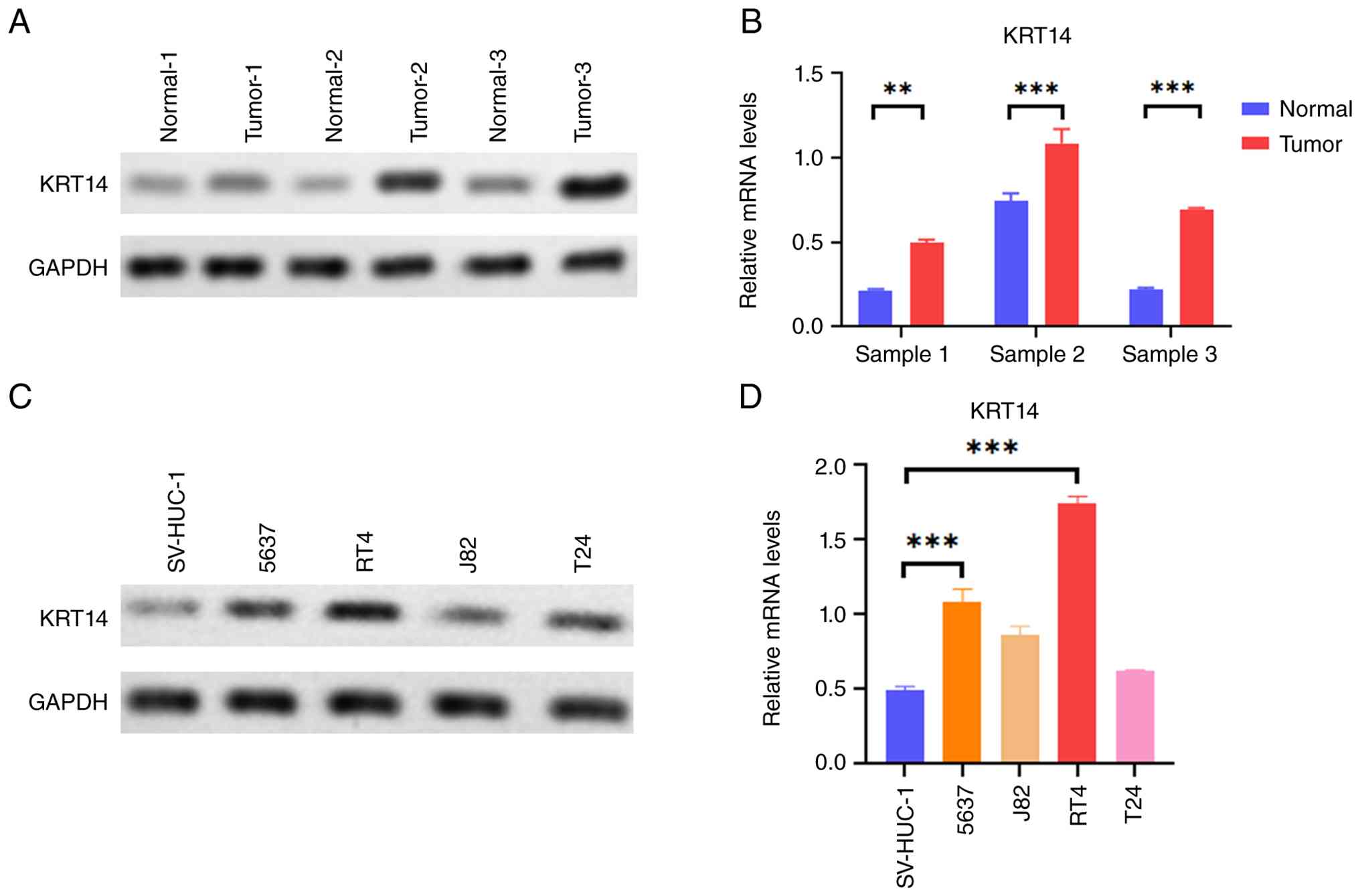
experiments. First, in order to verify the expression of KRT14 in
bladder cancer tissue, three pairs of tumor and adjacent normal
tissue samples were obtained from patients with bladder cancer. The
results demonstrated that KRT14 protein expression was markedly
elevated in bladder tumor tissues compared with that in the paired
normal adjacent tissues (Fig. 4A).
In addition, the mRNA expression level of KRT14 in bladder cancer
tissue was significantly higher than that in the adjacent normal
tissue, and the difference was significant (Fig. 4B). Subsequently, the expression of
KRT14 at the level of bladder cancer cells was assessed. First, the
protein expression level of KRT14 was compared between human normal
bladder epithelial cells (SV-HUC-1) and four human bladder cancer
cells (5637, J82, RT4 and T24). Consistent with the aforementioned
results, the expression level of KRT14 in bladder cancer cells was
significantly higher than that in the normal control cells
(Fig. 4C). Similarly, the mRNA
expression level of KRT14 in bladder cancer cells was significantly
higher than that in the normal control cells, and the difference
was statistically significant (Fig.
4D). Moreover, the findings revealed that KRT14 had the highest
expression level in 5637 and RT4 bladder cancer cells. Therefore,
these two bladder cancer cells were selected for further
experimentation.
Silencing KRT14 inhibits the
proliferation of bladder cancer cells
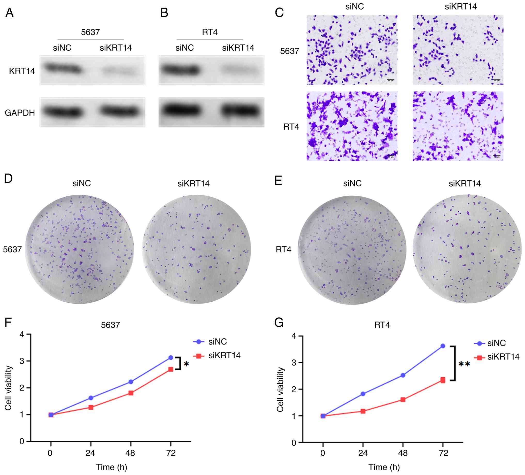
In order to further explore the effect of KRT14 on
the biological behavior of bladder cancer cells, KRT14 expression
was silenced in 5637 and RT4 bladder cancer cells (Fig. 5A and B). The results demonstrated
that KRT14 expression was significantly inhibited in the two
bladder cancer cell lines. In order to further evaluate the impact
of KRT14 on the progress of bladder cancer cells, the invasive
ability of cells was assessed using Transwell assays. The results
revealed that, compared with that in the control group cells, the
silencing of KRT14 in cells markedly attenuated migration (Fig. 5C). Cell clone formation assays
further revealed that knocking down KRT14 simultaneously inhibited
the colony numbers of 5637 and RT4 cells compared with that of the
control group cells (Fig. 5D and
E). Consistent with the aforementioned results, CCK-8 assay
results demonstrated that the cell viability of KRT14-silenced
bladder cancer cells 5637 and RT4 was significantly reduced at 24,
48 and 72 h. Furthermore, the results revealed a time-dependent
effect (Fig. 5F and G). These
results indicate that inhibition of KRT14 can significantly inhibit
the invasion and proliferation of bladder cancer cells.
Overexpression of KRT14 promotes the
progression of bladder cancer
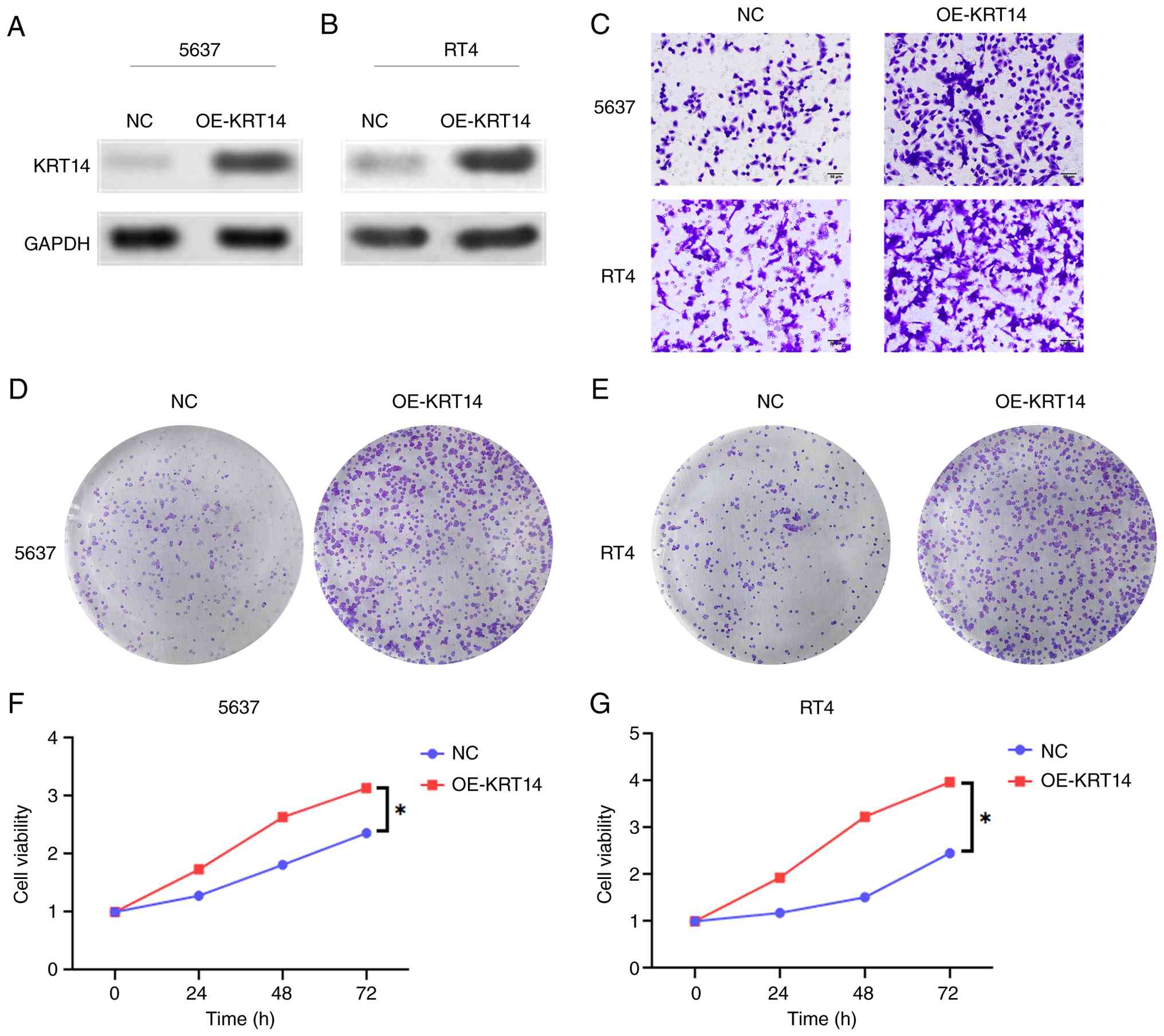
Subsequently, KRT14 was overexpressed in 5637 and
RT4 cells, and the findings revealed that KRT14 was markedly
upregulated in the two bladder cancer cells (Fig. 6A and B). In order to further explore
the impact of KRT14 on the progress of bladder cancer cells, the
migratory ability of cells was assessed using Transwell assays. The
results revealed that the migratory ability of cells overexpressing
KRT14 was notably higher (Fig. 6C).
Cell clone formation assays further demonstrated that
overexpression of KRT14 increased the number of colonies of 5637
and RT4 cells simultaneously compared with that in the control
group (Fig. 6D and E). Consistent
with the aforementioned results, CCK-8 assay results revealed that
the cell viability of KRT14-overexpressed bladder cancer cells 5637
and RT4 significantly increased at 24, 48 and 72 h, and revealed a
time-dependent effect (Fig. 6F and
G). These results indicate that KRT14 can significantly promote
the invasion and proliferation of bladder cancer cells.
KRT14 is directly regulated by
transcription factor E2F1
By searching the Genecards database, potential
upstream transcription factors that may regulate KRT14 were
identified. E2F1 was selected for further research as it serves an
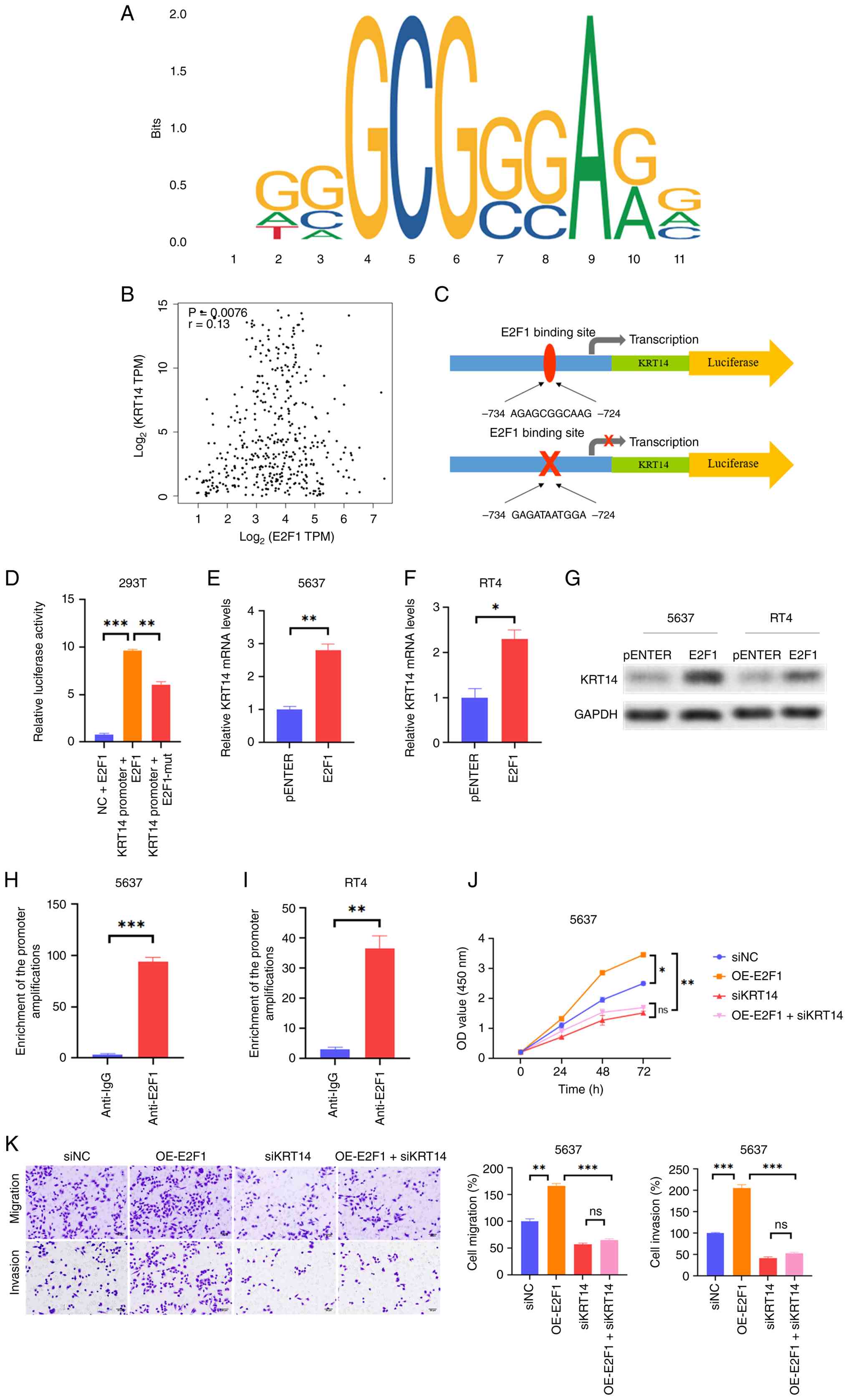
important role in human malignant tumors (26). The binding site of KRT14 promoter
and E2F1 were predicted using the meta site MOTIF. The E2F1 binding
sites from −734 to −724 on the KRT14 gene were identified (Fig. 7A). To assess whether the
transcriptional regulatory relationship between E2F1 and KRT14
observed in vitro exists in human bladder cancer clinical
tissues, co-expression correlation analysis was performed using
transcriptomic data from TCGA-BLCA cohort. The results demonstrated
that E2F1 and KRT14 exhibited a statistically significant positive
co-expression correlation in bladder tumor tissues (Fig. 7B).
In order to further assess the aforementioned
findings, the KRT14 promoter with binding sites was cloned into a
pGL3 basic vector (Fig. 7C). A
significant increase in luciferase activity was observed in cells
transfected with KRT14 promoter and E2F1 overexpression vector, and
revealed strong luciferase activity compared with that in cells
transfected with E2F1 mutant vectors (Fig. 7D). In order to provide a more
comprehensive explanation of the association between E2F1 and
KRT14, E2F1 was overexpressed in bladder cancer cells. Findings
revealed that E2F1 was significantly upregulated in the two bladder
cancer cells (Fig. S1). In 5637
and RT4 cells, overexpression of E2F1 significantly upregulated the
mRNA and protein levels of KRT14 (Fig.
7E-G). In addition, ChIP-PCR detection results revealed that
the promoter amplification of KRT14 at the E2F1 binding site were
significantly enriched using anti-E2F1 antibodies (Fig. 7H and I). Further research analyzed
the effect of E2F1 on KRT14 mediated phenotype of bladder cancer
cells. The results revealed that KRT14 silencing significantly
reduced the survival rate of 5637 cells, while transfection of
cells with E2F1 overexpression vectors significantly improved the
survival rate. In addition, overexpression of exogenous E2F1 did
not effectively restore cell proliferation (Fig. 7J), migration and invasion (Fig. 7K) abilities under KRT14 silencing
conditions. Collectively, these phenotypic rescue data functionally
support that E2F1 exerts its tumor-promoting effects through
transcriptionally upregulating KRT14 as a downstream effector
gene.
Drug sensitivity analysis and
molecular docking
The aforementioned results demonstrated the cancer
promoting effect of KRT14 in bladder cancer. Furthermore, in order
to explore the small molecular compounds that could be applied to
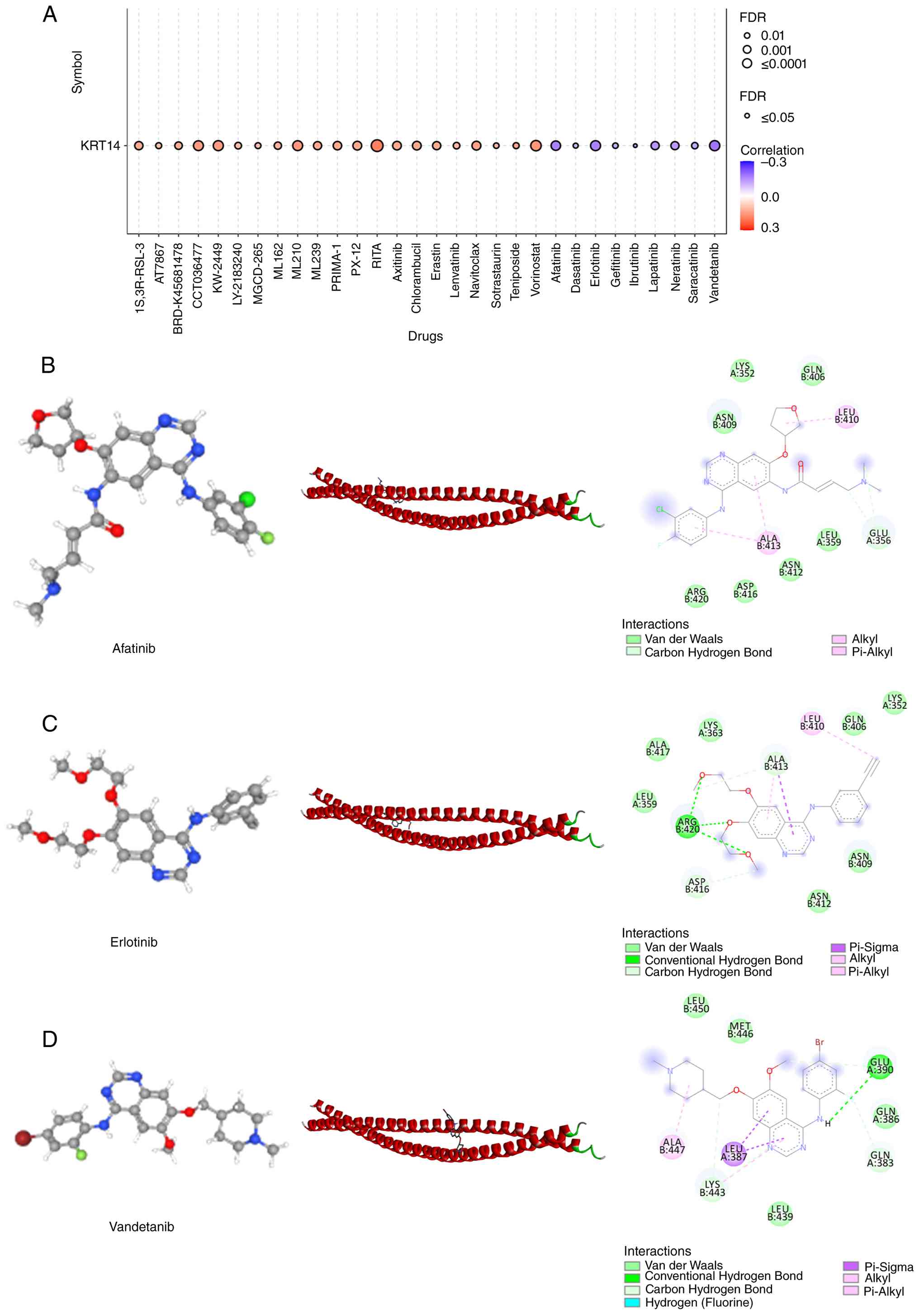
target KRT14, the drug sensitivity of KRT14 was analyzed. GSCA
correlation analysis was performed to screen small molecules whose
drug sensitivity status exhibited significant correlation with
KRT14 mRNA abundance in bladder cancer. The top three correlated
compounds (afatinib, erlotinib and vandetanib) were selected for
further in silico docking simulation (Fig. 8A). The structure of potential
compounds was determined based on Spearman correlation analysis. To
further validate the binding activity between KRT14 and these three
compounds, the potential of small molecules were predicted as
candidate compounds through computer simulation of molecular
docking. Subsequent molecular docking simulations predicted
favorable theoretical binding energy and potential intermolecular
interactions between each candidate compound and the KRT14 protein
structure, providing preliminary computational evidence for their
possible binding capacity with KRT14 (Fig. 8B-D).
Discussion
Bladder cancer is one of the most common types of
cancer in the world, with ~610, 000 new cases and ~220,000
mortalities each year (1).
Therefore, seeking effective treatment, improving the prognosis and
survival rate of patients with bladder cancer has become an urgent
clinical issue.
The occurrence and development of tumors is a
complex process that is regulated by numerous genes (27). The construction of high-throughput
sequencing technology and genomic databases, such as TCGA and GEO,
has provided new information about genomics and transcriptomics
(28), and bioinformatics and data
mining are increasingly being applied in medical research (29). Moreover, fully utilizing public
databases is a reliable method for assessing tumor progression and
prognosis. The present study first identified differentially
expressed genes from the GSE77883 dataset, and subsequently
validated KRT14 as a robust prognostic marker and oncogenic driver
across independent clinical tissue datasets and multiple cell
models.
KRT14 is a major cytoplasmic intermediate filament
protein and belongs to the type I keratin family KRT. Cancer cells
expressing KRT14 have been observed in several types of invasive
tumors, such as bladder and breast cancer (30–35),
which are specifically associated with invasive cancer cells and
contain invasive components (36–38).
Therefore, KRT14 may serve a specific role in the invasion process
beyond the basic maintenance of cytoskeleton integrity.
Cancer-related studies have reported that subpopulations of cells
expressing KRT14 regulate the formation of polyclonal tumor
deposits in vitro and in vivo (34,35).
KRT14 serves an important role in the occurrence and development of
several cancers. It is reported to be upregulated in numerous
cancers and is considered to be a biomarker of certain malignant
tumors, such as lung adenocarcinoma (39) and breast cancer (40). KRT14 contributes to chemoresistance
in bladder cancer not only via its structural roles, but also by
directly regulating translational machinery through eukaryotic
translation initiation factor 4H, leading to upregulation of the
metabolic enzyme acyl-CoA oxidase 2 (41). Previous research reported that
cisplatin-resistant ovarian cancer cell lines had elevated KRT14
expression, and KRT14 knockdown reduced cisplatin resistance by
lowering low-density lipoprotein receptor-related protein 11
expression. Therefore, KRT14 may serve a crucial role in mediating
cisplatin resistance in ovarian cancer (42).
However, little is known about the expression and
potential role of KRT14 in bladder cancer. The results of the
present study demonstrated that KRT14 is highly expressed in
bladder cancer. Kaplan Meier Plotter survival curve analysis
revealed that the prognosis of patients with bladder cancer with
high expression of KRT14 was worse than those with low KRT14
expression. In addition, ROC curve analysis suggested that KRT14
could be an effective indicator to predict the poor prognosis of
bladder cancer. Moreover, single cell sequencing demonstrated that
KRT14 is typically at high levels in tumor associated cells. KRT14
was also revealed to be highly expressed in a pathological tissue
microarray of bladder cancer. Furthermore, in order to confirm the
role of KRT14 in bladder cancer in vitro, the high
expression of KRT14 in bladder cancer at the tissue and cell levels
was assessed. The results revealed that silencing KRT14 could
significantly inhibit cell invasion and proliferation, and
overexpression of KRT14 could significantly promote the invasion
and proliferation of 5637 and RT4 cells.
E2F1 is a widely studied transcription factor that
can transcribe and regulate SH3 domain binding kinase 1 (43), RCC1 domain containing 1 (44) and pyruvate dehydrogenase kinase 1
(45), mediating several malignant
behaviors by regulating cell cycle progression, tumor metastasis
and treatment response through these target genes. In the research
of bladder cancer, E2F1 has been reported to induce myelin and
lymphocyte protein 2 transcriptional activation, thus promoting the
progress of bladder cancer and inhibiting sunitinib sensitivity
(46). In addition, as a pivotal
transcription factor, E2F1 promotes the transcription of disks
large-associated protein 5 (DLGAP5) and establishes a positive
feedback loop between DLGAP5 and E2F1, thus accelerating the
progress of bladder cancer (47).
This indicates the carcinogenic effect of E2F1 and its potential as
a molecular drug target. In the present work, multiple lines of
molecular evidence including motif prediction, dual-luciferase
reporter assay and ChIP-PCR consistently demonstrated that E2F1
acts as an upstream transcriptional activator of KRT14: E2F1
directly binds the −734 to −724 region of the KRT14 promoter and
markedly elevates KRT14 mRNA and protein abundance in bladder
cancer cells. Notably, phenotypic rescue experiments demonstrated
that the tumor-promoting capacity of E2F1 was highly dependent on
intact KRT14 expression.
Several limitations of the present study should be
acknowledged. First, the initial differentially expression gene
screening relied on GSE77883, a cell-line microarray dataset
comparing gemcitabine-resistant T24 cells with parental T24 cells,
rather than primary human bladder tumor and adjacent normal
tissues. Transcriptional changes identified in this dataset may
partially reflect adaptive transcriptomic remodeling induced by
long-term gemcitabine exposure, which is specifically associated
with chemoresistance rather than general bladder tumorigenesis.
Another limitation is the potential confounding effect of altered
cell proliferation on Transwell migration and invasion readouts. As
KRT14 knockdown robustly suppressed cell proliferation and
viability, the reduced number of migrated/invaded cells may have
partially arisen from slower cell growth rather than a specific
impairment of cell motility. Follow-up experiments such as
time-lapse live cell imaging to track single-cell migration speed,
or normalization of Transwell counts to parallel viable cell
numbers, will be performed in future work to fully decouple cell
motility from proliferation. Finally, the paired human bladder
tumor and adjacent normal tissue cohort used for RT-qPCR and
western blot analysis only contained 3 patient pairs, which
provides limited statistical power for drawing definitive
tissue-level expression conclusions. This small sample size
restricts the generalizability of the tissue western blot and
RT-qPCR results. Future validation with an expanded clinical cohort
containing at least 10–15 paired tissue samples will be performed
to further consolidate the tissue expression pattern of KRT14.
In conclusion, KRT14 is upregulated in bladder
cancer and is associated with an unfavorable clinical prognosis.
KRT14 depletion suppresses multiple malignant phenotypes of bladder
cancer cells, highlighting its candidacy as a promising prognostic
biomarker and therapeutic target. Mechanistically, the present
study defined a linear oncogenic cascade where upstream
transcription factor E2F1 directly binds the KRT14 promoter to
transcriptionally activate KRT14 expression. Functional rescue
assays further demonstrated that the oncogenic activity of E2F1
relies on downstream KRT14 to drive bladder cancer progression.
These findings advance the mechanistic insight into KRT14-mediated
bladder tumorigenesis and provide novel clues for developing
targeted therapeutic strategies against bladder cancer.
Supplementary Material
Supporting Data
Supporting Data
Acknowledgements
Not applicable.
Funding
The present study was funded by Nantong University Special
Research Fund for Clinical Medicine (grant no. 2025LQ024), the
Scientific Research Development Fund Project of Kangda College of
Nanjing Medical University (grant no. KD2025KYJJ180), and the
Nantong Science and Technology Program Guidance Project (grant no.
MSZ2024128).
Availability of data and materials
The data generated in the present study may be
requested from the corresponding author.
Authors' contributions
CLW and XC designed the study, and drafted and
revised the manuscript. XHC, SNL and XSZ performed the experiments
and analyzed the data. CLW and XC oversaw the manuscript and gave
approval for the final submitted version. CLW and XHC confirm the
authenticity of all the raw data. All authors read and approved the
final version of the manuscript.
Ethics approval and consent to
participate
All experimental procedures were performed in
accordance with the ethical standards of the Declaration of
Helsinki and were approved by the Ethics Committee of Nantong City
No. 1 People's Hospital (approval no. 2026-KT102-01). All patients
signed a written informed consent form, which included consent to
participate in the present study and use of their medical data for
research purposes.
Patient consent for publication
All patients signed a written informed consent form,
which included consent to for the publication of anonymized
findings.
Competing interests
The authors declare that they have no competing
interests.
References
|
1
|
Bray F, Laversanne M, Sung H, Ferlay J,
Siegel RL, Soerjomataram I and Jemal A: Global cancer statistics
2022: GLOBOCAN estimates of incidence and mortality worldwide for
36 cancers in 185 countries. CA Cancer J Clin. 74:229–263.
2024.PubMed/NCBI
|
|
2
|
Bhanvadia SK: Bladder cancer survivorship.
Curr Urol Rep. 19:1112018. View Article : Google Scholar : PubMed/NCBI
|
|
3
|
Knowles MA and Hurst CD: Molecular biology
of bladder cancer: New insights into pathogenesis and clinical
diversity. Nat Rev Cancer. 15:25–41. 2015. View Article : Google Scholar : PubMed/NCBI
|
|
4
|
Anderson B: Bladder cancer: Overview and
management. Part 2: Muscle-invasive and metastatic bladder cancer.
Br J Nurs. 27 (Suppl):S8–S20. 2018. View Article : Google Scholar : PubMed/NCBI
|
|
5
|
Woldu SL, Bagrodia A and Lotan Y:
Guideline of guidelines: Non-muscle-invasive bladder cancer. BJU
Int. 119:371–380. 2017. View Article : Google Scholar : PubMed/NCBI
|
|
6
|
D Elia C, Pycha A, Folchini DM, Mian C,
Hanspeter E, Schwienbacher C, Vjaters E, Pycha A and Trenti E.:
Diagnostic predictive value of Xpert bladder cancer monitor in the
follow-up of patients affected by non-muscle invasive bladder
cancer. J Clin Pathol. 72:140–144. 2019. View Article : Google Scholar : PubMed/NCBI
|
|
7
|
Babjuk M, Burger M, Capoun O, Cohen D,
Compérat EM, Dominguez Escrig JL, Gontero P, Liedberg F,
Masson-Lecomte A, Mostafid AH, et al: European association of
urology guidelines on Non-muscle-invasive bladder cancer (Ta, T1,
and Carcinoma in situ). Eur Urol. 81:75–94. 2022. View Article : Google Scholar : PubMed/NCBI
|
|
8
|
Hurle R, Casale P, Saita A, Colombo P,
Elefante GM, Lughezzani G, Fasulo V, Paciotti M, Domanico L,
Bevilacqua G, et al: Clinical performance of Xpert Bladder Cancer
(BC) Monitor, a mRNA-based urine test, in active surveillance (AS)
patients with recurrent non-muscle-invasive bladder cancer (NMIBC):
Results from the Bladder Cancer Italian Active Surveillance (BIAS)
project. World J Urol. 38:2215–2220. 2020. View Article : Google Scholar : PubMed/NCBI
|
|
9
|
Matulewicz RS, DeLancey JO and Meeks JJ:
Cystoscopy. JAMA. 317:11872017. View Article : Google Scholar : PubMed/NCBI
|
|
10
|
Gezginci E, Iyigun E, Kibar Y and Bedir S:
Three distraction methods for pain reduction during cystoscopy: A
randomized controlled trial evaluating the effects on pain,
Anxiety, and satisfaction. J Endourol. 32:1078–1084. 2018.
View Article : Google Scholar : PubMed/NCBI
|
|
11
|
Kwon WA, Lee JW, Seo HK, Oh TH, Park SC,
Jeong HJ and Seo IY: Hand-holding during cystoscopy decreases
patient anxiety, pain, and dissatisfaction: A pilot randomized
controlled trial. Urol Int. 100:222–227. 2018. View Article : Google Scholar : PubMed/NCBI
|
|
12
|
Valenberg FJPV, Hiar AM, Wallace E, Bridge
JA, Mayne DJ, Beqaj S, Sexton WJ, Lotan Y, Weizer AZ, Jansz GK, et
al: Prospective validation of an mRNA-based urine test for
surveillance of patients with bladder cancer. Eur Urol. 75:853–860.
2019. View Article : Google Scholar : PubMed/NCBI
|
|
13
|
Tomczak K, Czerwińska P and Wiznerowicz M:
The cancer genome atlas (TCGA): An immeasurable source of
knowledge. Contemp Oncol (Pozn). 19:A68–A77. 2015.PubMed/NCBI
|
|
14
|
Edgar R, Domrachev M and Lash AE: Gene
expression omnibus: NCBI gene expression and hybridization array
data repository. Nucleic Acids Res. 30:207–210. 2002. View Article : Google Scholar : PubMed/NCBI
|
|
15
|
Clough E, Barrett T, Wilhite SE, Ledoux P,
Evangelista C, Kim IF, Tomashevsky M, Marshall KA, Phillippy KH,
Sherman PM, et al: NCBI GEO: Archive for gene expression and
epigenomics data sets: 23-year update. Nucleic Acids Res.
52:D138–D144. 2024. View Article : Google Scholar : PubMed/NCBI
|
|
16
|
Zhang H, Zhang X, Du Y, Xiong W, Zhang X,
Zhang B and Shang P: High FSTL1 expression promotes bladder cancer
progression by enhancing tumor cell migration. Clin Transl Oncol.
28:265–276. 2026. View Article : Google Scholar : PubMed/NCBI
|
|
17
|
Aizezi X, Silafu B, Ma Y, Wang Z, Li J and
Huang J: Analysis of IGFL2 gene expression and prognostic value in
bladder cancer based on TCGA database. Asia Pac J Clin Oncol.
22:129–139. 2026. View Article : Google Scholar : PubMed/NCBI
|
|
18
|
Bilandzic M, Rainczuk A, Green E,
Fairweather N, Jobling TW, Plebanski M and Stephens AN: Keratin-14
(KRT14) positive leader cells mediate mesothelial clearance and
invasion by ovarian cancer cells. Cancers (Basel). 11:12282019.
View Article : Google Scholar : PubMed/NCBI
|
|
19
|
Verma A, Singh A, Singh MP, Nengroo MA,
Saini KK, Satrusal SR, Khan MA, Chaturvedi P, Sinha A, Meena S, et
al: EZH2-H3K27me3 mediated KRT14 upregulation promotes TNBC
peritoneal metastasis. Nat Commun. 13:73442022. View Article : Google Scholar : PubMed/NCBI
|
|
20
|
Wang S, Gao P, Wang X, Duan L, He X and Qu
J: Clinical utility of keratin 14 expression measurement in
reflecting the tumor properties and prognosis in patients with
renal cell carcinoma: A study with long-term follow-up. Int Urol
Nephrol. 56:2045–2053. 2024. View Article : Google Scholar : PubMed/NCBI
|
|
21
|
Azhati B, Maolakuerban N, Ma T, Li X and
Rexiati M: Up-regulation of DRAM2 promotes tolerance of bladder
transitional cell carcinoma to gemcitabine. Arch Med Sci.
16:1207–1217. 2020. View Article : Google Scholar : PubMed/NCBI
|
|
22
|
Tian S, Wang C, Zhao X, Xuan Y, Wei W,
Dong Y, Tao W, Zhang C, Cai T, Liu C, et al: A CEBPB/TYMP/GDF15
signaling axis mediates tumor growth and cisplatin resistance in
bladder cancer. Transl Oncol. 61:1025372025. View Article : Google Scholar : PubMed/NCBI
|
|
23
|
Matsumoto R, Tsuda M, Yoshida K, Tanino M,
Kimura T, Nishihara H, Abe T, Shinohara N, Nonomura K and Tanaka S:
Aldo-keto reductase 1C1 induced by interleukin-1β mediates the
invasive potential and drug resistance of metastatic bladder cancer
cells. Sci Rep. 6:346252016. View Article : Google Scholar : PubMed/NCBI
|
|
24
|
Huang P, Wang J, Yu Z, Lu J, Sun Z and
Chen Z: Redefining bladder cancer treatment: Innovations in
overcoming drug resistance and immune evasion. Front Immunol.
16:15378082025. View Article : Google Scholar : PubMed/NCBI
|
|
25
|
Livak KJ and Schmittgen TD: Analysis of
relative gene expression data using real-time quantitative PCR and
the 2(−Delta Delta C(T)) method. Methods. 25:402–408. 2001.
View Article : Google Scholar : PubMed/NCBI
|
|
26
|
Abe T, Masuda T, Tobo T, Hashimoto M,
Nakano Y, Saito H, Ofuchi T, Ikehara T, Tsujimoto A, Nakano T, et
al: ZNF282 promotes colorectal cancer progression possibly via E2F1
activation. Cancer Sci. 117:90–104. 2026. View Article : Google Scholar : PubMed/NCBI
|
|
27
|
Wang L, Wang Y, Wang J, Li L and Bi J:
Identification of a Prognosis-related risk signature for bladder
cancer to predict survival and immune landscapes. J Immunol Res.
2021:32363842021. View Article : Google Scholar : PubMed/NCBI
|
|
28
|
Zhang Y, Luo J, Liu Z, Liu X, Ma Y, Zhang
B, Chen Y, Li X, Feng Z, Yang N, et al: Identification of hub genes
in colorectal cancer based on weighted gene co-expression network
analysis and clinical data from The Cancer Genome Atlas. Biosci
Rep. 41:BSR202112802021. View Article : Google Scholar : PubMed/NCBI
|
|
29
|
Zhu Q, Zhang L, Deng Y and Tang L:
Identification of immune-related and autophagy-related genes for
the prediction of survival in bladder cancer. BMC Genom Data.
23:602022. View Article : Google Scholar : PubMed/NCBI
|
|
30
|
Chu PG, Lyda MH and Weiss LM: Cytokeratin
14 expression in epithelial neoplasms: A survey of 435 cases with
emphasis on its value in differentiating squamous cell carcinomas
from other epithelial tumours. Histopathology. 39:9–16. 2001.
View Article : Google Scholar : PubMed/NCBI
|
|
31
|
Papafotiou G, Paraskevopoulou V, Vasilaki
E, Kanaki Z, Paschalidis N and Klinakis A: KRT14 marks a
subpopulation of bladder basal cells with pivotal role in
regeneration and tumorigenesis. Nat Commun. 7:119142016. View Article : Google Scholar : PubMed/NCBI
|
|
32
|
Cheah MT, Chen JY, Sahoo D,
Contreras-Trujillo H, Volkmer AK, Scheeren FA, Volkmer JP and
Weissman IL: CD14-expressing cancer cells establish the
inflammatory and proliferative tumor microenvironment in bladder
cancer. Proc Natl Acad Sci USA. 112:4725–4730. 2015. View Article : Google Scholar : PubMed/NCBI
|
|
33
|
Volkmer JP, Sahoo D, Chin RK, Ho PL, Tang
C, Kurtova AV, Willingham SB, Pazhanisamy SK, Contreras-Trujillo H,
Storm TA, et al: Three differentiation states risk-stratify bladder
cancer into distinct subtypes. Proc Natl Acad Sci USA.
109:2078–2083. 2012. View Article : Google Scholar : PubMed/NCBI
|
|
34
|
Cheung KJ, Padmanaban V, Silvestri V,
Schipper K, Cohen JD, Fairchild AN, Gorin MA, Verdone JE, Pienta
KJ, Bader JS and Ewald AJ: Polyclonal breast cancer metastases
arise from collective dissemination of keratin 14-expressing tumor
cell clusters. Proc Natl Acad Sci USA. 113:E854–E863. 2016.
View Article : Google Scholar : PubMed/NCBI
|
|
35
|
Cheung KJ, Gabrielson E, Werb Z and Ewald
AJ: Collective invasion in breast cancer requires a conserved basal
epithelial program. Cell. 155:1639–1651. 2013. View Article : Google Scholar : PubMed/NCBI
|
|
36
|
Lichtner RB, Julian JA, North SM, Glasser
SR and Nicolson GL: Coexpression of cytokeratins characteristic for
myoepithelial and luminal cell lineages in rat 13762NF mammary
adenocarcinoma tumors and their spontaneous metastases. Cancer Res.
51:5943–5950. 1991.PubMed/NCBI
|
|
37
|
Gordon LA, Mulligan KT, Maxwell-Jones H,
Adams M, Walker RA and Jones JL: Breast cell invasive potential
relates to the myoepithelial phenotype. Int J Cancer. 106:8–16.
2003. View Article : Google Scholar : PubMed/NCBI
|
|
38
|
Petrocca F, Altschuler G, Tan SM, Mendillo
ML, Yan H, Jerry DJ, Kung AL, Hide W, Ince TA and Lieberman J: A
genome-wide siRNA screen identifies proteasome addiction as a
vulnerability of basal-like triple-negative breast cancer cells.
Cancer Cell. 24:182–196. 2013. View Article : Google Scholar : PubMed/NCBI
|
|
39
|
Hossein Zadeh R, Hossein Zadeh R,
Hajimoradi M, Islampanah M, Zarimeidani F, Rahmati R, Ahmadinia M,
Bahrami N, Mohamadnia A, Shafaghi S and Nazari E: Identification of
diagnostic and prognostic biomarkers in lung adenocarcinoma through
integrated bioinformatics analysis and real time PCR validation.
Sci Rep. 16:66792026. View Article : Google Scholar : PubMed/NCBI
|
|
40
|
Dong J, Kong L, Wang S, Xia M, Zhang Y, Wu
J, Yang F, Zuo S and Wei J: Oncolytic adenovirus encoding
apolipoprotein A1 suppresses metastasis of triple-negative breast
cancer in mice. J Exp Clin Cancer Res. 43:1022024. View Article : Google Scholar : PubMed/NCBI
|
|
41
|
Liu S, Liu T, Yang C, Mou Z, Zou L and
Jiang H: Cytoskeletal protein KRT14 governs cisplatin resistance by
modulating eIF4H-dependent ACOX2 translation and lipid metabolism
in bladder cancer. Cell Death Dis. 17:1342025. View Article : Google Scholar : PubMed/NCBI
|
|
42
|
Liu Z, Weng T, Cheng M, Lei T, Xiao D,
Deng Q and Wu T: KRT14 knockdown reduces cisplatin resistance by
lowering LRP11 expression levels in cisplatin-resistant ovarian
cancer cell lines. Transl Cancer Res. 14:1786–1798. 2025.
View Article : Google Scholar : PubMed/NCBI
|
|
43
|
Wang Q, Deng Y, Yu F and Liu Z: The
E2F1/SBK1 axis activates the Notch signaling pathway to accelerate
the malignant progression of breast cancer. Naunyn Schmiedebergs
Arch Pharmacol. 398:16045–16058. 2025. View Article : Google Scholar : PubMed/NCBI
|
|
44
|
Han S, Lin M, Wu L, Lin X, Chen M, Hu C,
Bao A, Fang Z and Zhu F: E2F1 modulates RCCD1 expression to
participate in the initiation and progression of EMT in colorectal
cancer. Pathol Res Pract. 260:1554292024. View Article : Google Scholar : PubMed/NCBI
|
|
45
|
Zhuo S, Yang L, Chen Z, Chen S, Yang S,
Chen T, Wu WS, Wang K and Yang K: Druggable target ATAD2 enhances
the malignant progression and cooperates with E2F1 to up-regulate
PDK1 expression in glioma. Genes Dis. 13:1018102025. View Article : Google Scholar : PubMed/NCBI
|
|
46
|
Su M, Chen W, Luo J, Li X, Ye D, Han D and
Fu G: E2F1-induced transcriptional activation of MAL2 inhibits
sunitinib sensitivity and promotes the malignant progression of
bladder cancer. J Chemother. 1-12:2025.doi:
10.1080/1120009X.2025.2561275 (Epub ahead of print). PubMed/NCBI
|
|
47
|
Zhou F, Deng Z, Shen D, Lu M, Li M, Yu J,
Xiao Y, Wang G, Qian K, Ju L and Wang X: DLGAP5 triggers
proliferation and metastasis of bladder cancer by stabilizing E2F1
via USP11. Oncogene. 43:594–607. 2024. View Article : Google Scholar : PubMed/NCBI
|