Introduction
Oral squamous cell carcinoma (OSCC) is a
heterogeneous malignant neoplasm originating from the epithelial
tissue of the oral mucosa, which remains one of the leading causes
of cancer-associated mortality worldwide (1). The 5-year cumulative overall survival
rate for surgically treated patients with OSCC ranges from 47 to
71% across different published studies (2,3). OSCC
accounts for 90% of all malignant tumors in the oral cavity and
serves as the most common subtype of head and neck squamous cell
carcinoma (HNSCC) globally (2,3).
Clinically, drug resistance, markedly reduces the efficacy of
traditional chemotherapeutics such as cisplatin, fluorouracil and
paclitaxel (4,5). Furthermore, while high-dose
radiotherapy is often combined with drug therapy, it causes acute
toxicities (such as soft-tissue necrosis and xerostomia) that
develop into long-term sequelae, further compromising treatment
tolerance and the quality of life of patients (6). Thus, current OSCC drug-based
treatments are far from optimal, warranting further effective and
tailored solutions in the future.
Branched-chain amino acids (BCAAs; including
leucine, isoleucine and valine) are essential amino acids that
require catabolism. Branched-chain amino acid transaminase (BCAT)
is the rate-limiting enzyme driving BCAA transamination, a reaction
that transfers the amino group of BCAAs (leucine, isoleucine and
valine) to α-ketoglutarate (α-KG), generating branched-chain
α-ketoacids (BCKAs) and glutamate (7). BCKAs enter the tricarboxylic acid
(TCA) cycle to produce ATP for tumor energy needs, while glutamate
acts as a key precursor for glutathione (GSH) synthesis: BCKAs
promote GSH production (supporting the first rate-limiting step via
glutamate-cysteine ligase), scavenge reactive oxygen species (ROS)
to protect tumors from oxidative stress and contribute to
chemoresistance (8–10). This BCAT-mediated metabolism (energy
supply via BCKAs and antioxidant defense via glutamate-GSH) is a
tumor-specific adaptation, making BCAT1 a potential therapeutic
target for tumor therapy (11). A
recent study further validated the clinical and functional
importance of BCAT1 in OSCC, demonstrating its involvement in
malignant progression and metabolic reprogramming (12).
N6-methyladenosine (m6A) modification is
the most abundant epigenetic modification in eukaryotic mRNA, which
is co-regulated by methyltransferase complexes (‘writers’) that
catalyze m6A deposition, demethylases (‘erasers’) that remove the
methylation mark, and m6A-binding proteins (‘readers’)
that recognize m6A and mediate downstream effects such as mRNA
splicing, translation or stability (13,14).
By influencing mRNA splicing, transport, translation and
degradation, m6A serves a key role in physiological
processes such as cell proliferation and differentiation (15). Increasing evidence has also
implicated multiple m6A regulatory factors in OSCC
tumorigenesis, prognosis and therapeutic response (16,17).
Heterogeneous nuclear ribonucleoprotein C (hnRNPC) is a classic
RNA-binding protein that mainly recognizes uracil-rich sequences in
precursor mRNA to participate in RNA splicing, stability
maintenance and nucleocytoplasmic transport, serving as a key
regulatory factor in the RNA metabolic network (18,19).
Previous studies have demonstrated that hnRNPC consistently exerts
oncogenic functions. hnRNPC promotes tumor progression through
multiple mechanisms, such as regulating the stability of mRNA or
microRNA and mediating alternative splicing, with well-documented
roles in glioma, acute myeloid leukemia and breast cancer (20–23).
Although both m6A regulators and BCAT1
have been individually implicated in OSCC pathogenesis (12,16,17,24),
no previous studies have established a mechanistic or functional
link associating these two pathways, to the best of our knowledge.
Notably, it remains to be elucidated whether hnRNPC governs BCAT1
expression and BCAA metabolism through m6A modification
in OSCC. Therefore, the novelty of the present study does not lie
in identifying m6A regulators or BCAT1 as individual
OSCC-associated molecules, but in establishing a novel functional
axis associating hnRNPC, m6A modification and
BCAT1-dependent BCAA metabolism.
The present study utilized The Cancer Genome Atlas
(TCGA) datasets to conduct an integrated bioinformatics analysis,
systematically exploring the relationship between m6A
modification patterns and OSCC malignancy.
Materials and methods
Collection of m6A
transcriptome data and expression profiling
Transcriptome datasets, comprising 515 HNSCC and 44
normal tissue samples, were retrieved from the TCGA database via
the UCSC Xena database (https://xenabrowser.net) and the GDC portal
(https://portal.gdc.cancer.gov).
Specifically, the Genomic Data Commons (GDC) TCGA Head and Neck
Cancer cohort was accessed through the UCSC Xena database, and the
Spliced Transcripts Alignment to a Reference-Fragments Per Kilobase
of transcript per Million mapped reads data under the gene
expression RNA sequencing module were downloaded. Clinical
information, including pathological stage, T stage, N stage and M
stage, was extracted from the TCGA-HNSC clinical data files. Tumor
staging was based on the American Joint Committee on Cancer staging
system (25). A total of 23
m6A-associated genes were selected from the dataset based on
findings from previous studies (15,16).
All bioinformatics analyses were performed using R software
(version 4.2.1; http://www.r-project.org/) and RStudio (version
2023.06.1; https://posit.co/; Posit Software, PBC). The following R
packages (all obtained from CRAN, http://cran.r-project.org/) were used: i) ‘ggplot2’
(version 3.5.2) and ‘boxplot’ (version 1.3.1) for constructing
heatmaps and boxplots to visualize gene expression patterns; ii)
‘survival’ (version 3.7–0) for Cox regression analysis;
‘forestplot’ (version 3.13) and ‘pheatmap’ (version 1.0.12) for
hazard ratio visualization; iii) ‘survminer’ (version 0.5.1) for
Kaplan-Meier curves; iv) ‘rms’ (version 6.8–2) for nomogram
construction; v) ‘gghalves’ (version 0.1.4) and ‘Hmisc’ (version
5.1-3) for clinical correlation analysis; and vi) ‘ggExtra’
(version 0.10.1), ‘cowplot’ (version 1.1.3), ‘ggpubr’ (version
0.6.0), ‘ggstatsplot’ (version 0.14.0) and ‘ggside’ (version 0.3.1)
for Pearson's correlation analysis between hnRNPC and BCAT1. To
identify genes associated with hnRNPC expression, patients with
HNSCC from TCGA database were stratified into hnRNPC-high and
hnRNPC-low groups based on the median expression level of hnRNPC.
Differentially expressed genes between the two groups were
identified using the ‘limma’ package (version 3.50.0) in R
software. Genes with |log2 fold-change|>1 and adjusted P-value
(false discovery rate) <0.05 were considered significantly
differentially expressed.
Development of Cox risk regression
model
Study samples were randomly allocated into a
training cohort and a validation cohort at a 3:7 ratio. Univariate
and multivariate Cox regression analyses were applied to identify
independent prognostic factors for patients with HNSCC. The
robustness of these factors was further assessed using the
validation cohort. Therefore, three genes, insulin-like growth
factor 2 mRNA-binding protein 2 (IGF2BP2), hnRNPC and YTHDC2, were
incorporated into the final Cox risk regression model.
Cell culture and lentivirus
preparation
The human HNSCC cell lines CAL-27 and SCC-15 were
acquired from The Cell Bank of Type Culture Collection of The
Chinese Academy of Sciences. These cell lines were maintained in
Dulbecco's Modified Eagle's Medium (DMEM) containing 10% fetal
bovine serum (FBS; Corning, Inc.). For stem cell (27-CSC)
induction, OSCC cells were cultured in DMEM/F12 medium supplemented
with basic fibroblast growth factor (bFGF) and epidermal growth
factor (EGF). These induced stem cells were grown as tumor spheres
in DMEM/F12 medium with 20 ng/ml each of bFGF (cat. no. HY-P7331;
MedChemExpress) and EGF (cat. no. AF-100-15-100; Peprotech, Inc.;
Thermo Fisher Scientific, Inc.).
For lentivirus-mediated knockdown, two independent
short hairpin RNA (shRNA) sequences targeting distinct regions of
hnRNPC and methyltransferase-like 3 (METTL3) transcripts were
designed and cloned into the pLKO.1-puro lentiviral vector [Hanheng
Biotechnology (Shanghai) Co., Ltd.]. Lentiviral particles were
produced by co-transfection of 293T cells with the pLKO.1-shRNA
transfer plasmid, the psPAX2 packaging plasmid and the pMD2.G
envelope plasmid. Transfection was performed when 293T cells
reached 70–80% confluence using Lipofectamine 3000 (Thermo Fisher
Scientific, Inc.) with 3 µg of total plasmid DNA
(pLKO.1-shRNA:psPAX2:pMD2.G=4:3:1) at 37°C for 6 h, after which the
culture medium was replaced. Viral supernatants were collected at
48 and 72 h post-transfection, filtered through 0.45 µm filters and
used immediately or stored at −80°C. For transduction, CAL-27 and
SCC-15 cells were incubated with viral supernatants supplemented
with 8 µg/ml polybrene for 24–48 h at 37°C. The negative control
(NC) group was transduced with lentiviral particles carrying a
non-targeting scrambled shRNA sequence cloned into the same
pLKO.1-puro backbone. Stably transduced cells were selected and
maintained in medium containing puromycin. The corresponding shRNA
sequences are provided in Table
SI.
Subcutaneous tumor xenograft
model
Male BALB/c nude mice (6 weeks old; 18–22 g) were
purchased from Cyagen Biosciences, Inc. A total of 15 mice were
randomly assigned to three groups (n=5 per group: sh-NC control,
sh-hnRNPC#1 and sh-hnRNPC#2). Mice were housed in a specific
pathogen-free animal facility under controlled environmental
conditions: Temperature, 20–26°C; relative humidity, 40–70%; a
12/12 h light/dark cycle; and free access to food and water. The
present study was approved by the Ethics Committee of Zhenjiang
Stomatological Hospital (approval no. PJ2023-08-10; Zhenjiang,
China). SCC-15 cells stably expressing sh-NC or sh-hnRNPC were
collected, resuspended at a density of 1×10⁷ cells per
100 µl in a 1:1 mixture of PBS and Matrigel (as recommended by ATCC
for subcutaneous tumor formation in nude mice; http://www.atcc.org/products/crl-1623),
and injected subcutaneously into the right flank of each mouse.
Tumor growth was monitored every 3 days starting from 1 week
post-injection; tumor volume was calculated using calipers with the
formula: Volume=(length × width2)/2. Humane endpoints
were predefined as: i) Tumor diameter >20 mm; ii) tumor
ulceration; iii) body weight loss >20%; or iv) signs of severe
distress. No mice reached these endpoints before day 21 and tumor
growth remained within humane limits throughout the experiment. At
21 days post-injection, mice were euthanized by gradual
CO2 inhalation (30–50% in an airtight chamber) followed
by cervical dislocation after loss of consciousness. Death was
confirmed by cessation of heartbeat and respiration, absence of
corneal/pedal reflexes and graying of mucous membranes. Tumors were
then dissected, weighed and processed for subsequent analyses. All
procedures complied with institutional animal welfare
guidelines.
Reagents
For reagent treatment, the indicated cells were
treated with various concentrations (5, 10, 20 and 30 µM) of ERG240
(cat. no. HY-W193545A; MedChemExpress). Dimethyl 2-oxoglutarate
were purchased from MedChemExpress (cat. no. HY-44134). The
m6A RNA methylation assay kit were purchased from Abcam
(cat. no. ab185912) and the assays were conducted according to the
manufacturer's instructions.
RNA extraction and reverse
transcription-quantitative PCR (RT-qPCR)
Total RNA was isolated from cultured cells with the
Axygen® RNA Extraction Kit (cat. no. AP-MN-MS-RNA-250;
Corning, Inc.). Subsequent to extraction, the RNA samples were
converted into complementary DNA via reverse transcription, a
process carried out using the Takara PrimeScript™ RT
Master Mix Kit (cat. no. RR036A; Takara Bio, Inc.) according to the
manufacturer's protocol (37°C for 15 min, followed by 85°C for 5
sec). For the analysis of gene expression, reactions were run in
triplicate using the Takara SYBR® Premix Ex
Taq™ II kit (cat. no. RR036A; Takara Bio, Inc.) on an
Applied Biosystems™ 7500 instrument (Thermo Fisher
Scientific, Inc.). The thermocycling conditions were as follows:
95°C for 30 sec, followed by 40 cycles of 95°C for 5 sec and 60°C
for 34 sec, with a final dissociation stage to generate melting
curves. The 2−ΔΔCq algorithm was employed to determine
the relative expression levels of each target gene (26). β-actin served as the endogenous
reference gene to normalize the data. Detailed information
regarding the primers utilized in the qPCR assays is provided in
Table SII.
Western blotting
SDS-PAGE and western blotting procedures were
conducted according to a previous study (27). For cell lysis, RIPA buffer (cat. no.
P0013D; Beyotime Biotechnology) was used, supplemented with both
protease inhibitors (cat. no. 43002700; Roche Diagnostics) and
phosphatase inhibitors (cat. no. 43002700; Roche Diagnostics).
Protein concentrations was determined using the BCA Protein Assay
kit (cat. no. K813-2500; BioVision, Inc.; Abcam) according to the
manufacturer's protocol. Equal protein aliquots were separated by
SDS-PAGE and transferred to 0.45 µm PVDF membranes (cat. no.
IPVH00010; MilliporeSigma; Merck KGaA). Following membranes were
blocked with skim milk before incubation with primary antibodies,
followed by a 1-h incubation with horseradish peroxidase-conjugated
secondary antibodies (Beijing Zhongshan Jinqiao Biotechnology Co.,
Ltd.). Immunoreactive bands were visualized using an ECL detection
kit (MilliporeSigma; Merck KGaA), with β-actin serving as the
loading control. Primary antibodies included: Rabbit anti-hnRNPC
(cat. no. ab133607; 1:1,000; Abcam), rabbit anti-BCAT1 (cat. no.
ab197941; 1:1,000; Abcam) and mouse anti-β-actin (cat. no. sc58673;
1:10,000; Santa Cruz Biotechnology, Inc.).
Colony formation assay
Cells were seeded in 6-well plates at a density of
1,000 cells per well and maintained in a 5% CO2
atmosphere at 37°C for 14 days in adherent culture (without soft
agar). Following PBS washes, cells were fixed with 4%
paraformaldehyde at room temperature for 15 min and stained with
0.5% crystal violet at room temperature for 15 min. Colony images
were captured using a Nikon bright-field microscope. Colony number
and area were quantified using ImageJ software (version 1.54f;
National Institutes of Health). A distinct cluster was scored as a
positive colony only if it contained ≥50 cells. To ensure the
accuracy of the analysis in distinguishing adjacent or overlapping
colonies, all software-based quantification was subjected to manual
verification. Results are expressed as mean ± standard deviation
(SD) from three independent biological replicates.
Cell proliferation assay
Relative cell proliferation was evaluated through
viability measurements using the Cell Counting Kit-8 (CCK-8; cat.
no. CK04; Dojindo Laboratories, Inc.) and 5-ethynyl-20-deoxyuridine
(EdU) incorporation assays with the EdU Cell Proliferation Assay
Kit (Guangzhou RiboBio, Co., Ltd.), following previously described
methods (28).
Limiting dilution assay
Isolated stem cells were plated in 96-well plates at
varying densities: 5, 10, 20, 50, 100 or 200 cells per well. After
an 8-day incubation period, tumor sphere formation in each well was
examined. Stem cell frequency was calculated using the extreme
limiting dilution analysis web tool (http://bioinf.wehi.edu.au/software/elda).
Seahorse-based bioenergetic
analysis
Oxygen consumption rate (OCR) was assessed using the
Seahorse XFe24 extracellular flux analyzer (Agilent Technologies,
Inc.) following the manufacturer's protocol. Cells were seeded onto
collagen-coated 24-well Seahorse plates at a density of
1×10⁵ cells per well and allowed to adhere overnight at
37°C. OCR measurements were performed in basal assay medium
containing 1 mM pyruvate, 10 mM glucose and 2 mM glutamine. This
was followed by sequential injections of 1 µM oligomycin, 2 µM FCCP
and 0.5 µM rotenone/antimycin A into each well.
BCAA and glutamate production
quantification
BCAA and glutamate production were measured using
the BCAA Assay Kit (cat. no. ab83374; Abcam) and Glutamate Assay
Kit (cat. no. MAK004-1KT; MilliporeSigma; Merck KGaA),
respectively, following the manufacturer's instructions. BCAA was
determined by subtracting the measured BCAA concentration in the
medium from the initial concentration. All results were normalized
to cell number.
Metabolite level detection
ATP levels were quantified using the ATP Assay Kit
(colorimetric/fluorometric; cat. no. ab83355). The ATP Assay kit
employs a standard protocol for fluorometric and colorimetric ATP
determination involving three key reactions: i) Glycerol
phosphorylation by glycerol kinase (ATP-dependent) to form glycerol
3-phosphate; ii) conversion of glycerol 3-phosphate to glycerone
phosphate and hydrogen peroxide by glycerol phosphate oxidase; and
iii) peroxidase-mediated reaction of hydrogen peroxide with a probe
to generate a red color (λ=570 nm) and fluorescence
(excitation/emission=535/587 nm). Succinate levels were measured
using the Succinate Assay Kit (colorimetric; cat. no. ab204718,
Abcam) following the manufacturer's instructions. This assay
detects succinate through its conversion (along with ATP and CoA)
to succinyl-CoA, ADP and inorganic phosphate by succinyl-CoA
synthase, with colorimetric detection at 450 nm. Fumarate
concentrations were determined using the Fumarate Assay Kit (cat.
no. MAK492; Merck KGaA), which utilizes a colorimetric method
(λ=450 nm) for quantification. GSH levels were measured with the
GSH Assay Kit (colorimetric; cat. no. ab239709; Abcam), where GSH
concentration is determined by absorbance readings at 412 nm.
RNA stability assay
CAL-27 and SCC-15 cells (4×10⁵) were
seeded in 6-well plates at 37°C overnight and treated with 5 µg/ml
actinomycin D (cat. no. HY-17559; MedChemExpress) at 37°C for
various durations (0, 2, 4 and 6 h). At each designated time point,
cells were harvested for RNA isolation and subsequent RT-qPCR
analysis as aforementioned.
RNA immunoprecipitation (RIP)
RIP assays were performed using the
PureBinding® RNA Immunoprecipitation Kit (cat. no.
P0101/P0102; Guangzhou Geneseed Biotech. Co., Ltd.) according to
the manufacturer's protocol. Briefly, 5 µg of hnRNPC antibody (cat.
no. ab133607; 1:1,000; Abcam) was bound to 200 µl protein A/G
magnetic beads (provided in the kit) at room temperature for 30
min, followed by three washes with wash buffer (prepared from the
kit components). The antibody-conjugated beads were then incubated
with pre-cleared nuclear extracts in RIP buffer at 4°C overnight
with rotation (10 rpm). For each IP reaction, 400 µl of pre-cleared
nuclear lysate was used. After immunoprecipitation, the beads were
washed three times with wash buffer (prepared from the kit
components) with vortexing for 2 min each wash, followed by
magnetic separation using a magnetic stand. Protein complexes were
eluted by boiling in 1X loading buffer for 5 min and isolated via
magnetic separation. RNA was purified using the DR Columns and RC
Columns provided in the kit, with centrifugation steps performed
according to the manufacturer's protocol (including 10,000 × g for
1 min for DNA removal, 10,000 × g for 30–60 sec for column washing,
and 12,000 × g for 3 min for RNA elution). Total RNA served as the
input control and extracted RNA was analyzed by RT-qPCR, which
included U1 small nuclear RNA as a negative control. All RIP-qPCR
data were normalized to U1 levels to account for non-specific
background binding. All RIP experiments were conducted in
triplicate using three biological replicates. Primer sequences are
provided in Table SIII.
Methylated RNA immunoprecipitation
(MeRIP)-qPCR
The m6A modification status of a specific
gene was analyzed using the MeRIP Kit (MilliporeSigma; Merck KGaA)
following previously described procedures (2). Briefly, 5 µg of anti-m6A
antibody (cat. no. CS220007; MilliporeSigma; Merck KGaA) or normal
mouse IgG (cat. no. CS200621; MilliporeSigma; Merck KGaA) was
prewashed and then incubated with Magna chromatin IP protein A/G
magnetic beads (cat. no. CS203152; MilliporeSigma; Merck KGaA) for
30 min at room temperature. This antibody-bead complex was then
incubated with purified poly-(A) RNA. The degree of enrichment of
m6A-containing mRNA was subsequently quantified using
RT-qPCR as aforementioned, with the relevant primer sequences
listed in Table SIII.
Gene expression visualization via gene
expression profiling interactive analysis (GEPIA)
To evaluate the correlation between hnRNPC and BCAT1
expression across different cancer types and normal tissues,
expression data were analyzed using the GEPIA web server
(http://gepia.cancer-pku.cn/), which
integrates RNA-sequencing expression data from the TCGA and GTEx
projects. Pearson's correlation analysis between hnRNPC and BCAT1
expression was performed using the correlation analysis module of
GEPIA. Specifically, the pan-cancer correlation was assessed using
TCGA tumor samples across all available cancer types, and the
pan-tissue correlation was assessed using GTEx normal tissue
samples. Scatter plots were generated with log2(TPM+1)-transformed
expression values, and the Pearson's correlation coefficient (R)
and corresponding P-value were calculated by GEPIA to quantify the
strength and significance of the association.
Statistical analysis
All experiments were performed with at least three
independent biological replicates (n ≥3), unless otherwise
indicated in the figure legends. Data are presented as mean ± SD.
Normality of data distribution was assessed using the Shapiro-Wilk
test. Homogeneity of variances was assessed using Levene's test.
For comparisons between two groups, a two-tailed Student's t-test
was applied when both normality and variance homogeneity
assumptions were met; otherwise, the Mann-Whitney U test was used.
For comparisons among three or more groups, one-way ANOVA followed
by Tukey's post hoc test was used for normally distributed data
with equal variances; the Kruskal-Wallis test followed by Dunn's
post hoc test was used for non-normal distributions or unequal
variances. All statistical analyses were performed using GraphPad
Prism (version 9.0; Dotmatics) or R software (version 4.1.0; R
Development Core Team). P<0.05 was considered to indicate a
statistically significant difference.
Results
hnRNPC represents a novel independent
prognostic biomarker in HNSCC
To investigate the impact of m6A
regulatory genes on HNSCC, the present study analyzed transcriptome
data from 515 patients with HNSCC and 44 normal tissues from TCGA
database. Expression profiles of 23 m6A-associated genes
were extracted, among which 18 demonstrated aberrant expression in
HNSCC (Fig. 1A), suggesting a close
association between m6A modification and HNSCC
development. Feature selection of m6A regulators was
performed using univariate Cox regression analysis (Fig. 1B), identifying five genes (hnRNPC,
ALKBH5, IGF2BP1, IGF2BP2 and YTHDC2) significantly associated with
HNSCC survival. Subsequent multivariate Cox regression analysis of
these candidates revealed that three genes (hnRNPC, IGF2BP2 and
YTHDC2) were significantly associated with survival (Fig. 1C). Univariate analysis (Fig. 1D) was also conducted on clinical
variables from HNSCC samples and significant clinical factors
(including sex, age and tumor stage) along with the three candidate
genes were incorporated into a multifactorial Cox model (Fig. 1E). The results indicated that
hnRNPC, IGF2BP2 and YTHDC2 could serve as independent prognostic
factors for HNSCC. Specifically, YTHDC2 was identified as a
protective factor, while IGF2BP2 and hnRNPC were risk factors.
Patients were stratified into high- and low-risk groups based on a
risk score derived from the model, with the optimal cut-off value
(4.64) determined by maximally selected rank statistics based on
the log-rank test (Fig. S1).
Survival analysis demonstrated that patients in the high-risk group
had significantly shorter overall survival (OS; Fig. 1F), a finding consistent in the test
set (Fig. 1G and H). A nomogram and
calibration curves based on the final multivariate Cox model
further supported these results (Fig.
1I and J). Although the roles of YTHDC2 and IGF2BP2 in HNSCC
have been previously reported (29,30),
the present study focused on hnRNPC. Survival analysis based on
hnRNPC expression alone indicated that high expression was
associated with worse prognosis (Fig.
1K). Furthermore, hnRNPC expression was positively associated
with stage III and IV and T stage (Fig.
1L and M).
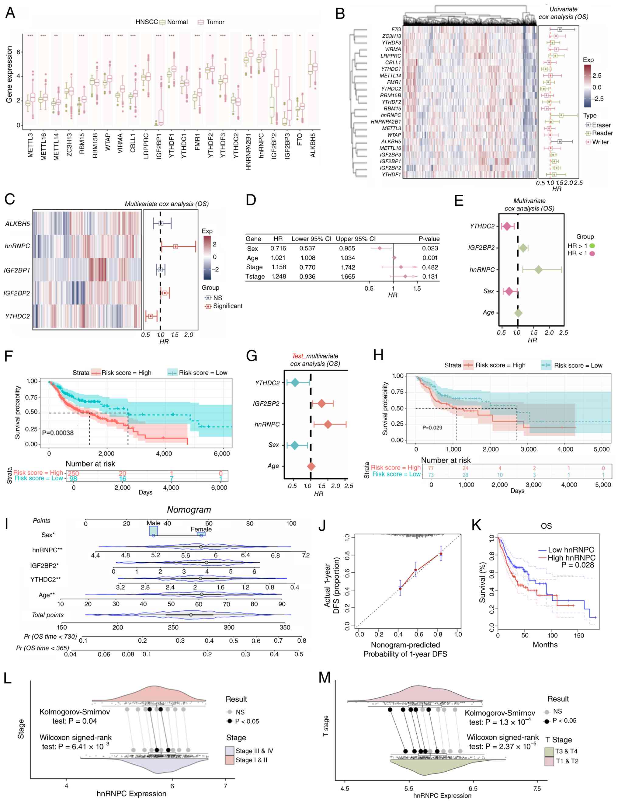 | Figure 1.hnRNPC represents a novel independent
prognostic biomarker in HNSCC. (A) Boxplot visualizing the
expression levels of 23 m6A-associated genes in 515
HNSCC and 44 normal tissues. (B) Heatmap (left) of the 23
m6A-associated genes and univariate Cox regression
analysis (right) based on m6A-associated genes
associated with the OS of HNSCC. (C; left) Heatmap of the
significant genes (hnRNPC, ALKBH5, IGF2BP1, IGF2BP2 and YTHDC2) in
the univariate Cox regression analysis and (right) multivariate Cox
regression analysis based on candidate genes with the OS of HNSCC.
(D) Forest plot presents the univariate Cox regression analysis of
the clinical information of the training set. (E) Forest plot
presents the multivariate regression analysis of the candidate
genes in the training set and clinical information (pink, HR <1;
green, HR >1). (F) Kaplan-Meier OS curves for patients in the
training set assigned to high- and low-risk groups. (G) Forest plot
presents the results of a multivariate regression analysis of
candidate genes in the test set and clinical information (red, HR
>1; blue, HR <1). (H) Kaplan-Meier OS curves for patients in
the test set assigned to high- and low-risk groups. (I) Nomogram
integrating the HNSCC risk score with age, sex and expression
levels of hnRNPC, IGF2BP2 and YTHDC2 to predict 1- and 2-year OS.
(J) Calibration curves confirming good agreement between
nomogram-predicted and -observed survival probabilities. (K)
Kaplan-Meier curves depicting OS according to the hnRNPC expression
based on The Cancer Genome Atlas data. (L) Box and whisker plots
comparing hnRNPC expression across pathological stages (stage I and
II vs. III and IV) and (M) T classifications (T3 and T4 vs. T1 and
T2). Boxes represent median and IQR; whiskers indicate 1.5X IQR.
Unless otherwise stated, data are expressed as mean ± SD.
*P<0.05, **P<0.01 and ***P<0.001. hnRNPC, heterogeneous
nuclear ribonucleoprotein C; HNSCC, head and neck squamous cell
carcinoma; NS, not significant; OS, overall survival; DFS,
disease-free survival; HR, hazard ratio; IQR, interquartile range;
IGF2BP2, insulin-like growth factor 2 mRNA-binding protein 2. |
hnRNPC exerts oncogenic functions in
OSCC
Prior to functional studies, the present study
examined the endogenous mRNA expression level of hnRNPC in a panel
of six commonly used OSCC cell lines (CAL-27, SCC-15, SCC-9,
SCC-25, HSC-3 and FaDu) by qPCR. Among these, CAL-27 and SCC-15
exhibited the markedly highest basal levels of hnRNPC mRNA
(Fig. S2A), the present study thus
knocked down hnRNPC expression in both cell lines using two
distinct shRNA sequences to verify hnRNPC function (Fig. 2A and B). Results from CCK-8 and
colony formation assays demonstrated that hnRNPC knockdown
significantly inhibited the proliferative capacity of OSCC cells
compared with the NC group, confirming the oncogenic function of
hnRNPC (Fig. 2C-F). Furthermore,
EdU incorporation assay indicated that hnRNPC knockdown
significantly suppressed the proliferation rate of OSCC cells
(Fig. 2G and H). Consistently, nude
mouse subcutaneous xenograft experiments demonstrated that hnRNPC
silencing led to a significant reduction in both volume and weight
of subcutaneously formed OSCC tumors (Fig. 2I-K). Furthermore, hnRNPC knockdown
significantly decreased the frequency of tumor sphere formation by
CAL-27-derived stem cells, as determined by an in vitro
limiting dilution assay (Fig. 2L).
The present study analyzed the expression levels of key stemness
genes, including Oct4, Sox2 and Nanog, which are well-known in
promoting tumor cells with self-renewal capabilities, using qPCR.
hnRNPC knockdown resulted in a significant downregulation of these
genes (Fig. 2M). Therefore, hnRNPC
emerges as a key regulator of OSCC cell proliferation and the
maintenance of stem-like properties.
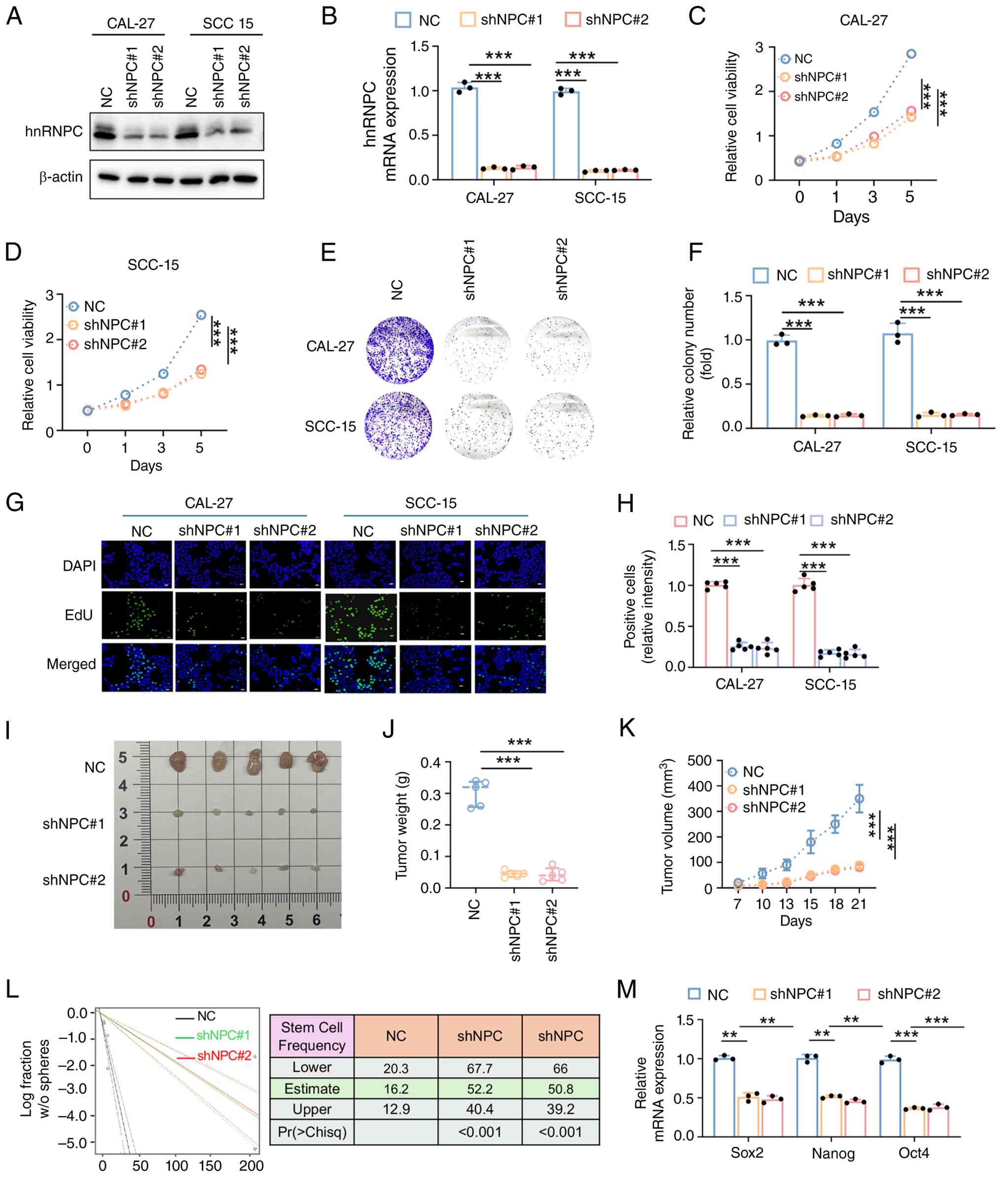 | Figure 2.hnRNPC exerts oncogenic functions in
oral SCC. (A) Western blotting and (B) qPCR were used to detect
protein expression after stable knockdown expression level of
hnRNPC in CAL-27 and SCC-15 cell lines. Cell Counting Kit-8 assay
was used to detect the effect of hnRNPC knockdown on the viability
of (C) CAL-27 and (D) SCC-15 cells. (E) Representative images and
(F) relative quantifications of colony formation assays of CAL-27
and SCC-15 cells. (G) 5-ethynyl-20-deoxyuridine solution was
incorporated for 2 h to evaluate the proliferation in CAL-27 and
SCC-15 cells. (H) Percentage of positive cells was quantified.
(I-K) Knockdown of hnRNPC can inhibit the growth of subcutaneous
tumors. SCC-15 cells with or without hnRNPC knockdown were
subcutaneously injected into nude mice. (I) Tumors were dissected
from the nude mice of each group and captured at 21 days after (J)
the weight of tumors was measured. (K) Tumor size was measured
every 3 days and growth curves were plotted. (L) Sphere formation
frequency of germline stem cells (27-CSC) with or without hnRNPC
knockdown was calculated with (left) extreme limiting dilution
assay analysis; (right) stem cell frequencies from 27-CSC cells
with or without hnRNPC knockdown were estimated as the ratio 1/x
with the upper and lower 95% confidence intervals, where 1=stem
cell and x=all cells. (M) qPCR was used to detect the Sox2, Nanog
and Oct4 mRNA expression levels. For (A-H) and (L-M), n=3
biologically independent experiments. For (I-K), n=5 mice per
group. Unless stated otherwise, data are expressed as mean ± SD.
**P<0.01 and ***P<0.001. qPCR, quantitative PCR; hnRNPC,
heterogeneous nuclear ribonucleoprotein C; SCC, squamous cell
carcinoma; CSC, cancer stem cell; NC, negative control; sh, short
hairpin RNA. |
hnRNPC promotes energy production and
antioxidant capacity in OSCC via BCAA metabolism
To investigate the mechanism underlying
hnRNPC-mediated regulation of OSCC progression, the present study
analyzed transcriptomic data of HNSC from TCGA database. The
results revealed that the BCAA metabolic pathway was active in HNSC
tissues with high hnRNPC expression (Fig. 3A). Since the BCAA metabolic pathway
is involved in energy production and redox homeostasis, the present
study also observed changes in pathways associated with oxidative
phosphorylation and stress (Fig. 3B and
C). Consistently, BCAA uptake capacity was significantly
impaired in OSCC cell lines with hnRNPC knockdown (Fig. 3D). Since BCAAs generate large
amounts of glutamate and BCKAs through catabolic pathways, where
glutamate contributes to the synthesis of reduced GSH for
antioxidant defense, and BCKAs promote the TCA cycle and oxidative
phosphorylation via acetyl-CoA production, the present study
examined these indicators in OSCC cells (31,32).
The present study findings demonstrated that hnRNPC knockdown led
to significantly decreased glutamate levels, reduced OCR and lower
GSH levels in OSCC cells compared with the negative control group
(Fig. 3E-J). In accordance, gene
set enrichment analysis (GSEA) based on TCGA data also revealed a
significant enrichment of GSH metabolism pathway in hnRNPC highly
expressed OSCC tissues (Fig. 3K and
L). These results indicated that hnRNPC can enhance tumor
energy production and antioxidant capacity by promoting the BCAA
metabolic pathway, thereby facilitating oxidative phosphorylation
and increasing GSH levels, which likely represents a key mechanism
underlying its tumor-promoting effects.
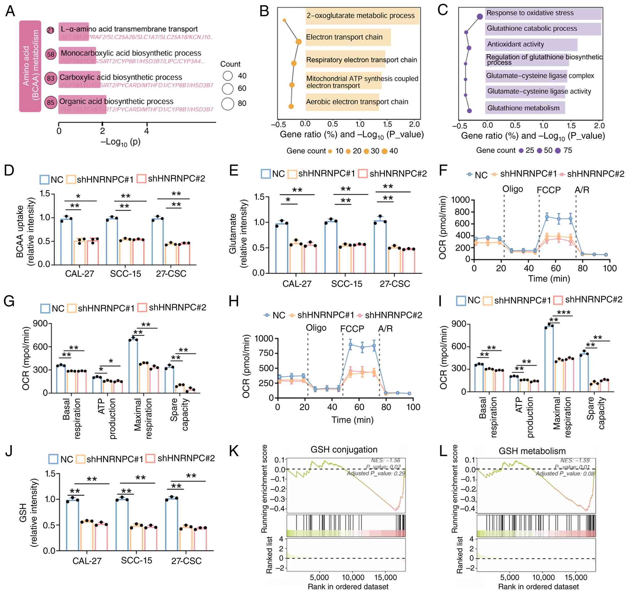 | Figure 3.hnRNPC promotes energy production and
antioxidant capacity in OSCC via BCAA metabolism. Gene Ontology
enrichment analysis of 1.5 fold-change differentially expressed
genes. (A) Metabolism of amino acids (BCAA), (B) energy metabolism
and (C) oxidative stress pathways. (D) BCAA uptake rate and (E)
internal glutamate levels were determined in CAL-27, SCC-15 and
27-CSC cells with or without hnRNPC knockdown. (F and H) Analysis
of OCR measurement of one representative experiment in indicated
cells after 4 h culture in basal condition and after sequential
addition of oligomycin, FCCP and A/R. (G and I) Quantification of
basal respiration, ATP-production-coupled respiration, maximal
respiration and spare capacity in CAL-27 and SCC-15 cells with or
without HRNPC knockdown. Box and whisker plots display median and
minimum to maximum; data in dot and bar plots are mean ± SD. (J)
GSH level was determined in CAL-27, SCC-15 and 27-CSC cells with or
without hnRNPC knockdown. (K and L) Gene set enrichment analysis
GSH metabolism pathway. Unless stated otherwise, data are expressed
as mean ± SD. *P<0.05, **P<0.01 and ***P<0.001. hnRNPC,
heterogeneous nuclear ribonucleoprotein C; A/R, antimycin/rotenone;
FCCP, carbonyl cyanide-p-trifluoromethoxyphenylhydrazone; GSH,
glutathione. |
hnRNPC regulates BCAT1 mRNA stability
in an m6A modification-dependent manner
To explore the specific mechanism by which hnRNPC
promotes tumor progression via regulation of the BCAA metabolism,
the present study analyzed differentially expressed genes between
hnRNPC-high and -low expressing samples from the HNSC transcriptome
database. The present study reported that BCAT1 was significantly
upregulated in OSCC tissues with high hnRNPC expression, suggesting
that hnRNPC may regulate BCAA metabolism by promoting BCAT1
expression (Fig. 4A and B).
Consistent with this observation, western blotting and qPCR results
demonstrated that BCAT1 expression was significantly suppressed in
OSCC cells following hnRNPC knockdown (Fig. 4C and D). Notably, treatment with the
BCAT1 inhibitor ERG240 further enhanced the tumor-suppressive
effects of hnRNPC knockdown (Fig. 4E
and F), indicating that combined targeting of hnRNPC and BCAT1
to interfere with BCAA metabolism could potentially represent a
promising therapeutic strategy for OSCC in the future.
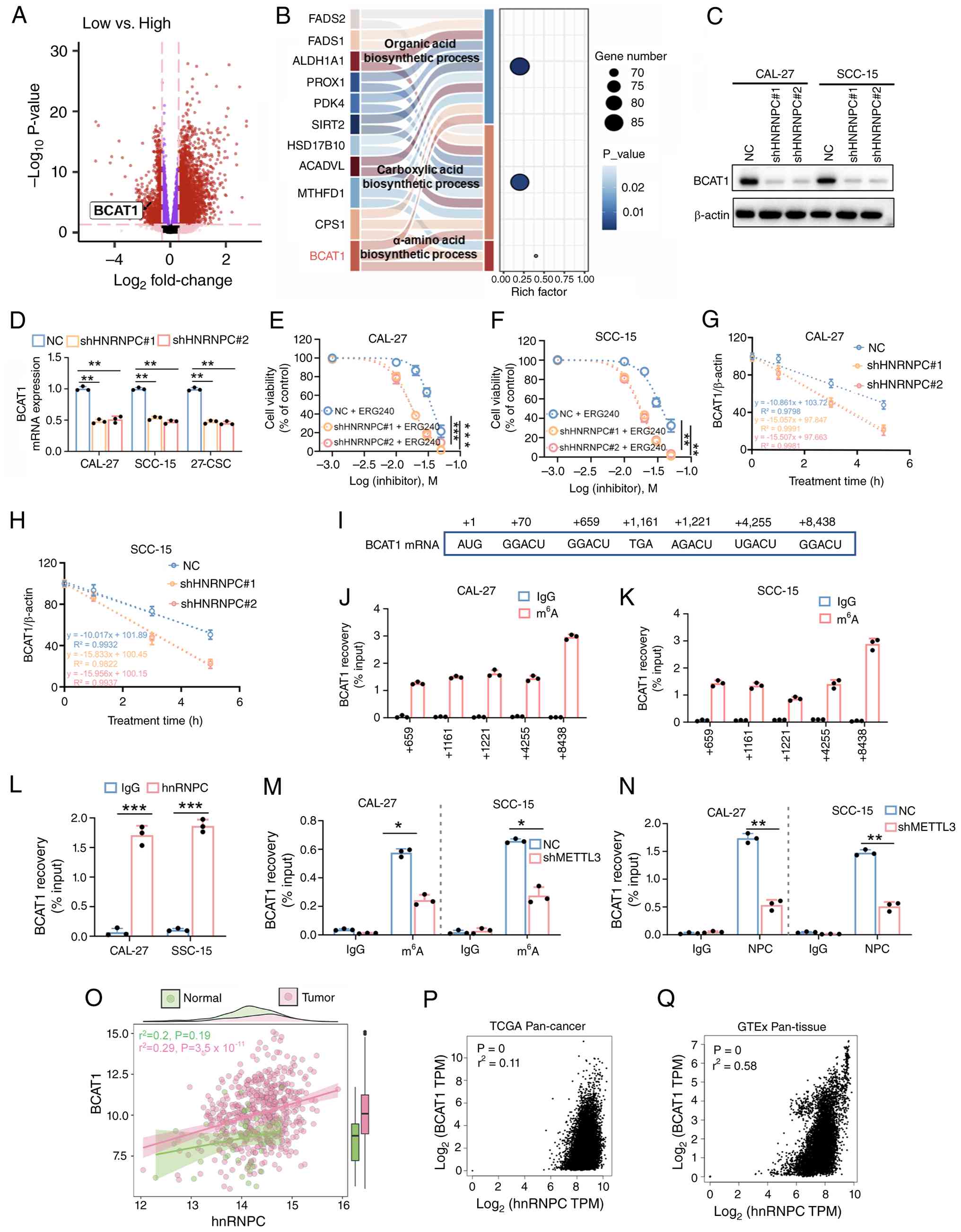 | Figure 4.hnRNPC regulates BCAT1 mRNA stability
in an m6A modification-dependent manner. (A) Volcano
plot displays the expression level of BCAT1 in hnRNPC with low vs.
high expression based on TCGA-sequencing data. (B) Sankey diagram
chart displays the expression levels of genes in different amino
acid metabolic pathways. BCAT1 denoted in pink. (C) Western
blotting and (D) qPCR analysis of BCAT1 protein and mRNA
expressions in in CAL-27, SCC-15 with or without hnRNPC expression.
Growth conditions of (E) CAL-27 and (F) SCC-15 cells with or
without hnRNPC knockdown after adding the BCAT1 inhibitor ERG240.
Detection of the stability of BCAT1 mRNA in (G) CAL-27 and (H)
SCC-15 cells after knockdown of hnRNPC. (I) Position map displays
the prediction of the top five m6A modification sites in
the BCAT1 mRNA using the ‘SRAMP’ algorithm. Analysis of MeRIP
assays detecting BCAT1 retrieved by a m6A antibody
around the high-confidence m6A sites in (J) CAL-27 and
(K) SCC-15 cells. (L) Assessment of RIP assays detecting BCAT1 mRNA
+8,438 site retrieved by a hnRNPC antibody or by IgG in CAL-27 and
SCC-15 cells. (M) Analysis of MeRIP assays detecting BCAT1 mRNA
+8,438 site recovered with m6A antibody in METTL3
knockdown in CAL-27 and SCC-15 cells. (N) Assessment of RIP assays
by a hnRNPC antibody or by IgG in CAL-27 and SCC-15 cells after
METTL3 knockdown. (O and P) Transcriptomic analysis of TCGA
database displays a significant positive correlation between hnRNPC
and BCAT1 expression in head and neck SCC tissues. (Q) GTEx
database also revealed the significant positive correlation between
hnRNPC and BCAT1 expression. Unless stated otherwise, data are
expressed as mean ± SD, *P<0.05, **P<0.01 and ***P<0.001.
m6A, N6-methyladenosine; hnRNPC, heterogeneous nuclear
ribonucleoprotein C; TCGA, The Cancer Genome Atlas; RIP, RNA
immunoprecipitation; MeRIP, methylated RIP; BCAT1, branched-chain
amino acid transaminase 1; qPCR, quantitative PCR; SCC, squamous
cell carcinoma; CSC, cancer stem cell; METTL3,
methyltransferase-like 3; sh, short hairpin RNA; TPM, transcripts
per million; GTEx, Genotype-Tissue Expression. |
Since hnRNPC functions as an m6A
modification reader protein to regulate mRNA stability of
downstream genes, the present study further investigated whether
hnRNPC-mediated regulation of BCAT1 is dependent on m6A
modification. mRNA stability assays demonstrated that BCAT1 mRNA
stability was significantly reduced after hnRNPC knockdown
(Fig. 4G and H). Using the ‘SRAMP’
algorithm, the present study predicted the top five m6A
modification sites in BCAT1 mRNA (Fig.
4I). Further MeRIP-qPCR experiments identified the most notable
m6A peak at position +8,438 within the BCAT1 3′-UTR
(Fig. 4J and K). RIP assays
confirmed the direct binding of hnRNPC to BCAT1 mRNA (Fig. 4L), while MeRIP analysis revealed
that METTL3 knockdown reduced m6A methylation on BCAT1
transcripts (Figs. 4M and S2B). Of note, METTL3 silencing also
diminished the association between hnRNPC and BCAT1 mRNA,
demonstrating that m6A marks are required for hnRNPC
recognition (Fig. 4N). Furthermore,
transcriptomic analysis of TCGA database revealed a significant
positive correlation between hnRNPC and BCAT1 expression in HNSC
tissues (Fig. 4O). Consistent with
this finding, pan-cancer transcriptomic data from TCGA database and
pan-tissue transcriptomic data from the GTEx database also revealed
the significant positive correlation between hnRNPC and BCAT1
expression (Fig. 4P and Q).
Therefore, hnRNPC may promote BCAT1 expression in an
m6A-dependent manner during OSCC progression, thereby
driving the malignant evolution of OSCC dependent on BCAA
metabolism.
α-KG rescues hnRNPC-mediated
BCAA-dependent energy deficiency
Since α-KG serves as a key donor for BCAT1-catalyzed
glutamate generation in the BCAA metabolic pathway and functions as
a key intermediate in mitochondrial TCA cycle, the present study
investigated whether exogenous supplementation of α-KG could
reverse the hnRNPC-induced suppression of BCAA metabolism and
subsequent energy deficiency. Results from the CCK-8 and colony
formation assays in the present study demonstrated that α-KG
supplementation significantly restored cell viability in
hnRNPC-knockdown OSCC cells (Fig.
5A-D). Stem cell viability experiments revealed that α-KG
supplementation notably increased the frequency of tumor sphere
formation by CAL-27-derived stem cells with hnRNPC knockdown
(Fig. 5E), accompanied by
significant recovery of Oct4, Sox2 and Nanog expression levels
which were downregulated by hnRNPC silencing (Fig. 5F). Furthermore, Seahorse assay
results indicated that α-KG supplementation led to significant
restoration of OCR and increased ATP levels in OSCC cells (Fig. 5G-K). Therefore, hnRNPC binds to
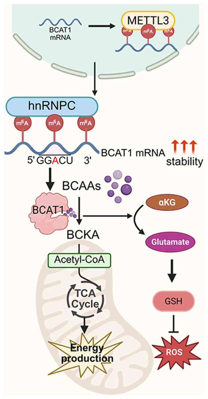
m6A-modified BCAT1 mRNA in a METTL3-dependent manner,
stabilizing BCAT1 transcripts and enhancing BCAT1 expression. This
upregulates BCAA catabolism, producing BCKAs for TCA cycle-driven
energy supply and glutamate for GSH-mediated antioxidant defense,
ultimately promoting OSCC cell proliferation, stemness and tumor
growth (Fig. 6). These findings
confirmed that BCAT1-mediated BCAA metabolic pathway for energy
production represents one of the key mechanisms underlying
hnRNPC-promoted tumor progression.
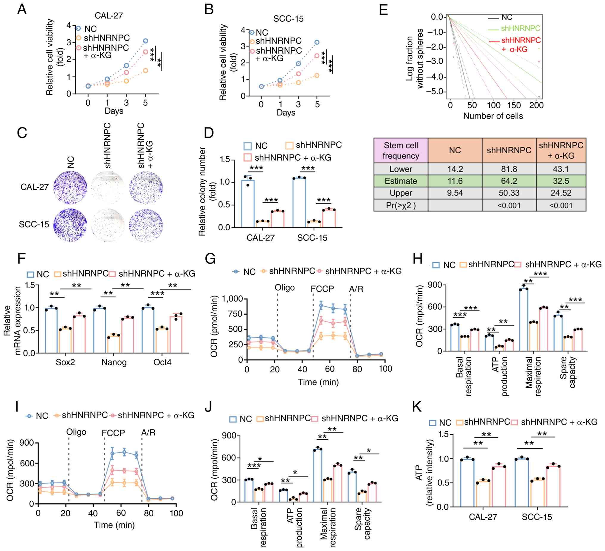 | Figure 5.α-KG rescues hnRNPC-mediated
BCAA-dependent energy deficiency. Cell Counting Kit-8 assay was
used to investigate the growth conditions of α-KG in CAL-27 (A) and
SCC-15 (B) cells with knocked-down hnRNPC. Representative images
(C) and relative quantifications (D) of colony formation assays of
indicated cells. (E) Sphere formation frequency in 27-CSC of
knocked-down hnRNPC with conditions of α-KG was calculated with
extreme limiting dilution assay analysis (top). Stem cell
frequencies from 27-CSC cells were estimated as the ratio 1/x with
the upper and lower 95% confidence intervals, where 1=stem cell and
x=all cells (bottom). (F) Quantitative PCR was used to detect the
Sox2, Nanog and Oct4 mRNA expression levels. (G and I) Analysis of
OCR measurement of one representative experiment in indicated cells
after 4 h culture in basal condition and after sequential addition
of oligomycin, FCCP and A/R. (H and J) Quantification of basal
respiration, ATP-production-coupled respiration, maximal
respiration and spare capacity in CAL-27 and SCC-15 of knocked-down
hnRNPC with conditions of α-KG. Box-and-whisker plots show median
and minimum to maximum; data in dot and bar plots are mean ± SD.
(K) ATP level was determined in CAL-27 and SCC-15 of knocked-down
hnRNPC with conditions of α-KG. Unless stated otherwise, data are
expressed as mean ± SD. *P<0.05, **P<0.01 and ***P<0.001.
m6A, N6-methyladenosine; hnRNPC, heterogeneous nuclear
ribonucleoprotein C; α-KG, α-ketoglutarate; NC, negative control;
sh, short hairpin RNA; FCCP, carbonyl
cyanide-p-trifluoromethoxyphenylhydrazone; A/R, antimycin/rotenone;
BCAA, branched-chain amino acid; SCC, squamous cell carcinoma; CSC,
cancer stem cell. |
Discussion
The present study systematically explored the role
and mechanism of hnRNPC in OSCC by integrating bioinformatics
analysis and experimental validation. Using data mining for
datasets of HNSCC in TCGA database, with OSCC as the predominant
subtype, the present study first identified hnRNPC as an
associative independent prognostic biomarker for HNSCC. The high
expression level of hnRNPC was associated with advanced
pathological stages, poor OS and served as a risk factor for
clinical outcomes. Functional experiments further confirmed the
oncogenic role of hnRNPC in OSCC. Knockdown of hnRNPC inhibited
cell proliferation, colony formation and tumor sphere formation
in vitro, and reduced tumor growth in nude mouse xenografts,
while also downregulating stemness-associated genes (Oct4, Sox2 and
Nanog). Mechanistically, the present study revealed that hnRNPC
promotes OSCC progression by regulating BCAA metabolism. hnRNPC
enhances BCAA uptake, supports BCAT1-dependent energy production
(via BCKAs entering the TCA cycle) and redox homeostasis (via
glutamate-mediated GSH synthesis). Of note, this regulation of
BCAT1 by hnRNPC is dependent on m6A modification; hnRNPC
binds to m6A-modified BCAT1 mRNA (predominantly at the
+8,438 site in the 3′-UTR) to maintain its stability and METTL3 is
required for this recognition. Lastly, exogenous supplementation of
α-KG, a key metabolite in TCA cycle, rescued the energy deficiency
and functional impairment of OSCC cells caused by hnRNPC knockdown,
further validating the BCAA metabolic pathway as a downstream
effector of hnRNPC.
A key innovation of the present study lies in
establishing a novel regulatory axis,
hnRNPC/m6A/BCAT1/BCAA metabolism, that drives OSCC
progression, filling key gaps in current knowledge. Previous
studies have separately highlighted the roles of m6A
modification and BCAA metabolism (for example, BCAT1) in OSCC, but
their functional association remained uncharacterized (12,30,33).
The present study findings demonstrated that hnRNPC, a classic
RNA-binding protein with established oncogenic roles in numerous
types of cancer, such as cervical and pancreatic cancer (34,35),
acts as an m6A reader in OSCC to stabilize BCAT1 mRNA.
This finding not only expands the functional spectrum of hnRNPC,
beyond mRNA splicing and stability regulation to
m6A-dependent metabolic control but also reveals a
unique epigenetic-metabolic crosstalk in OSCC. Specifically,
m6A modification, by marking BCAT1 mRNA, enables hnRNPC
to fine-tune BCAA metabolism, an adaptation that supports tumor
energy demands (via BCKA-driven oxidative phosphorylation) and
antioxidant defense (via glutamate-GSH axis). Although the present
study identified hnRNPC as a key m6A reader that
stabilizes BCAT1 mRNA in OSCC, emerging evidence indicates that
multiple m6A readers may coordinately regulate target
gene expression (16,17). Previous studies have confirmed that
YTHDF1 and IGF2BP2 act as key m6A readers involved in
OSCC progression (16,17). Therefore, the possibility that
YTHDF1, IGF2BP2 or other m6A readers may also contribute
to the regulation of BCAT1 mRNA stability or translation in OSCC
should not be excluded. The potential crosstalk or redundant
functions among these m6A readers warrant further
exploration in future studies.
Notably, this axis also explains the clinical
challenge of chemoresistance in OSCC. Previous studies have
reported that BCAT1-mediated GSH synthesis reduces drug-induced ROS
and DNA damage (9,36). The present study extended this
finding by demonstrating that hnRNPC upregulation enhances this
pathway, providing a molecular basis on why high hnRNPC expression
associates with poor prognosis. This mechanistic association offers
a novel perspective in overcoming chemoresistance, suggesting that
targeting hnRNPC or BCAT1 may hold potential to disrupt the
BCAA-GSH antioxidant system and sensitize OSCC cells to cisplatin
and other ROS-inducing therapies.
The hnRNPC/m6A/BCAT1 axis identified in
the present study may hold preliminary clinical relevance for the
management of OSCC in the future. From a prognostic perspective,
hnRNPC may serve as a prognostic biomarker. When combined with
pathological stage, hnRNPC expression may help improve risk
stratification for patients with OSCC and assist clinicians in
identifying individuals at higher risk of recurrence or
progression. In terms of treatment, the present study preclinical
findings suggested that targeting this axis may represent a
promising investigative strategy. Combined inhibition of hnRNPC
(such as, using small interfering RNAs or small-molecule
inhibitors) and BCAT1 (for example, using ERG240) may theoretically
synergistically disrupt BCAA metabolism. Such disruption could
reduce tumor energy supply, impair antioxidant capacity and
increase chemosensitivity in preclinical models. Furthermore,
METTL3 shRNA can block the m6A modification of BCAT1
mRNA, thereby eliminating the stabilization of BCAT1 mRNA mediated
by hnRNPC. This approach may represent a potential alternative to
avoid potential off-target effects associated with direct hnRNPC
inhibition.
Despite its contributions, the present study had
several limitations. First, the functional experiments in the
present study were performed in only two OSCC cell lines (CAL-27
and SCC-15). The selection of these two lines was based on the
following rationale: i) The present study initially screened a
panel of six OSCC cell lines (CAL-27, SCC-15, SCC-9, SCC-25, HSC-3
and FaDu) by qPCR and identified that CAL-27 and SCC-15 express
endogenously high levels of hnRNPC mRNA, providing a
physiologically relevant background for loss-of-function
experiments. Furthermore, both cell lines have been extensively
used as validated models for m6A RNA methylation
research in OSCC, including functional studies of METTL3, METTL14
and Fat mass and Obesity-associated (37,38).
Furthermore, a recent study specifically demonstrated that hnRNPC
knockdown in CAL-27 cells significantly impairs proliferation,
invasion and malignant transformation (39), directly supporting the suitability
of this cell line in investigating hnRNPC-dependent mechanisms.
Nevertheless, the present study acknowledges that the number of
OSCC models were limited. Validation in additional OSCC cell lines
and patient-derived xenograft models, which further recapitulate
the heterogeneity of clinical OSCC, would strengthen the
translational relevance of the present study findings. Second,
formal in vivo metabolic intervention studies (for example,
using BCAT1 inhibitors or m6A modulators) and validation
using clinical patient samples remain lacking, to the best of our
knowledge. Therefore, the translational and therapeutic
implications stated in the present study should be considered
preliminary and exploratory. Third, the association between hnRNPC
expression and clinical chemoresistance was not validated using
patient samples, which is key to translating the present study
findings into clinical practice. Future studies should focus on
in vivo metabolic validation, patient cohort verification
and preclinical investigation of combination therapies targeting
the hnRNPC/m6A/BCAT1 axis to further establish its
clinical utility.
In conclusion, the present study revealed a novel
epigenetic-metabolic axis in OSCC, demonstrating that hnRNPC
promotes BCAA metabolism via m6A-dependent stabilization
of BCAT1. These findings not only enhance current understanding of
OSCC progression but also highlight promising candidate markers and
prospective therapeutic targets that warrant further validation
toward improvement of OSCC management in the future.
Supplementary Material
Supporting Data
Supporting Data
Acknowledgements
Not applicable.
Funding
The present study was supported by the Guidance Program of the
Jiangsu Provincial Health Commission (grant no. Z2022002) and the
Natural Science Basic Research Program of Shaanxi (grant no.
2025JC-YBQN-1095).
Availability of data and materials
The data generated in the present study may be
requested from the corresponding author.
Authors' contributions
YA designed the present study. ZZ, XG, TH, FC and MG
performed the experiments. ZZ, XG and TH analyzed the data. YA
wrote the manuscript. YA and ZZ confirm the authenticity of all the
raw data. All authors have read and approved the final
manuscript.
Ethics approval and consent to
participate
The animal experiments were approved by the Ethics
Committee of Zhenjiang Stomatological Hospital (approval no.
PJ2023-08-10; Zhenjiang, China) and complied with the National
Institutes of Health Guide For The Care And Use Of Laboratory
Animals (National Institutes of Health publications no. 8023;
revised 1978).
Patient consent for publication
Not applicable.
Competing interests
The authors declare that they have no competing
interests.
References
|
1
|
Hsu CW, Chen YT, Hsieh YJ, Chang KP, Hsueh
PC, Chen TW, Yu JS, Chang YS, Li L and Wu CC: Integrated analyses
utilizing metabolomics and transcriptomics reveal perturbation of
the polyamine pathway in oral cavity squamous cell carcinoma. Anal
Chim Acta. 1050:113–122. 2019. View Article : Google Scholar : PubMed/NCBI
|
|
2
|
Chamoli A, Gosavi AS, Shirwadkar UP,
Wangdale KV, Behera SK, Kurrey NK, Kalia K and Mandoli A: Overview
of oral cavity squamous cell carcinoma: Risk factors, mechanisms,
and diagnostics. Oral Oncol. 121:1054512021. View Article : Google Scholar : PubMed/NCBI
|
|
3
|
Tan Y, Wang Z, Xu M, Li B, Huang Z, Qin S,
Nice EC, Tang J and Huang C: Oral squamous cell carcinomas: State
of the field and emerging directions. Int J Oral Sci. 15:442023.
View Article : Google Scholar : PubMed/NCBI
|
|
4
|
Patil VM, Noronha V, Joshi A, Abhyankar A,
Menon N, Dhumal S and Prabhash K: Beyond conventional chemotherapy,
targeted therapy and immunotherapy in squamous cell cancer of the
oral cavity. Oral Oncol. 105:1046732020. View Article : Google Scholar : PubMed/NCBI
|
|
5
|
Parmar A, Macluskey M, Mc Goldrick N,
Conway DI, Glenny AM, Clarkson JE, Worthington HV and Chan KK:
Interventions for the treatment of oral cavity and oropharyngeal
cancer: Chemotherapy. Cochrane Database Syst Rev.
12:CD0063862021.PubMed/NCBI
|
|
6
|
Zhou Y, Wang L, Liu M, Jiang H and Wu Y:
Oral squamous cell carcinoma: Insights into cellular heterogeneity,
drug resistance, and evolutionary trajectories. Cell Biol Toxicol.
41:1012025. View Article : Google Scholar : PubMed/NCBI
|
|
7
|
Choi BH, Hyun S and Koo SH: The role of
BCAA metabolism in metabolic health and disease. Exp Mol Med.
56:1552–1559. 2024. View Article : Google Scholar : PubMed/NCBI
|
|
8
|
Qian L, Li N, Lu XC, Xu M, Liu Y, Li K,
Zhang Y, Hu K, Qi YT, Yao J, et al: Enhanced BCAT1 activity and
BCAA metabolism promotes RhoC activity in cancer progression. Nat
Metab. 5:1159–1173. 2023. View Article : Google Scholar : PubMed/NCBI
|
|
9
|
McBrayer SK, Mayers JR, DiNatale GJ, Shi
DD, Khanal J, Chakraborty AA, Sarosiek KA, Briggs KJ, Robbins AK,
Sewastianik T, et al: Transaminase inhibition by 2-hydroxyglutarate
impairs glutamate biosynthesis and redox homeostasis in glioma.
Cell. 175:101–116.e25. 2018. View Article : Google Scholar : PubMed/NCBI
|
|
10
|
Xu E, Ji B, Jin K and Chen Y:
Branched-chain amino acids catabolism and cancer progression: Focus
on therapeutic interventions. Front Oncol. 13:12206382023.
View Article : Google Scholar : PubMed/NCBI
|
|
11
|
Ananieva EA and Wilkinson AC:
Branched-chain amino acid metabolism in cancer. Curr Opin Clin Nutr
Metab Care. 21:64–70. 2018.PubMed/NCBI
|
|
12
|
Yuan Z, Li M and Tang Z: BCAT1 promotes
cell proliferation, migration, and invasion via the PI3K-Akt
signaling pathway in oral squamous cell carcinoma. Oral Dis.
31:364–375. 2025. View Article : Google Scholar : PubMed/NCBI
|
|
13
|
Deng X, Qing Y, Horne D, Huang H and Chen
J: The roles and implications of RNA m6A modification in
cancer. Nat Rev Clin Oncol. 20:507–526. 2023. View Article : Google Scholar : PubMed/NCBI
|
|
14
|
Hong J, Xu K and Lee JH: Biological roles
of the RNA m6A modification and its implications in
cancer. Exp Mol Med. 54:1822–1832. 2022. View Article : Google Scholar : PubMed/NCBI
|
|
15
|
Jiang X, Liu B, Nie Z, Duan L, Xiong Q,
Jin Z, Yang C and Chen Y: The role of m6A modification in the
biological functions and diseases. Signal Transduct Target Ther.
6:742021. View Article : Google Scholar : PubMed/NCBI
|
|
16
|
Zhao W, Cui Y, Liu L, Ma X, Qi X, Wang Y,
Liu Z, Ma S, Liu J and Wu J: METTL3 facilitates oral squamous cell
carcinoma tumorigenesis by enhancing c-Myc stability via
YTHDF1-mediated m6A modification. Mol Ther Nucleic
Acids. 20:1–12. 2020. View Article : Google Scholar : PubMed/NCBI
|
|
17
|
Xu L, Li Q, Wang Y, Wang L, Guo Y, Yang R,
Zhao N, Ge N, Wang Y and Guo C: Yang: m6A
methyltransferase METTL3 promotes oral squamous cell carcinoma
progression through enhancement of IGF2BP2-mediated SLC7A11 mRNA
stability. Am J Cancer Res. 11:5282–5298. 2021.PubMed/NCBI
|
|
18
|
De Kesel J, Fijalkowski I, Pieters T,
Borin C, Christensen KT, Wittouck M, Van Laere J, Guerrero L,
Reunes L, Caruso M, et al: HNRNPC and m6A RNA methylation control
oncogenic transcription and metabolism in T-cell leukemia. Blood.
146:275–290. 2025. View Article : Google Scholar : PubMed/NCBI
|
|
19
|
Bi Z, Liu Y, Zhao Y, Yao Y, Wu R, Liu Q,
Wang Y and Wang X: A dynamic reversible RNA
N6-methyladenosine modification: Current status and
perspectives. J Cell Physiol. 234:7948–7956. 2019. View Article : Google Scholar : PubMed/NCBI
|
|
20
|
Navickas A, Asgharian H, Winkler J, Fish
L, Garcia K, Markett D, Dodel M, Culbertson B, Miglani S, Joshi T,
et al: An mRNA processing pathway suppresses metastasis by
governing translational control from the nucleus. Nat Cell Biol.
25:892–903. 2023. View Article : Google Scholar : PubMed/NCBI
|
|
21
|
Wu Y, Zhao W, Liu Y, Tan X, Li X, Zou Q,
Xiao Z, Xu H, Wang Y and Yang X: Function of HNRNPC in breast
cancer cells by controlling the dsRNA-induced interferon response.
EMBO J. 37:e990172018. View Article : Google Scholar : PubMed/NCBI
|
|
22
|
Su Z, Liu X, Xu Y, Hu W, Zhao C, Zhao H,
Feng X, Zhang S, Yang J, Shi X and Peng J: Novel reciprocal fusion
genes involving HNRNPC and RARG in acute promyelocytic leukemia
lacking RARA rearrangement. Haematologica. 105:e376–e378. 2020.
View Article : Google Scholar : PubMed/NCBI
|
|
23
|
Chen JJ, Lu TZ, Wang T, Yan WH, Zhong FY,
Qu XH, Gong XC, Li JG, Tou FF, Jiang LP and Han XJ: The m6A reader
HNRNPC promotes glioma progression by enhancing the stability of
IRAK1 mRNA through the MAPK pathway. Cell Death Dis. 15:3902024.
View Article : Google Scholar : PubMed/NCBI
|
|
24
|
Qin Z, Bai J, He H and Li B: Expression
and significance of m6A-RNA-methylation in oral cancer and
precancerous lesion. Front Oncol. 13:10130542023. View Article : Google Scholar : PubMed/NCBI
|
|
25
|
Amin MB, Greene FL, Edge SB, Compton CC,
Gershenwald JE, Brookland RK, Meyer L, Gress DM, Byrd DR and
Winchester DP: The eighth edition AJCC cancer staging manual:
Continuing to build a bridge from a population-based to a more
‘personalized’ approach to cancer staging. CA Cancer J Clin.
67:93–99. 2017.PubMed/NCBI
|
|
26
|
Livak KJ and Schmittgen TD: Analysis of
relative gene expression data using real-time quantitative PCR and
the 2(−Delta Delta C(T)) method. Methods. 25:402–408. 2001.
View Article : Google Scholar : PubMed/NCBI
|
|
27
|
Shen L, Qu X, Li H, Xu C, Wei M, Wang Q,
Ru Y, Liu B, Xu Y, Li K, et al: NDRG2 facilitates colorectal cancer
differentiation through the regulation of Skp2-p21/p27 axis.
Oncogene. 37:1759–1774. 2018. View Article : Google Scholar : PubMed/NCBI
|
|
28
|
Liu H, Li D, Sun L, Qin H, Fan A, Meng L,
Graves-Deal R, Glass SE, Franklin JL, Liu Q, et al: Interaction of
lncRNA MIR100HG with hnRNPA2B1 facilitates m6A-dependent
stabilization of TCF7L2 mRNA and colorectal cancer progression. Mol
Cancer. 21:742022. View Article : Google Scholar : PubMed/NCBI
|
|
29
|
Tang D, Cao C, Huang S, He Q and Wang A:
YTHDF2 drives oral squamous cell carcinoma progression via
m6A-dependent degradation of MTUS1/ATIP1 mRNA and
mitochondrial dysregulation. Cell Signal. 136:1121452025.
View Article : Google Scholar : PubMed/NCBI
|
|
30
|
Zhang D, Mai L, Zhang L, Huang G, Lin Z,
Wang S, Rao G, Xie S and Pan C: The m6A reader IGF2BP2
promotes oral squamous cell carcinoma progression by maintaining
UCA1 stability. Recent Pat Anticancer Drug Discov. 20:697–709.
2025. View Article : Google Scholar : PubMed/NCBI
|
|
31
|
Nie C, He T, Zhang W, Zhang G and Ma X:
Branched Chain amino acids: Beyond nutrition metabolism. Int J Mol
Sci. 19:9542018. View Article : Google Scholar : PubMed/NCBI
|
|
32
|
Duan Y, Li F, Li Y, Tang Y, Kong X, Feng
Z, Anthony TG, Watford M, Hou Y, Wu G and Yin Y: The role of
leucine and its metabolites in protein and energy metabolism. Amino
Acids. 48:41–51. 2016. View Article : Google Scholar : PubMed/NCBI
|
|
33
|
Su Y, Hu Y, Qu B, Lei R and Guo G: METTL3
promotes OSCC progression by down-regulating WEE1 in a
m6A-YTHDF2-dependent manner. Mol Biotechnol. 67:1867–1879. 2025.
View Article : Google Scholar : PubMed/NCBI
|
|
34
|
Liu YY, Xia M, Chen ZB, Liao YD, Zhang CY,
Yuan L, Pan YW, Huang H, Lu HW and Yao SZ: HNRNPC mediates
lymphatic metastasis of cervical cancer through m6A-dependent
alternative splicing of FOXM1. Cell Death Dis. 15:7322024.
View Article : Google Scholar : PubMed/NCBI
|
|
35
|
Yang N, Liu L, Liu X, Chen Y, Lu J and
Wang Z: hnRNPC promotes malignancy in pancreatic cancer through
stabilization of IQGAP3. Biomed Res Int. 2022:63196852022.
View Article : Google Scholar : PubMed/NCBI
|
|
36
|
Wang Y, Zhang J, Ren S, Sun D, Huang HY,
Wang H, Jin Y, Li F, Zheng C, Yang L, et al: Branched-chain amino
acid metabolic reprogramming orchestrates drug resistance to EGFR
tyrosine kinase inhibitors. Cell Rep. 28:512–525.e6. 2019.
View Article : Google Scholar : PubMed/NCBI
|
|
37
|
Wen H, Tang J, Cui Y, Hou M and Zhou J:
m6A modification-mediated BATF2 suppresses metastasis and
angiogenesis of tongue squamous cell carcinoma through inhibiting
VEGFA. Cell Cycle. 22:100–116. 2023. View Article : Google Scholar : PubMed/NCBI
|
|
38
|
Chen X, Liu Y, Sun D, Sun R, Wang X, Li M,
Song N, Ying J, Guo T and Jiang Y: Long noncoding RNA lnc-H2AFV-1
promotes cell growth by regulating aberrant m6A RNA modification in
head and neck squamous cell carcinoma. Cancer Sci. 113:2071–2084.
2022. View Article : Google Scholar : PubMed/NCBI
|
|
39
|
Zhang Y, Wang Y, Peng J, Zhao K, Li L,
Zhang Y, Zhai Z, Yuan S, Li S, Ye F and Wang L: Expression and
prognostic significance of the m6A RNA methylation regulator HNRNPC
in HNSCC. Front Oncol. 15:15168672025. View Article : Google Scholar : PubMed/NCBI
|