Evaluation of contrast-enhanced ultrasonography using perfluorobutane (Sonazoid®) in patients with small hepatocellular carcinoma: Comparison with dynamic computed tomography

  • Authors:
    • Miki Kan
    • Atsushi Hiraoka
    • Takahide Uehara
    • Satoshi Hidaka
    • Misa Ichiryu
    • Hiromasa Nakahara
    • Hironori Ochi
    • Atsushi Tanabe
    • Akihiro Kodama
    • Aki Hasebe
    • Yasuyuki Miyamoto
    • Tomoyuki Ninomiya
    • Masanori Abe
    • Yoichi Hiasa
    • Bunzo Matsuura
    • Morikazu Onji
    • Yuki Shinbata
    • Chieko Kameoka
    • Shigekazu Doi
    • Hiromi Tamura
    • Keizou Furuya
    • Kojiro Michitaka
  • View Affiliations

  • Published online on: May 1, 2010     https://doi.org/10.3892/ol_00000085
  • Pages: 485-488
Metrics: Total Views: 0 (Spandidos Publications: | PMC Statistics: )
Total PDF Downloads: 0 (Spandidos Publications: | PMC Statistics: )


Abstract

This study aimed to elucidate the efficacy of contrast-enhanced ultrasonography (CEUS) with perfluorobutane (Sonazoid®) in the diagnosis of hepatocellular carcinomas (HCCs), particularly small HCCs, by comparing the results with dynamic computed tomography (Dy-CT). Seventy-nine nodules in 69 patients with chronic liver disease, suspected as HCCs were studied. The nodules were selected based on the results of B-mode ultrasonography and/or Dy-CT conducted between January and August 2007. The nodules were divided into two groups: the S-group with tumors ≤2 cm (49 nodules), and the L-group with tumors >2 cm (30 nodules). Typical HCCs were defined, and the nodules were enhanced and shown as defects in the arterial and late phase of Dy-CT, respectively. Target lesions were scanned using CEUS, and the results were compared with those of Dy-CT. The L-group nodules diagnosed as HCCs using Dy-CT were also diagnosed as HCCs using CEUS. In the S-group, the diagnostic sensitivity of CEUS was 94.7% and the specificity was 81.8%. We diagnosed two liver tumors that were detected by CEUS but not by Dy-CT; biopsies revealed one tumor to be a well-differentiated HCC and the other to be an atypical adenomatous hyperplasia. The sensitivity and specificity of CEUS against HCC were high even in the small-size HCCs. Thus, Sonazoid is useful in the screening for small HCCs.
View Figures
View References

Related Articles

Journal Cover

May-June 2010
Volume 1 Issue 3

Print ISSN: 1792-1074
Online ISSN:1792-1082

Sign up for eToc alerts

Recommend to Library

Copy and paste a formatted citation
x
Spandidos Publications style
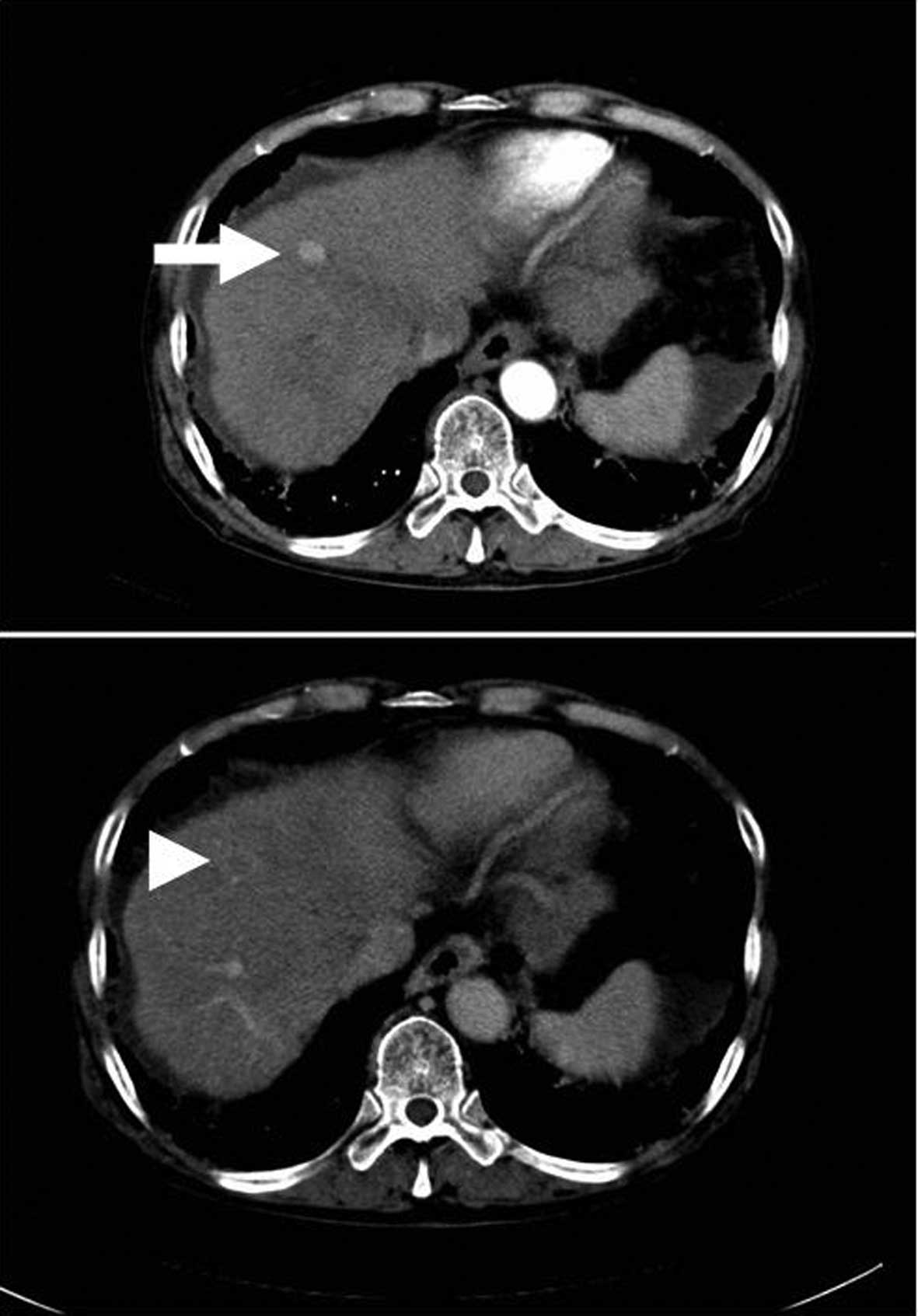
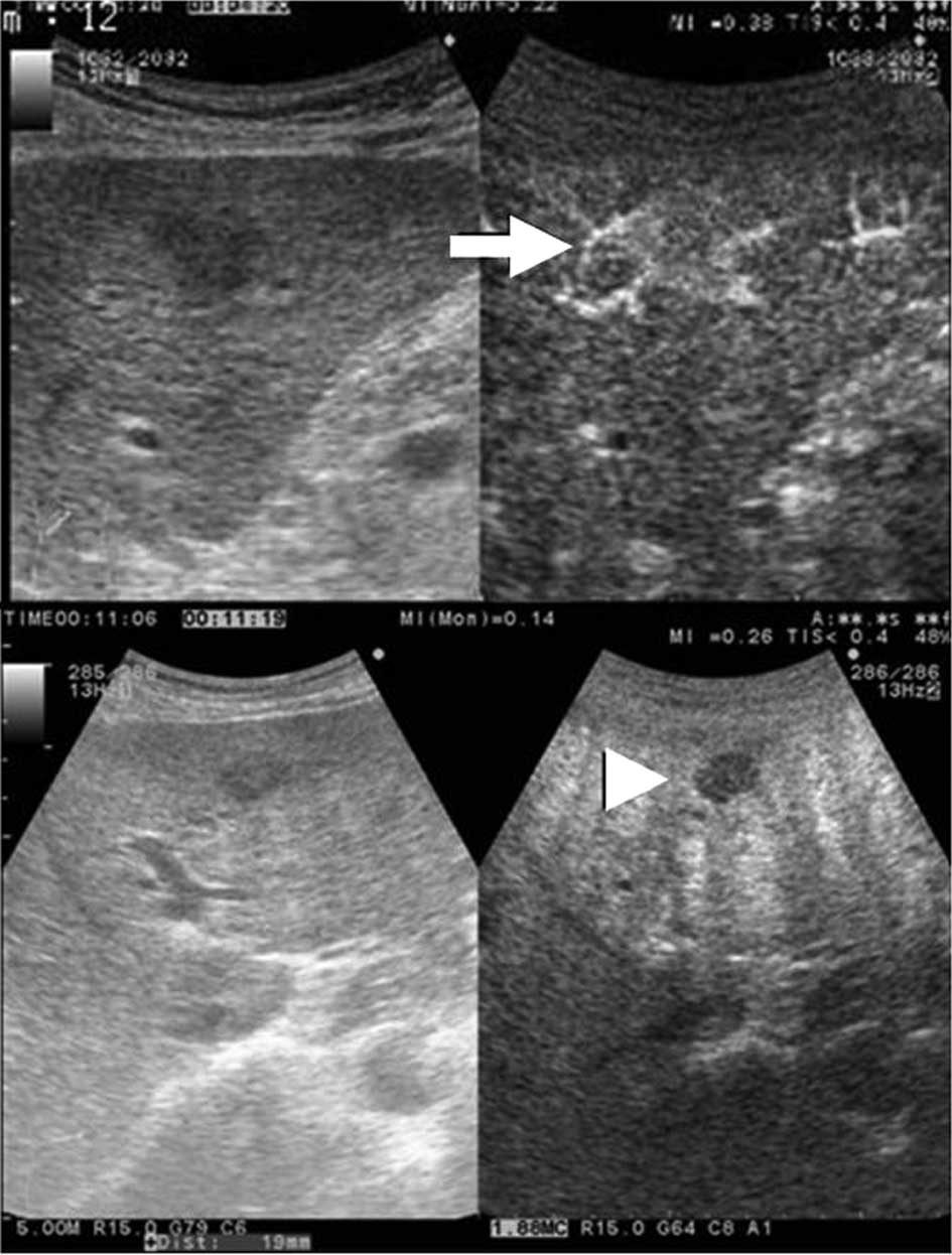
Kan M, Hiraoka A, Uehara T, Hidaka S, Ichiryu M, Nakahara H, Ochi H, Tanabe A, Kodama A, Hasebe A, Hasebe A, et al: Evaluation of contrast-enhanced ultrasonography using perfluorobutane (Sonazoid®) in patients with small hepatocellular carcinoma: Comparison with dynamic computed tomography . Oncol Lett 1: 485-488, 2010
APA
Kan, M., Hiraoka, A., Uehara, T., Hidaka, S., Ichiryu, M., Nakahara, H. ... Michitaka, K. (2010). Evaluation of contrast-enhanced ultrasonography using perfluorobutane (Sonazoid®) in patients with small hepatocellular carcinoma: Comparison with dynamic computed tomography . Oncology Letters, 1, 485-488. https://doi.org/10.3892/ol_00000085
MLA
Kan, M., Hiraoka, A., Uehara, T., Hidaka, S., Ichiryu, M., Nakahara, H., Ochi, H., Tanabe, A., Kodama, A., Hasebe, A., Miyamoto, Y., Ninomiya, T., Abe, M., Hiasa, Y., Matsuura, B., Onji, M., Shinbata, Y., Kameoka, C., Doi, S., Tamura, H., Furuya, K., Michitaka, K."Evaluation of contrast-enhanced ultrasonography using perfluorobutane (Sonazoid®) in patients with small hepatocellular carcinoma: Comparison with dynamic computed tomography ". Oncology Letters 1.3 (2010): 485-488.
Chicago
Kan, M., Hiraoka, A., Uehara, T., Hidaka, S., Ichiryu, M., Nakahara, H., Ochi, H., Tanabe, A., Kodama, A., Hasebe, A., Miyamoto, Y., Ninomiya, T., Abe, M., Hiasa, Y., Matsuura, B., Onji, M., Shinbata, Y., Kameoka, C., Doi, S., Tamura, H., Furuya, K., Michitaka, K."Evaluation of contrast-enhanced ultrasonography using perfluorobutane (Sonazoid®) in patients with small hepatocellular carcinoma: Comparison with dynamic computed tomography ". Oncology Letters 1, no. 3 (2010): 485-488. https://doi.org/10.3892/ol_00000085