Introduction
Although radiotherapy has proven to be effective and
is among the most common treatment modalities (1), much effort is being invested to
further improve its effectiveness. Besides the development of new
treatment techniques, the effects of radiotherapy can be improved
by concurrent administration of radiosensitizing agents.
Previously, it was shown that chemotherapeutic agents such as
cisplatin (cis-diamminodi-chloroplatinum II) and gemcitabine
(difluorodeoxycytidine) are potent radioenhancers both in
vitro and in vivo (2–4).
Halogenated pyrimidines such as bromodeoxyuridine (BrdU) and
iodo-deoxyuridine (IdU) are also potent radiosensitizers (5–8). The
aforementioned agents affect cells at the DNA level and are either
directly incorporated into the DNA (BrdU, gemcitabine), form DNA
adducts/crosslinks (cisplatin) or inhibit the production of
deoxynucleotides, necessary building blocks for DNA syntheses and
repair (gemcitabine).
Another group of potential radiosensitizers consists
of agents that influence chromatin structure, thereby affecting DNA
damage repair. Chromatin is a highly dynamic, yet efficiently
organized structure that regulates almost all DNA-associated
processes, including transcription, replication and repair
(9). The basic chromatin unit is
the nucleosome that consists of approximately 146 bp of DNA wrapped
around a protein octamer containing two molecules each of histones
H2A, H2B, H3 and H4 (10). The
histone core contains highly conserved tail regions that can be
covalently modified by phosphorylation, methylation and acetylation
(11,12). Histone acetylation is
antagonistically regulated by histone acetylases (HATs) and histone
deacetylases (HDACs). In general, histone acetylation is associated
with a more open chromatin structure and active gene transcription,
whereas deacetylation is associated with condensed chromatin and
gene repression (12). Due to the
intimate relationship between chromatin and DNA repair, disruption
of the balance between HDACs and HATs may play a role in
antineoplastic therapy. Subsequently, multiple HDAC inhibitors are
currently undergoing clinical trials (13). HAT inhibitors have been less
extensively studied, but may also possess anticancer potential.
Based on sequence and structure homology, three main
classes of HATs have been described, including GCN5/PCAF, p300/CBP
and the MYST family (MOZ, YBF2/SAS3, SAS2 and Tip60) (14). The exact functions that HATs play in
cell biology have yet to be investigated in more detail, but it is
clear that HATs play a role in gene transcription and that the
effects of HATs can be both global and gene-specific. HATs are
often part of large multiprotein complexes that determine their
specificity. HATs primarily acetylate histones, but other proteins
can be affected as well (15). The
cell permeable salicylic acid analog anacardic acid (AA) is a
potent, non-competitive inhibitor of p300 and PCAF HAT activities
(16). Sun et al (17) demonstrated that AA is an effective
inhibitor of the MYST HAT Tip60. Tip60 was reported to play a role
in the acetylation and activation of the ataxia telangiectasia
mutant, an important mediator of DNA damage response (18). Ikura et al (19) reported that the inactivation of
Tip60 resulted in a defective double strand break (DSB) repair.
This defective repair may be due to the failure of Tip60 to induce
local relaxation of chromatin at DSB sites, which may be necessary
for proper DSB responses (9). This
failure is supported by the finding that compact chromatin
structure (heterochromatin) poses a (physical) barrier to the
propagation of DSB signaling (20).
Moreover, Biade et al (21)
previously showed that chemicals that promote chromatin compaction
were able to induce significant radiosensitization in tumour cells.
Due to the influence of chromatin structure on DNA repair, HAT
inhibitors are likely to posses radiosensitizing properties.
Subsequently, the radiosensitizing effects of low concentrations of
AA on a set of mammalian cell lines were studied.
Materials and methods
Cell lines
The human squamous lung carcinoma SW1573 cell line
was cultured in Leibowitz-15 medium (Gibco-BRL Life Technologies,
Breda, The Netherlands) supplemented with 10% heat-inactivated
fetal bovine serum (FBS) and 2 mM glutamine, 100 U/ml penicillin
and 100 μg/ml streptomycin (Gibco). The human osteosarcoma U2OS
cell line was cultured in Dulbecco’s modified Eagle’s medium (DMEM)
(Gibco) supplemented with 10% heat-inactivated FBS and 2 mM
glutamine, 100 U/ml penicillin and 100 μg/ml streptomycin. The
Chinese hamster V79 lung fibroblasts were cultured in minimum
essential medium (MEM) containing Hanks salts (Gibco) supplemented
with 10% heat-inactivated FBS and 2 mM glutamine, 100 U/ml
penicillin and 100 μg/ml streptomycin. SW1573, U2OS and V79 cells
were maintained in a 37°C humidified incubator with an air
atmosphere of 0, 10 and 2% CO2, respectively. The cell
lines were cultured as a monolayer in tissue culture flasks (Costar
Europe Ltd., Badhoevedorp, The Netherlands) and passaged twice a
week to ensure exponential growth.
Irradiation
Irradiation was performed with γ-rays from a
137Cs source, yielding a dose rate of about 0.6 Gy/min.
Cells were irradiated with doses of up to 8 Gy.
Anacardic acid
A 100 mM stock solution of AA (Calbiochem, cat no.
172050) was dissolved in dimethylsulfoxide (DMSO) just before use.
For the experiments, AA was pre-diluted in medium at appropriate
concentrations ranging from 50 to 300 μM and then added to the
cells. AA concentrations of up to 300 μM were used for the
cytotoxicity measurement.
Clonogenic assay
Cytotoxicity of AA alone, ionizing radiation (IR)
alone and combination treatment of AA and IR was measured using
clonogenic survival assays (22).
Cytotoxicity of AA alone was determined after a 4-h incubation at
concentrations of 0, 50, 100, 200 and 300 μM. Control experiments
with DMSO were accomplished. Cells were plated in appropriate
densities. After 3 h, when cells were attached, they were treated
with AA for 4 h. In case of combined treatment, cells were
irradiated with 0, 2, 4, 6 and 8 Gy 1 h after the onset of 100 μM
AA incubation. Following treatment, the cells were washed twice
with phosphate buffered saline, and fresh medium was added. Cells
were allowed to form colonies for 10 days and were fixed and
stained using a solution of 0.05% crystal violet in 6%
glutaraldehyde. Colonies of ≥50 cells were scored as originating
from a single clonogenic cell (22).
Surviving fractions (S(D)/(S(0)) after dose D,
corrected for the toxicity of AA alone (S(0)), were calculated and
survival curves were analysed using SPSS statistical software
(Chicago, IL, USA) by fitting the data using weighted, stratified,
linear regression, according to the linear-quadratic formula:
S(D)/S(0) = exp-(αD+βD2) (5,23,24).
A procedure described by Franken et al (22) was used to test for a statistically
significant difference between IR alone and the combined treatment
of AA and IR curves.
Results and Discussion
To investigate the radiosensitizing properties of
HAT inhibitors the commercially available AA was selected. To
determine the appropriate dose for radiosensitization studies,
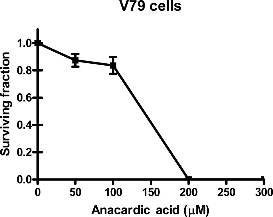
cytotoxicity experiments were performed. Fig. 1 shows the cytotoxicity of AA for
V79, SW1573 and U2OS cells incubated with concentrations up to 300
μM for 4 h.
It was noted that in the three cell lines the
cytotoxicity of AA increases rapidly with concentrations above 100
μM. When incubated at a concentration of 200 μM, V79 and U2OS cell
lines show no remaining viable clonogenic cells. A similar effect
is achieved for SW1573 cells at a concentration of 300 μM. This
effect indicates that the cytotoxicity of AA depends on the cell
line tested. To evaluate the clinical applicability of AA as a
radiosensitizer, a 4-h incubation at a concentration of 100 μM was
selected for further experiments. Treatment with this concentration
was selected as it resulted in relatively low cytotoxicity in the
examined cell lines showing a clonogenic survival (±SEM) of
0.84±0.06, 0.84±0.04 and 0.84±0.09 for V79, SW1573 and U2OS cells,
respectively. These data were corrected for the toxicity of the AA
solvent, DMSO.
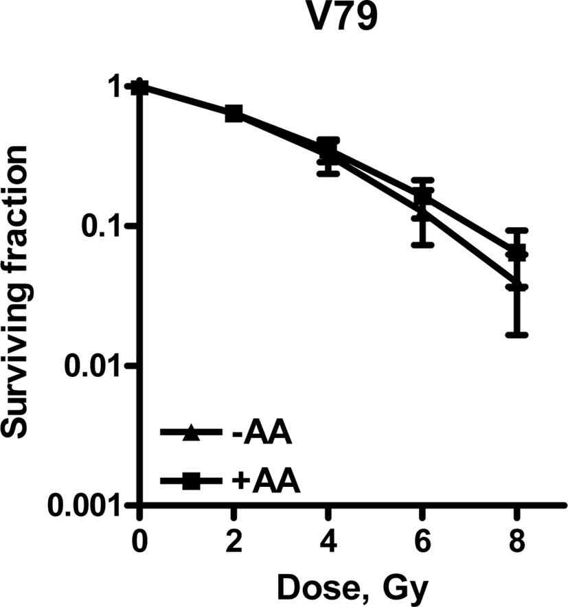
To establish the radiosensitizing properties of AA,
clonogenic assays were performed. Survival curves are shown in
Fig. 2. Cells were plated in
appropriate densities prior to the commencement of AA treatment and
irradiation. In this experimental set-up, the cells were allowed to
repair the radiation-induced DNA damage.
The survival curves were analysed according to the
linear-quadratic model (LQ) describing lethal events as a function
of the dose (D) with the parameters α and β as constants: F(D) =
αD+ βD2 (5,23,24).
This model describes lethal events induced in the
low-dose range (≤2Gy; α) separately from damage induced in the
high-dose range (≥2Gy; β). In the low-dose range, damage is
primarily induced by single-hit particle tracks, represented as
lethal (LD) and potentially lethal damage (PLD). If not repaired,
the latter type of damage becomes lethal, representing fixated
(P)LD.
In the higher dose range LD is partly caused by the
accumulation or interaction of multiple particle-induced sublethal
damage (SLD) events. Table I shows
the linear-quadratic parameters α and β.
 | Table ILinear- quadratic parameters. |
Table I
Linear- quadratic parameters.
| LQ parameter | α, Gy-1 | β, Gy-2 |
|---|
| V-79 | radiation only | 0.164 ± 0.044 | 0.030 ± 0.008 |
| radiation + AA | 0.182 ± 0.031 | 0.020 ± 0.005 |
| SW1573 | radiation only | 0.015 ± 0.026 | 0.055 ± 0.004 |
| radiation + AA | 0.089 ± 0.041 | 0.043 ± 0.007 |
| U2OS | radiation only | 0.475 ± 0.050 | 0.018 ± 0.024 |
| radiation + AA | 0.635 ± 0.065 | 0.020 ± 0.020 |
Fig. 2 shows that
U2OS cells are radiosensitized by incubation with 100 μM AA for 4
h, since the survival curves with and without AA showed a
significant difference.
A significant increase of the α value by a factor of
1.3 was observed. The β value was not significantly changed,
indicating an intact repair of SLD (5). As repair function is unaffected, a
likely explanation for the enhanced expression of LD induced by AA
is due to an increase in the amount of directly lethal lesions.
One explanation for the increased number of lethal
lesions induced by AA may be due to the hypoacetylated status and
subsequent condensed structure of the chromatin. Biade et al
(21) previously showed that
compacted chromatin is a radiation hypersensitive target associated
with the single-hit (low-dose) killing of tumour cells.
In contrast to U2OS cells, neither V79 nor SW1573
cells were radiosensitized by incubation with 100 μM AA. SW1573
cells showed a significant increase of the α value by a factor of
5.9. This increase was accompanied, however, by a decrease of the β
value by a factor of 0.78, indicating the enhanced repair of SLD.
This decrease explains why the complete survival curves with and
without AA were not significantly different for SW1573 cells.
Few studies have been published regarding the effect
of AA on radiosensitization. In contrast to our results, Sun et
al (17) found a distinct
radiosensitizing effect of a 4-h and 40 min incubation in the
presence of 30 μM AA in HeLa, SCC35 and SQ20B cells. These authors
suggested that the radiosensitizing properties of AA may be due to
the inhibition of the Tip60-dependent activation of ataxia
telangiectasia mutant and DNA-PKcs protein kinases, proteins
essential in cellular responses to DNA damage.
The hypoacetylation of H4 histones and a moderate
increase in sensitivity to ionizing radiation by the HAT inhibitors
copper sulfate (CuSO4) and nickel chloride
(NiCl2) was demonstrated by Song et al (25) when using non-toxic concentrations on
yeast cells.
Bandyopadhyay et al (26) reported an ultraviolet-sensitizing
effect in the human lung H358 cancer cell line when using the
bisubstrate adduct spermidine-CO-CH2-CoA
(Spd(N1)-CoA). The radiosensitizing effect correlated
with an increased inhibition of histone acetylation and was
accompanied by a transient arrest of DNA synthesis, transient delay
in S-phase progression and the inhibition of nucleotide excision
repair and DNA double strand break repair.
Although we did not find radiosensitization in all
of the cell lines studied, the likelihood of AA to increase α
suggests potential advantages for clinical application, especially
for low-dose fractionated radiotherapy. However, further intensive
studies extended to other cell lines are required to elucidate
whether radiosensitization is due to an increased (P)LD and/or the
effects on DNA repair. Insight into the mechanism of
radiosensitization is required if it is to be used in a therapeutic
setting.
Acknowledgements
The authors would like to thank Professor G.W.
Barendsen for his critical comments and useful suggestions on the
manuscript. The Maurits and Anna de Kock and the Nijbakker Morra
foundations are acknowledged for sponsoring laboratory
equipment.
References
|
1
|
Joiner M, van der Kogel A and Steel G:
Introduction: the significance of radiobiology and radiotherapy for
cancer treatment. Basic Clinical Radiobiology. 4th edition. Joiner
and van der Kogel A: University Press/Hodder; Arnold, Oxford: pp.
1–10. 2009, View
Article : Google Scholar
|
|
2
|
Haveman J, Castro Kreder N, Rodermond HM,
Franken NA, Stalpers LJ, Zdzienicka MZ and Peters GJ: Cellular
response of X-ray sensitive hamster mutant cell lines to
gemcitabine, cisplatin and 5-fluorouracil. Oncol Rep. 12:187–192.
2004.PubMed/NCBI
|
|
3
|
Bergs JWJ, Franken NA, ten Cate R, van
Bree C and Haveman J: Effect of cisplatin and gamma irradiation on
cell survival, the induction of chromosomal aberrations and
apoptosis in SW-1573 cells. Mutat Res. 594:148–154. 2006.
View Article : Google Scholar : PubMed/NCBI
|
|
4
|
Bergs JWJ, Franken NA, Haveman J, Geijsen
ED, Crezee J and van Bree C: Hyperthermia, cisplatin and radiation
trimodality treatment: a promising cancer treatment? A review from
preclinical studies to clinical application. Int J Hyperthermia.
23:329–341. 2007. View Article : Google Scholar
|
|
5
|
Franken NA, van Bree C, Kipp JBA and
Barendsen GW: Modification of potentially lethal damage in
irradiated Chinese hamster V79 cells after incorporation of
halogenated pyrimidines. Int J Radiat Biol. 72:101–109. 1997.
View Article : Google Scholar : PubMed/NCBI
|
|
6
|
Franken NA, van Bree C, Veltmaat MAT,
Rodermond HM, Haveman J and Barendsen GW: Radiosensitization by
bromodeoxyuridine and hyperthermia: Analysis of linear and
quadratic parameters of radiation survival curves of two human
tumor cell lines. J Radiat Res. 42:179–190. 2001. View Article : Google Scholar : PubMed/NCBI
|
|
7
|
Iliakes G, Pantelias and Okayasu R:
Mechanism of radiosensitization by halogenated pyrimidines: effect
of BrdU on radiation induction of DNA and chromosome damage and its
correlation with cell killing. Radiat Res. 119:286–304. 1989.
View Article : Google Scholar : PubMed/NCBI
|
|
8
|
Franken NA, van Bree C, Veltmaat MA,
Ludwików G, Kipp JB and Barendsen GW: Increased chromosome exchange
frequencies in iodo-deoxyuridine-sensitized human SW-1573 cells
after γ-irradiation. Oncol Rep. 6:59–63. 1999.PubMed/NCBI
|
|
9
|
Squatrito M, Gorrini C and Amati B: Tip60
in DNA damage response and growth control: many tricks in one HAT.
Trends Cell Biol. 16:433–442. 2006. View Article : Google Scholar : PubMed/NCBI
|
|
10
|
Luger K, Mäder AW, Richmond RK, Sargent DF
and Richmond TJ: Crystal structure of the nucleosome core particle
at 2.8 Å resolution. Nature. 389:251–260. 1997.
|
|
11
|
Kouzarides Tony: Chromatin modifications
and their function. Cell. 128:693–705. 2007. View Article : Google Scholar : PubMed/NCBI
|
|
12
|
Shahbazian MD and Grunstein M: Functions
of site-specific histone acetylation and deacetylation. Annu Rev
Biochem. 76:75–100. 2007. View Article : Google Scholar : PubMed/NCBI
|
|
13
|
Kristensen LS, Nielsen HM and Hansen LL:
Epigenetics and cancer treatment. Eur J Pharmacol. 625:131–142.
2009. View Article : Google Scholar : PubMed/NCBI
|
|
14
|
Lee KK and Workman JL: Histone
acetyltransferase complexes: one size doesn’t fit all. Nat Rev Mol
Cell Biol. 8:284–295. 2007.PubMed/NCBI
|
|
15
|
Dekker FJ and Haisma HJ: Histone acetyl
transferases as emerging drug targets. Drug Discov Today.
14:942–948. 2009. View Article : Google Scholar : PubMed/NCBI
|
|
16
|
Balasubramanyam K, Swaminathan V,
Ranganathan A and Kundu TK: Small molecule modulators of histone
acetyltransferase p300. J Biol Chem. 278:19134–19140. 2003.
View Article : Google Scholar : PubMed/NCBI
|
|
17
|
Sun Y, Jiang X, Chen S and Price BD:
Inhibition of histone acetyltransferase activity by anacardic acid
sensitizes tumor cells to ionizing radiation. FEBS Lett.
580:4353–4356. 2006. View Article : Google Scholar : PubMed/NCBI
|
|
18
|
Sun Y, Jiang X, Chen S, Fernandes N and
Price BD: A role for the Tip60 histone acetyltransferase in the
acetylation and activation of ATM. Proc Natl Acad Sci.
102:13182–13187. 2005. View Article : Google Scholar : PubMed/NCBI
|
|
19
|
Ikura T, Ogryzko VV, Grigoriev M, Groisman
R, Wang J, Horikoshi M, Scully R, Qin J and Nakatani Y: Involvement
of the Tip60 histone acetylase complex in DNA repair and apoptosis.
Cell. 102:463–473. 2000. View Article : Google Scholar : PubMed/NCBI
|
|
20
|
Goodarzi AA, Noon AT and Jeggo PA: The
impact of heterochromatin on DSB repair. Biochem Soc Trans.
37:569–576. 2009. View Article : Google Scholar : PubMed/NCBI
|
|
21
|
Biade S, Stobbe CC, Boyd JT and Chapman
JD: Chemical agents that promote chromatin compaction
radiosensitize tumour cells. Int J Radiat Biol. 77:1033–1042. 2001.
View Article : Google Scholar : PubMed/NCBI
|
|
22
|
Franken NA, Rodermond HR, Stap J, Haveman
J and van Bree C: Clonogenic assay of cells in vitro. Nat Protoc.
1:2315–2319. 2006. View Article : Google Scholar : PubMed/NCBI
|
|
23
|
Barendsen GW: Parameters of
linear-quadratic radiation dose-effect relationships: dependence on
LET and mechanisms of reproductive cell death. Int J Radiat Biol.
71:649–655. 1997. View Article : Google Scholar : PubMed/NCBI
|
|
24
|
Barendsen GW, van Bree C and Franken NA:
Importance of cell proliferative state and potentially lethal
damage repair on radiation effectiveness: implications for combined
tumor treatments. Int J Oncol. 19:247–256. 2001.
|
|
25
|
Song S, McCann KE and Brown JM:
Radiosensitization of yeast cells by inhibition of histone H4
acetylation. Radiat Res. 170:618–627. 2008. View Article : Google Scholar : PubMed/NCBI
|
|
26
|
Bandyopadhyay K, Banères JL, Martin A,
Blonski C, Parello J and Gjerset RA: Spermidinyl-CoA-based HAT
inhibitors block DNA repair and provide cancer-specific chemo- and
radiosensitization. Cell Cycle. 8:2779–2788. 2009. View Article : Google Scholar : PubMed/NCBI
|