Introduction
Definitive chemoradiotherapy (CRT) is considered to
be an alternative, standard, non-surgical treatment for esophageal
cancer, since it shows comparable clinical outcomes to
esophagectomy (1,2). However, persistent or recurrent
locoregional disease commonly occurs after CRT and remains an
unresolved issue (1,2). The survival of patients who do not
achieve complete response (CR) is dismal (3). Therefore, improvement of local control
is one of the major factors for better survival of esophageal
cancer patients who are treated with CRT. Salvage esophagectomy has
been a curative treatment of choice, but it is a more difficult
procedure than primary esophagectomy, and the postoperative
mortality rates are relatively higher (4). Moreover, there are no curative
chemotherapy protocols currently available for the treatment of
residual esophageal tumors. Again, salvage endoscopic mucosal
resection (EMR) is used for locoregional disease without distant
metastasis primarily or following CRT failure (5). Nevertheless, EMR has limitations with
respect to resection size and is difficult to perform in cases with
fibrous scar lesions (6,7). Endoscopic submucosal dissection (ESD)
allows for the removal of larger gastrointestinal tumors en
bloc, but advanced skill is required. ESD is also associated
with a substantial risk of complications (8,9).
Photodynamic therapy (PDT) is an ablative treatment
for rapidly proliferating tissues, including dysplastic and
malignant lesions (1). It involves
administration of a photosensitizing drug followed by the
application of a specific wavelength of light, leading to
intracellular photoexcitation and injury (11). Theoretically, PDT can cure
gastrointestinal neoplasms contained within the submucosal layer
(10,12,13).
It is a highly effective, painless, and usually well-tolerated
procedure that is simple to perform (10,13).
Unlike resection, PDT has the advantage of preserving the integrity
of the esophagus. This study aimed to investigate PDT as definitive
management for non-metastatic superficial esophageal squamous cell
carcinoma (ESCC). In addition, the in vitro study and our
case series suggest that PDT is more effective for ESCC when
combined with chemotherapy, such as fluorouracil.
Patients and methods
Clinical setting of PDT
Between April 2007 and March 2010, 15 patients with
ESCC were treated by PDT at Nagasaki University Hospital. Although
all persistent or recurrent tumors were surgically resectable, the
decision to undergo non-surgical treatment was based on the
patients' refusal of surgery or severe concomitant disease. The
criteria for PDT were: i) lack of detection of lymph node or
distant metastases; ii) the ESCC tumor invasion was within the
mucosal and/or submucosal layer on endoscopic ultrasonography; iii)
other non-surgical treatments including EMR and ESD were not
indicated for reasons of difficulty or non-curability; and iv)
written informed consent was obtained from each patient. A total of
13 patients had CRs with CRT, but the tumor was recurrent at the
primary site. In addition, there were 2 naïve cases of ESCC. The
CRT consisted of 60 Gy irradiation, along with 2 cycles of
continuous infusion with 5-fluorouracil (5-FU) and cisplatin
(CDDP). 5-FU (700 mg/m2, 24-h intravenous infusion) was
administered on days 1 to 4. CDDP (70 mg/m2, 2-h
intravenous infusion) was administered with hydration on day 1.
This schedule was repeated twice, every 4 weeks. Radiotherapy was
initiated concurrently on the first day of the first and second
course of chemotherapy and was delivered in 30 fractions of 2 Gy
for a total of 60 Gy. In addition, 2 courses of the same
chemotherapy were added. The definition of CR after CRT was: i)
disappearance of the tumor lesion or ulcer of the primary site with
confirmed cancer-negative histology, and ii) disappearance of
measurable or assessable metastatic lesions confirmed on computed
tomography (CT) (14).
The PDT procedure began with an intravenous
administration of 2 mg/kg of Photofrin (Wyeth, Tokyo, Japan),
followed by dye laser irradiation. The 630-nm wavelength laser beam
was provided by an excimer dye laser (EDL-1; Hamamatsu Photonics,
Hamamatsu, Japan). The laser treatment was performed 48 and/or 72 h
after injection of the photosensitizer. The laser was delivered via
a free-cut fiber introduced into the operative channel of the
fiberscope. The distal tip of the fiber was kept ~1 cm from the
surface of the lesion. The total light density was 80
J/cm2, with a 4-mJ/pulse maximum pulse energy and a
40-Hz pulse frequency. All of the patients were instructed to avoid
direct exposure to sunlight for 4 weeks after the injection.
Endoscopic examination with biopsy was repeated 7 days later, at 1,
3, 6 and 12 months after PDT, and then annually. The effectiveness
of PDT was classified as CR when there was no macroscopic or
microscopic evidence of ESCC, or non-CR when a tumor was observed
at endoscopy and confirmed histologically. Local recurrence was
defined as a relapse after achieving CR (14). Cervical/thoracic/abdominal CT was
performed at 3, 6 and 12 months after PDT and then annually. Blood
samples were obtained from each patient before and 7 days after PDT
for measurement of serum total reactive oxygen species (ROS) to
monitor whether the total ROS values could predict the efficacy of
PDT (15).
For patients with submucosal ESCC, 50 mg of S-1
(Taiho Pharmaceutical, Tokyo, Japan), an oral fluorouracil, was
administered twice daily for 28 consecutive days, followed by 14
days of rest for 12 months. S-1 consists of a 1:0.4:1 molar ratio
mixture of tegafur and two modulating substances: gimeracil
(5-chloro-2,4-dihydroxypyrimidine; CDHP) and oteracil (potassium
oxonate) (16).
In vitro study
The cytotoxic effect of combination treatment with
PDT and 5-FU or CDDP on a human ESCC cell line, OE21, was
investigated. OE21 cells were obtained from the American Type
Culture Collection (Manassas, VA, USA) and grown in RPMI-1640
(Nissui Ceutical, Tokyo, Japan) with 10% fetal bovine serum,
glutamine (0.6 mg/ml), penicillin (100 U/ml) and streptomycin (100
mg/ml) at 37°C in a humidified atmosphere of 5% CO2 in
air. OE21 cells were exposed to 5-FU (Sigma Aldrich, St. Louis, MO,
USA) or CDDP (Nippon Kayaku, Tokyo, Japan) at various
concentrations for 24 h to set the half maximal (50%) inhibitory
concentration (IC50) for each chemotherapy drug.
Subsequently, the cells were exposed to Photofrin at various
concentrations in the presence of CDDP or 5-FU at the selected
IC50 for each drug for 24 h, followed by irradiation
with an Nd:YAG-pumped dye laser (630 nm, 5.0 J/cm2)
(17) (Quanta-Ray® DCR-3
and PDL-2, Spectra Physics, Mountain View, CA, USA).
Cellular survival was assessed by the
3-(4,5-dimethylthiazol-2-yl)-5-(3-carboxymethoxyphenyl)-2-(4-sulfophenyl)-2H-tetrazolium
(MTS) assay (Promega, Madison, WI, USA). OE21 cells were plated at
a density of 3×10³ cells/well on 96-well plates and
grown overnight. The assay was initiated 24 h later by adding 20 μl
of MTS solution reagent to 100 μl of culture medium for each well.
After incubation for 3 h at 37°C, the plates were read in a
microplate autoreader (Molecular Devices, Sunnyvale, CA, USA) at a
wavelength of 490 nm. The results were expressed as the mean
optical density for selected paradigms performed in duplicate. All
assays were set up in triplicate, and the results were expressed as
the means ± standard deviation (SD). Statistical significance was
determined by the unpaired Student's t-test using the statistical
package StatView (Abacus Concepts, Inc., Berkeley, CA, USA).
P<0.01 was considered to be significant.
Results
Clinical outcomes
Patient baseline characteristics prior to PDT are
summarized in Table I. The median
age was 71 years (range 54–86). There were 14 males and 1 female.
Of the 15 patients, 9 had intramucosal ESCC, and the remaining
patients had submucosally invasive ESCC. Clinical outcomes with PDT
are summarized in Table II. The
median total light dosage delivered was 600 J (range 280–1065).
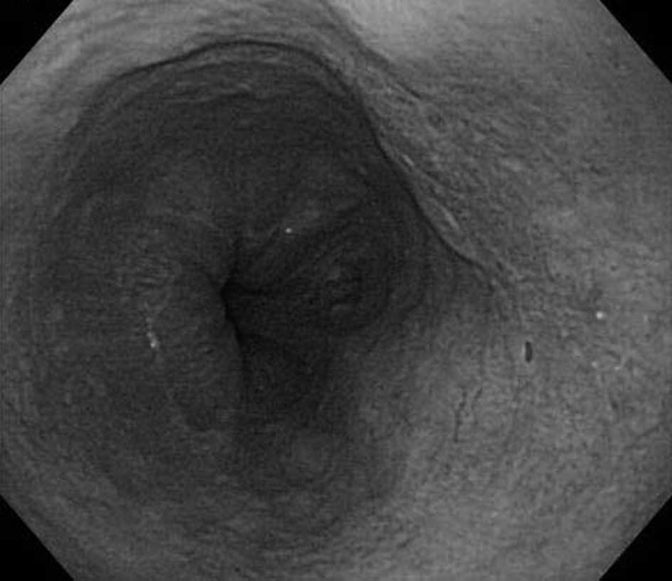
Patient tumors showed a response to PDT. CR was achieved in 11
(73%) of the 15 patients, but 2 patients experienced local
recurrence after PDT. Thus, the 6 patients were treated again with
PDT, and the recurrent/residual lesions were eliminated in all 6
cases. Initial PDT was successfully performed in 4 of the 6
submucosal ESCC cases (Fig. 1),
while CR was achieved in 7 of the 9 mucosal tumor cases. Otherwise,
no clinicopathological factors had a significant impact on PDT
outcomes. At a median follow-up period of 23 months (range 4–35),
13 patients were alive, and 11 were disease-free. Two patients with
intramucosal ESCC succumbed due to metastatic disease despite
having no local recurrence in the irradiated esophagus. The 5
patients treated with S-1 were alive, and 3 were disease-free,
although the median follow-up period was 8 months. One patient with
submucosal ESCC was unable to commence chemotherapy treatment due
to sustained bone marrow suppression following CRT.
 | Table IBackground characteristics of the
patients with superficial esophageal squamous carcinoma treated by
photodynamic therapy (PDT). |
Table I
Background characteristics of the
patients with superficial esophageal squamous carcinoma treated by
photodynamic therapy (PDT).
| Case no. | Age | Gender | Esophageal
location | Endoscopic
findings | Invasive depth | Chemoradiation |
|---|
| 1 | 73 | M | Thorax | IIc | sm | Recurrence |
| 2 | 54 | M | Thorax | III | sm | Recurrence |
| 3 | 85 | M | Thorax | IIc | m | Recurrence |
| 4 | 71 | M | Cervix | IIb | m | Recurrence |
| 5 | 75 | M | Cervix | IIb | m | Recurrence |
| 6 | 69 | M | Abdomen | IIa | sm | Recurrence |
| 7 | 59 | M | Thorax | IIb | m | Recurrence |
| 8 | 76 | M | Abdomen | IIc | m | Recurrence |
| 9 | 79 | M | Thorax | I | sm | Recurrence |
| 10 | 69 | M | Thorax | IIc | m | Recurrence |
| 11 | 70 | M | Thorax | IIb | m | Recurrence |
| 12 | 59 | M | Thorax | IIb | m | Naïve |
| 13 | 60 | F | Cervix | IIb | m | Recurrence |
| 14 | 86 | M | Thorax | IIc | sm | Recurrence |
| 15 | 76 | M | Thorax | IIc | sm | Naïve |
| Table IIClinical outcomes of photodynamic
therapy. |
Table II
Clinical outcomes of photodynamic
therapy.
| Case no. | PDT dose (J) | Complications | Response to initial
PDT | Repeated PDT | Chemotherapy with
PDT | Follow-up
(months) | Prognosis |
|---|
| 1 | 400 | High-grade fever,
chest pain | CR | | | 35 | Alive |
| 2 | 480 | Chest pain | Non-CR | Performed | S-1 | 33 | Alive |
| 3 | 620 | High-grade
fever | Non-CR | Performed | | 33 | Alive |
| 4 | 940 | High-grade
fever | CR | | | 31 | Deceased |
| 5 | 355 | Esophageal
stenosis | CR | | | 30 | Alive |
| 6 | 1065 | Skin
phototoxicity | CR | | S-1 | 28 | Alive |
| 7 | 520 | Chest pain,
high-grade fever | CR | | | 23 | Deceased |
| 8 | 540 | Chest pain,
high-grade fever | CR | | | 23 | Alive |
| 9 | 900 | Esophageal
stenosis | Non-CR | Performed | S-1 | 9 | Alive |
| 10 | 600 | High-grade fever,
mediastinitis | CR | | S-1 | 8 | Alive |
| 11 | 400 | Esophageal
stenosis | Non-CR | Performed | | 8 | Alive |
| 12 | 1000 | None | Recurrence after
CR | Performed | | 7 | Alive |
| 13 | 280 | None | CR | | | 4 | Alive |
| 14 | 740 | High-grade fever,
mediastinitis | Recurrence after
CR | Performed | | 4 | Alive |
| 15 | 860 | None | CR | | S-1 | 33 | Alive |
No significant difference was found in pretreatment
serum total ROS levels between the CR (156.3±36.2 units) and non-CR
groups (177.0±28.0 units) at the initial PDT. No significant
difference was noted in the 7-day post-treatment total ROS values
irrespective of the treatment outcome (205.2±56.5 and 204.7±27.2
units, respectively). When limited to patients who had successful
PDT, the serum total ROS values were significantly increased from
156.3±36.2 units before PDT to 205.2±56.5 units after PDT
(p<0.05).
In all cases, Photofrin administration was well
tolerated. There were no allergic reactions or injection site
irritation. As for acute complications within 7 days after PDT,
high fever (>38.0°C) and chest pain that required analgesic
treatment were observed in 10 and 4 patients, respectively. A total
of 6 patients experienced significant complications: 2 had
mediastinitis; 3 had esophageal stenosis that required repeated
endoscopic balloon dilation; and 1 had cutaneous phototoxicity.
Each complication was successfully managed with medical treatment.
No deaths were attributable to the PDT procedure itself.
In vitro study
The two chemotherapeutic agents substantially
affected survival of the OE21 cells with the IC50 of
single 5-FU and CDDP set at 3.5 and 9.0 μM, respectively. PDT
showed clear cytotoxic activity against the OE21 cells. To induce
IC50, a Photofrin concentration of 10 μg/ml was required
for this ESCC cell line at a laser power of 5 J/cm2.
Following the combination of 3.5 μM of 5-FU with PDT using
Photofrin at various concentrations, a significant synergistic
effect was observed with the IC50 of Photofrin at 0.83
μg/ml for the same PDT setting. The IC50 at a laser
power of 5 J/cm2 decreased 3-fold (3.3 μg/ml) in the
presence of 9 μM of CDDP. When PDT was combined with 10 μg/ml of
Photofrin at a laser power of 5 J/cm2 with the two
chemotherapeutic agents at various concentrations, the
IC50s were substantially reduced from 3.5 and 9.0 μM to
0.75 and 1.2 μM for CDDP and 5-FU, respectively.
Discussion
In the present study, 12 (80%) of the 15 patients
with superficial ESCC achieved CR with Photofrin-mediated PDT. The
group consisted of no less than 13 patients with recurrent ESCC
after CRT. In a similar setting of ESCC cases, Yano et al
reported that the CR rate was 62% (8 of 13 patients) by salvage PDT
after CRT failure. In their study, the overall survival rate 1 year
after salvage PDT was 68.4% (14).
In this study, 13 of the 15 patients were alive, and 11 were
disease-free at the median follow-up period of 23 months. Two
patients succumbed due to metastatic disease although no local
recurrence of ESCC was noted after PDT. Hattori et al
reported that the overall survival rate of patients treated by
salvage EMR for locoregional failure after CRT was 56% at 3 years
(5). On the other hand, previous
data showed that overall 3-year survival for patients with non-CR
with definitive CRT was no more than 6% (3). Collectively, the results suggest that
local treatment by endoscopic modalities such as EMR and PDT is a
treatment option for superficial ESCC without metastasis.
From a technical point of view, PDT appears to be
superior to EMR. When the corresponding lesion exhibits ulceration,
severe fibrosis, or stenosis, salvage EMR is difficult or
impossible to perform (6). Little
information is available on clinical outcomes of ESD in the salvage
setting, although ESD permits the removal of esophageal epithelial
neoplasms en bloc, irrespective of size (7). Nevertheless, in ESCC cases with
invasion in the submucosal layer, ESD/EMR cannot be used due to its
lack of curative potential. Even in such cases, PDT may yield
relatively high CR rates, as indicated by the present and previous
data (14). Surgical resection has
been considered a salvage modality for these patients. However,
Swisher et al reported that patients treated with salvage
esophagectomy had a significantly higher incidence of anastomotic
leaks (39 vs. 7%) and a longer hospital stay (29 vs. 18 days) than
those treated with planned esophagectomy (4). ESCC cases treated with CRT may
occasionally be unresectable due to concomitant diseases (13). Thus, PDT offers an attractive
alternative for patients with ESCC tumors without metastasis who
would not otherwise be referred to a surgeon for treatment.
Moreover, in selected patients with primary superficial ESCC, PDT
as definitive therapy may avoid the risks associated with
esophageal resection (13).
PDT was performed safely in the present and previous
studies, and the majority of complications were manageable with
medical treatment (13,14). However, it is of clinical relevance
that life-threatening complications are rarely noted. An
esophagotracheal fistula may develop with PDT even in naïve early
esophageal cancer cases; the reported incidence is 6.5% (14). Similarly, severe mediastinitis and
pericardial effusion were previously documented following salvage
PDT (13,14). Possible reasons for the
complications include radiation-induced esophageal damage and heart
disease as well as transmural necrosis potentiated by PDT.
Stricture formation at the irradiated esophageal site was common
following PDT, occurring in 20–42% of cases. The majority of
patients who underwent 3 or more PDT treatments developed a
stricture requiring dilation, and the number of attempts varied
(range 1–20, mean 2.7) (13).
Nevertheless, the indications for prophylactic pneumatic dilation
warrant further evaluation.
Oral S-1, the 5-FU modulating drug, was administered
to patients with submucosal ESCC with a potential risk of
metastatic disease in the present study. The patients were still
alive without significant complications despite their short-term
follow-up. Keeley et al applied chemotherapy and/or
radiation therapy in 16 (32%) of 50 patients with Barrett's
high-grade dysplasia and esophageal cancer, varying from T1 to T3
tumor, to supplement primary PDT (13). Of the 16 patients who received
concurrent chemoradiation, 6 remained alive at a mean interval of
16 months. Four of the patients who received PDT/chemo/radiation
showed no evidence of disease at a mean interval of 13 months, and
2 were alive with disease. Conversely, all 10 patients succumbing
to the disease had local recurrence. Due to superior survival and
local control, esophagectomy may remain the preferred treatment for
patients without physiological impairment. Nevertheless, the
preliminary data on chemotherapy using the single fluorouracil
along with PDT prompted us to investigate their combined effects
in vitro. 5-FU and CDDP are widely used in chemotherapy
regimens against various types of cancers. Combined chemotherapy
consisting of the two key drugs has been a representative standard
regimen against ESCC (19).
Therefore, we focused on 5-FU and CDDP to enhance the cytotoxicity
of Photofrin-mediated PDT in cellular experiments.
Upon activation by the special wavelength light,
such Photofrin-derived photosensitizers undergo photochemical
reactions to transfer electrons or hydrogen or to form singlet
oxygen and generate excessive ROS (11,20).
This leads to oxidative damage to proteins, lipids and DNA,
resulting in apoptotic or necrotic cell death (11,20,21).
In line with this theoretical concept, the patients who achieved CR
had a significant increase in circulating ROS levels following PDT,
which was not observed in non-CR cases. Our in vitro study
showed that the combination of Photofrin-mediated PDT and 5-FU or
CDDP resulted in a significantly lower cell survival than the
single-mode treatment. Notably, with combined treatment, each
IC50 dosage was significantly decreased; in particular,
much lower concentrations of Photofrin and 5-FU were required to
obtain sufficient cell killing and vice versa. Promising results
using similar or other chemotherapeutic drugs have been reported in
combination with PDT (17,22,23).
The synergistic effects of the two treatments may suggest diverse
potential mechanisms for cellular death, one directly associated
with PDT action and the other associated with each cytotoxic drug
(17,22–24).
Based on the in vitro results and our study, it is evident
that more studies are warranted to determine the manner in which
chemotherapeutic drugs and PDT interact and can be combined in
order to result in increased cell killing with reduced side
effects.
In conclusion, PDT demonstrated acceptable
short-term outcomes, feasible curative properties, and safety for
the treatment of superficial ESCC. Although further long-term
follow-up studies are required, PDT is a promising treatment option
for selected ESCC cases, particularly local recurrence following
CRT. Combination therapy with PDT and 5-FU or CDDP may result in
enhanced cytotoxic effects on ESCC, possibly reducing the effective
dosage of each drug and decreasing side effects.
References
|
1
|
Cooper JS, Guo MD, Herskovic A, et al:
Chemoradiotherapy of locally advanced esophageal cancer: long-term
follow-up of a prospective randomized trial (RTOG 85-01). Radiation
Therapy Oncology Group. JAMA. 281:1623–1627. 1999. View Article : Google Scholar : PubMed/NCBI
|
|
2
|
Herskovic A, Martz K, Al-Sarraf M, et al:
Combined chemotherapy and radiotherapy compared with radiotherapy
alone in patients with cancer of the esophagus. N Engl J Med.
326:1593–1598. 1992. View Article : Google Scholar : PubMed/NCBI
|
|
3
|
Ishikura S, Nihei K, Ohtsu A, et al:
Long-term toxicity after definitive chemoradiotherapy for squamous
cell carcinoma of the thoracic esophagus. J Clin Oncol.
21:2697–2702. 2003. View Article : Google Scholar : PubMed/NCBI
|
|
4
|
Swisher SG, Wynn P, Putnam JB, et al:
Salvage esophagectomy for recurrent tumors after definitive
chemotherapy and radiotherapy. J Thorac Cardiovasc Surg.
123:175–183. 2002. View Article : Google Scholar : PubMed/NCBI
|
|
5
|
Hattori S, Muto M, Ohtsu A, et al: EMR as
salvage treatment for patients with locoregional failure of
definitive chemoradiotherapy for esophageal cancer. Gastrointest
Endosc. 58:65–70. 2003. View Article : Google Scholar : PubMed/NCBI
|
|
6
|
Gotoda T, Kondo H, Ono H, et al: A new
endoscopic mucosal resection procedure using an insulation-tipped
electrosurgical knife for rectal flat lesions: report of two cases.
Gastrointest Endosc. 50:560–563. 1999. View Article : Google Scholar : PubMed/NCBI
|
|
7
|
Isomoto H and Yamaguchi N: Endoscopic
submucosal dissection in the era of proton pump inhibitors. J Clin
Biochem Nutr. 44:205–211. 2009. View Article : Google Scholar : PubMed/NCBI
|
|
8
|
Fujishiro M, Kodashima S, Goto O, et al:
Endoscopic submucosal dissection for esophageal squamous cell
neoplasms. Dig Endosc. 21:109–115. 2009. View Article : Google Scholar : PubMed/NCBI
|
|
9
|
Isomoto H, Nishiyama H, Yamaguchi N, et
al: Clinicopathological factors associated with clinical outcomes
of endoscopic submucosal dissection for colorectal epithelial
neoplasms. Endoscopy. 41:679–683. 2009. View Article : Google Scholar
|
|
10
|
Wolfsen HC: Uses of photodynamic therapy
in premalignant and malignant lesions of the gastrointestinal tract
beyond the esophagus. J Clin Gastroenterol. 39:653–664. 2005.
View Article : Google Scholar : PubMed/NCBI
|
|
11
|
Robertson CA, Evans DH and Abrahamse H:
Photodynamic therapy (PDT): a short review on cellular mechanisms
and cancer research applications for PDT. J Photochem Photobiol B.
96:1–8. 2009. View Article : Google Scholar : PubMed/NCBI
|
|
12
|
Hahn SM, Putt ME, Metz J, et al: Photofrin
uptake in the tumor and normal tissues of patients receiving
intraperitoneal photodynamic therapy. Clin Cancer Res.
12:5464–5470. 2006. View Article : Google Scholar : PubMed/NCBI
|
|
13
|
Keeley SB, Pennathur A, Gooding W,
Landreneau RJ, Christie NA and Luketich J: Photodynamic therapy
with curative intent for Barrett's esophagus with high grade
dysplasia and superficial esophageal cancer. Ann Surg Oncol.
14:2406–2410. 2007. View Article : Google Scholar
|
|
14
|
Yano T, Muto M, Minashi K, Ohtsu A and
Yoshida S: Photodynamic therapy as salvage treatment for local
failures after definitive chemoradiotherapy for esophageal cancer.
Gastrointest Endosc. 62:31–36. 2005. View Article : Google Scholar : PubMed/NCBI
|
|
15
|
Hayashi I, Morishita Y, Imai K, Nakamura
M, Nakachi K and Hayashi T: High-throughput spectrophotometric
assay of reactive oxygen species in serum. Mutat Res. 63:55–61.
2007. View Article : Google Scholar : PubMed/NCBI
|
|
16
|
Fujii M, Kochi M and Takayama T: Recent
advances in chemotherapy for advanced gastric cancer in Japan. Surg
Today. 40:295–300. 2010. View Article : Google Scholar : PubMed/NCBI
|
|
17
|
Nonaka M, Ikeda H and Inokuchi T: Effect
of combined photodynamic and chemotherapeutic treatment on lymphoma
cells in vitro. Cancer Lett. 184:171–178. 2002. View Article : Google Scholar : PubMed/NCBI
|
|
18
|
Veinot JP and Edwards WD: Pathology of
radiation-induced heart disease: a surgical and autopsy study of 27
cases. Hum Pathol. 27:766–773. 1996. View Article : Google Scholar : PubMed/NCBI
|
|
19
|
Ohtsu A, Yoshida S and Saijo N:
Disparities in gastric cancer chemotherapy between the East and
West. J Clin Oncol. 24:2188–2196. 2006. View Article : Google Scholar : PubMed/NCBI
|
|
20
|
Plaetzer K, Krammer B, Berlanda J, Berr F
and Kiesslich T: Photophysics and photochemistry of photodynamic
therapy: fundamental aspects. Lasers Med Sci. 24:259–268. 2009.
View Article : Google Scholar : PubMed/NCBI
|
|
21
|
Wang J and Yi J: Cancer cell killing via
ROS: to increase or decrease, that is the question. Cancer Biol
Ther. 7:1875–1884. 2008. View Article : Google Scholar : PubMed/NCBI
|
|
22
|
Park S, Hong SP, Oh TY, Bang S, Chung JB
and Song SY: Paclitaxel augments cytotoxic effect of photodynamic
therapy using verteporfin in gastric and bile duct cancer cells.
Photochem Photobiol Sci. 7:769–774. 2008. View Article : Google Scholar : PubMed/NCBI
|
|
23
|
Zimmermann A, Walt H, Haller U, Baas P and
Klein SD: Effects of chlorin-mediated photodynamic therapy combined
with fluoropyrimidines in vitro and in a patient. Cancer Chemother
Pharmacol. 51:147–154. 2003.PubMed/NCBI
|
|
24
|
Takahira K, Sano M, Arai H and Hanai H:
Apoptosis of gastric cancer cell line MKN45 by photodynamic
treatment with photofrin. Lasers Med Sci. 19:89–94. 2004.
View Article : Google Scholar : PubMed/NCBI
|