Introduction
Reactive oxygen species (ROS) include hydrogen
peroxide (H2O2), the superoxide anion
(O2•−) and the hydroxyl radical
(•OH). ROS are involved in the regulation of many
important cellular events, including transcription factor
activation, gene expression, differentiation and cell proliferation
(1,2). ROS are generated as by-products of
mitochondrial respiration or by oxidases, such as the nicotine
adenine diphosphate (NADPH) oxidase and the xanthine oxidase (XO)
(3). A change in the redox state of
tissues or cells alters the generation or metabolism of ROS. The
principal metabolic pathways involved in redox defense, include
superoxide dismutases (SOD), which involve 3 isoforms, the
extracellular (SOD3), cytoplasmic (SOD1), and the mitochondrial
(SOD2) isoforms (4), which
metabolize O2•− to
H2O2. Further metabolism by peroxidases,
including catalase (CAT) and glutathione (GSH) peroxidase (GPX),
yields O2 and H2O (5). Cells have various antioxidant systems
to manage their redox state, which is important for their survival.
The thioredoxin (TXN) system consists of TXN, TXN reductase and
NADPH and is critically involved in maintaining cellular redox
homeostasis (6). TXN as a thiol
reductase is a potent antioxidant and acts as a scavenger of ROS
(6). Excessive production of ROS
can be induced by endogenous and/or exogenous sources, which then
initiates events that lead to cell death depending on the cell type
(7–9).
The ubiquitin-dependent proteasomal system presents
the foremost non-lysosomal corridor through which intracellular
proteins involved in cell cycling, proliferation, differentiation
and apoptosis are degraded in eukaryotic cells (10,11).
Transformed cells including cancer cells accumulate more
misfolded/mutated/damaged proteins due to the elevated replication
rate of malignant cells (12).
Thus, these cells can be much more susceptible to proteasome
inhibition than normal cells. Apoptosis in cancer cells is closely
connected with the activity of the ubiquitin/proteasome pathways
(13,14). Accordingly, the inhibition of
proteasome function has emerged as a useful strategy to control
apoptosis. The peptide aldehyde MG132
(carbobenzoxy-Leu-Leu-leucinal) efficiently prevents the
proteolytic activity of the proteasome complex (15). Various proteasome inhibitors
including MG132 have been demonstrated to stimulate apoptotic cell
death through the induction of ROS (16,17).
ROS formation and GSH depletion by proteasome inhibitors may
trigger mitochondrial dysfunction and subsequent cytochrome C
release, which can lead to cell death (18,19).
The mechanism underlying ROS generation after inhibition of the
proteasome is still imprecise.
Lung cancer is a main cause of cancer death in
developed countries. Various novel remedial strategies including
new drug development are currently under consideration due to
intrinsic or acquired resistance and toxicity of conventional drugs
(20). Specifically, drugs that aim
at specific intracellular pathways related to the distinctive
properties of cancer cells continue to be developed. Recently, it
has been reported that a proteasome inhibitor bortezomib (PS-341,
Velcade) inhibits lung cancer cells (21,22).
The toxicological mechanism of MG132 in lung cancer cells has not
been fully understood. We recently demonstrated that MG132 reduced
the growth of Calu-6 and A549 lung cancer cells via apoptosis and
GSH depletion (23,24). On the other hand, little is known
about the cellular effects of MG132 on normal primary lung cells in
relation to cell death. Because we observed that MG132 induced the
growth inhibition and death in human pulmonary fibroblast (HPF)
cells via a caspase-independent manner (unpublished data), in the
present study, we investigated the effects of N-acetyl cysteine
(NAC) and vitamin C (well known antioxidants) or L-buthionine
sulfoximine (BSO; an inhibitor of GSH synthesis) (25) on MG132-treated HPF cells in relation
to cell growth, death, ROS and GSH levels. Furthermore, we examined
the effects of antioxidant-related siRNAs on cell death, ROS and
GSH levels in MG132-treated HPF cells.
Materials and methods
Cell culture
The human pulmonary fibroblast (HPF) cells from
PromoCell GmbH (Heidelberg, Germany) were maintained in humidified
incubator containing 5% CO2 at 37°C. HPF cells were
cultured in RPMI-1640 supplemented with 10% fetal bovine serum
(FBS) and 1% penicillin-streptomycin (Gibco-BRL, Grand Island, NY).
HPF cells were used between passages four and eight.
Reagents
MG132 was purchased from Calbiochem (San Diego, CA)
and was dissolved in dimethyl sulfoxide (DMSO; Sigma-Aldrich, St.
Louis, MO) solution buffer. NAC and BSO were obtained from
Sigma-Aldrich. NAC was dissolved in 20 mM HEPES (pH 7.0) buffer.
BSO was dissolved in water. Vitamin C purchased from Riedel-de Haen
(Hannover, Germany) was also dissolved in water. Based on previous
studies (26,27) cells were pretreated with 2 mM NAC or
10 μM BSO or 0.4 mM vitamin C for 1 h prior to MG132 treatment.
DMSO (0.2%) was used as a control vehicle and it did not appear to
affect cell growth or death.
Detection of intracellular ROS and
O2•− levels
Intracellular ROS levels were detected by means of
an oxidation-sensitive fluorescent probe dye,
2′,7′-dichlorodihydrofluorescein diacetate (H2DCFDA,
Ex/Em of 495 nm/529 nm; Invitrogen Molecular Probes, Eugene, OR) as
previously described (28).
H2DCFDA is poorly selective for superoxide anion radical
(O2•−). On the other hand, dihydroethidium
(DHE) (Ex/Em of 518 nm/605 nm; Invitrogen Molecular Probes) is a
fluorogenic probe that is highly selective for
O2•− (28).
Mitochondrial O2•− levels were detected using
the MitoSOX™ Red mitochondrial O2•− indicator
(Ex/Em of 510 nm/580 nm; Invitrogen Molecular Probes) as previously
described (28). In brief,
1×10⁶ cells in 60-mm culture dish (Nunc) were incubated
with the indicated doses of MG132 with or without NAC, BSO, vitamin
C or antioxidant-related siRNA duplexes for 24 h. Cells were then
washed in PBS and incubated with 20 μM H2DCFDA, 20 μM
DHE or 5 μM MitoSOX Red at 37°C for 30 min. DCF, DHE and MitoSOX
Red fluorescence intensities were detected using a FACStar flow
cytometer (Becton-Dickinson, Franklin Lakes, NJ). ROS and
O2•− levels were expressed as mean
fluorescence intensity (MFI), using the CellQuest software
(Becton-Dickinson).
Detection of intracellular glutathione
(GSH) levels
Cellular GSH levels were analyzed using a
5-chloromethylfluorescein diacetate dye (CMFDA, Ex/Em of 522 nm/595
nm; Invitrogen Molecular Probes) as previously described (28). In brief, 1×10⁶ cells in
60-mm culture dishes (Nunc) were incubated with the indicated doses
of MG132 with or without NAC, BSO, vitamin C or antioxidant-related
siRNA duplexes for 24 h. Cells were then washed with PBS and
incubated with 5 μM CMFDA at 37°C for 30 min. CMF fluorescence
intensity was determined using a FACStar flow cytometer
(Becton-Dickinson). Negative CMF staining (GSH depleted) cells were
expressed as the percent of (−) CMF cells.
Cell growth inhibition assays
The effect of drugs on HPF cell growth was
determined by the
3-(4,5-dimethylthiazol-2-yl)-2,5-diphenyltetrazolium bromide (MTT)
assay as previously described (29). In brief, 5×10³ cells/well
were seeded in 96-well microtiter plates (Nunc). After exposure to
the indicated doses of MG132 with or without NAC, BSO or vitamin C
for 24 h, 20 μl of MTT solution [2 mg/ml in phosphate-buffered
saline (PBS)] were added to each well of the 96-well plates. The
plates were incubated for 4 additional hours at 37°C. Media in
plates were withdrawn by pipetting and 200 μl of DMSO was added to
each well to solubilize the formazan crystals. Optical density was
measured at 570 nm using a microplate reader (SpectraMAX 340,
Molecular Devices Co., Sunnyvale, CA).
Annexin-V/PI staining for cell death
detection
Apoptosis was determined by staining cells with
Annexin-V-fluorescein isothiocyanate (FITC, Ex/Em of 488 nm/519 nm;
Invitrogen Molecular Probes) and propidium iodide (PI, Ex/Em of 488
nm/617 nm; Sigma-Aldrich). In brief, 1×10⁶ cells in
60-mm culture dishes (Nunc) were incubated with the indicated doses
of MG132 with or without NAC, BSO, vitamin C or antioxidant-related
siRNA duplex for 24 h. Cells were washed twice with cold PBS and
then resuspended in 500 μl binding buffer (10 mM HEPES/NaOH pH 7.4,
140 mM NaCl, 2.5 mM CaCl2) at a concentration of
1×10⁶ cells/ml. Annexin-V-FITC (5 μl) and PI (1 μg/ml)
were then added to these cells, which were analyzed with a FACStar
flow cytometer (Becton-Dickinson). Viable cells were negative for
both PI and Annexin-V; apoptotic cells were positive for Annexin-V
and negative for PI, whereas late apoptotic dead cells displayed
both high Annexin-V and PI labeling. Non-viable cells, which
underwent necrosis, were positive for PI and negative for
Annexin-V.
Measurement of MMP (ΔΨm)
MMP (ΔΨm) levels were measured using a
rhodamine 123 fluorescent dye (Sigma-Aldrich; Ex/Em of 485 nm/535
nm) as previously described (30).
In brief, 1×10⁶ cells in 60-mm culture dishes (Nunc)
were incubated with the indicated doses of MG132 with or without
NAC, BSO or vitamin C for 24 h. Cells were washed twice with PBS
and incubated with rhodamine 123 (0.1 μg/ml) at 37°C for 30 min.
Rhodamine 123 staining intensity was determined by flow cytometry
(Becton-Dickinson). An absence of rhodamine 123 from cells
indicated the loss of MMP (ΔΨm) in HPF cells.
Western blot analysis
The patterns of ubiquitinated proteins were
evaluated using Western blot analysis. In brief, 1×10⁶
cells in 60-mm culture dish (Nunc) were incubated with 10 μM MG132
with or without NAC for 24 h. The cells were then washed in PBS and
suspended in five volumes of lysis buffer (20 mM HEPES, pH 7.9, 20%
glycerol, 200 mM KCl, 0.5 mM EDTA, 0.5% NP-40, 0.5 mM DTT and 1%
protease inhibitor cocktail). Supernatant protein concentrations
were determined using the Bradford method. Samples containing 40 μg
total protein were resolved by 12.5% SDS-PAGE gels, transferred to
Immobilon-P PVDF membranes (Millipore, Billerica, MA) by
electroblotting and then probed with anti-ubiquitin and
anti-β-actin antibodies (Santa Cruz Biotechnology, Santa Cruz, CA).
Membranes were incubated with horseradish peroxidase-conjugated
secondary antibodies. Blots were developed using an ECL kit
(Amersham, Arlington Heights, IL).
Transfection of cells with
antioxidant-related siRNAs
Gene silencing of SOD1, SOD2, CAT, GPX, TXN was
performed as previously described (31). The siRNA duplexes consisted of a
non-specific control siRNA duplex
[5′-CCUACGCCACCAAUUUCGU(dTdT)-3′], the SOD1
[5′-GAAAACACGGUGGGCCAAA(dTdT)-3′], the SOD2 [5′-CUGGGAGAAUGUAAC
UGAA (dTdT)-3′], the CAT [5′-CACUGAUUUCACAACAGAU (dTdT)-3′], the
GPX [5′-CAAGCUCAUCACCUGGUCU (dTdT)-3′] and the TXN
[5′-GCAUGCCAACAUUCCAGUU (dTdT)-3′] siRNA duplexes which were
purchased from the Bioneer Corp. (Daejeon, South Korea). In brief,
2.5×10⁵ cells in 6-well plates (Nunc) were incubated in
RPMI-1640 supplemented with 10% FBS. The next day, cells (30–40%
confluence) in each well were transfected with the control or each
siRNA duplex [80 picomoles in Opti-MEM (Gibco-BRL)] using
Lipofectamine 2000, according to the manufacturer's instructions
(Invitrogen, Brandford, CT). Two days later, cells were treated
with or without 30 μM MG132 for 24 additional hours. The
transfected cells were collected and used for the measurement of
Annexin-V-FITC/PI staining cells, ROS and GSH depletion levels.
Statistical analysis
The results represent the mean of at least 3
independent experiments (mean ± SD). The data were analyzed using
Instat software (GraphPad Software, Inc., San Diego, CA). The
Student's t-test or one-way analysis of variance (ANOVA) with
post-hoc analysis using Tukey's multiple comparison test was used
for parametric data. Statistical significance was defined as
P<0.05.
Results
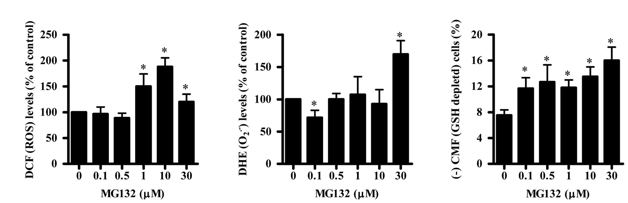
MG132 alters ROS and GSH levels in HPF
cells
To assess intracellular ROS and GSH levels in
MG132-treated HPF cells, we used (0.1–30 μM) of MG132 based on our
unpublished findings that 0.5–30 μM MG132 dose-dependently
inhibited the growth of HPF cells with an IC50 of ~20 μM
at 24 h. As shown in Fig. 1A,
intracellular ROS (DCF) levels were not altered in HPF cells
treated with 0.1 or 0.5 μM MG132 but were increased in 1–30 μM
MG132-treated HPF cells. Intracellular O2•−
(DHE) level was decreased in HPF cells treated with 0.1 μM MG132
and was not significantly changed by 0.5, 1 and 10 μM MG132
(Fig. 1B). An increase in
O2•− levels was observed in 30 μM
MG132-treated HPF cells (Fig. 1B).
In relation to GSH levels in MG132-treated HPF cells, MG132
increased the number of GSH-depleted HPF cells at 24 h in a
dose-dependent manner as compared with those of the control cells
(Fig. 1C).
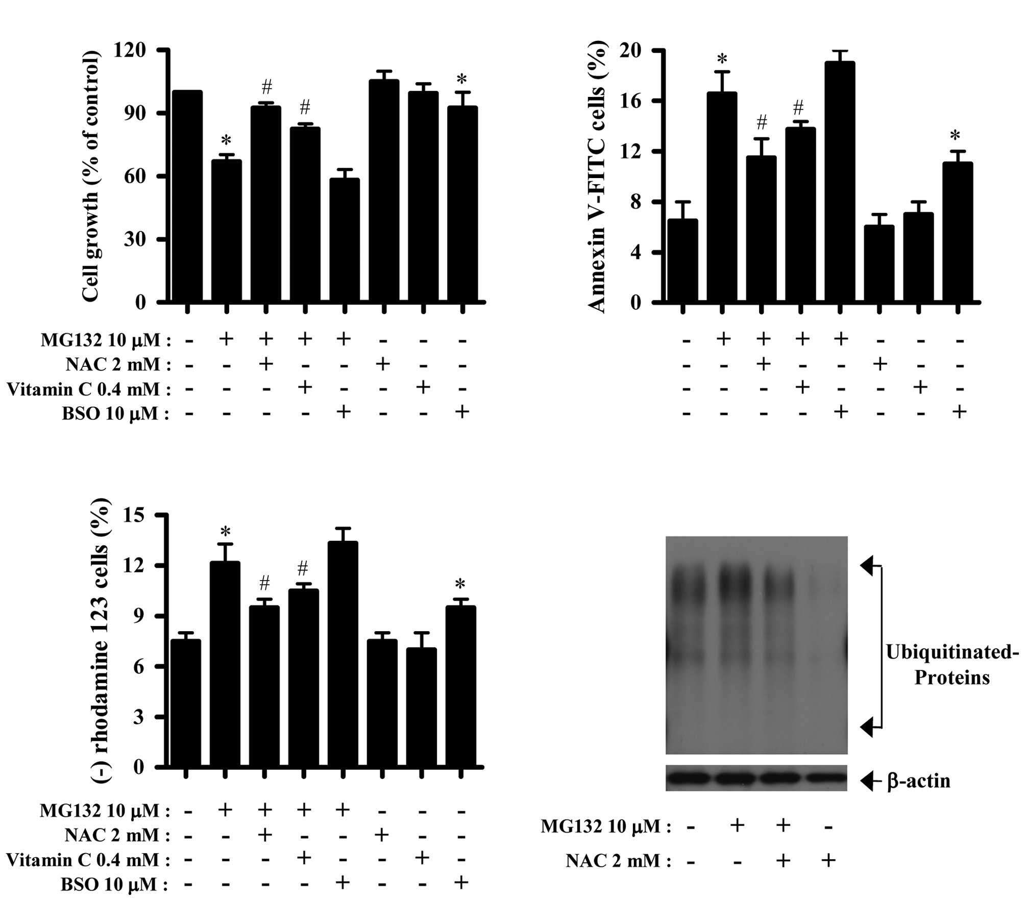
NAC, vitamin C or BSO influences the
growth inhibition and death of MG132-treated HPF cells
We examined the effect of NAC, vitamin C and BSO on
the growth and death of MG132-treated HPF cells. For this
experiment, 10 μM MG132 was chosen as a suitable dose to examine
cell growth inhibition and death in the presence or absence of NAC,
vitamin C or BSO. Based on the MTT assay, 10 μM MG132 inhibited the
growth of HPF cells by about 35% at 24 h (Fig. 2A). Treatment with NAC and vitamin C
significantly prevented the growth inhibition by MG132 whereas BSO
slightly enhanced the growth inhibition (Fig. 2A). BSO alone inhibited HPF cell
growth (Fig. 2A). In relation to
cell death, MG132 induced cell death in HPF cells at 24 h, as
evidenced by Annexin-V staining (Fig.
2B). Both NAC and vitamin C significantly rescued HPF cells
from the MG132 insult (Fig. 2B).
BSO slightly increased the cell death by MG132 and this agent alone
also induced cell death in HPF control cells (Fig. 2B).
Apoptosis is closely related to the collapse of MMP
(ΔΨm) (32). Therefore,
we determined the loss of MMP (ΔΨm) in MG132-treated HPF
cells. Similarly to the results of Annexin-V staining; both NAC and
vitamin C attenuated the loss of MMP (ΔΨm) in
MG132-treated HPF cells, whereas BSO mildly enhanced the loss in
these cells (Fig. 2C). BSO alone
induced MMP (ΔΨm) loss in HPF control cells (Fig. 2C). Moreover, we observed that MG132
increased the level of anonymous ubiquitinated proteins in HPF
cells (Fig. 2C). NAC showing a
strong antiapoptotic effect attenuated the ubiquitinated protein
levels in MG132-treated HPF cells (Fig.
2D). NAC also strongly decreased the basal ubiquitinated
protein levels in HPF control cells (Fig. 2D).
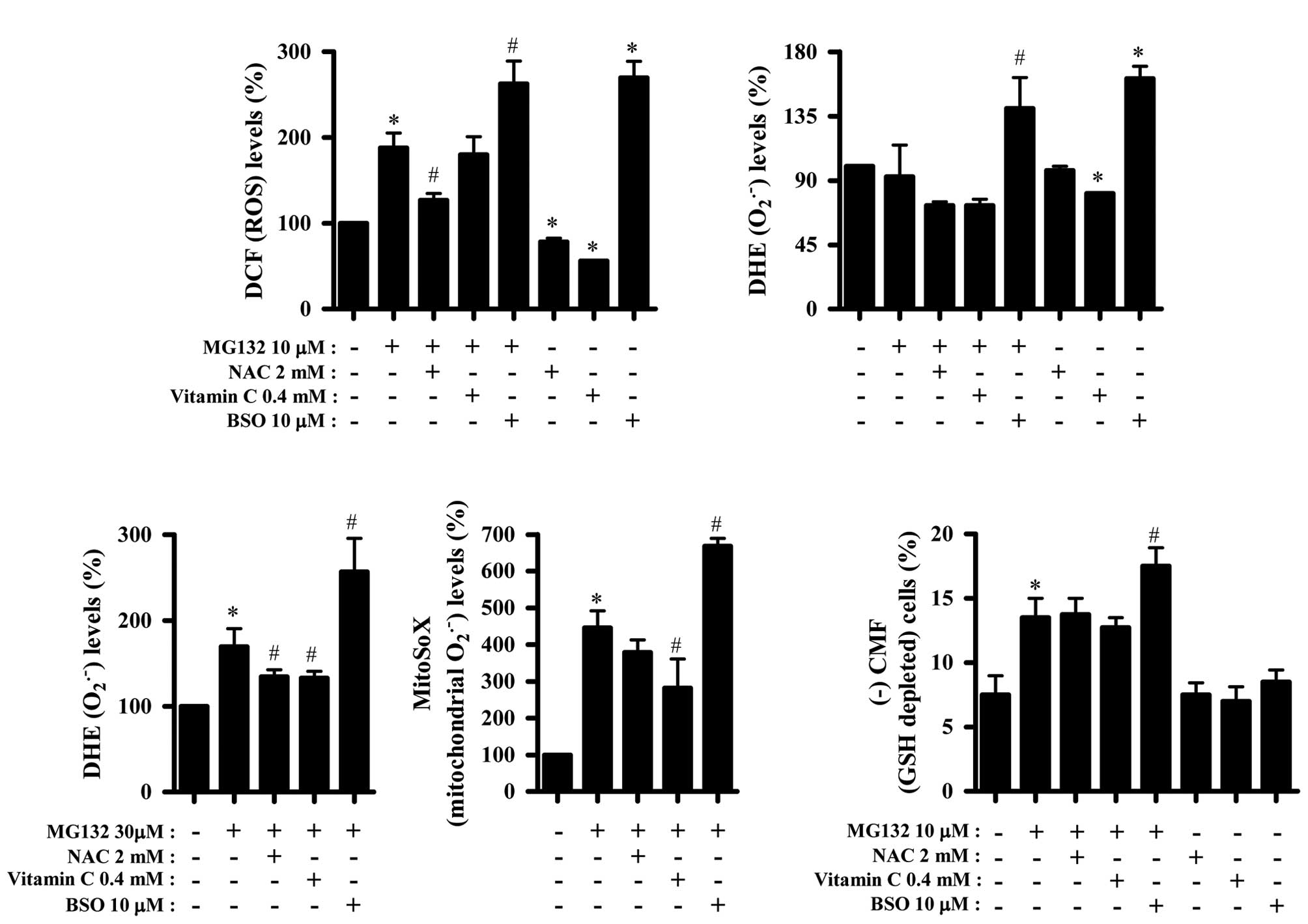
NAC, vitamin C or BSO affect ROS and GSH
levels in MG132-treated HPF cells
Next, ROS and GSH levels in HPF cells treated with
10 or 30 μM MG132with or without NAC, vitamin C or BSO were
assessed. As shown in Fig. 3A, ROS
(DCF) level in MG132-treated HPF cells was significantly decreased
by NAC, but that was not significantly altered by vitamin C. Both
NAC and vitamin C decreased basal ROS (DCF) levels in HPF control
cells (Fig. 3A). In contrast, BSO
strongly increased ROS (DCF) levels in MG132-treated or -untreated
HPF cells (Fig. 3A). Both NAC and
vitamin C seemed to decrease O2•− levels in
MG132-treated and -untreated HPF cells (Fig. 3B). However, BSO significantly
increased O2•− levels in MG132-treated or
-untreated HPF cells (Fig. 3B).
In addition, we assessed the effect of NAC, vitamin
C or BSO on O2•− levels in 30 μM
MG132-treated HPF cells. As shown in Fig. 3C, NAC and vitamin C attenuated
O2•− levels in these cells, but BSO strongly
intensified the level. Furthermore, MitoSOX Red fluorescence
levels, which specifically indicate O2•−
levels in the mitochondria, were strongly increased in 30 μM
MG132-treated HPF cells after 24 h (Fig. 3C). Both NAC and vitamin C decreased
the mitochondrial O2•− levels in
MG132-treated HPF cells, whereas BSO enhanced them (Fig. 3C). In relation to GSH levels, NAC
did not affect the number of GSH-depleted cells among MG132-treated
HPF cells, but vitamin C slightly decreased this number (Fig. 3D). BSO seemed to increase the
numbers of GSH-depleted cells among MG132-treated cells (Fig. 3D). NAC, vitamin C or BSO alone did
not significantly affect the percent of GSH depletion in HPF
control cells (Fig. 3D).
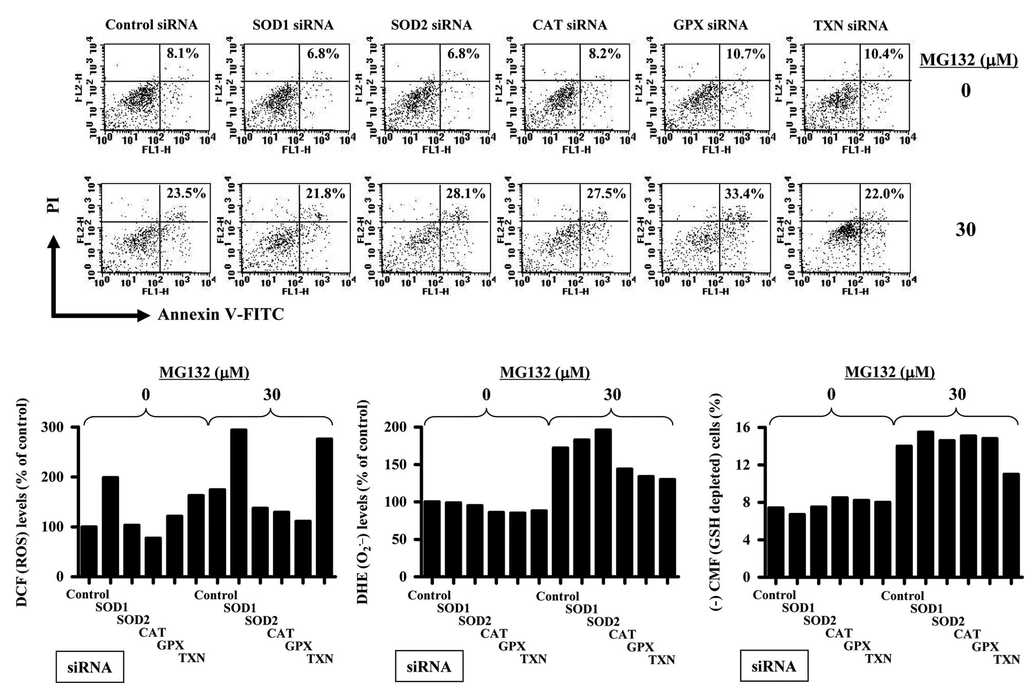
Antioxidant-related siRNAs affect cell
death, ROS and GSH levels in MG132-treated HPF cells
Furthermore, it was determined whether antioxidant
(SOD1, SOD2, CAT, GPX or TXN)-related siRNAs changed cell death,
ROS and GSH levels in MG132-treated HPF cells. As shown in Fig. 4, 30 μM MG132 increased the
proportion of Annexin-V-stained cells about 15% compared with that
in control siRNA-treated HPF cells. Treatment with 10 μM MG132 did
not clearly increase Annexin V-stained cell number in this system
(data not shown). Probably, addition of Lipofectamine 2000 in the
medium seemed to attenuate the biological activity of MG132. All
the siRNAs of antioxidant-related proteins did not significantly
alter Annexin V-stained cell number in HPF control cells for 72 h
(Fig. 4A). Administration of SOD1
or TXN siRNA did not affect cell death in MG132-treated HPF cells
whereas SOD2, CAT or GPX siRNA increased the Annexin-V-stained cell
number in these cells (Fig. 4A).
Especially, GPX siRNA treatment showed a strong pro-apoptotic
effect on MG132-treated HPF cells (Fig.
4A).
In relation to ROS levels, SOD1, GPX or TXN siRNA
increased ROS (DCF) levels in HPF control cells but CAT siRNA
decreased the levels at 72 h (Fig.
4B). SOD1 or TXN siRNAs intensified ROS (DCF) levels in
MG132-treated HPF cells whereas SOD2, CAT or GPX siRNA relatively
decreased the level in these cells (Fig. 4B). CAT, GPX or TXN siRNA seemed to
decrease O2•− levels in HPF control cells
(Fig. 4C). SOD2 siRNA slightly
increased O2•− levels in MG132-treated HPF
cells whereas CAT, GPX or TX siRNA attenuated the level in these
cells (Fig. 4C). In view of the GSH
levels, all siRNAs of antioxidant-related proteins did not affect
the number of GSH-depleted cells in HPF control cells for 72 h
(Fig. 4D). While SOD1, SOD2, CAT or
GPX siRNA did not clearly alter the GSH-depleted cell number in
MG132-treated HPF cells, TNX siRNA prevented GSH deletion in these
cells (Fig. 4D).
Discussion
Various proteasome inhibitors including MG132 have
been demonstrated to stimulate apoptotic cell death through the
induction of ROS (16,17). Because MG132 induced growth
inhibition and death in HPF cells, in the present study we focused
on evaluating the molecular mechanism of MG132-induced HPF cell
death in relation to ROS and GSH. According to our result, ROS
level (as determined by DCF) were increased in HPF cells treated
with 1, 10 or 30 μM MG132. However, O2•−
levels in HPF cells was only increased by 30 μM MG132. Thus,
although MG132 generally seemed to increase intracellular ROS
levels in HPF cells, it affected different ROS levels depending on
its concentration. It is reported that ROS formation due to
proteasome inhibitors may cause mitochondrial dysfunction and
subsequent cytochrome C release, which leads to cell viability loss
(18,19). The collapse of MMP (ΔΨm)
occurs during apoptosis (32).
Correspondingly, MG132 induced the loss of MMP (ΔΨm) in
HPF cells. Furthermore, mitochondrial O2•−
levels in HPF cells were increased by 30 μM MG132. Although the
mechanism underlying ROS generation after MG132 treatment is not
clearly explained, our results suggest that increased
O2•− in MG132-treated HPF cells mainly
originates from the mitochondria.
Treatment with NAC and vitamin C significantly
prevented the growth inhibition of MG132-treated HPF cells and also
decreased the number of Annexin-V-FITC positive cells in these
cells. Both antioxidants attenuated the loss of MMP
(ΔΨm) in MG132-treated HPF cells. Conversely, BSO
slightly enhanced growth inhibition, cell death and MMP
(ΔΨm) loss in MG132-treated HPF cells. These results
implied that changes in ROS or GSH levels by NAC, vitamin C or BSO
affected the growth inhibition and death in MG132-treated HPF
cells. Thus, we assessed ROS or GSH levels in MG132-treated HPF
cells in the presence or absence of NAC, vitamin C or BSO.
As expected, both antioxidants of NAC and vitamin C
attenuated ROS levels including mitochondrial
O2•− levels in MG132-treated or -untreated
HPF cells. BSO showing a slight enhancement in cell death and MMP
(ΔΨm) loss in MG132-treated HPF cells intensified ROS
levels, including mitochondrial O2•− in these
cells. In addition, diethyldithiocarbamate, known to be an
inhibitor of SOD (33), augmented
growth inhibition, cell death, MMP (ΔΨm) loss and
O2•− levels in MG132-treated HPF cells (data
not shown). Therefore, MG132 seemed to induce HPF cell death
through the induction of ROS. Because NAC and vitamin C
individually affected different ROS levels in MG132-treated or
-untreated HPF cells, each antioxidant may be exerting its effects
on the prevention of MG132-induced HPF cell death via different
pathways. BSO alone induced cell growth inhibition, cell death and
MMP (ΔΨm) loss in HPF control cells and strongly
increased ROS levels. Therefore, an increased ROS by BSO treatment
seemed to be tightly related to the HPF cell growth inhibition and
death. Furthermore, we observed that MG132 blocked the activity of
the proteasome in HPF cells, which was efficiently attenuated by
NAC. These results suggest that proteasome inhibition by MG132
influences growth inhibition and death in HPF cells.
In relation to the administration of
antioxidant-related siRNAs in MG132-treated HPF cells, SOD2, CAT or
GPX siRNAs increased the number of Annexin V-stained cells.
However, these siRNAs did not increase, but rather decreased ROS
(DCF) levels in MG132-treated HPF cells. In addition, SOD1 and TXN
siRNA, which did not enhance HPF cell death by MG132, strongly
increased ROS (DCF) levels in these cells. CAT and GPX siRNAs
attenuated O2•− levels in MG132-treated HPF
cells. Furthermore, SOD2 siRNA slightly increased
O2•− levels in MG132-treated HPF cells,
whereas TXN siRNA decreased the level in these cells. Therefore,
the alterations of MG132-induced HPF cell death by
antioxidant-related siRNAs are not correlated with the ROS changes
induced by these siRNAs. Moreover, administration with SOD1, GPX or
TXN siRNA increased ROS (DCF) levels in HPF control cells, but SOD2
or CAT siRNA did not. None of these siRNAs increased
O2•− levels in HPF control cells. Because a
change in the generation or metabolism of ROS in the cells is
influenced by various pro-oxidant or antioxidant enzymes as well as
activities in various cellular organelles, such as the mitochondria
and the endoplasmic reticulum, our results suggest that the
downregulation of each antioxidant protein by its corresponding
siRNA does not simply increase ROS levels in HPF cells and can
individually affect different ROS levels. Therefore, the effects of
ROS level alterations induced by antioxidant-related siRNAs in
MG132-treated HPF cells on cell death need to be further
studied.
The redox status of cellular GSH is a crucial
regulatory element in the protein ubiquitination system (34). GSH depletion due to proteasome
inhibitors can lead to cell death (18,19).
Likewise, MG132 dose-dependently increased the number of
GSH-depleted cells in the HPF cells. BSO as a GSH synthesis
inhibitor increased the numbers of GSH-depleted cells in
MG132-treated HPF cells. However, 10 μM BSO showing a cell death
effect in HPF control cells did not induce GSH depletion. Other
reports definitely show that 100 μM or 1 mM BSO decreased GSH
levels in MCF breast cancer cells (35) or U937 leukemia cells (36). These data imply that BSO differently
influences GSH levels depending on the cell types or the incubation
doses. In addition, although it is known that NAC containing a
thiol group is a GSH precursor, NAC used in this study did not seem
to be a GSH precursor since NAC did not affect GSH depletion in
MG132-treated HPF cells. However, because we demonstrated that NAC
significantly prevented GSH depletion in propyl gallate-treated
HeLa cells (26), it is considered
that NAC can be a GSH precursor or not depending on the
co-incubated agents or cell lines. Moreover, all the siRNAs of
antioxidant-related proteins did not influence GSH depletion in HPF
control cells. These siRNAs except for TXN siRNA did not affect the
number of GSH-depleted cells in MG132-treated HPF cells. Therefore,
the downregulation of antioxidant proteins by their targeting
siRNAs seems to not strongly alter GSH levels in HPF cells. Because
TXN as a potent antioxidant can stimulate cell proliferation or may
confer resistance to anticancer drugs (37,38),
the downregulation of TXN may render cells sensitive to several
cytotoxic drugs. However, our results showed that TXN siRNA did not
enhance HPF cell death by MG132 but prevented GSH deletion in
MG132-treated HPF cells. Therefore, the mechanism of the TRX
siRNA-induced effects on the prevention of GSH depletion rather
than on the enhancement of cell death in MG132-treated HPF cells
needs to be further clarified. Taken together, our results suggest
that the intracellular GSH levels seem to play a decisive role on
MG132-induced HPF cell death, but changes of the content are not
sufficient to predict cell death.
In conclusion, MG132 induced the growth inhibition
and death of HPF cells, which were accompanied by increasing ROS
levels and GSH depletion. The changes of ROS or GSH levels by NAC,
vitamin C or BSO appeared to affect cell growth inhibition and
death in MG132-treated HPF cells. In addition, administration of
antioxidant-related siRNAs did not affect cell death, or ROS and
GSH levels in MG132-treated or MG132-untreated HPF cells. Our
present data provide useful information for understanding the
cytotoxic or toxicological effects of MG132 in normal lung cells in
relation to ROS and GSH levels.
Acknowledgements
This study was supported by a grant from the
Ministry of Science and Technology (MoST)/Korea Science and
Engineering Foundation (KOSEF) through the Diabetes Research Center
at Chonbuk National University (2011-0028226) and the National
Research Foundation of Korea Grant funded by the Korean Government
(MEST) (2010-0021808).
Abbreviations:
|
HPF
|
human pulmonary fibroblast
|
|
MG132
|
carbobenzoxy-Leu-Leu-leucinal
|
|
ROS
|
reactive oxygen species
|
|
MMP (ΔΨm)
|
mitochondrial membrane potential
|
|
NADPH
|
nicotine adenine diphosphate
|
|
XO
|
xanthine oxidase
|
|
SOD
|
superoxide dismutase
|
|
CAT
|
catalase
|
|
GPX
|
GSH peroxidase
|
|
TXN
|
thioredoxin
|
|
FBS
|
fetal bovine serum
|
|
PI
|
propidium iodide
|
|
H2DCFDA
|
2′,7′-dichlorodihydrofluorescein
diacetate
|
|
DHE
|
dihydroethidium
|
|
GSH
|
glutathione
|
|
FITC
|
fluorescein isothiocyanate
|
|
CMFDA
|
5-chloromethylfluorescein
diacetate
|
|
MTT
|
3-(4,5-dimethylthiazol-2-yl)-2,5-diphenyltetrazolium bromide
|
|
siRNA
|
small interfering RNA
|
|
NAC
|
N-acetyl cysteine
|
|
BSO
|
L-buthionine sulfoximine
|
References
|
1
|
Gonzalez C, Sanz-Alfayate G, Agapito MT,
Gomez-Nino A, Rocher A and Obeso A: Significance of ROS in oxygen
sensing in cell systems with sensitivity to physiological hypoxia.
Respir Physiol Neurobiol. 132:17–41. 2002. View Article : Google Scholar : PubMed/NCBI
|
|
2
|
Baran CP, Zeigler MM, Tridandapani S and
Marsh CB: The role of ROS and RNS in regulating life and death of
blood monocytes. Curr Pharm Des. 10:855–866. 2004. View Article : Google Scholar : PubMed/NCBI
|
|
3
|
Zorov DB, Juhaszova M and Sollott SJ:
Mitochondrial ROS-induced ROS release: an update and review.
Biochim Biophys Acta. 1757:509–517. 2006. View Article : Google Scholar : PubMed/NCBI
|
|
4
|
Zelko IN, Mariani TJ and Folz RJ:
Superoxide dismutase multigene family: a comparison of the CuZn-SOD
(SOD1), Mn-SOD (SOD2), and EC-SOD (SOD3) gene structures,
evolution, and expression. Free Radic Biol Med. 33:337–349. 2002.
View Article : Google Scholar : PubMed/NCBI
|
|
5
|
Wilcox CS: Reactive oxygen species: roles
in blood pressure and kidney function. Curr Hypertens Rep.
4:160–166. 2002. View Article : Google Scholar : PubMed/NCBI
|
|
6
|
Marks PA: Thioredoxin in cancer - role of
histone deacetylase inhibitors. Semin Cancer Biol. 16:436–443.
2006. View Article : Google Scholar : PubMed/NCBI
|
|
7
|
Chen TJ, Jeng JY, Lin CW, Wu CY and Chen
YC: Quercetin inhibition of ROS-dependent and -independent
apoptosis in rat glioma C6 cells. Toxicology. 223:113–126. 2006.
View Article : Google Scholar : PubMed/NCBI
|
|
8
|
Dasmahapatra G, Rahmani M, Dent P and
Grant S: The tyrphostin adaphostin interacts synergistically with
proteasome inhibitors to induce apoptosis in human leukemia cells
through a reactive oxygen species (ROS)-dependent mechanism. Blood.
107:232–240. 2006. View Article : Google Scholar
|
|
9
|
Wallach-Dayan SB, Izbicki G, Cohen PY,
Gerstl-Golan R, Fine A and Breuer R: Bleomycin initiates apoptosis
of lung epithelial cells by ROS but not by Fas/FasL pathway. Am J
Physiol Lung Cell Mol Physiol. 290:L790–L796. 2006. View Article : Google Scholar : PubMed/NCBI
|
|
10
|
Orlowski RZ: The role of the
ubiquitin-proteasome pathway in apoptosis. Cell Death Differ.
6:303–313. 1999. View Article : Google Scholar : PubMed/NCBI
|
|
11
|
Voges D, Zwickl P and Baumeister W: The
26S proteasome: a molecular machine designed for controlled
proteolysis. Annu Rev Biochem. 68:1015–1068. 1999. View Article : Google Scholar : PubMed/NCBI
|
|
12
|
Adams J: The proteasome: a suitable
antineoplastic target. Nat Rev Cancer. 4:349–360. 2004. View Article : Google Scholar : PubMed/NCBI
|
|
13
|
Drexler HC: Activation of the cell death
program by inhibition of proteasome function. Proc Natl Acad Sci
USA. 94:855–860. 1997. View Article : Google Scholar : PubMed/NCBI
|
|
14
|
Shah SA, Potter MW and Callery MP:
Ubiquitin proteasome pathway: implications and advances in cancer
therapy. Surg Oncol. 10:43–52. 2001. View Article : Google Scholar : PubMed/NCBI
|
|
15
|
Lee DH and Goldberg AL: Proteasome
inhibitors: valuable new tools for cell biologists. Trends Cell
Biol. 8:397–403. 1998. View Article : Google Scholar : PubMed/NCBI
|
|
16
|
Wu HM, Chi KH and Lin WW: Proteasome
inhibitors stimulate activator protein-1 pathway via reactive
oxygen species production. FEBS Lett. 526:101–105. 2002. View Article : Google Scholar : PubMed/NCBI
|
|
17
|
Perez-Galan P, Roue G, Villamor N,
Montserrat E, Campo E and Colomer D: The proteasome inhibitor
bortezomib induces apoptosis in mantle-cell lymphoma through
generation of ROS and Noxa activation independent of p53 status.
Blood. 107:257–264. 2006. View Article : Google Scholar
|
|
18
|
Ling YH, Liebes L, Zou Y and Perez-Soler
R: Reactive oxygen species generation and mitochondrial dysfunction
in the apoptotic response to Bortezomib, a novel proteasome
inhibitor, in human H460 non-small cell lung cancer cells. J Biol
Chem. 278:33714–33723. 2003. View Article : Google Scholar : PubMed/NCBI
|
|
19
|
Qiu JH, Asai A, Chi S, Saito N, Hamada H
and Kirino T: Proteasome inhibitors induce cytochrome
c-caspase-3-like protease-mediated apoptosis in cultured cortical
neurons. J Neurosci. 20:259–265. 2000.PubMed/NCBI
|
|
20
|
Petty RD, Nicolson MC, Kerr KM,
Collie-Duguid E and Murray GI: Gene expression profiling in
non-small cell lung cancer: from molecular mechanisms to clinical
application. Clin Cancer Res. 10:3237–3248. 2004. View Article : Google Scholar : PubMed/NCBI
|
|
21
|
Mortenson MM, Schlieman MG, Virudachalam S
and Bold RJ: Effects of the proteasome inhibitor bortezomib alone
and in combination with chemotherapy in the A549 non-small-cell
lung cancer cell line. Cancer Chemother Pharmacol. 54:343–353.
2004.PubMed/NCBI
|
|
22
|
Ling YH, Liebes L, Jiang JD, Holland JF,
Elliott PJ, Adams J, Muggia FM and Perez-Soler R: Mechanisms of
proteasome inhibitor PS-341-induced G(2)-M-phase arrest and
apoptosis in human non-small cell lung cancer cell lines. Clin
Cancer Res. 9:1145–1154. 2003.PubMed/NCBI
|
|
23
|
Han YH and Park WH: MG132 as a proteasome
inhibitor induces cell growth inhibition and cell death in A549
lung cancer cells via influencing reactive oxygen species and GSH
level. Hum Exp Toxicol. 29:607–614. 2010. View Article : Google Scholar : PubMed/NCBI
|
|
24
|
Han YH and Park WH: MG132, a proteasome
inhibitor decreased the growth of Calu-6 lung cancer cells via
apoptosis and GSH depletion. Toxicol In Vitro. 24:1237–1242. 2010.
View Article : Google Scholar : PubMed/NCBI
|
|
25
|
Bailey HH: L-S,R-buthionine sulfoximine:
historical development and clinical issues. Chem Biol Interact.
111–112:239–254. 1998.PubMed/NCBI
|
|
26
|
Han YH and Park WH: Propyl gallate
inhibits the growth of HeLa cells via regulating intracellular GSH
level. Food Chem Toxicol. 47:2531–2538. 2009. View Article : Google Scholar : PubMed/NCBI
|
|
27
|
You BR and Park WH: Gallic acid-induced
lung cancer cell death is related to glutathione depletion as well
as reactive oxygen species increase. Toxicol In Vitro.
24:1356–1362. 2010. View Article : Google Scholar : PubMed/NCBI
|
|
28
|
Han YH, Kim SZ, Kim SH and Park WH:
Pyrogallol as a glutathione depletor induces apoptosis in HeLa
cells. Int J Mol Med. 21:721–730. 2008.PubMed/NCBI
|
|
29
|
Park WH, Seol JG, Kim ES, Hyun JM, Jung
CW, Lee CC, Kim BK and Lee YY: Arsenic trioxide-mediated growth
inhibition in MC/CAR myeloma cells via cell cycle arrest in
association with induction of cyclin-dependent kinase inhibitor,
p21, and apoptosis. Cancer Res. 60:3065–3071. 2000.
|
|
30
|
Han YH, Kim SZ, Kim SH and Park WH:
Arsenic trioxide inhibits growth of As4.1 juxtaglomerular cells via
cell cycle arrest and caspase-independent apoptosis. Am J Physiol
Renal Physiol. 293:F511–F520. 2007. View Article : Google Scholar : PubMed/NCBI
|
|
31
|
Elbashir SM, Harborth J, Lendeckel W,
Yalcin A, Weber K and Tuschl T: Duplexes of 21-nucleotide RNAs
mediate RNA interference in cultured mammalian cells. Nature.
411:494–498. 2001. View Article : Google Scholar : PubMed/NCBI
|
|
32
|
Yang J, Liu X, Bhalla K, Kim CN, Ibrado
AM, Cai J, Peng TI, Jones DP and Wang X: Prevention of apoptosis by
Bcl-2: release of cytochrome c from mitochondria blocked. Science.
275:1129–1132. 1997. View Article : Google Scholar : PubMed/NCBI
|
|
33
|
Cocco D, Calabrese L, Rigo A, Argese E and
Rotilio G: Re-examination of the reaction of diethyldithiocarbamate
with the copper of superoxide dismutase. J Biol Chem.
256:8983–8986. 1981.PubMed/NCBI
|
|
34
|
Jahngen-Hodge J, Obin MS, Gong X, Shang F,
Nowell TR Jr, Gong J, Abasi H, Blumberg J and Taylor A: Regulation
of ubiquitin-conjugating enzymes by glutathione following oxidative
stress. J Biol Chem. 272:28218–2826. 1997. View Article : Google Scholar : PubMed/NCBI
|
|
35
|
Lewis-Wambi JS, Kim HR, Wambi C, Patel R,
Pyle JR, Klein-Szanto AJ and Jordan VC: Buthionine sulfoximine
sensitizes antihormone-resistant human breast cancer cells to
estrogen-induced apoptosis. Breast Cancer Res. 10:R1042008.
View Article : Google Scholar
|
|
36
|
Ramos AM and Aller P: Quercetin decreases
intracellular GSH content and potentiates the apoptotic action of
the antileukemic drug arsenic trioxide in human leukemia cell
lines. Biochem Pharmacol. 75:1912–1923. 2008. View Article : Google Scholar : PubMed/NCBI
|
|
37
|
Gallegos A, Gasdaska JR, Taylor CW,
Paine-Murrieta GD, Goodman D, Gasdaska PY, Berggren M, Briehl MM
and Powis G: Transfection with human thioredoxin increases cell
proliferation and a dominant-negative mutant thioredoxin reverses
the transformed phenotype of human breast cancer cells. Cancer Res.
56:5765–5770. 1996.
|
|
38
|
Kim SJ, Miyoshi Y, Taguchi T, Tamaki Y,
Nakamura H, Yodoi J, Kato K and Noguchi S: High thioredoxin
expression is associated with resistance to docetaxel in primary
breast cancer. Clin Cancer Res. 11:8425–8430. 2005. View Article : Google Scholar : PubMed/NCBI
|