Introduction
Most solid tumors show resistance to chemotherapy.
To exert efficacy, anticancer drugs should reach their target cells
at lethal concentrations. To reach all tumor cells, drugs must
penetrate multiple cell layers, which is a significant hurdle
imposed by a solid tumor microenvironment. In contrast to normal
tissues, solid tumors have a complex microenvironment. The tumor
microenvironment consists of various cell types and an enriched
extracellular matrix, as well as an abnormal vasculature and
hypoxic regions (1–3). Besides poor perfusion, extravascular
compartments of solid tumors pose additional conditions that make
diffusion of drug through cell layers difficult, due to increased
distance between blood vessels and an increased interstitial fluid
pressure (1). The majority of
studies on drug resistance have focused on the cellular mechanism
via genetic alteration (4).
Recently, much attention has been paid to the impact of drug
penetration and distribution into solid tumor tissues, as shown by
a considerable amount of data published on the limited distribution
of various anticancer agents and on novel strategies for
improvement of drug distribution, which may ultimately result in
greater efficacy.
Paclitaxel (PTX) is one of the most important
chemotherapeutic drugs used in the treatment of human solid tumors,
including ovarian, breast, and head and neck cancers (5). The excessive accumulation of PTX in
the periphery of tissue fragments (histocultures) and multicellular
tumor spheroids (MCS), with very limited penetration into the
interior has been reported (6,7). The
limited penetration has been attributed to tissue adhesion
(specific and unspecific), cellularity, and expression of
P-glycoprotein (P-gp) (6,8,9).
Doxorubicin (DOX) is a first line antineoplastic agent against many
solid tumors, as well as leukemias and lymphomas (10). Limited availability of DOX due to
its insufficient distribution in solid tumors in association with
efflux by the P-gp pump, increases sequestration in endosomes and
tumor cell packing density (3,9,11,12).
The potential contribution of tissue penetration of
PTX and DOX to their limited efficacy has not been fully evaluated.
This may be due to the absence of proper methods for in
vitro investigation of tissue penetration and distribution and
cell survival. As in vitro models, multicellular layers
(MCL) and MCS have become the most commonly used tools for
qualitative and quantitative assessment of drug
penetration/distribution (3). In
MCLs, a drug is added on one side of the MCL and its appearance on
the other side is measured by appropriate analytical methods. MCLs
have been used successfully in the study of the pharmacokinetics of
anticancer drugs, such as tirapazamine and other DNA alkylators
(13–16). MCS also demonstrate many of the
properties of solid tumors in vivo, including expression of
extracellular matrix (ECM), tight junctions, and lower cell
proliferation to the center, and have been used in examination of
the kinetics of drug penetration (17,18).
Earlier studies evaluated penetration of anticancer
agents including PTX and DOX in 3D models and the influence of P-gp
expression, cell density, and differential expression of the ECM
(9,19,20).
The effect of the drug exposure schedule, however, has not been
fully studied and controversy still exists over the importance of
drug concentration vs. exposure duration for increased efficacy
(7,21). In the present study, we investigated
a detailed penetration/distribution profile of PTX and DOX in MCL
of human colorectal cancer cells as an in vitro model for
avascular regions of human solid tumors. We compared the
penetration, distribution, and retention of PTX and DOX in MCL
following a long period of exposure up to 72 h as well as under
different exposure conditions. Drug penetration and retention in
MCL showed not only concentration- and time-dependency, but also
schedule-dependency. Our data demonstrate the different penetration
kinetics between PTX and DOX, and the relative importance of long
exposure time in terms of penetration and retention in
multicellular layer cultures. The present study may provide the
rationale for the need of pharmacokinetic modulation of drug
distribution, which may in turn lead to efficacy modulation.
Materials and methods
Cell lines and chemical reagents
The human colorectal cancer cell line, DLD-1, was
obtained from the Korea cell line bank (Seoul, Korea). Cells were
grown as monolayers at 37˚C in RPMI-1640 (Gibco-BRL, Rockville, MD)
supplemented with 100 μg/ml streptomycin (Sigma Chemical Co., St.
Louis, MO), 100 units/ml penicillin (Sigma Chemical Co.), and 10%
heat-inactivated fetal bovine serum (WelGene, Daegu, Korea) in a
humidified atmosphere of 95% air plus 5% CO2.
Paclitaxel-rhodamine (PTX-rd) was synthesized as described in the
previous study (22). DOX was a
generous gift from Dong-A Pharmaceutical (Giheung, Yongin,
Gyeonggi, Korea). Calcein-AM was purchased from Molecular Probes,
Inc. (Eugene, OR) and other reagents, unless otherwise noted, were
purchased from Sigma Chemical Co.
Growth and characterization of MCLs
Exponentially growing cells (3×10⁵) were
seeded on a collagen-coated, microporous (0.4 μm) membrane in
Transwell inserts (Corning Costar, Acton, MA), as previously
reported (22). Cells were allowed
to attach for 4 h and the membranes were then submerged in a
culture jar supplemented with 150 ml of RPMI-1640 medium with
intermittent stirring. MCL were allowed to grow up to 8 days.
Frozen sections (20 μm) were prepared in a vertical direction
(perpendicular to the membrane) using Tissue-Tek O.C.T compound
(Sakura, Torrance, CA):20% sucrose (1:2). Sections were stained
with H&E and uniformity of MCL growth was assessed under a
light microscope: only MCL batches with uniform growth across the
membrane were used for data collection.
Penetration experiments
MCLs, after 8 days culture were transferred into
24-well plates or 6-well plates, as needed, depending on
experiments. At the beginning of the penetration assay, the medium
in the donor chamber was replaced with fresh medium containing each
agent, i.e., calcein-AM, PTX-rd, DOX, or ethidium homodimer-1
(EthD-1). The medium volume in the top and bottom chamber was 200
and 700 μl in 24-well plates, and 200 μl and 7 ml in 6-well plates,
respectively. Under these conditions, fluid levels between the
bottom chamber (BC) and top chamber (TC) were even, and,
consequently, penetration was driven by the concentration
gradient.
For drug uptake in monolayers, 1×10⁶
cells/well were plated in an 8-well chamber slide system (Lab-Tek
II, Nalgene, Nunc International, Naperville, IL) and cells were
allowed to attach for 24 h. Cells were then exposed to drugs for 3
h and observed under a confocal microscope (Bio-Rad Laboratories,
Hercules, CA) at λEx/Em of 482/528 nm for PTX-rd and at
λEx/Em of 480/590 nm for DOX.
Image acquisition and analysis
Frozen sections of MCLs were examined under a
fluorescence microscope (Olympus, AX70, TR-6A02, Tokyo, Japan) at
λEx/Em of 488/517 nm for calcein-AM and at
λEx/Em of 482/528 nm for DOX, PTX-rd, and EthD-1. The
images of interest were obtained using the DP70-BSW software as an
average of 1360×1024 μm size (0.2 μm2/pixel) with ISO
200 and exposure time between 1/4–1/30 using intra-group control.
Line morphometric analysis of the fluorescence intensity was
performed using OPTIMAS version 6.5 (Media Cybernetics®,
Silver Spring, MD). A minimum signal level just below threshold was
set for each tissue section based on an average background reading
from regions without staining. Data were normalized for tissue
autofluorescence (background) and plotted against the relative
distance (%) from the drug exposure side (either top or bottom
side) of the MCL. When plotting, fluorescence within 5–10% distance
from the membrane was manually deleted in order to eliminate a
spill-over effect of high fluorescence intensity from the membrane.
Horizontal images were obtained by optical sectioning of MCL using
confocal laser scanning setup (LSM 510 Meta, Carl Zeiss, Jena,
Germany), which was connected to an inverted microscope (Axiovert
200M, Carl Zeiss).
Measurement of cell cycle distribution
using a fluorescence-activated cell sorter (FACS)
After drug exposure, MCLs were washed with cold-PBS,
and treated with trypsin-EDTA (0.05% w/v) on ice for 1 h at 37˚C
for 5 min. Cells were then suspended as single cells. For
monolayers, cells were collected as a single cell suspension after
drug exposure. Cells were then fixed with 70% cold-ethanol and
stored at −20˚C until FACS analysis. Upon analysis, fixed cells
were washed with cold-PBS, treated with RNase A (50 μg/ml) and PI
(50 μg/ml) at 37˚C for 5 min, and then immediately analyzed using a
flow cytometer (FACScan, Becton-Dickinson Immunocytometry System,
San Jose, CA). Cell cycle analysis was performed using Modifit
(Verity Software House, Topsham, ME).
Results
Growth of MCL of DLD-1 cells
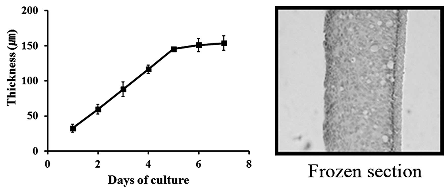
DLD-1 MCL growth was evaluated for 8 days. The
thickness of MCL reached ~150 μm, as observed on frozen sections
(Fig. 1). A stable multi-cell layer
culture of 15–17 cells was formed with no necrotic part, which
appeared to be an appropriate model for avascular regions of human
solid tumors.
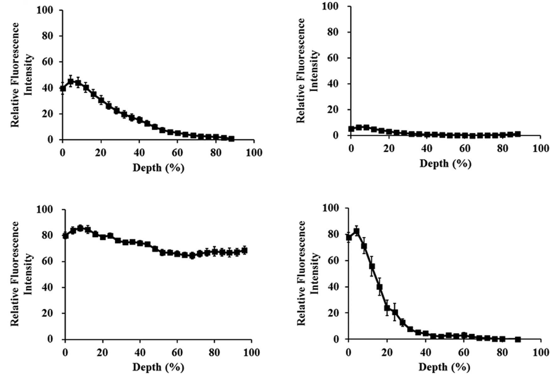
Distribution of compounds with different
physicochemical properties in DLD-1 MCL
P-gp is believed to cause multi-drug resistance
(MDR) by reducing drug uptake into cells and tissues. On the other
hand, several studies have contradictorily reported a positive
effect of P-gp on tissue penetration (3,6,8,11,19).
We compared MCL penetration of PTX (50 μM), DOX (100 μM),
calcein-AM (a vital dye, 40 μM), and EthD-1 (20 μM) after 2-h
exposure in the top chamber of a transwell. All compounds, except
EthD-1, are known as substrates of P-gp. Penetration of DOX was
complete, whereas PTX showed insignificant penetration, and
calcein-AM and EthD-1 showed an intermediate level of penetration
with localization within 20 and 40% depth from the exposure side,
respectively (Fig. 2). DOX and PTX
are both hydrophobic substrates of P-gp, yet, the two compounds
showed drastically different profiles of penetration. We selected
these two compounds for further investigation of the detailed
penetration kinetics.
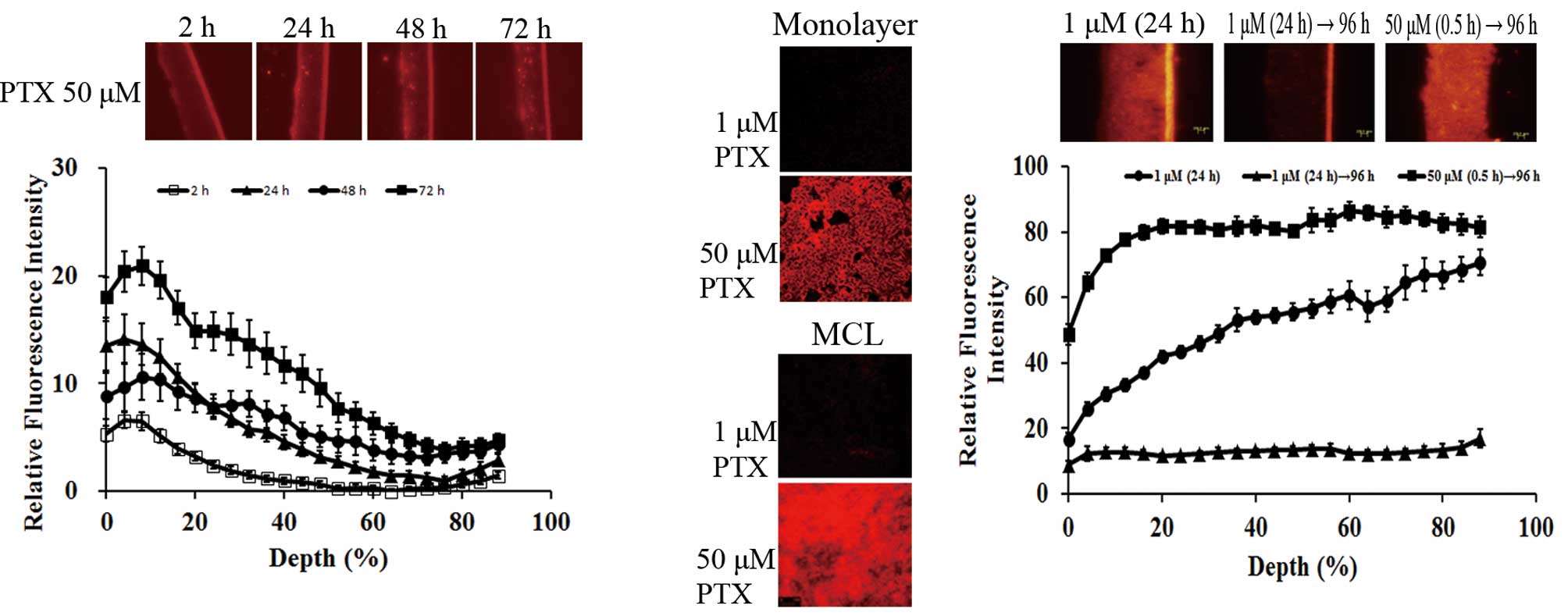
Penetration and distribution of PTX-rd in
DLD-1 MCL
Exposure of MCL to 50 μM PTX-rd resulted in
time-dependent penetration; fluorescence above the background level
was observed within 20% in 2 h and within >90% by 72 h (Fig. 3A). Based on the AUC of the
fluorescence profile, drug distribution increased by 3- and 6-fold
at 24 and 72 h, respectively, compared with that of 2 h.
Distribution profiles of 24 and 48 h showed similar AUC;
nonetheless, an increased level of distribution in the deeper layer
of MCL at 48 h indicated that distribution into deeper layers
occurred only after 48 h, and, before that, preferential
accumulation of PTX-rd was limited to the upper 40% of the layers,
with minimal distribution to the lower 40% of layers.
PTX accumulation was compared between the monolayers
and the MCL. Both cultures were exposed to 1 and 50 μM of PTX-rd
(Fig. 3B). No fluorescence above
the background was observed at 1 μM in either of the cultures. When
exposed to 50 μM PTX-rd, the fluorescence intensity in monolayers
was maximal at 3 h and the level of intensity was comparable to
that of the MCL (optical section at 40–60% depth, 60–90 μm from the
exposure side) after 48-h exposure. The significantly longer time
taken for MCL penetration, as well as the limited penetration
profile, indicated the presence of a penetration barrier for PTX in
DLD-1 MCL (Fig. 3A and B).
In order to gain an understanding of the effect of
exposure conditions on drug penetration and retention, we compared
PTX-rd distribution after exposure to 1 μM (for 24 h) or 50 μM (for
0.5 h), followed by wash-out until 96 h (bottom side exposure,
Fig. 3C). PTX showed preferential
accumulation at the exposure side (100% depth side) after 24 h
exposure at 1 μM. However, the level of fluorescence dropped to
less than 20% throughout the MCL depth following incubation in
drug-free media until 96 h, which indicated complete wash-out,
resulting in no drug retention. Dramatically contrasting data were
obtained after exposure to a 50 μM concentration for 0.5 h,
followed by a wash-out period, where PTX-rd showed homogeneous
distribution throughout the MCL layers with a 6.3-fold higher level
of fluorescence intensity, compared with that of 1 μM × 24 h. These
data clearly indicate the advantage of higher drug concentration
rather than longer exposure time in tissue penetration of PTX. Note
that the drug exposure AUC was comparable between these two groups,
i.e., 1 μM × 24 h vs. 50 μM × 0.5 h. Note also that drug exposure
conditions shown in Fig. 3C
differed from those of Fig. 3A due
to a 35-fold larger volume of donor compartment, i.e., drug was
given in either the top chamber (200 μl) or the bottom chamber (7
ml) for Fig. 3A and 3C,
respectively.
Penetration and distribution of DOX in
DLD-1 MCL
Exposure of MCL to 100 μM DOX resulted in
time-dependent drug penetration, as shown with PTX-rd (Fig. 4A). At earlier times (≤30 min), DOX
showed preferential accumulation in the upper 40% of the layers,
similarly to that observed with PTX-rd. Full penetration was
obtained after 1 h and the accumulation level showed a further
increase as exposure time increased, up to 3 h. Distribution
profiles at 2 and 3 h were similar throughout the MCL depth.
Comparison using the AUC of the fluorescence-depth profile showed
that drug distribution at 1 and 3 h was 1.5 and 4 times greater
than that of 30 min, respectively. Compared with the steep-slope
profile of PTX-rd, DOX showed rapid penetration, resulting in a
rather flat distribution profile throughout the MCL depth by 1 h
(Fig. 3A vs. 4A).
DOX accumulation was compared between the monolayers
and the MCL after exposure to 10 and 100 μM DOX, respectively. In
MCL, the level of intensity was measured on an optical section of
MCL at 60% depth (90 μm from the exposure side). Fluorescence
intensity in monolayers was maximal at 3 h for both concentrations.
No significant signal was detected in MCL exposed to 10 μM DOX. The
fluorescence intensity of the optical section of MCL was comparable
with that of monolayers when exposed to 100 μM DOX. The data
indicate that DOX accumulation into MCL required a higher
concentration, compared with that in monolayers (Fig. 4B).
In order to gain an understanding of the effect of
exposure conditions on drug penetration and retention, we compared
the distribution of DOX fluorescence after exposure to 1 μM (for 24
h) and 50 μM (for 0.5 h), followed by wash-out until 72 h (the
bottom side exposure, Fig. 4C). DOX
distribution within MCL was rather flat after 24 h of exposure at 1
μM (compared with PTX, Fig. 3C) and
decreased to half-level following 48 h wash-out. Exposure of MCL to
50 μM (0.5 h) resulted in a 2.5-fold increase of DOX distribution
in terms of AUC of the fluorescence intensity profile, compared
with 1 μM (24 h). These data also indicate the relative importance
of drug concentration over exposure time for DOX, as seen in PTX.
Note that the drug exposure AUC was comparable between these two
groups, i.e., 1 μM × 24 h vs. 50 μM × 0.5 h. Note also that drug
exposure conditions shown in Fig.
4C differed from those of Fig.
4A due to a 35-fold larger volume of donor compartment, i.e.,
drug was given in either the top chamber (200 μl) or the bottom
chamber (7 ml) for Fig. 4A and C,
respectively.
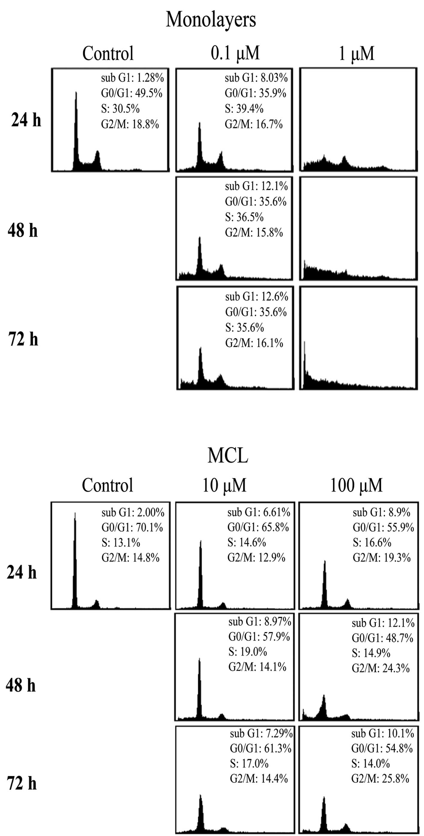
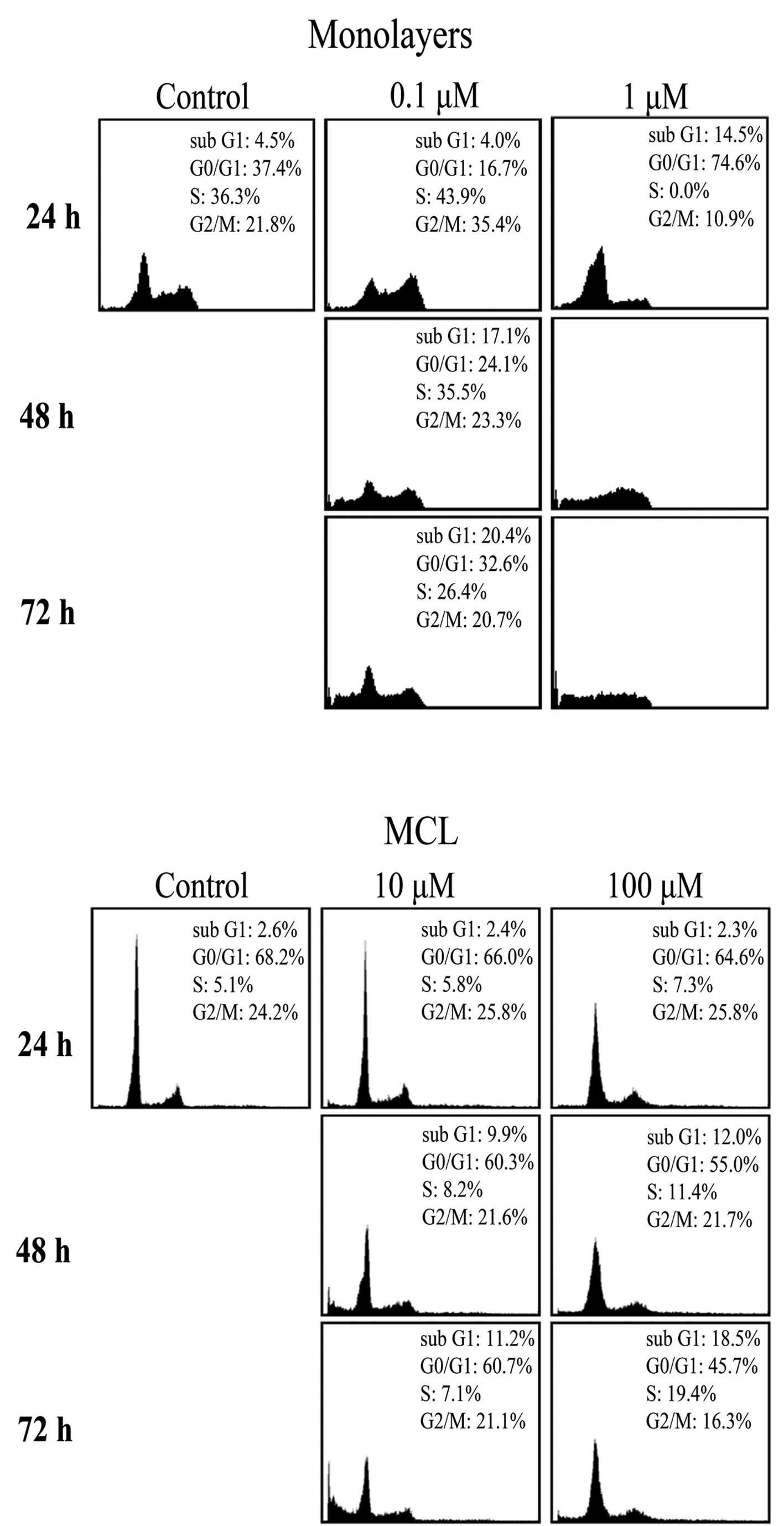
Cell cycle distributions following drug
exposure
The limited distribution of PTX and DOX in MCL
(Figs. 3 and 4) prompted us to examine its correlation
with compromised antitumor efficacy by comparison of the growth
inhibition between monolayers and MCLs. DLD-1 cells grown as
monolayers showed a typical cell cycle distribution and the
distribution was completely destroyed upon exposure to 1 μM (10 ×
IC50) PTX (Fig. 5A). On
the other hand, no significant changes in cell cycle distribution
were observed in DLD-1 cells grown in MCL, even at concentrations
as high as 10 or 100 μM, except for a small increase in the
G2/M phase after exposure to 100 μM (19.3–25.8%)
(Fig. 5B). Note that
%G0/G1 was higher (49.5 vs. 70.1%) in MCL,
compared with monolayers, indicating significantly slow cell
proliferation. These data combined with the drug distribution
profile (Fig. 3) suggest that
limited penetration of PTX may contribute to the lack of
anti-proliferative activity in DLD-1 cells grown in MCL (Fig. 5). In the same way, changes in cell
proliferation were determined following exposure to 0.1–10 μM of
DOX in DLD-1 cells grown in monolayers and MCLs. Cell cycle
distribution was completely abolished at 1 μM (10 ×
IC50) DOX in monolayers (Fig. 6A). Exposure of DLD-1 MCL to DOX at
10 and 100 μM resulted in a significant increase in the
sub-G1 fraction (2.3–18.5%) (Fig. 6B); nonetheless, the changes were
marginal, compared with that of monolayers (Fig. 6A). Hence, the limited distribution
of DOX in MCL also contributed to the limited anti-proliferation
activity, as shown for PTX.
Discussion
Different from agents targeting tumor vasculature,
interstitial drug delivery is the main determinant of anticancer
efficacy for agents targeting tumor parenchymal tissue. The solid
tumor microenvironment plays a critical role as a barrier to
interstitial delivery; hence, many studies have focused on defining
the major mechanisms for and strategies to overcome interstitial
delivery. MCS has been utilized in the study of penetration of
anticancer agents, including DOX, vinblastine, PTX, and
methotrexate (9,18,23–25).
MCL models have been used for a more quantitative study via a
direct assessment of drug penetration into multicellular layers
(12,14,22,26–28).
These multicellular cultures show many characteristics of in
vivo tumors, including the prevalence of extracellular matrix
and presence of hypoxia and desmosomes between cells (9,13,29).
If a substance is added to one side of the MCL, the appearance of
the substance on the other side is measured for determination of
its penetration kinetics (9,12).
Penetration kinetics of PTX, methothrexate, 5-fluorouracil (5-FU),
DOX, and tirapazamine have been studied using these MCL models
(9,14,22,28).
Tumors typically contain irregular, tortuous
networks of leaky microvessels with heterogeneous blood flow and
intervessel distances that fall between 50 and 200 μm (15–20 cell
diameters from the nearest blood vessel), which is significantly
greater than that of normal tissue (a few cell diameters) (30,31).
In this respect, the DLD-1 MCL cultures used in this study closely
represent the avascular region of human solid tumors because
8-day-cultured DLD-1 MCLs showed ~150 μm thickness, with 15–17 cell
layers (Fig. 1). We used static
chamber conditions, which may also represent the tumor
microenvironment of high interstitial pressure and little
convection movement. Although quantitative extrapolation may not be
possible, a relative comparison should be feasible.
In the present study, we evaluated and compared the
penetration profiles of PTX and DOX in DLD-1 MCL. For measurement
of time- and dose-dependent distribution within MCL, we used drug
fluorescence tracing either by the drug itself (DOX) or by a
rhodamine-derivative (PTX). We also compared drug uptake and
drug-induced cell cycle changes between cells grown as monolayers
and MCL. As previously reported, PTX-rd shows physicochemical and
biological properties similar to those of the parent PTX and is
considered suitable for both pharmacokinetics and efficacy
evaluation (8,22,32,33).
For P-gp substrates, it is well known that cellular
uptake decreases in P-gp-overexpressing cells grown in monolayers
because substrates are subject to efflux by P-gp after entering
cells (19). On the other hand,
tissue penetration of P-gp substrates has been reported to increase
as shown with BODIPY-taxol in 3-dimensional cultures of
P-gp-overexpressing cells (8). When
P-gp efflux was inhibited by efflux inhibitors, drug sequestration
at the drug exposure side occurred and the availability of the drug
to penetrate into deeper layers decreased (19). DLD-1 cells used in the present study
are P-gp (+) and penetration of DOX (100 μM) was completed within 1
h. However, the other two P-gp substrates, calcein-AM and PTX-Rd,
showed limited penetration, compared with DOX (Fig. 2). These data suggest that
penetration of P-gp-substrates is influenced by other factors, such
as physiochemical properties of the agents, besides their
interaction with P-gp.
We used fluorescence microscopic analysis for drug
penetration. Images of representative regions of the MCL sections
were acquired and quantitative line morphometric analysis was
performed. In this line morphometric analysis, areas adjacent to
membranes were intentionally deleted in order to eliminate the
spill-over effect, as described in Materials and methods.
Fluorescence intensity in the very top layers of MCL showed
slightly decreased levels, i.e., the maximum level of fluorescence
was observed in 5–10% of layers. This was observed in either case
when drug was added to the top or bottom chamber. Reasons for this
lower level in the exposure layers may be attributed to drug-efflux
by P-gp; however, it may also be due to loss of drug during
wash-out for sample harvest. Note that this effect was not observed
on the data from the bottom side exposure (Figs. 3C and 4C) due to manual deletion of the
spill-over range.
PTX penetration into MCL showed time-dependency
(Fig. 3A). Its penetration into
whole layers was observed after 48 h and the tissue level increased
further until 72 h, suggesting slow penetration. Until 24 h, drug
accumulation was limited to the upper layer and insignificant
fluorescence intensity was observed in deep layers. The
distribution profile was ‘flip-flopped’ at 48 h, i.e., the level in
the upper layers decreased, as the level in the lower layers
increased, while the AUC of the tissue distribution profile
remained the same. Hence, our data suggest that PTX penetration was
so slow to show a two-step pattern of distribution, i.e.,
accumulation during the first 24 h and movement through the layers
during the next 24 h. A similar phenomenon has been reported for
PTX distribution in histocultures: PTX accumulates in the
peripheral part of the tissue (histocultures) and the break-through
penetration to the center region is caused by cell density
reduction induced by drug-induced apoptosis after 24 h (6,9). As
expected from limited penetration of PTX in MCL, cell cycle
dysregulation was minimal in MCL, even at the exposure to 10 or
100-fold higher drug concentration compared to monolayers (Fig. 5B).
Contrary to PTX, DOX showed relatively rapid
penetration into MCL layers, resulting in homogeneous distribution
within 1 h, after which the accumulation level increased and a
plateau level was reached at 3 h (Fig.
4A). As DOX showed homogeneous and significant accumulation in
MCL within 3 h compared to PTX, growth inhibition measured by cell
cycle dysregulation was greater than that of PTX, showing a
significant increase in sub-G1 fraction (18.5%) by 72 h
(Fig. 6B). However, growth
inhibition as as measurement of the increased sub-G1
fraction was much lower and significantly more delayed than
expected. It has been suggested that cells grown in a 3D
microenvironment resembling the in vivo conditions exhibit a
significantly greater resistance to drugs than cells grown as
single cell suspension or monolayers (multicellular resistance)
(1,34–36).
Three-dimensional cultures have fewer of actively proliferating
cells, which in turn requires a greater drug concentration and a
longer exposure for an effect to occur (37,38). A
further study is warranted to measure the actual drug concentration
and pharmacodynamics in MCL conditions.
Our data suggest an advantage of high drug
concentration over longer exposure time under the same level of
exposure (CxT) in tissue accumulation of PTX and DOX (Figs. 3C and 4C). Drug retention in MCL after a long
wash-out period (≥72 h) was significantly greater when exposed to a
higher drug concentration over a short duration (50 μM × 0.5 h),
compared with a lower drug concentration over a long duration (1 μM
× 24 h). This effect was much pronounced for PTX compared to DOX,
and comparable with the observation made by another group that drug
accumulation was much greater when exposed to a higher
concentration under the same CxT exposure condition (21,39).
This can be attributed to saturation of the P-gp pump by high drug
concentration and subsequent intracellular accumulation, leading to
apoptosis induction and reduced cell density (9,21,39).
Our data may not be compared with a study that suggested a cell
kill advantage of longer exposure, in which the cell kill was
measured after disaggregation of spheroids into single cell
suspension (7).
In this study, we demonstrated that: i) the drug
penetration into multicellular layers was not only dependent on the
substrate's specificity for the P-gp pump but also dependent on
other factors including the physiochemical properties of the drugs;
and ii) the penetration and retention of PTX and DOX in MCL was not
only concentration- and time-dependent, but also
schedule-dependent. It can be suggested that slow releasing
formulations or a slow infusion regimen may not necessarily be
desirable, especially for PTX, due to insufficient penetration and
accumulation which may result from a low local concentration at the
target site.
Acknowledgements
This study was supported by the Mid-career
Researcher Program through NRF grant funded by the MEST (no.
2011-0027565).
References
|
1
|
Trédan O, Galmarini CM, Patel K and
Tannock IF: Drug resistance and the solid tumor microenvironment. J
Natl Cancer Inst. 99:1441–1454. 2007.
|
|
2
|
Primeau AJ, Rendon A, Hedley D, Lilge L
and Tannock IF: The distribution of the anticancer drug doxorubicin
in relation to blood vessels in solid tumors. Clin Cancer Res.
11:8782–8788. 2005. View Article : Google Scholar : PubMed/NCBI
|
|
3
|
Minchinton AI and Tannock IF: Drug
penetration in solid tumours. Nat Rev Cancer. 6:583–592. 2006.
View Article : Google Scholar : PubMed/NCBI
|
|
4
|
Xia Y and Lee K: Targeting multidrug
resistance with small molecules for cancer therapy. Biomol Therap.
18:375–385. 2010. View Article : Google Scholar
|
|
5
|
Rowinsky EK: Clinical pharmacology of
Taxol. J Natl Cancer Inst Monogr. 15:25–37. 1993.PubMed/NCBI
|
|
6
|
Kuh HJ, Jang SH, Wientjes MG, Weaver JR
and Au JL: Determinants of paclitaxel penetration and accumulation
in human solid tumor. J Pharmacol Exp Ther. 290:871–880.
1999.PubMed/NCBI
|
|
7
|
Nicholson KM, Bibby MC and Phillips RM:
Influence of drug exposure parameters on the activity of paclitaxel
in multicellular spheroids. Eur J Cancer. 13:1291–1298. 1997.
View Article : Google Scholar : PubMed/NCBI
|
|
8
|
Martin C, Walker J, Rothnie A and
Callaghan R: The expression of P-glycoprotein does influence the
distribution of novel fluorescent compounds in solid tumour models.
Br J Cancer. 89:1581–1589. 2003. View Article : Google Scholar : PubMed/NCBI
|
|
9
|
Grantab R, Sivananthan S and Tannock IF:
The penetration of anticancer drugs through tumor tissue as a
function of cellular adhesion and packing density of tumor cells.
Cancer Res. 66:1033–1039. 2006. View Article : Google Scholar : PubMed/NCBI
|
|
10
|
Outomuro D, Grana DR, Azzato F and Milei
J: Adriamycin-induced myocardial toxicity: new solutions for an old
problem? Int J Cardiol. 117:6–15. 2007. View Article : Google Scholar : PubMed/NCBI
|
|
11
|
Patel KJ and Tannock IF: The influence of
P-glycoprotein expression and its inhibitors on the distribution of
doxorubicin in breast tumors. BMC Cancer. 9:3562009. View Article : Google Scholar : PubMed/NCBI
|
|
12
|
Lee CM and Tannock IF: Inhibition of
endosomal sequestration of basic anticancer drugs: influence on
cytotoxicity and tissue penetration. Br J Cancer. 94:863–869. 2006.
View Article : Google Scholar : PubMed/NCBI
|
|
13
|
Hicks KO, Ohms SJ, van Zijl PL, Denny WA,
Hunter PJ and Wilson WR: An experimental and mathematical model for
the extravascular transport of a DNA intercalator in tumours. Br J
Cancer. 76:894–903. 1997. View Article : Google Scholar : PubMed/NCBI
|
|
14
|
Hicks KO, Fleming Y, Siim BG, Koch CJ and
Wilson WR: Extravascular diffusion of tirapazamine: effect of
metabolic consumption assessed using the multicellular layer model.
Int J Radiat Oncol Biol Phys. 42:641–649. 1998. View Article : Google Scholar : PubMed/NCBI
|
|
15
|
Hicks KO, Pruijn FB, Baguley BC and Wilson
WR: Extravascular transport of the DNA intercalator and
topoisomerase poison
N-[2-(dimethylamino)ethyl]acridine-4-carboxamide (DACA): diffusion
and metabolism in multicellular layers of tumor cells. J Pharmacol
Exp Ther. 297:1088–1098. 2001.PubMed/NCBI
|
|
16
|
Hicks KO, Pruijn FB, Sturman JR, Denny WA
and Wilson WR: Multicellular resistance to tirapazamine is due to
restricted extravascular transport: a
pharmacokinetic/pharmacodynamic study in HT29 multicellular layer
cultures. Cancer Res. 63:5970–5977. 2003.
|
|
17
|
Sutherland RM and Durand RE: Radiation
response of multicell spheroids: an in vitro tumour model. Curr Top
Radiat Res Q. 11:87–139. 1976.PubMed/NCBI
|
|
18
|
Sutherland RM: Cell and environment
interactions in tumor microregions: the multicell spheroid model.
Science. 240:177–184. 1988. View Article : Google Scholar : PubMed/NCBI
|
|
19
|
Tunggal JK, Melo T, Ballinger JR and
Tannock IF: The influence of expression of P-glycoprotein on the
penetration of anticancer drugs through multicellular layers. Int J
Cancer. 86:101–107. 2000. View Article : Google Scholar : PubMed/NCBI
|
|
20
|
Tannock IF, Lee CM, Tunggal JK, Cowan DS
and Egorin MJ: Limited penetration of anticancer drugs through
tumor tissue: a potential cause of resistance of solid tumors to
chemotherapy. Clin Cancer Res. 8:878–884. 2002.PubMed/NCBI
|
|
21
|
Jang SH, Wientjes MG and Au JL:
Enhancement of paclitaxel delivery to solid tumors by
apoptosis-inducing pretreatment: effect of treatment schedule. J
Pharmacol Exp Ther. 296:1035–1042. 2001.PubMed/NCBI
|
|
22
|
Al-Abd AM, Lee JH, Kim SY, Kun N and Kuh
HJ: Novel application of multicellular layers culture for in situ
evaluation of cytotoxicity and penetration of paclitaxel. Cancer
Sci. 99:423–431. 2008. View Article : Google Scholar : PubMed/NCBI
|
|
23
|
West GW, Weichselbaum R and Little JB:
Limited penetration of methotrexate into human osteosarcoma
spheroids as a proposed model for solid tumor resistance to
adjuvant chemotherapy. Cancer Res. 40:3665–3668. 1980.
|
|
24
|
Nederman T and Carlsson J: Penetration and
binding of vinblastine and 5-fluorouracil in cellular spheroids.
Cancer Chemother Pharmacol. 13:131–135. 1984.PubMed/NCBI
|
|
25
|
Durand RE: Distribution and activity of
antineoplastic drugs in a tumor model. J Natl Cancer Inst.
81:146–152. 1989. View Article : Google Scholar : PubMed/NCBI
|
|
26
|
Tunggal JK, Cowan DS, Shaikh H and Tannock
IF: Penetration of anticancer drugs through solid tissue: a factor
that limits the effectiveness of chemotherapy for solid tumors.
Clin Cancer Res. 5:1583–1586. 1999.PubMed/NCBI
|
|
27
|
Hicks KO, Pruijn FB, Secomb TW, Hay MP,
Hsu R, Brown JM, Denny WA, Dewhirst MW and Wilson WR: Use of
three-dimensional tissue cultures to model extravascular transport
and predict in vivo activity of hypoxia-targeted anticancer drugs.
J Natl Cancer Inst. 98:1118–1128. 2006. View Article : Google Scholar : PubMed/NCBI
|
|
28
|
Kyle AH, Huxham LA, Yeoman DM and
Minchinton AI: Limited tissue penetration of taxanes: a mechanism
for resistance in solid tumors. Clin Cancer Res. 13:2804–2810.
2007. View Article : Google Scholar : PubMed/NCBI
|
|
29
|
Wilson WR and Hicks KO: Measurement of
extravascular drug diffusion in multicellular layers. Br J Cancer.
79:1623–1626. 1999.PubMed/NCBI
|
|
30
|
Wijffels KI, Kaanders JH, Rijken PF,
Bussink J, van den Hoogen FJ, Marres HA, de Wilde PC, Raleigh JA
and van der Kogel AJ: Vascular architecture and hypoxic profiles in
human head and neck squamous cell carcinomas. Br J Cancer.
83:674–683. 2000. View Article : Google Scholar : PubMed/NCBI
|
|
31
|
Huxham LA, Kyle AH, Baker JH, Nykilchuk LK
and Minchinton AI: Microregional effects of gemcitabine in HCT-116
xenografts. Cancer Res. 64:6537–6541. 2004. View Article : Google Scholar : PubMed/NCBI
|
|
32
|
Guy R, Scott Z, Sloboda R and Nicolaou K:
Fluorescent toxoids. Chem Biol. 3:1021–1031. 1996. View Article : Google Scholar
|
|
33
|
Baloglu E, Kingston DG, Patel P,
Chatterjee SK and Bane SL: Synthesis and microtubule binding of
fluorescent paclitaxel derivatives. Bioorg Med Chem Lett.
11:2249–2252. 2001. View Article : Google Scholar : PubMed/NCBI
|
|
34
|
Mellor HR, Ferguson DJ and Callaghan R: A
model of quiescent tumour microregions for evaluating multicellular
resistance to chemotherapeutic drugs. Br J Cancer. 93:302–309.
2005. View Article : Google Scholar : PubMed/NCBI
|
|
35
|
Olive PL and Durand RE: Drug and radiation
resistance in spheroids: cell contact and kinetics. Cancer
Metastasis Rev. 13:121–138. 1994. View Article : Google Scholar : PubMed/NCBI
|
|
36
|
Croix BS, Rak JW, Kapitain S, Sheehan C,
Graham CH and Kerbel RS: Reversal by hyaluronidase of
adhesion-dependent multicellular drug resistance in mammary
carcinoma cells. J Natl Cancer Inst. 88:1285–1296. 1996. View Article : Google Scholar : PubMed/NCBI
|
|
37
|
Barbone D, Ryan JA, Kolhatkar N, Chacko
AD, Jablons DM, Sugarbaker DJ, Bueno R, Letai AG, Coussens LM,
Fennell DA and Broaddus VC: The Bcl-2 repertoire of mesothelioma
spheroids underlies acquired apoptotic multicellular resistance.
Cell Death Dis. 2:e1742011. View Article : Google Scholar : PubMed/NCBI
|
|
38
|
Lee SY, Jeong EK, Jeon HM, Kim CH and Kang
HS: Implication of necrosis-linked p53 aggregation in acquired
apoptotic resistance to 5-FU in MCF-7 multicellular tumour
spheroids. Oncol Rep. 24:73–79. 2010.PubMed/NCBI
|
|
39
|
Jang SH, Wientjes MG and Au JL:
Determinants of paclitaxel uptake, accumulation and retention in
solid tumors. Invest New Drugs. 19:113–123. 2001. View Article : Google Scholar : PubMed/NCBI
|