Introduction
Breast cancer is the most prevalent cancer in women
worldwide, and the leading cause of cancer death in women,
accounting for 23% of the total new cancer cases and 14% of total
cancer deaths (1). About 12% of
women in the United States will develop invasive breast cancer over
the course of their lifetime. About 39,520 women in the US were
expected to die in 2011 from breast cancer (2). Though treatable in early stages, once
metastasis has occurred the survival rate is drastically reduced to
a median of 2–3 years and treatment focuses on palliative care
(3). Endometrial cancer is the most
common cancer of the female reproductive organs, with most cases
diagnosed in the 50 to 69 age group. Approximately 46, 470 new
cases are estimated to be diagnosed in 2011 and 8,120 deaths
(4). Cervical cancer is the third
most commonly diagnosed cancer and the fourth leading cause of
deaths in females worldwide, with more than 85% of associated
deaths occurring in developing countries (1). Cervical cancer develops slowly, taking
10–15 years to develop into cancer from a pre-cancerous condition
called dysplasia. Though fully treatable in early stages, once the
cancer has metastasized, patient outcome is poor. Ovarian
carcinoma, which occurs mainly in post-menopausal women, is the
ninth most common cancer among women and the fifth leading cause of
death among women worldwide (5).
Since ovarian cancer often remains clinically silent, the majority
of patients with ovarian carcinoma have advanced intraperitoneal
metastatic disease at diagnosis, resulting in a poor prognosis.
About 80% of ovarian cancer cases are diagnosed at an advanced
stage after metastasis has occurred (5).
Progression of metastasis occurs secondary to cancer
cell detachment from the primary tumor, basement membrane
degradation, cancer cell invasion into the surrounding stroma, and
entry into and transport through the vascular or lymphatic system
to distal sites such as the liver, lungs, and brain, and
extravasation, tumor cell proliferation and angiogenesis at distal
sites (6–10). Tumor cell invasion depends upon
degradation of the extracellular matrix (ECM), which is composed of
collagen, proteoglycans, fibronectin, laminin and other
glycoproteins, and, when intact, acts as a barrier to block cancer
cell invasion (11–13). Two families of proteases, the MMPs
and urokinase plasminogen activators (u-PA) are involved in tumor
invasion and metastasis. Numerous clinical and experimental studies
have demonstrated that elevated levels of u-PA and MMPs are
associated with tumor growth, cancer progression, metastasis and
shortened survival in patients (14–27).
MMPs, especially MMP-2 and MMP-9 play key roles in
tumor cell invasion and metastasis due to their ability to degrade
type IV collagen, a major component of the ECM (13,28,29).
Secreted in their latent zymogenic form as inactive pro-enzymes,
MMP-2 and MMP-9 are cleaved by other MMPs or proteases to yield the
activated forms of 68, 58, and 54 kDa for MMP-2, and 84 kDa for
MMP-9. Proteolytic activities of MMP-2 and MMP-9 are inhibited by
specific inhibitors, tissue inhibitors of metalloproteinases
(TIMPs). Thus, a critical determinant of net proteolytic
degradation is the balance between MMP and TIMP levels. Clinical
studies note the association of MMP expression with progression of
breast (18–20), cervical (21,22),
uterine (24) and ovarian (23,25)
cancers.
The serine protease u-PA consists of two disulfide
bridges linked to polypeptides, which are cleaved to the active
chain (33kDa) by various stimuli. The protease u-PA converts
plasminogen to plasmin, which is capable of promoting tumor growth
and angiogenesis, degrading the ECM and basement membrane and
activating pro-MMPs (30).
Synthetic u-PA inhibitors have been reported to inhibit metastasis
of mammary carcinoma cell lines (31). Clinical studies have shown that high
uPA levels are correlated with progression of female cancers
(25–27,32,33).
Rath and Pauling (34) proposed that nutrients such as lysine
and ascorbic acid be utilized to target plasmin-mediated connective
tissue degradation as a universal approach to tumor growth and
expansion. Binding to plasminogen active sites, lysine blocks
plasminogen activation into plasmin by tissue plasminogen activator
(t-PA). Thus it modulates the plasmin-induced MMP activation
cascade (35). Subsequent studies
confirmed this approach and resulted in identifying a novel
formulation composed of lysine, ascorbic acid, proline and green
tea extract and other micronutrients (NM), which has shown
significant anticancer activity against a large number (~40) of
cancer cell lines, blocking cancer growth, tissue invasion and MMP
expression both in vitro and in vivo(36). In this study, we focused on the
modulating effect of NM on the activities of MMP-2 and -9, TIMPs
and u-PA in human breast (MBA-MD-231, MCF-7), cervical (HeLa),
uterine (SK-UT-1) and ovarian (SK-OV-3) cancer cell lines.
Methods and materials
Materials
Human breast cancer cells MDA-MB-231 and MCF-7,
cervical cancer cells HeLa, uterine leimyosarcoma SK-UT-1 and
ovarian cancer cells SK-OV-3, along with their culture media were
obtained from ATCC. Antibiotics, penicillin, and fetal bovine serum
(FBS), were obtained from Gibco (BRL, Long Island, NY).
Twenty-four-well tissue culture plates were obtained from Costar
(Cambrdige, MA). Gelatinase zymography was performed in 10% Novex
pre-cast SDS polyacrylamide gel (Invitrogen Inc.) with 0.1% gelatin
in non-reducing conditions. The nutrient mixture (NM), prepared by
VitaTech (Hayward, CA) was composed of the following ingredients in
the relative amounts indicated: vitamin C (as ascorbic acid and as
Mg, Ca, and palmitate ascorbate) 700 mg; L-lysine 1000 mg;
L-proline 750 mg; L-arginine 500 mg; N-acetyl cysteine 200 mg;
standardized green tea extract (80% polyphenol) 1000 mg; selenium
30 μg; copper 2 mg; manganese 1 mg. All other reagents used were of
high quality and were obtained from Sigma, unless otherwise
indicated.
Cell cultures
Human breast cancer cell lines MDA-MB-231 and MCF-7
and cervical cancer cell line HeLa were grown in MEM, uterine cell
line SK-UT-1 in DEME and ovarian cancer cell line SK-OV-3 in McCoy
medium supplemented with 10% FBS, penicillin (100 U/ml), and
streptomycin (100 μg/ml) in 24-well tissue culture plates. The
cells were plated at a density of 1×10⁵ cells/ml and
grown to confluency in a humidified atmosphere at 5% CO2
at 37°C. Serum-supplemented media were removed and the cell
monolayer was washed once with PBS with the recommended serum-free
media. The cells were treated with the nutrient mixture, dissolved
in media and tested at 0, 50, 100, 250, 500, and 1000 μg/ml in
triplicate at each dose. Parallel sets of cultures were treated
with PMA (100 ng/ml) for induction of MMP-9. Control and PMA
treatments were done in triplicates. The plates were then returned
to the incubator. The conditioned media were collected separately,
pooled, and centrifuged at 4°C for 10 min at 3000 rpm to remove
cells and cell debris. The supernatant was collected and used to
assess for u-PA activity (by fibrin zymography on 10% SDS-PAGE gels
containing fibrinogen and plasminogen), MMP-2 and -9 (by gelatinase
zymography), and TIMPs (by reverse zymography).
Fibrin zymography
Fibrin zymography was used to analyze u-PA activity
on 10% SDS-PAGE gels containing fibrinogen (5.5 mg/ml) and
plasminogen (50 μg/ml). After electrophoresis, the gels were washed
twice with 2.5% Triton X-100 for 30 min. The gels were then
incubated overnight at 37°C with 0.1% glycine buffer pH 7.5 and
then stained with 0.5% Coomassie Brilliant Blue R250 and destained.
Electrophoresis of u-PA and t-PA were conducted for comparison.
Fibrin zymograms were scanned using CanoScan 9950F Canon
Scanner.
Gelatinase zymography
Gelatinase zymography was performed in 10% NOVEX
Pre-Cast SDS polyacrylamide gel (Invitrogen Corporation) in the
presence of 0.1% gelatin under non-reducing conditions. Culture
media (20 μl) were mixed with sample buffer and loaded for SDS-PAGE
with tris glycine SDS buffer as suggested by the manufacturer
(Novex). Samples were not boiled before electrophoresis. Following
electrophoresis the gels were washed twice in 2.5% Triton X-100 for
30 min at room temperature to remove SDS. The gels were then
incubated at 37°C overnight in substrate buffer containing 50 mM
Tris-HCl and 10 mM CaCl2 at pH 8.0 and stained with 0.5%
Coomassie Blue R250 in 50% methanol and 10% glacial acetic acid for
30 min and destained. Upon renaturation of the enzyme, the
gelatinases digest the gelatin in the gel and give clear bands
against an intensely stained background. Protein standards were run
concurrently and approximate molecular weights were determined by
plotting the relative mobilities of known proteins.
Reverse zymography
TIMPS were analyzed by reverse zymography on 15% SDS
gels containing serum-free conditioned medium from cells. After
electrophoresis the gels were washed twice with 2.5% Triton-X for
30 min at room temperature to remove SDS. The gels were then
incubated at 37°C overnight in 50 mM Tris-HCl and 10 mM
CaCl2 at pH 7.6 and stained with 0.5% Coomassie Blue
R25, destained and scanned.
Scanning of gelatinase and fibrin
zymograms
Gelatinase and fibrin zymograms were scanned using
CanoScan 9950F Canon scanner at 300 dpi. The intensity of the bands
was evaluated using the pixel-based densitometer program
Un-Scan-It, Version 5.1, 32-bit, by Silk Scientific Corporation
(Orem, UT, USA), at a resolution of 1 Scanner Unit (1/100 of an
inch for an image that was scanned at 100 dpi). The pixel
densitometer calculates the optical density of each pixel (values 0
to 255) using the darkly stained background of the gel as a pixel
value of 0. A logarithmic optical density scale was used since the
optical density of films and gels is logarithmically proportional
to the concentration. The pixel densitometer sums the optical
density of each pixel to give a band’s density. In all graphs, band
densities were reported as percentages of the sums of all pixels in
a given lane (treatment) of a gel.
Statistical analysis
Pearson’s correlation coefficient was determined
between mean MMP-9, u-PA and TIMP-2 expression of breast cancer
cell lines MDA-MB-231 and MCF-7 and uterine cell line SK-UT-1 and
between mean MMP-2 and TIMP-2 expression of cervical cell line HeLa
and ovarian cancer SK-OV-3 using MedCalc Software (Mariakerke,
Belgium).
Results
Effect of NM on u-PA activity in human
breast, cervical, uterine and ovarian cell lines
Activity of u-PA was detected in both breast cancer
cell lines and in the uterine cell line showing two bands
corresponding to 55 and 33 kD. NM exerted dose response inhibition
with virtual block of u-PA activity at 100 μg/ml in SK-UT-1 cells
(linear trend R2=0.461) and in MCF-7 cells (linear trend
R2=0.656), and at 500 μg/ml (linear trend
R2=0.813) in MDA-MB-231 cells. See Fig. 1 for respective fibrin zymograms and
densitometry analyses. However, no bands corresponding to u-PA were
detected for HeLa and SK-OV-3 cell lines.
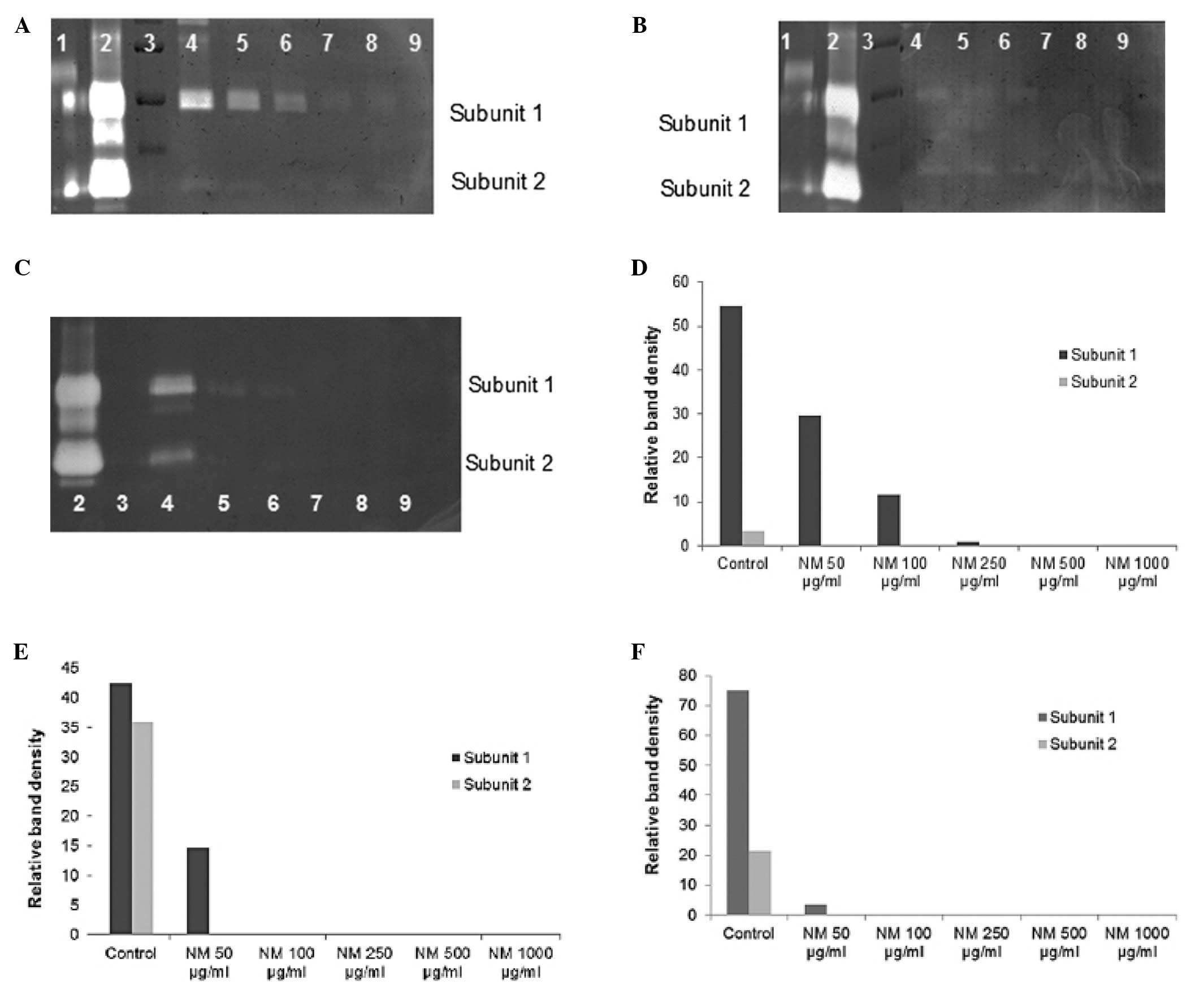 | Figure 1Effect of NM on breast cancer cell
lines MDA-MB-231 and MCF-7 and uterine cancer cell line SK-UT-1
u-PA expression. Fibrin zymograms of MDA-MB-231 (A), MCF-7 (B) and
SK-UT-1 (C) u-PA expression. Lane: 1, t-PA; 2, u-PA; 3, Markers; 4,
Control, 5–9 NM 50, 100, 250, 500, 1000 μg/ml. Densitometric
analyses of MDA-MB-231 (D), MCF-7 (E) and SK-UT-1 (F) u-PA
expression. |
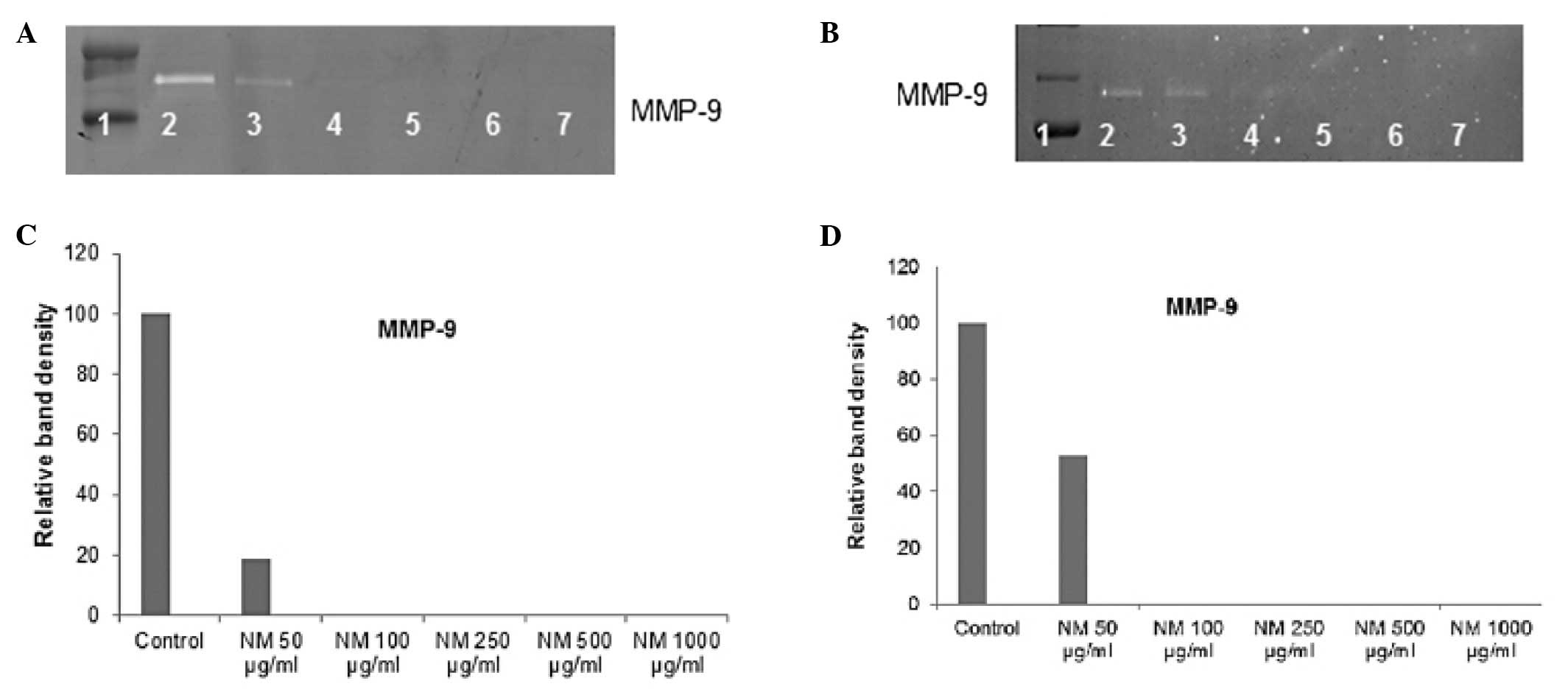
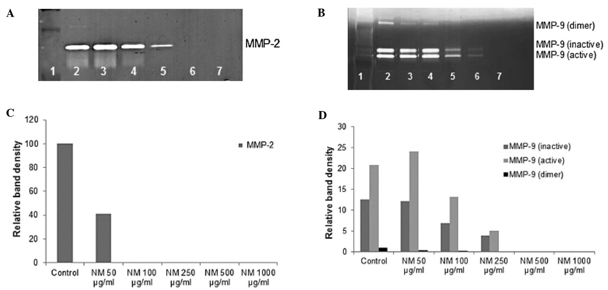
Effect of NM on MMP-2 and MMP-9
expression by human breast cervical and ovarian cancer cell
lines
On gelatinase zymography, a band corresponding to
MMP-9 was detected in both MDA-MB-231 and MCF-7 cell lines.
Cervical HeLa cells showed two bands, an intense band corresponding
to MMP-2 and a faint band corresponding to MMP-9, which was
enhanced with PMA treatment. Normal uterine SK-UT-1 cells did not
express MMP-2 or MMP-9; however, MMP-9 was induced with PMA.
Ovarian SK-OV-3 cells showed only a band corresponding to MMP-2. NM
inhibited MMP expression in all cell lines, with complete block of
MMP-9 in breast cancer cells at 100 μg/ml and in uterine and
cervical cells at 500 μg/ml. NM blocked MMP-2 expression in ovarian
and cervical cell lines at 100 and 1000 μg/ml, respectively. See
Figs. 2–4 for gelatinase zymograms and densitometry
analyses.
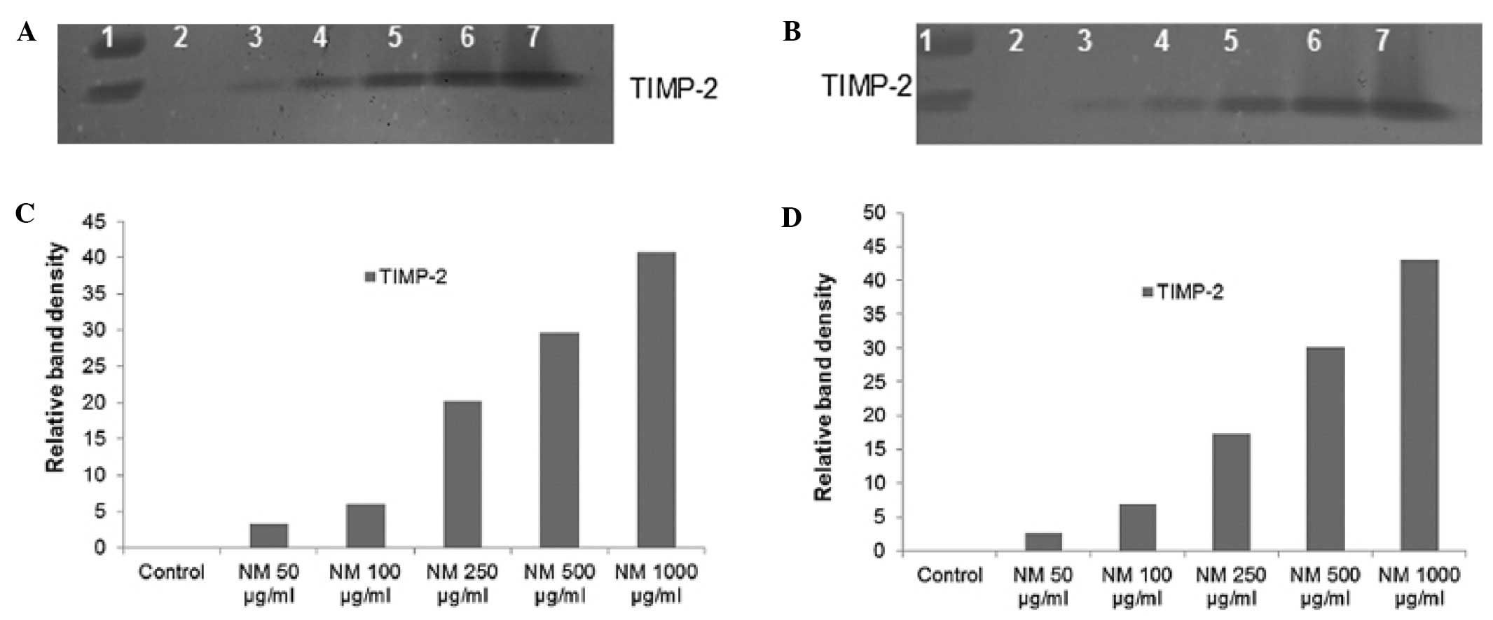
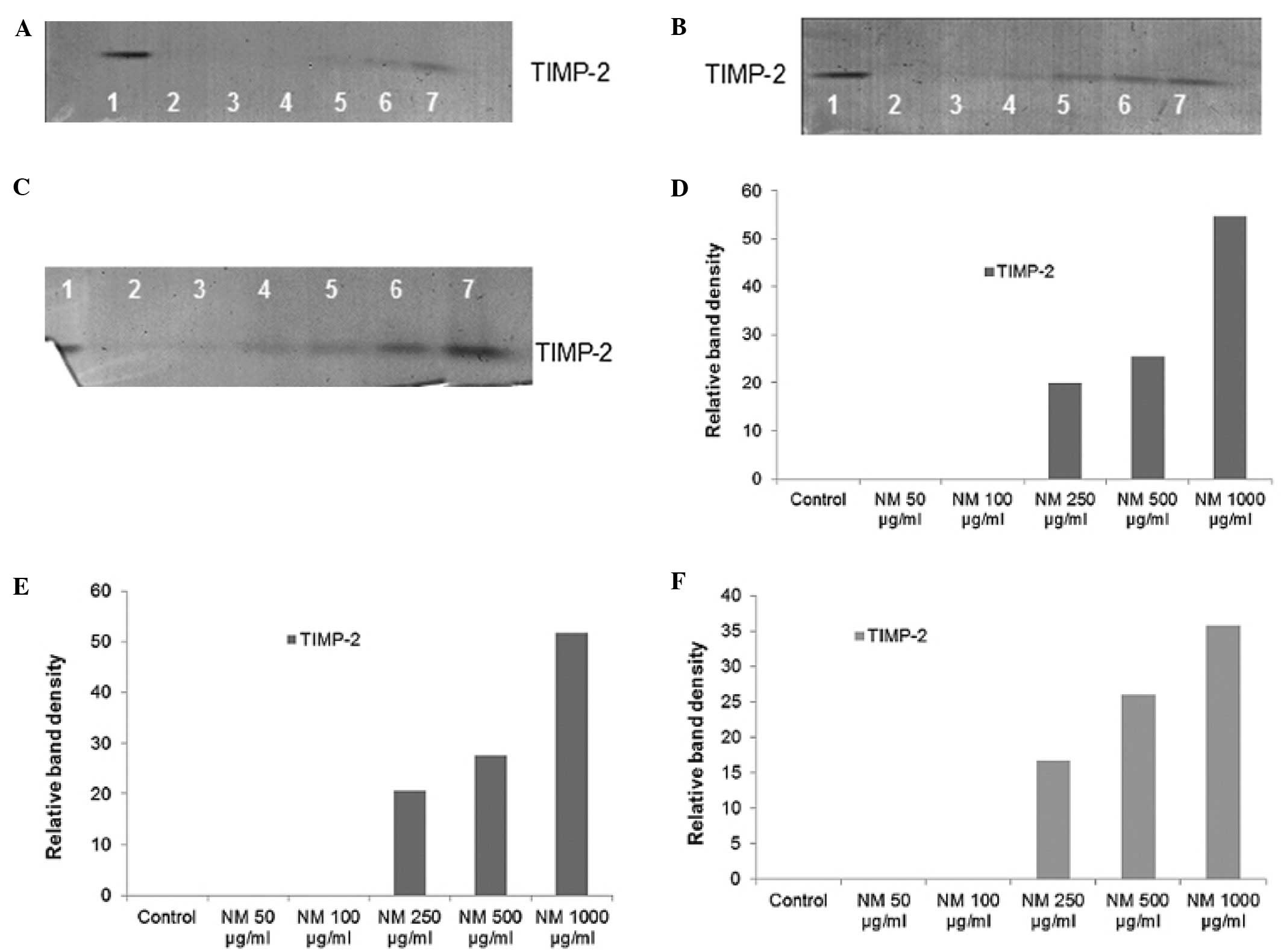
Effect of NM on TIMPs activity in human
breast, cervical and ovarian cancer cell lines
Reverse zymography revealed up regulation of TIMP-2
activity with NM treatment in all cancer cell lines in a
dose-dependent manner. Minimum activity was expressed at 50 and
maximum at 1000 μg/ml NM. See Figs.
5 and 6 for respective reverse
zymograms and densitometry analyses.
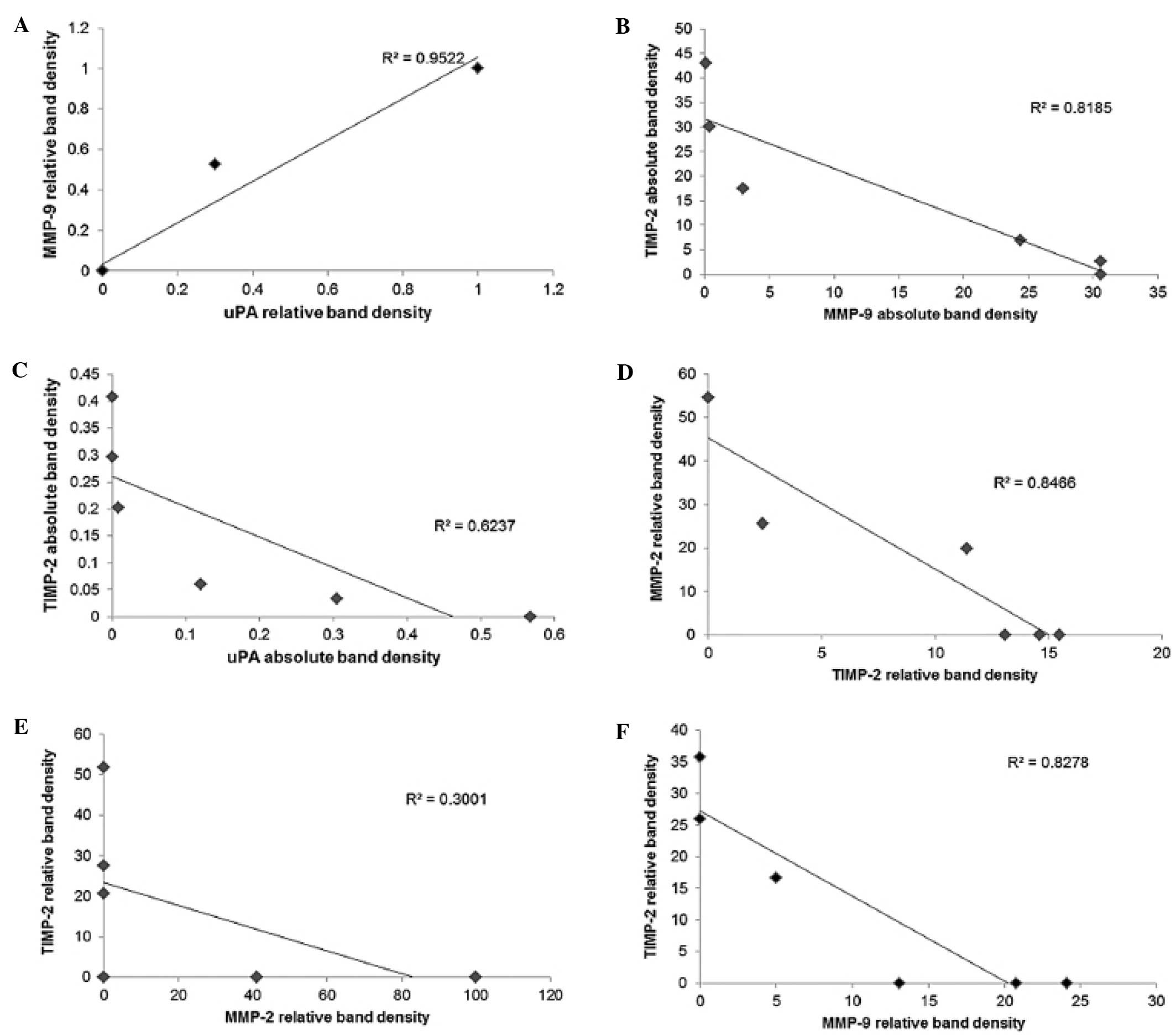
Correlation between female cancer cell
lines u-PA, TIMP-2 and MMP expressions
Analysis revealed a positive correlation between
NM-treated breast cancer cell line MCF-7 u-PA and MMP expressions,
as shown in Fig. 7A, with a
correlation coefficient r=0.976. A negative correlation
(correlation coefficient r= −0.904) was found between the
expressions of MCF-7 u-PA and MMP-9 (Fig. 7B). A negative correlation
(correlation coefficient r= −0.790) was found between MBA-MB-231
expression of TIMP-2 and u-PA (Fig.
7C). Negative correlations were found between HeLa expression
of TIMP-2 and MMP-2 (correlation coefficient r= −0.820) and between
SK-OV-3 TIMP-2 and MMP-2 (correlation coefficient r= −0.548), as
shown in Fig. 7D and E,
respectively. A negative correlation was found between uterine
SK-UT-1 cell TIMP-2 and MMP-9 (r= −0.910), as shown in Fig. 7F; Table
I).
 | Table IOverview of MMP-2 and -9, u-PA and
TIMP-2 expression of female cancer cell lines. |
Table I
Overview of MMP-2 and -9, u-PA and
TIMP-2 expression of female cancer cell lines.
| Cancer cell
line | MMP-2 | MMP-9 | u-PA | TIMP-2 |
|---|
| Breast cancer
MDA-MB-231 | − | + | + | + |
| Breast cancer
MCF-7 | − | + | + | + |
| Cervical cancer
HeLa | + | + | − | + |
| Ovarian cancer
SK-OV-3 | + | − | − | + |
| Uterine cancer
SK-UT-1 | − | With PMA
induction | + | + |
Discussion
Critical events in tumor cell invasion include cell
attachment, degradation of the ECM and migration through the
disrupted matrix. The two families of proteases, matrix
metalloproteinases and urokinase plasminogen activators play key
roles in tumor cell invasion. Experimental studies have
demonstrated the role of urokinase plasminogen, especially cell
surface u-PA, as an initiator of ECM proteolysis and associated
tumor cell invasion (35). The
protease u-PA converts plasminogen to plasmin, which is capable of
promoting tumor growth and angiogenesis, degrading the ECM and
basement membrane and activating pro-MMPs (30). Duffy first reported the prognostic
value of u-PA in breast cancer patients, showing a positive
correlation between high levels of u-PA and cancer progression
(32). High u-PA levels have been
reported to be prognostic indicators of increased risk of
endometrial cancer progression (26,27).
High levels of u-PA in cervical cancer patients have also been
reported to be correlated with pelvic lymph node metastasis
(17) and were reported to predict
survival in advanced ovarian cancer patients after radical surgery
and chemotherapy (33). Matrix
metalloproteinases, especially MMP-2 and MMP-9 play pivotal roles
in tumor cell invasion and metastasis due to their ability to
degrade type IV collagen, a major component of the ECM.
Overproduction of MMPs, especially MMP-2 and -9 has been associated
with a more aggressive behavior of female cancers (18,22–24).
Our study demonstrated that the specific mixture of
nutrients tested significantly inhibited breast cancer cell
MDA-MB-231 and MCF-7 u-PA secretion. (Cervical cancer HeLa and
ovarian cancer SK-OV-3 cells were not found to secrete u-PA in this
study). Furthermore, the NM demonstrated dose-dependent decrease in
MMP secretion and increase in TIMP-2 secretion by all these female
cancer cells. As expected, a significant positive correlation was
found between the secretion of u-PA and MMPs and a significant
negative correlation between u-PA and TIMP-2 secretion by NM
treatment of breast cancer cells. As anticipated, a significant
negative correlation was found between MMP and TIMP-2 secretion by
all the female cancer cell lines tested. Furthermore, a previous
study demonstrated significant correlation between NM inhibition of
Matrigel invasion and NM modulation of the MMP-2 and -9 activity of
the female cancer cells lines studied (37). A significant negative correlation
was found between NM modulation of Matrigel invasion inhibition and
MMP-9 secretion with breast cancer MDA-MB-231 (r= − 0.851) and
MCF-7 (r= −0.993) cell lines and with uterine cancer SK-UT-1 cell
line (r= −0.910). For cervical HeLa cells and ovarian SK-OV-3
cells, negative correlations (r= −0.924 and r= −0.812,
respectively) were found between NM modulation of Matrigel invasion
inhibition and MMP-2 secretion. A previous in vivo study of
the effects of NM on breast cancer supports these results in that
it demonstrated significant inhibition of MDA-MB-231 xenograft
tumor growth in nude mice and inhibition of MMP-9 and VEGF
secretion and mitosis in the tissue of nutrient-supplemented mice
(38).
In contrast to the associated toxicity and limited
efficacy of standard cancer chemotherapy and radiation therapy,
extensive research has documented the efficacy and safety of
dietary and botanical natural compounds in cancer prevention
(39). The nutrient mixture was
formulated by selecting nutrients that act on critical
physiological targets in cancer progression and metastasis, as
documented in both clinical and experimental studies. Combining
these micronutrients expands metabolic targets, maximizing
biological impact with lower doses of components. For example, a
previous study of the comparative effects of NM, green tea extract
and EGCG on inhibition of MMP-2 and MMP-9 secretion of different
cancer cell lines with varying MMP secretion patterns, documented
the superior potency of NM over GTE and EGCG at equivalent doses
(40). These results can be
understood from the more comprehensive treatment offered by the
combination of nutrients in NM over individual components of NM
since MMP-2 and MMP-9 are mediated by differential pathways.
Optimal ECM structure depends upon adequate supplies
of ascorbic acid and the amino acids lysine and proline to ensure
proper synthesis and hydroxylation of collagen fibers. In addition,
lysine contributes to ECM stability as a natural inhibitor of
plasmin-induced proteolysis (34,41).
Manganese and copper are also essential for collagen formation.
There is considerable documentation of the potency of green tea
extract in modulating cancer cell growth, metastasis, angiogenesis,
and other aspects of cancer progression (42–48).
N-acetyl cysteine and selenium have demonstrated inhibition of
tumor cell MMP-9 and invasive activities, as well as migration of
endothelial cells through ECM (49–51).
Ascorbic acid demonstrates cytotoxic and antimetastatic actions on
malignant cell lines (52–56) and cancer patients have been found to
have low levels of ascorbic acid (57,58).
Low levels of arginine, a precursor of nitric oxide (NO), can limit
the production of NO, which has been shown to predominantly act as
an inducer of apoptosis (59).
In conclusion, the NM demonstrated potent anticancer
activity by targeting primary mechanisms responsible for the
aggressive spread of breast, uterine, cervical and ovarian cancer.
In this in vitro study, the NM significantly inhibited
breast cancer cell lines MDA-MB-231 and MCF-7 and uterine cell line
SK-UT-1 secretion of u-PA and MMP-9 and increased their secretion
of TIMP-2, suggesting its potential in modulating breast and
uterine cancer invasion and metastasis. Cervical HeLa and ovarian
SK-OV-3 cell lines did not secrete u-PA; however, secretion by
these cell lines of MMP-2 was inhibited by NM and secretion of
TIMP-2 was enhanced by NM. With all these female cancer cell lines,
NM inhibition of MMP secretion was found to be correlated
significantly with Matrigel invasion of these cell lines.
Furthermore, use of the nutrient mixture would not pose any toxic
effect clinically, especially in the relevant doses, as in
vivo safety studies demonstrate. An in vivo toxicology
study showed that NM had no adverse effects on vital organs (heart,
liver, and kidney), or on the associated functional serum enzymes
(60).
Acknowledgements
Mr. J. Monterrey provided assistance in scanning the
gels. The study was funded by Dr. Rath Health Foundation (Santa
Clara, CA, USA) a non-profit organization.
References
|
1
|
Jemal A, Bray F, Center MM, Ferley J, Ward
E and Forman D: Global cancer statistics. CA Cancer J Clin.
61:69–90. 2011. View Article : Google Scholar
|
|
2
|
Breastcancer.org. U.S. Breast Cancer
Statistics. http://www.breastcancer.org/symptoms/understand_bc/statistics.jsp.
Accessed December 21, 2011
|
|
3
|
Ali SM, Harvey HA and Lipton A: Metastatic
breast cancer: overview of treatment. Clin Orthop. 414:S132–S137.
2003. View Article : Google Scholar
|
|
4
|
Endometrial (Uterine) Cancer. What are the
key statistics about endometrial cancer? http://www.cancer.org/Cancer/EndometrialCancer/DetailedGuide/endometrial-uterine-cancer-key-statistics.
Accessed January 20, 2012
|
|
5
|
Ovarian Cancer National Alliance. Ovarian
Cancer Statistics. http://www.ovariancancer.org/about-ovarian-cancer/statsitics/.
Accessed December 28, 2011
|
|
6
|
Fidler IJ: Molecular biology of cancer:
invasion and metastasis. Cancer: Principles and Practice of
Oncology. De Vita VT, Hellman S and Rosenberg SA: 5th edition.
Lippincott-Raven; Philadelphia, PA: pp. 135–152. 1997
|
|
7
|
Egeblad M and Werb Z: New functions for
the matrix metalloproteinases in cancer progression. Nat Rev
Cancer. 2:161–174. 2002. View
Article : Google Scholar : PubMed/NCBI
|
|
8
|
Folkman J: Role of angiogenesis in tumor
growth and metastasis. Semin Oncol. 29(Suppl 16): 15–18. 2002.
View Article : Google Scholar : PubMed/NCBI
|
|
9
|
Chambers AF and Matrisian LM: Changing
views on the role of matrix metalloproteinases in metastasis. J
Natl Cancer Inst. 89:1260–1270. 1997. View Article : Google Scholar : PubMed/NCBI
|
|
10
|
Kleiner DL and Stetler-Stevenson WG:
Matrix metalloproteinases and metastasis. Cancer Chemother
Pharmacol. 43:S42–S51. 1999. View Article : Google Scholar
|
|
11
|
Yurchenko PD and Schitny JC: Molecular
architecture of basement membranes. FASEB J. 4:1577–1590.
1990.PubMed/NCBI
|
|
12
|
Barsky SH, Siegel GP, Jannotta F and
Liotta LA: Loss of basement membrane components by invasive tumors
but not by their benign counterparts. Lab Invest. 49:140–147.
1983.PubMed/NCBI
|
|
13
|
Liotta LA, Tryggvason K, Garbisa A, Hart
I, Foltz CM and Shafie S: Metastatic potential correlates with
enzymatic degradation of basement membrane collagen. Nature.
284:67–68. 1980. View
Article : Google Scholar : PubMed/NCBI
|
|
14
|
Nelson AR, Fingleton B, Rothenberg ML and
Matrisian LM: Matrix metalloproteinases: biologic activity and
clinical implications. J Clin Oncol. 18:1135–1149. 2000.PubMed/NCBI
|
|
15
|
Bérubé M, Deschambeault A, Boucher M,
Germain L, Petitclerc E and Guérin SL: MMP-2 expression in uveal
melanoma: differential activation status dictated by the cellular
environment. Mol Vis. 11:1101–1111. 2005.PubMed/NCBI
|
|
16
|
Garzetti G, Ciavattini A, Lucarini G,
Goteri G, de Nicolis M, Garbisa S, Masiero L, Romanini C and
Graziella B: Tissue and serum metalloproteinase (MMP-2) expression
in advanced ovarian serous cystadenocarcinomas: clinical and
prognostic implications. Anticancer Res. 15:2799–2804.
1995.PubMed/NCBI
|
|
17
|
Sugimura M, Kobayashi H, Kanayama N and
Terao T: Clinical significance of urokinase-type plasminogen
activator (uPA) in invasive cervical cancer of the uterus. Gynecol
Oncol. 46:330–336. 1992. View Article : Google Scholar : PubMed/NCBI
|
|
18
|
Bachmeier BE, Nerlich AG, Lichtinghagen R
and Sommerhoff CP: Matrix metalloproteinases (MMPs) in breast
cancer cell lines of different tumorigenicity. Anticancer Res.
6A:3821–3828. 2001.PubMed/NCBI
|
|
19
|
Pellikainen JM, Ropponen KM, Kataja VV,
Kellokoski JK, Eskelinen MJ and Kosma VM: Expression of matrix
metalloproteinase (MMP)-2 and MMP-9 in breast cancer with a special
reference to activator protein-2, HER-2, and prognosis. Clin Cancer
Res. 10:7621–7628. 2004. View Article : Google Scholar : PubMed/NCBI
|
|
20
|
Scorilas A, Karameris A, Arnogiannaki N,
Ardavanis A, Bassilopoulos P, Tangas T and Tlieri M: Overexpression
of matrix-metalloproteinase-9 in human breast cancer: a potential
favourable indicator in node-negative patients. Br J Cancer.
84:1488–1496. 2001. View Article : Google Scholar : PubMed/NCBI
|
|
21
|
Asha Nair S, Karunagaran D, Nair MB and
Sudhakaran PR: Changes in matrix metalloproteinases and their
endogenous inhibitors during tumor progression in the uterine
cervix. J Cancer Res Clin Oncol. 129:123–131. 2003.PubMed/NCBI
|
|
22
|
Zhou CY, Yao JF and Chen XD: Expression of
matrix metalloproteinase-2,9 and their inhibitor-TIMP 1,2 in human
squamous cell carcinoma of uterine cervix. Al Zhen. 21:735–739.
2002.PubMed/NCBI
|
|
23
|
Lopata A, Agresta F, Quinn MA, Smith C,
Ostor AG and Salamonsen LA: Detection of endometrial cancer by
determination of matrix metalloproteinases in the uterine cavity.
Gynecol Oncol. 90:318–324. 2003. View Article : Google Scholar : PubMed/NCBI
|
|
24
|
Aglund K, Rauvala M, Puistola U, Angström
T, Turpeeniemi-Hujanen T, Zackrisson B and Stendahl U: Gelatinases
A and B (MMP-2 and MMP-9) in endometrial cancer - MMP-9 correlates
to the grade and the stage. Gynecol Oncol. 94:699–704. 2004.
View Article : Google Scholar : PubMed/NCBI
|
|
25
|
Torng PL, Mao TL, Chan WY, Huang SC and
Lin CT: Prognostic significance of stromal metalloproteinase-2 in
ovarian adenocarcinoma and in relation to carcinoma progression.
Gynecol Oncol. 92:559–567. 2004. View Article : Google Scholar : PubMed/NCBI
|
|
26
|
Memarzedeh S, Kozak KR, Chang L, Natarajan
S, Shintaku P, Reddy ST and Farias-Eisner R: Urokinase plasminogen
activator receptor: prognostic biomarker for endometrial cancer.
Proc Natl Acad Sci USA. 99:10647–10652. 2002. View Article : Google Scholar : PubMed/NCBI
|
|
27
|
Steiner E, Pollow K, Hasenclever D,
Schormann W, Hermes M, Schmidt M, Puhl A, Brulport M, Bauer A,
Petry IB, Koelbl H and Hengstler JG: Role of urokinase-type
plasminogen activator (uPA) and plasminogen activator inhibitor
type 1 (PAI-1) for prognosis in endometrial cancer. Gynecol Oncol.
108:569–576. 2008. View Article : Google Scholar : PubMed/NCBI
|
|
28
|
Stetler-Stevenson WG: The role of matrix
metalloproteinases in tumor invasion, metastasis and angiogenesis.
Surg Oncol Clin N Am. 10:383–392. 2001.PubMed/NCBI
|
|
29
|
Stetler-Stevenson WG: Type IV collagenases
in tumor invasion and metastasis. Cancer Metastasis Rev. 9:289–303.
1990. View Article : Google Scholar : PubMed/NCBI
|
|
30
|
Dano K, Andreasen PA, Grondahl-Hansen J,
Kristensen P, Nielsen LS and Skriver L: Plasminogen activators,
tissue degradation and cancer. Adv Cancer Res. 44:139–266. 1985.
View Article : Google Scholar : PubMed/NCBI
|
|
31
|
Alonso DF, Farias EF, Ladeda V, Davel L,
Puricelli L and Bal de Kier Joffé E: Effects of synthetic urokinase
on local invasion and metastasis in a murine mammary tumor model.
Breast Cancer Res Treat. 40:209–223. 1996. View Article : Google Scholar : PubMed/NCBI
|
|
32
|
Duffy MJ, Duggan C, Mulcahy HE, McDermott
EW and O’Higgins NJ: Urokinase plasminogen activator: a prognostic
marker in breast cancer including patients with axillary
node-negative disease. Clin Chem. 44:1177–1183. 1998.PubMed/NCBI
|
|
33
|
Kuhn W, Pache L, Schmaltfeldt B, Dettmar
P, Schmitt M, Jänicke F and Graeff H: Urokinase (uPA) and Pal-1
predict survival in advanced ovarian cancer patients (FIGO III)
after radical surgery and platinum-based chemotherapy. Gynecol
Oncol. 55:401–409. 1994. View Article : Google Scholar : PubMed/NCBI
|
|
34
|
Rath M and Pauling L: Plasmin-induced
proteolysis and the role of apoprotein(a), lysine and synthetic
analogs. J Orthomolecular Med. 7:17–23. 1992.
|
|
35
|
Andreasen PA, Kjøller L, Christensen L and
Duffy MJ: The urokinase-type plasminogen activator system in cancer
metastasis: a review. Int J Cancer. 72:1–22. 1997. View Article : Google Scholar : PubMed/NCBI
|
|
36
|
Niedzwiecki A, Roomi MW, Kalinovsky T and
Rath M: Micronutrient synergy - a new tool in effective control of
metastasis and other key mechanisms of cancer. Cancer Metastasis
Rev. 29:529–543. 2010. View Article : Google Scholar : PubMed/NCBI
|
|
37
|
Roomi MW, Monterrey JC, Kalinovsky T,
Niedzwiecki A and Rath M: Inhibition of invasion and MMPs by a
nutrient mixture in human cancer cell lines: a correlation study.
Exp Oncol. 32:243–248. 2010.PubMed/NCBI
|
|
38
|
Roomi MW, Ivanov V, Kalinovsky T,
Niedzwiecki A and Rath M: In vitro and in vivo antitumorigenic
activity of a mixture of lysine, proline, ascorbic acid, and green
tea extract on human breast cancer lines MDA-MB-231 and MCF-7. Med
Oncol. 22:129–138. 2005. View Article : Google Scholar : PubMed/NCBI
|
|
39
|
Amin ARMR, Kucek O, Khuri FR and Shin DM:
Perspectives for cancer prevention with natural compounds. J Clin
Oncol. 27:2712–2725. 2009. View Article : Google Scholar : PubMed/NCBI
|
|
40
|
Roomi MW, Monterrey JC, Kalinovsky T, Rath
M and Niedzwiecki A: Comparative effects of EGCG, green tea and a
nutrient mixture on the patterns of MMP-2 and MMP-9 expression in
cancer cell lines. Oncol Rep. 24:747–757. 2010.PubMed/NCBI
|
|
41
|
Sun Z, Chen YH, Wang P, Zhang J, Gurewich
V, Zhang P and Liu JN: The blockage of high-affinity lysine binding
sites of plasminogen by EACA significantly inhibits
prourokinase-induced plasminogen activation. Biochem Biophys Acta.
1596:182–192. 2002.PubMed/NCBI
|
|
42
|
Kemberling JK, Hampton JA, Keck RW, Gomez
MA and Selman SH: Inhibition of bladder tumor growth by the green
tea derivative epigallocatechin-3-gallate. J Urol. 170:773–776.
2003. View Article : Google Scholar : PubMed/NCBI
|
|
43
|
Sato D and Matsushima M: Preventive
effects of urinary bladder tumors induced by
N-butyl-N-(4-hydroxybutyl)-nitrosamine in rat by green tea leaves.
Int J Urol. 10:160–166. 2003. View Article : Google Scholar : PubMed/NCBI
|
|
44
|
Valcic S, Timmermann BN, Alberts DS,
Wachter GA, Krutzsch M, Wymer J and Guillen JM: Inhibitory effect
of six green tea catechins and caffeine on the growth of four
selected human tumor cell lines. Anticancer Drugs. 7:461–468. 1996.
View Article : Google Scholar : PubMed/NCBI
|
|
45
|
Mukhtar H and Ahmed N: Tea polyphenols:
prevention of cancer and optimizing health. Am J Clin Nutr.
71:S1698–S1704. 2000.PubMed/NCBI
|
|
46
|
Yang GY, Liao J, Kim K, Yurtow EJ and Yang
CS: Inhibition of growth and induction of apoptosis in human cancer
cell lines by tea polyphenols. Carcinogenesis. 19:611–616. 1998.
View Article : Google Scholar : PubMed/NCBI
|
|
47
|
Taniguchi S, Fujiki H, Kobayashi H, Go H,
Miyado K, Sadano H and Shimikawa R: Effect of (−) epigallocatechin
gallate, the main constituent of green tea, on lung metastasis with
mouse B16 melanoma cell lines. Cancer Lett. 65:51–54. 1992.
|
|
48
|
Hara Y: Green Tea: Health Benefits and
Applications. Marcel Dekker Inc; New York: 2001, View Article : Google Scholar
|
|
49
|
Kawakami S, Kageyama Y, Fujii Y, Kihara K
and Oshima H: Inhibitory effects of N-acetyl cysteine on invasion
and MMP 9 production of T24 human bladder cancer cells. Anticancer
Res. 21:213–219. 2001.PubMed/NCBI
|
|
50
|
Morini M, Cai T, Aluigi MG, Noonan DM,
Masiello L, De Floro S, D’Agostinin F, Albini A and Fassima G: The
role of the thiol N-acetyl cysteine in the prevention of tumor
invasion and angiogenesis. Int J Biol Markers. 14:268–271.
1999.PubMed/NCBI
|
|
51
|
Yoon SO, Kim MM and Chung AS: Inhibitory
effects of selenite on invasion of HT 1080 tumor cells. J Biol
Chem. 276:20085–20092. 2001. View Article : Google Scholar : PubMed/NCBI
|
|
52
|
Naidu KA, Karl RC and Coppola D:
Antiproliferative and proapoptotic effect of ascorbyl stearate in
human pancreatic cancer cells: association with decreased
expression of insulin-like growth factor 1 receptor. Dig Dis Sci.
48:230–237. 2003. View Article : Google Scholar
|
|
53
|
Anthony HM and Schorah CJ: Severe
hypovitaminosis C in lung-cancer patients: The utilization of
vitamin C in surgical repair and lymphocyte-related host
resistance. Br J Cancer. 46:354–367. 1982. View Article : Google Scholar : PubMed/NCBI
|
|
54
|
Maramag C, Menon M, Balaji KC, Reddy PG
and Laxmanan S: Effect of vitamin C on prostate cancer cells in
vitro: effect on cell number, viability and DNA synthesis.
Prostate. 32:188–195. 1997. View Article : Google Scholar : PubMed/NCBI
|
|
55
|
Koh WS, Lee SJ, Lee H, Park C, Park MH,
Kim WS, Yoon SS, Park K, Hong SI, Chung MH and Park CH:
Differential effects and transport kinetics of ascorbate
derivatives in leukemic cell lines. Anticancer Res. 8:2487–2493.
1998.PubMed/NCBI
|
|
56
|
Chen Q, Espey MG, Krishna MC, Mitchell JB,
Corpe CP, Buettner GR, Shacter E and Levine M: Pharmacologic
ascorbic acid concentrations selectively kill cancer cells: Action
as a pro-drug to deliver hydrogen peroxide to tissues. Proc Natl
Acad Sci USA. 102:13604–13609. 2005. View Article : Google Scholar : PubMed/NCBI
|
|
57
|
Nunez C, Ortiz de Apodaca Y and Ruiz A:
Ascorbic acid in the plasma and blood cells of women with breast
cancer. The effect of consumption of food with an elevated content
of this vitamin. Nutr Hosp. 10:368–372. 1995.(In Spanish).
|
|
58
|
Kurbacher CM, Wagner U, Kolster B,
Andreotti PE, Krebs D and Bruckner HW: Ascorbic acid (vitamin C)
improves the antineoplastic activity of doxorubicin, cisplatin and
paclitaxel in human breast carcinoma cells in vitro. Cancer Lett.
103:183–189. 1996. View Article : Google Scholar : PubMed/NCBI
|
|
59
|
Cooke JP and Dzau VJ: Nitric oxide
synthase: Role in the genesis of vascular disease. Annu Rev Med.
48:489–509. 1997. View Article : Google Scholar : PubMed/NCBI
|
|
60
|
Roomi MW, Ivanov V, Netke SP, Niedzwiecki
A and Rath M: Serum markers of the liver, heart, and kidney and
lipid profile and histopathology in ODS rats treated with nutrient
synergy. J AM Coll Nutr. 22:4772003.
|