In vivo real-time imaging of chemotherapy response on the liver metastatic tumor microenvironment using multiphoton microscopy

  • Authors:
    • Koji Tanaka
    • Masato Okigami
    • Yuji Toiyama
    • Yuki Morimoto
    • Kohei Matsushita
    • Mikio Kawamura
    • Kiyoshi Hashimoto
    • Susumu Saigusa
    • Yoshinaga Okugawa
    • Yasuhiro Inoue
    • Keiichi Uchida
    • Toshimitsu Araki
    • Yasuhiko Mohri
    • Akira Mizoguchi
    • Masato Kusunoki
  • View Affiliations

  • Published online on: August 23, 2012     https://doi.org/10.3892/or.2012.1983
  • Pages: 1822-1830
Metrics: Total Views: 0 (Spandidos Publications: | PMC Statistics: )
Total PDF Downloads: 0 (Spandidos Publications: | PMC Statistics: )


Abstract

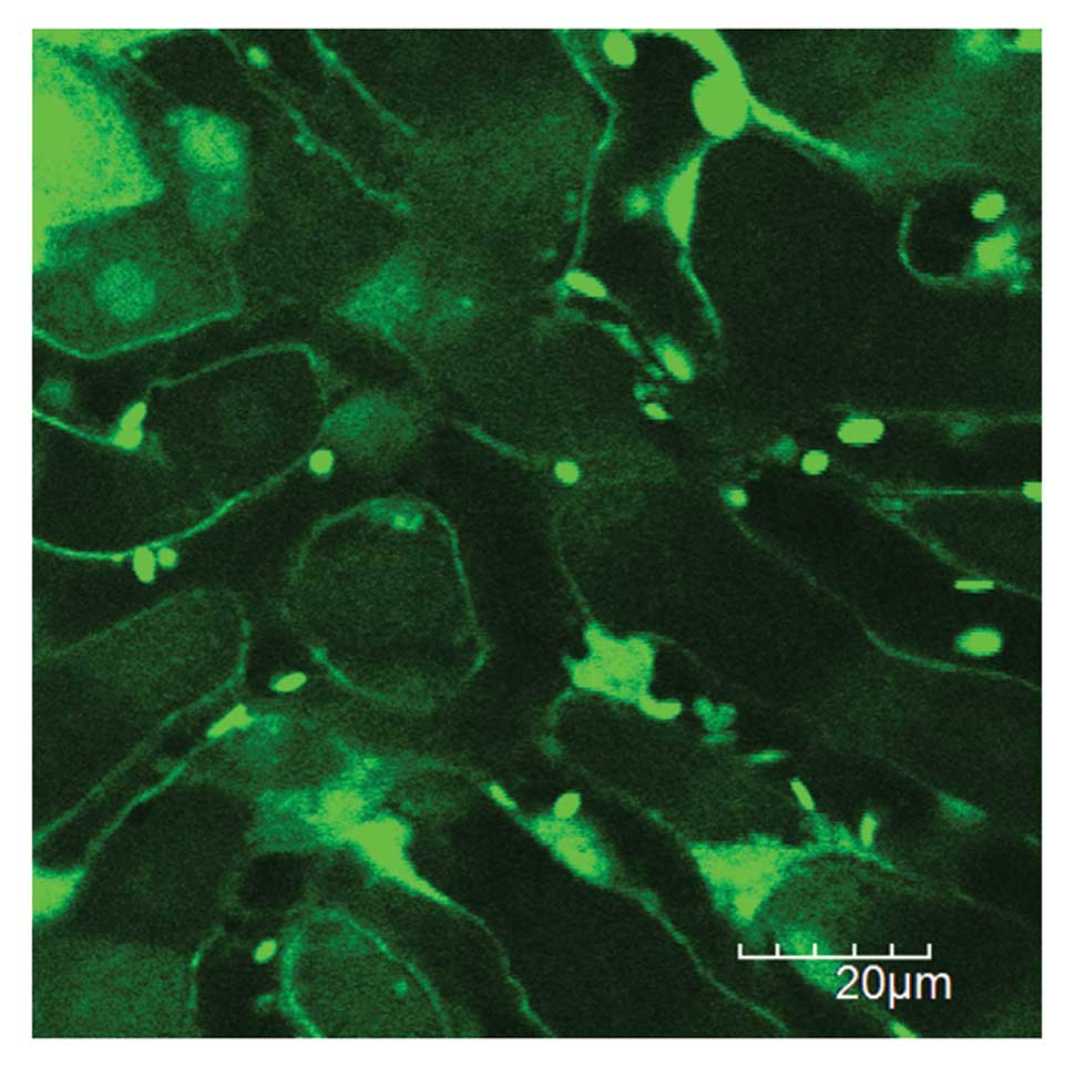
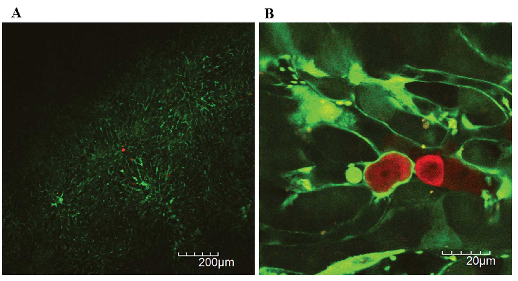
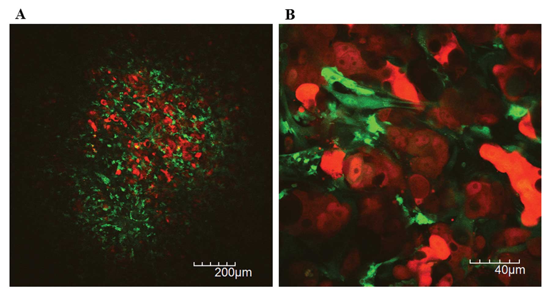
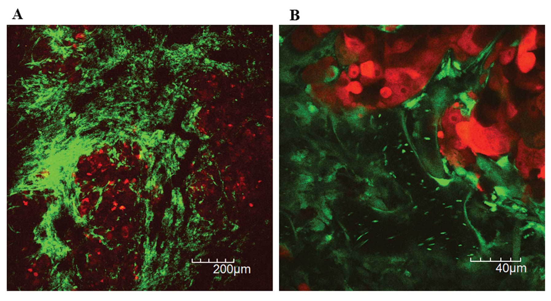
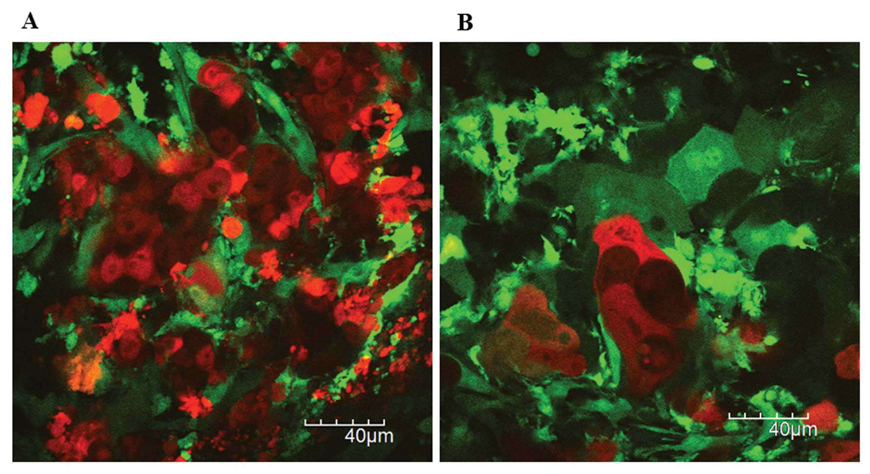
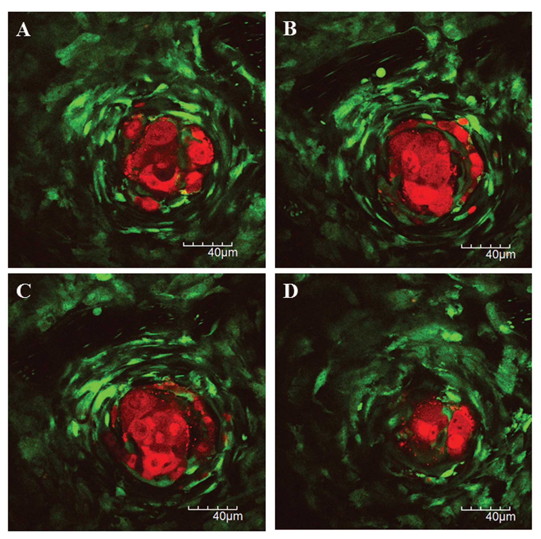
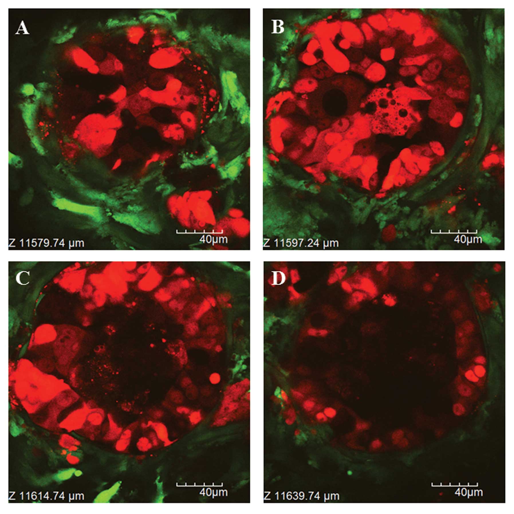
In vivo real-time visualization of chemotherapy response at the cellular level provides us with direct evidence of what happens on the tumor microenvironment of metastatic organs. We imaged the response of metastatic tumor cells and host stromal cells to chemotherapeutics on liver metastatic xenografts in living mice using intravital two-photon laser scanning microscopy (TPLSM). Red fluorescent protein-expressing human colorectal cancer cells (HT29) was inoculated to the spleen of green fluorescent protein-expressing nude mice. 5-Fluorouracil or irinotecan was intraperitoneally administered after the formation of macroscopic liver metastases. Intravital TPLSM was performed at multiple time-points for time-series imaging of liver metastatic xenografts in the same mice. Under the 1st TPLSM, HT29 cells were visualized in hepatic sinusoids at the single cell level. Liver metastatic nodules consisting of viable cancer cells and surrounding stroma with tumor vessels were visualized under the 2nd TPLSM. After chemotherapy, tumor cell fragmentation, condensation, swelling and intracellular vacuoles were observed under the 3rd TPLSM. There was no obvious morphological difference in tumor response between these chemotherapeutics. Time-series intravital TPLSM imaging on the metastatic tumor xenografts may be useful for screening and evaluating new chemotherapeutics with less interindividual variability.
View Figures
View References

Related Articles

Journal Cover

November 2012
Volume 28 Issue 5

Print ISSN: 1021-335X
Online ISSN:1791-2431

Sign up for eToc alerts

Recommend to Library

Copy and paste a formatted citation
x
Spandidos Publications style
Tanaka K, Okigami M, Toiyama Y, Morimoto Y, Matsushita K, Kawamura M, Hashimoto K, Saigusa S, Okugawa Y, Inoue Y, Inoue Y, et al: In vivo real-time imaging of chemotherapy response on the liver metastatic tumor microenvironment using multiphoton microscopy. Oncol Rep 28: 1822-1830, 2012
APA
Tanaka, K., Okigami, M., Toiyama, Y., Morimoto, Y., Matsushita, K., Kawamura, M. ... Kusunoki, M. (2012). In vivo real-time imaging of chemotherapy response on the liver metastatic tumor microenvironment using multiphoton microscopy. Oncology Reports, 28, 1822-1830. https://doi.org/10.3892/or.2012.1983
MLA
Tanaka, K., Okigami, M., Toiyama, Y., Morimoto, Y., Matsushita, K., Kawamura, M., Hashimoto, K., Saigusa, S., Okugawa, Y., Inoue, Y., Uchida, K., Araki, T., Mohri, Y., Mizoguchi, A., Kusunoki, M."In vivo real-time imaging of chemotherapy response on the liver metastatic tumor microenvironment using multiphoton microscopy". Oncology Reports 28.5 (2012): 1822-1830.
Chicago
Tanaka, K., Okigami, M., Toiyama, Y., Morimoto, Y., Matsushita, K., Kawamura, M., Hashimoto, K., Saigusa, S., Okugawa, Y., Inoue, Y., Uchida, K., Araki, T., Mohri, Y., Mizoguchi, A., Kusunoki, M."In vivo real-time imaging of chemotherapy response on the liver metastatic tumor microenvironment using multiphoton microscopy". Oncology Reports 28, no. 5 (2012): 1822-1830. https://doi.org/10.3892/or.2012.1983