Introduction
Cervical cancer is the third most common cancer in
women worldwide, with global estimates of 529,800 new cases and
275100 deaths in 2008 (1). In the
United States, cervical cancer is the second leading cause of
cancer related deaths in young women as approximately 12,000 women
were diagnosed with cervical cancer in 2010, about 4,000 of whom
died from this disease (2).
Although advanced cervical cancer can be treated by radical surgery
with or without radiotherapy and/or chemotherapy, some patients
with high risk factors will still have an unfavorable prognosis
(3). The 5-year survival rate is
~70% and has not improved in the last decade (2). Therefore, new strategies, such as
immunotherapy and molecular-targeted therapy, may prove useful in
improving the prognosis for cervical cancer patients.
Indoleamine-2,3-dioxygenase (IDO) is an enzyme that
catalyzes the first and rate-limiting step in the kynurenine
pathway of tryptophan catabolism. IDO was originally discovered in
1967 (4,5) in rabbit small intestines and was
purified in 1978 (6). Subsequently,
it was reported that the enzyme could be induced in the mouse lung
by either viral infection (7) or
endotoxin shock (8).
Proinflammatory mediators, such as interferon or other cytokines,
can also stimulate IDO induction (9). A study revealing that IDO in the mouse
placenta prevented rejection of the allogeneic fetus exposed its
immunosuppressive properties (10).
Recently, it was demonstrated that IDO can induce immunotolerance
in patients with autoimmune diseases (11) and chronic infections (12). Most human malignant tumors express
IDO (13), and IDO can contribute
to tumor-induced immunosuppression by starving T-cells, which are
sensitive to tryptophan deficiency. In this environment, tumor
cells can escape immune surveillance via the action of IDO
(10).
Natural killer (NK) cells are important members of
the innate immune system, which plays a role in inhibiting the
growth of several types of tumors (14). Tryptophan-derived catabolic
kynurenine can reduce NK cell number and weaken NK cell
cytotoxicity by inhibiting the expression of NK cell receptors,
thus contributing to tumor progression (15). In gynecology, IDO expression has
been observed in cervical, endometrial and ovarian cancers
(13), and associations between IDO
expression and prognosis of in these cancers have been reported
(16–19).
RNA interference (RNAi) is a technique for gene
silencing and involves a post-transcriptional gene-silencing
mechanism (20). Among the
different types of RNAi techniques, the use of small interfering
RNAs (siRNAs) effectively suppresses gene expression in a transient
manner (21). Short hairpin RNAs
(shRNAs) driven by polymerase III promoters have been developed to
attain long-term stable target gene silencing (22,23).
In this study, we used an shRNA vector to silence
IDO expression in an IDO-expressing cervical cancer cell line to
further elucidate the relationship between expression and cervical
cancer growth. Moreover, we investigated the function of NK cells
in cervical cancer progression in order to develop an IDO-targeted
molecular therapy for cervical cancer.
Materials and methods
Cell culture
The 9 cervical cancer cell lines used in this study
(SKG-I, -II, -IIIa, -IIIb, SiHa, CaSki, BOKU, HCS-2 and ME-180)
(24–30) were obtained as follows: the SKG-I,
-II, -IIIa and IIIb lines were obtained from Dr Daisuke Aoki (Keio
University, Tokyo, Japan); the SiHa line was purchased from the
American Type Culture Collection (ATCC, Manassas, VA); and the
CaSki, BOKU, HCS-2 and ME-180, were all purchased from the Japanese
Collection of Research Bioresources (JCRB, Osaka, Japan). These
cell lines were maintained in D-MEM/Ham’s F-12 medium (DMEM/F12,
Gibco, Grand Island, NY) containing 10% inactivated fetal calf
serum (Sigma, St. Louis, MO), 100 U/ml penicillin (Gibco) and 100
μg/ml streptomycin (Gibco) at 37°C in a 5% CO2
atmosphere for no longer than 8 weeks after recovery from frozen
stocks.
The NK cell line KHYG-1 (31) was purchased from the JCRB. Cells
were cultured in RPMI-1640 medium supplemented with 100 nM of human
interleukin-2 (R&D Systems, Minneapolis, MN) and 10%
inactivated fetal calf serum (Sigma), at 37°C in a 5%
CO2 atmosphere for no longer than 8 weeks after recovery
from frozen stocks.
Antibodies
The anti-human IDO monoclonal antibody was prepared
as previously reported (32).
Anti-human actin (SIGMA) and anti-mouse CD49b antibodies (R&D
Systems) were used according to manufacturer’s protocols.
shRNA stable cell line and control cell
line
The short hairpin RNA (shRNA) plasmid targeting IDO
gene expression (piGENE PURhU6/shIDO) and control plasmid (piGENE
PURhU6) have been previously described (33), and were transfected into the CaSki
cell line using Lipofectamine LTX and Plus Reagent (Invitrogen,
Carlsbad, CA) according to the manufacturer’s instructions.
Transfected cells were selected for using 0.5 μg/ml puromycin
(Calbiochem, Darmstadt, Germany). Resistant CaSki/shIDO and
CaSki/Mock clones were obtained after 4 weeks. The cells were
subsequently maintained in the presence of 0.5 μg/ml puromycin.
Western blot analysis
Before protein extraction for western blot analysis,
cervical cancer cells were cultured in DMEM/F12 with 100 ng/ml
interferon-γ (R&D Systems) for 24 h. Ten micrograms of protein
extracted from a cultured cell homogenate was mixed with 2X
SDS-PAGE sample buffer [120 mM Tris-HCl (pH 6.8), 4% SDS, 20%
glycerol, 0.004% bromophenol blue and 10% 2-mercaptoethanol]. The
mixture was heated at 95°C for 2 min, and electrophoresed on a 0.1%
SDS-10% polyacrylamide gel, before blotting the proteins onto a
polyfluorovinylidene membrane. The membrane were blocked with a
Non-Protein Blocking Agent (ATTO Corp., Tokyo, Japan) at room
temperature for 1 h, and incubated with the anti-human IDO
monoclonal (1:1000) and anti-human actin polyclonal antibodies
(1:200) for 1 h at room temperature. The membrane was washed with
phosphate-buffered saline (PBS)-Tween-20 3 times and then incubated
with a horseradish peroxidase-conjugated secondary anti-mouse
(Thermo, Rockford, IL) or anti-rabbit antibody (Thermo). Signals
were detected by chemiluminescence (ECL kit; Amersham Biosciences,
Piscataway, NJ) on X-ray film.
In vitro cell growth kinetics
Five-hundred CaSki/shIDO and CaSki/Mock cells were
seeded onto a 96-well plate and cultured in DMEM/F12 medium
containing 10% fetal calf serum. Every 24 h, cells were counted
using a colorimetric assay with the Cell Proliferation kit II (XTT)
(Boehringer Mannheim GmbH Biochemica, Mannheim, Germany) and a
growth curve was drawn from the results.
Sensitivity of transfectants to NK cells
in vitro
The sensitivity of CaSki/shIDO and CaSki/Mock cells
to NK cells was investigated by colorimetric assay using XTT.
Five-hundred CaSki/shIDO and CaSki/Mock cells were seeded onto a
96-well plate and co-cultured with KHYG-1 cells (0, 500, 1000, 2000
or 4000 cells) in DMEM/F12 medium containing 10% fetal calf serum
for 72 h. After 3 washes with PBS to completely remove KHYG-1
cells, viable cell count was determined by colorimetric assay and
calculated as the percent of control cells (respective cell lines
cultured without KHYG-1 cells).
Experimental animals
Four- to six-week-old female BALB/c nude mice (Japan
Clea Laboratories, Tokyo, Japan) were used in this study. All
animal experiments were conducted according to the institutional
and national guidelines for animal experiments.
Subcutaneous tumor growth in vivo
CaSki/shIDO and CaSki/Mock cells (5×10⁶
cells from each line) were inoculated subcutaneously into the back
of mice to induce tumor growth. The tumor volume [(long diameter) ×
(short diameter)2 × 1/2] was measured twice a week to
generate a tumor growth curve.
Immunohistochemical staining
One week after subcutaneous tumor cell inoculation,
mice were sacrificed under isoflurane anesthesia, and the tumor was
removed. After formalin fixation, paraffin sections were prepared,
deparaffinized, and treated with hydrogen peroxide for 30 min to
block endogenous peroxidase. The sections were then reacted with a
1:10 dilution (5 μg/ml) of anti-mouse CD49b primary antibody for 16
h at room temperature, washed 3 times washes with PBS, and then
incubated with enzyme-conjugated streptavidin for 30 min. The
sections were again washed with PBS 3 times, and color was
developed using the diaminobenzidine method. The number of stained
NK cells was counted under high-power magnification (×400).
Statistical analysis
The test of significance between the 2 groups was
performed using Student’s t-test. A P-value of <0.05 was
considered significant.
Results
IDO expression
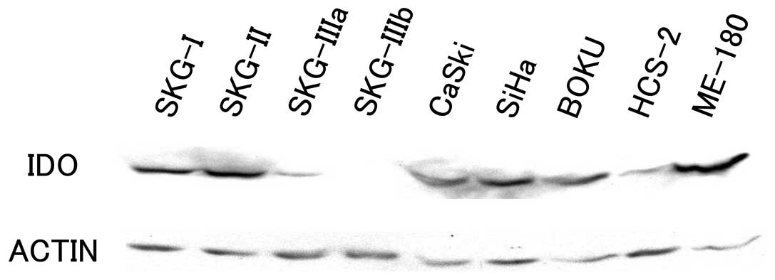
As shown in Fig. 1,
IDO expression was detected by western blotting at a position
corresponding to a molecular weight of 41 kDa in all cell lines
except for the SKG-IIIb line.
Establishing an IDO-downregulated cell
line
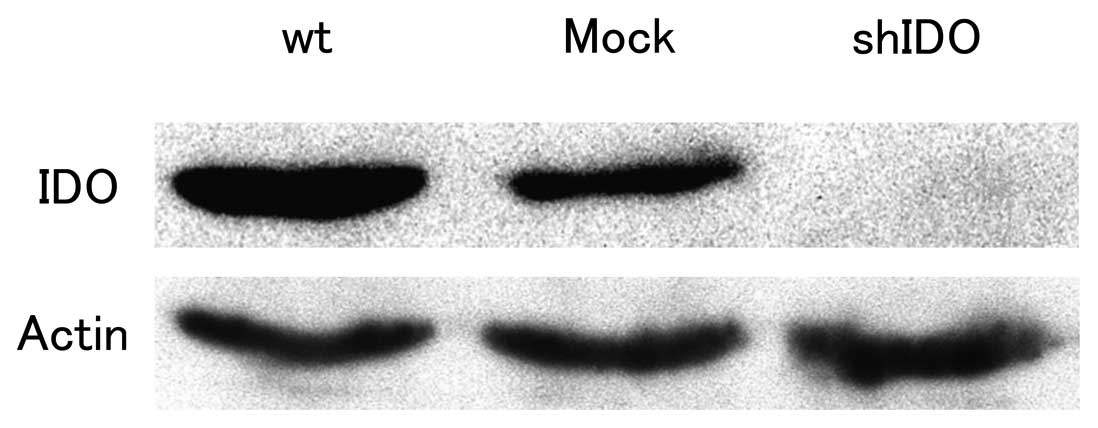
Fig. 2 shows the
results of western blot analysis of the CaSki cervical cancer cell
line transfected with either an shIDO expression vector or a
control vector. Parental cells (wt) and control vector-transfected
cells (Mock) expressed IDO. In contrast, the shIDO expression
vector-transfected cells (shIDO) did not show IDO expression,
confirming IDO downregulation in the CaSki/shIDO cell line.
In vitro cell growth kinetics
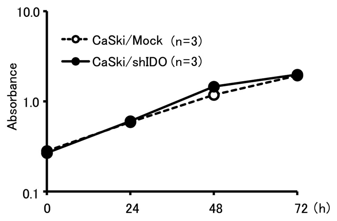
Growth curve analyses of CaSki/shIDO and CaSki/Mock
cells revealed no significant differences between the two groups,
suggesting that IDO downregulation did not affect cell growth in
vitro (Fig. 3).
Sensitivity of transfectants to NK cells
in vitro
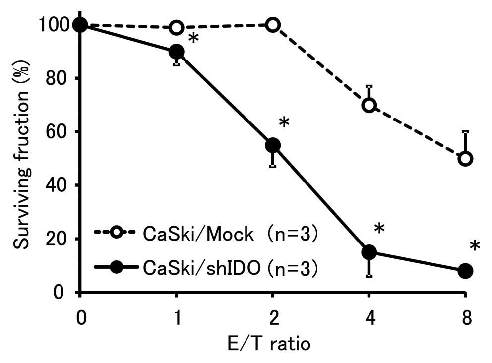
The proportion of viable tumor cells co-cultured
with NK cells is shown in Fig. 4.
The percent survival of CaSki/shIDO cells was significantly lower
than that of the control cells, indicating that the downregulation
of IDO increased the sensitivity of tumor cells to NK cells.
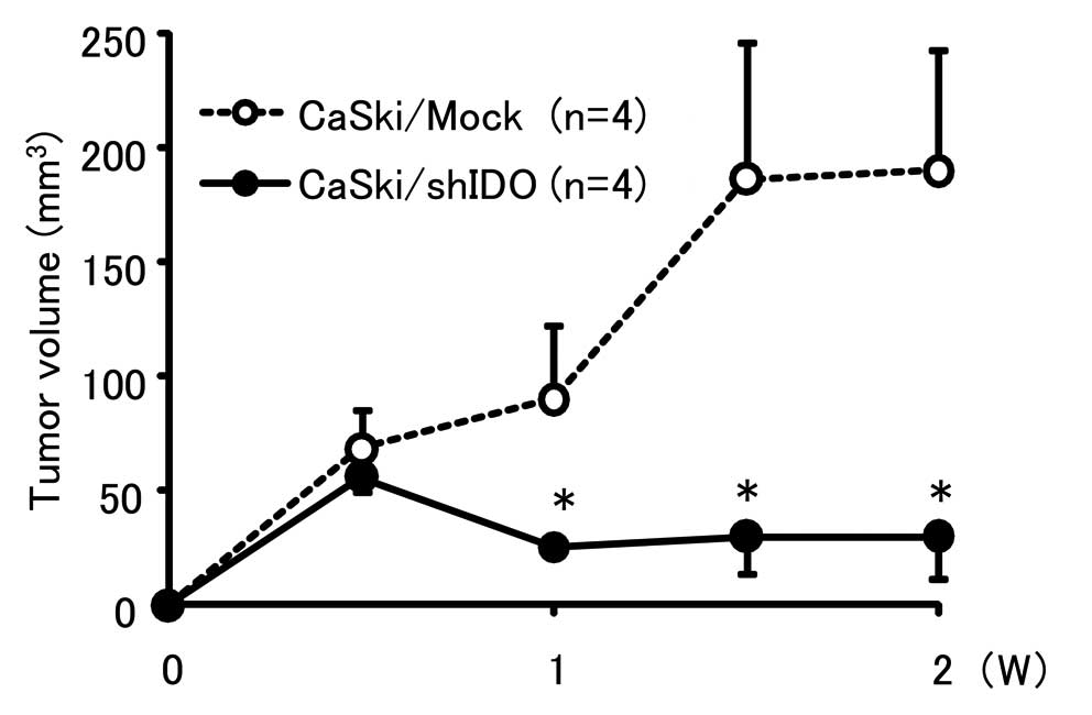
Tumor growth in vivo
Both CaSki/shIDO and control cells formed small
nodules 3 days after inoculation (Fig.
5). Subsequently, the tumors in the control group enlarged,
whereas those in the CaSki/shIDO group reduced in size, suggesting
that the downregulation of IDO inhibited tumor growth in
vivo.
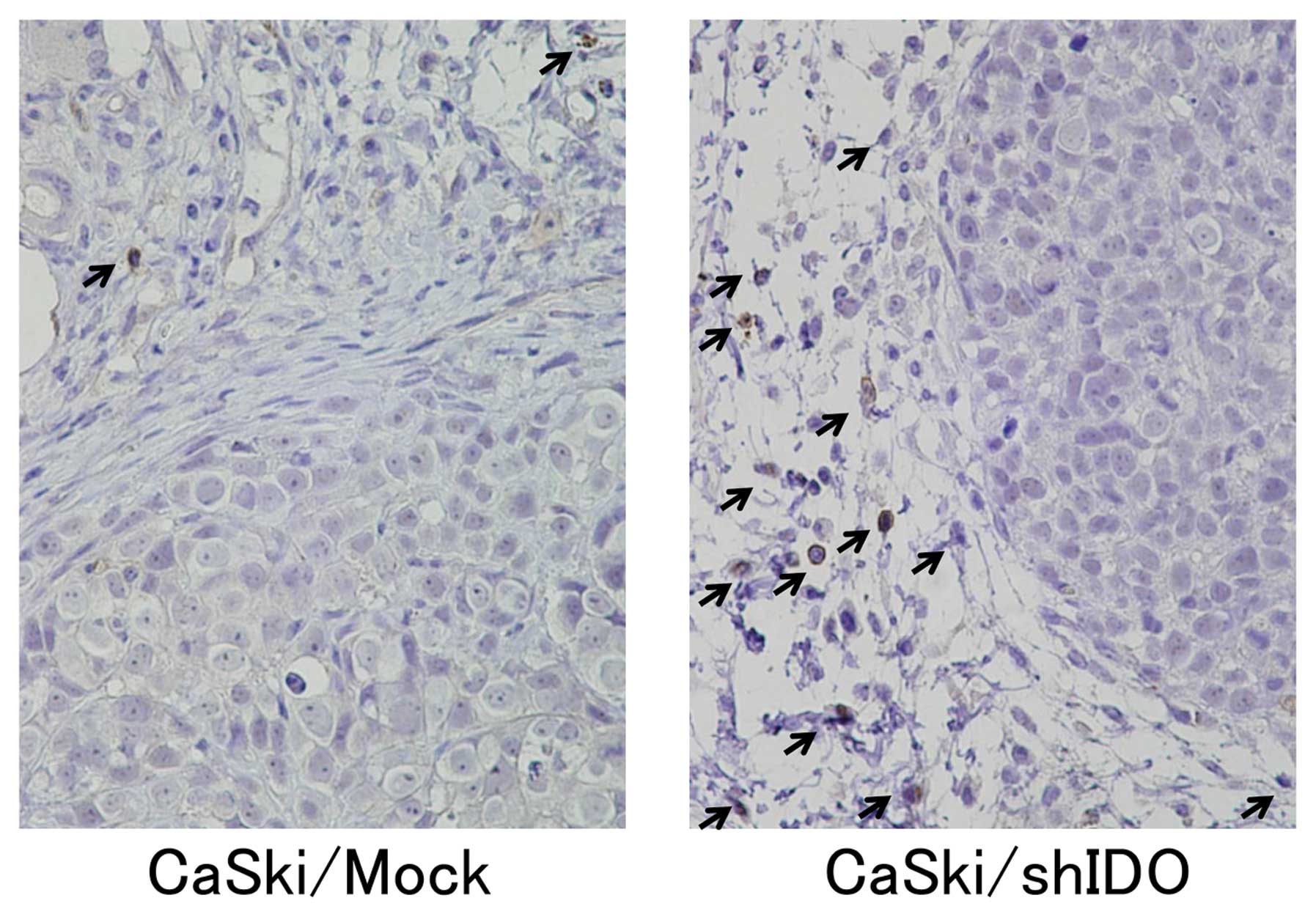
Number of NK cells in the tumor
stroma
NK cells immunostaining (black arrows) reveals an
accumulation of NK cells in the stroma of both CaSki/shIDO and
control subcutaneous tumors (Fig.
6). The number of NK cells (24±8) that accumulated in
CaSki/shIDO tumors was significantly higher than (2±2) in the
control tumors (P<0.01). These results suggest that the
downregulation of IDO promoted NK cell accumulation around the
tumor.
Discussion
The experiments described in this study aim to
clarify the relationship between the immunosuppressive enzyme IDO
and cervical cancer progression, as well as to develop a molecular
therapy targeting IDO. First, we investigated the expression of IDO
in 9 cervical cancer cell lines stimulated with interferon-γ and
observed that all of them, except the SKG-IIIb line, expressed IDO.
These results suggest that many cervical cancer cells produce IDO.
Next, we utilized an shRNA expression vector targeting the IDO gene
to examine whether the inhibition of IDO can control cervical
cancer tumor growth. We found that the downregulation of IDO
expression did not influence cervical cancer cell growth in
vitro, but controlled tumor growth in vivo. In addition,
the downregulation of IDO increased the sensitivity of cervical
cancer cells to NK cells in vitro and promoted NK cell
accumulation in the tumor stroma in vivo. These findings
indicate that downregulation of IDO effects cervical cancer tumor
growth by promoting NK cell accumulation in tumors, suggesting that
IDO may be a useful therapeutic target for patients with cervical
cancer.
There are a few reports that describe IDO expression
in cervical cancer. Inaba et al reported that IDO is
expressed in 52% of invasive cervical cancer cases as determined by
immunohistochemical staining (16).
On the other hand, Nakamura et al reported that IDO
expression was detected in all 25 cases of invasive cervical cancer
(34). In addition, both reports
observed that IDO expression in invasive cancer was confined to the
cancer cells at the invasive front (16,34).
Since these cells are in an environment where they are easily
exposed to proinflammatory mediators, such as interferon-γ or other
cytokines, they may be stimulated to produce IDO. In our study,
although only 2 of 9 cervical cancer cell lines constitutively
expressed IDO (CaSki and BOKU, data not shown), all cell lines
except for SKG-IIIb cells expressed IDO after stimulation with
interferon-γ. These results suggest that many cervical cancer cells
have the capability to produce IDO.
The lack of the essential amino acid tryptophan and
accumulation of its metabolite, kynurenine, inhibit cell growth and
induce cell death. T-cells are particularly sensitive to this type
of stress (10). Regarding the
mechanism of cancer cell immunotolerance, IDO has been shown to
promote local tryptophan depletion, resulting in T-cell function
inhibition in the vicinity of IDO-expressing cancer cells and
general local immunotolerance (13). The possibility that IDO expression
is involved in the immunotolerance of cervical cancer through such
a T-cell mediated mechanism cannot be excluded. However, a cervical
cancer cell line that can form a tumor in immunocompetent mice has
not been found. Therefore, we chose the human cervical cancer cell
line (CaSki) that constitutively expresses IDO and implanted this
cell line in nude mice. Since nude mice congenitally lack T cells,
in this experimental system, we were not able to examine the effect
of IDO on T-cell function.
It has been reported that IDO promotes the
accumulation of the tryptophan metabolite kynurenine, which
suppresses the expression of NK cell receptors, and thereby
inhibits the NK cell function (15). Similarly, in our previous
experiments using ovarian cancer cells, IDO expression inhibited
the cytotoxic activity of NK cells in vitro and suppressed
NK cell accumulation in the tumor stroma in vivo(35). Here, we demonstrated that IDO
downregulation enhanced the sensitivity of cervical cancer cells to
NK cells in vitro and promoted NK cell accumulation in the
cervical cancer stroma in vivo. Thus, IDO downregulation
reinforced the sensitivity of cancer cells to NK cells and
suppressed cervical cancer growth.
To date, chemically synthesized siRNA and
vector-mediated expression of shRNA are the most commonly used RNAi
gene silencing techniques (36,37).
Although siRNA can be more easily transfected into mammalian cells
and its silencing ability is more effective than shRNA, its effects
are transient. The remarkable advantage of shRNA is that the
inhibition of target genes can last for weeks or even months,
making it possible to elucidate the consequences of long-term,
stable gene silencing (36). In
actual clinical settings, viral-based expression vectors or
nanoparticle-based vectors (38)
could be used to deliver IDO shRNA to the cancer cells.
The results of this study demonstrate that the
downregulation of IDO in human cervical cancer cells that
constitutively express this enzyme inhibits cervical cancer
progression. This suggests that IDO-targeted shRNA is a potentially
effective molecular-targeted therapy for cervical cancer.
References
|
1
|
Jemal A, Bray F, Center MM, Ferlay J, Ward
E and Forman D: Global cancer statistics. CA Cancer J Clin.
61:69–90. 2011. View Article : Google Scholar
|
|
2
|
Jemal A, Siegel R, Xu J and Ward E: Cancer
statistics, 2010. CA Cancer J Clin. 60:277–300. 2010. View Article : Google Scholar
|
|
3
|
Monk BJ, Wang J, Im S, et al: Rethinking
the use of radiation and chemotherapy after radical hysterectomy: a
clinical-pathologic analysis of a Gynecologic Oncology
Group/Southwest Oncology Group/Radiation Therapy Oncology Group
trial. Gynecol Oncol. 96:721–728. 2005. View Article : Google Scholar
|
|
4
|
Higuchi K and Hayaishi O: Enzymic
formation of D-kynurenine from D-tryptophan. Arch Biochem Biophys.
120:397–403. 1967. View Article : Google Scholar : PubMed/NCBI
|
|
5
|
Yamamoto S and Hayaishi O: Tryptophan
pyrrolase of rabbit intestine. D- and L-tryptophan-cleaving enzyme
or enzymes. J Biol Chem. 242:5260–5266. 1967.PubMed/NCBI
|
|
6
|
Shimizu T, Nomiyama S, Hirata F and
Hayaishi O: Indoleamine 2,3-dioxygenase. Purification and some
properties. J Biol Chem. 253:4700–4706. 1978.PubMed/NCBI
|
|
7
|
Yoshida R, Urade Y, Tokuda M and Hayaishi
O: Induction of indoleamine 2,3-dioxygenase in mouse lung during
virus infection. Proc Natl Acad Sci USA. 76:4084–4086. 1979.
View Article : Google Scholar : PubMed/NCBI
|
|
8
|
Yoshida R and Hayaishi O: Induction of
pulmonary indoleamine 2,3-dioxygenase by intraperitoneal injection
of bacterial lipopolysaccharide. Proc Natl Acad Sci USA.
75:3998–4000. 1978. View Article : Google Scholar : PubMed/NCBI
|
|
9
|
Fujigaki S, Saito K, Sekikawa K, et al:
Lipopolysaccharide induction of indoleamine 2,3-dioxygenase is
mediated dominantly by an IFN-γ-independent mechanism. Eur J
Immunol. 31:2313–2318. 2001.PubMed/NCBI
|
|
10
|
Munn DH, Zhou M, Attwood JT, et al:
Prevention of allogeneic fetal rejection by tryptophan catabolism.
Science. 281:1191–1193. 1998. View Article : Google Scholar : PubMed/NCBI
|
|
11
|
Schroecksnadel K, Winkler C, Duftner C,
Wirleitner B, Schirmer M and Fuchs D: Tryptophan degradation
increases with stage in patients with rheumatoid arthritis. Clin
Rheumatol. 25:334–337. 2006. View Article : Google Scholar : PubMed/NCBI
|
|
12
|
Mellor AL and Munn DH: Ido expression by
dendritic cells: tolerance and tryptophan catabolism. Nat Rev
Immunol. 4:762–774. 2004. View
Article : Google Scholar : PubMed/NCBI
|
|
13
|
Uyttenhove C, Pilotte L, Theate I, et al:
Evidence for a tumoral immune resistance mechanism based on
tryptophan degradation by indoleamine 2,3-dioxygenase. Nat Med.
9:1269–1274. 2003. View
Article : Google Scholar : PubMed/NCBI
|
|
14
|
Vivier E, Tomasello E, Baratin M, Walzer T
and Ugolini S: Functions of natural killer cells. Nat Immunol.
9:503–510. 2008. View
Article : Google Scholar
|
|
15
|
Della Chiesa D, Carlomagno S, Frumento G,
et al: The tryptophan catabolite L-kynurenine inhibits the surface
expression of NKp46- and NKG2D-activating receptors and regulates
NK-cell function. Blood. 108:4118–4125. 2006.PubMed/NCBI
|
|
16
|
Inaba T, Ino K, Kajiyama H, et al:
Indoleamine 2,3-dioxygenase expression predicts impaired survival
of invasive cervical cancer patients treated with radical
hysterectomy. Gynecol Oncol. 117:423–428. 2010. View Article : Google Scholar
|
|
17
|
Ino K, Yoshida N, Kajiyama H, et al:
Indoleamine 2,3-dioxygenase is a novel prognostic indicator for
endometrial cancer. Br J Cancer. 95:1555–1561. 2006. View Article : Google Scholar : PubMed/NCBI
|
|
18
|
Takao M, Okamoto A, Nikaido T, et al:
Increased synthesis of indoleamine-2,3-dioxygenase protein is
positively associated with impaired survival in patients with
serous-type, but not with other types of, ovarian cancer. Oncol
Rep. 17:1333–1339. 2007.
|
|
19
|
Inaba T, Ino K, Kajiyama H, et al: Role of
the immunosuppressive enzyme indoleamine 2,3-dioxygenase in the
progression of ovarian carcinoma. Gynecol Oncol. 115:185–192. 2009.
View Article : Google Scholar : PubMed/NCBI
|
|
20
|
Gartel AL and Kandel ES: RNA interference
in cancer. Biomol Eng. 23:17–34. 2006. View Article : Google Scholar
|
|
21
|
Scherr M and Eder M: Gene silencing by
small regulatory RNAs in mammalian cells. Cell Cycle. 6:444–449.
2007. View Article : Google Scholar : PubMed/NCBI
|
|
22
|
Hannon GJ, Chubb A, Maroney PA, Hannon G,
Altman S and Nilsen TW: Multiple cis-acting elements are required
for RNA polymerase III transcription of the gene encoding H1 RNA,
the RNA component of human RNase P. J Biol Chem. 266:22796–22799.
1991.PubMed/NCBI
|
|
23
|
Walchli S and Sioud M: Vector-based
delivery of siRNAs: in vitro and in vivo challenges. Front Biosci.
13:3488–3493. 2008. View
Article : Google Scholar : PubMed/NCBI
|
|
24
|
Taguchi S: Establishment and
characterization of the human uterine cervical epidermoid cancer
cell line. Nippon Sanka Fujinka Gakkai Zasshi. 33:1180–1188.
1981.(In Japanese).
|
|
25
|
Ishikawa I, Nozawa S, Kikuchi K, Kurihara
S and Okumura H: Establishment of human uterine cervical cancer
cell line and comparative studies between normal and malignant
uterine cervical cells in vitro. Acta Obst Gynaec Jpn. 30:731–738.
1978.
|
|
26
|
Nozawa S, Udagawa Y, Ohta H, Kurihara S
and Fishman WH: Newly established uterine cervical cancer cell line
(SKG-III) with Regan isoenzyme, human chorionic gonadotropin
β-subunit, and pregnancy-specific β1-glycoprotein phenotypes.
Cancer Res. 43:1748–1760. 1983.PubMed/NCBI
|
|
27
|
Friedl F, Kimura I, Osato T and Ito Y:
Studies on a new human cell line (SiHa) derived from carcinoma of
uterus. I. Its establishment and morphology. Proc Soc Exp Biol Med.
135:543–545. 1970. View Article : Google Scholar : PubMed/NCBI
|
|
28
|
Pattillo RA, Hussa RO, Story MT, Ruckert
ACF, Shalaby MR and Mattingly RF: Tumor antigen and human chorionic
gonadotropin in CaSKi cells: a new epidermoid cervical cancer cell
line. Science. 196:1456–1458. 1977. View Article : Google Scholar : PubMed/NCBI
|
|
29
|
Morisawa T, Kuramoto H, Shimoda T,
Sakamoto I, Kato Y and Hamano M: Establishment and characterization
of a new SCC antigen producing cell line (HCS-2) from a carcinoma
of the uterine cervix. Hum Cell. 1:308–314. 1988.PubMed/NCBI
|
|
30
|
Sykes JA, Whitescarver J, Jernstrom P,
Nolan JF and Byatt P: Some properties of a new epithelial cell line
of human origin. J Natl Cancer Inst. 45:107–122. 1070
|
|
31
|
Yagita M, Huang CL, Umehara H, et al: A
novel natural killer cell line (KHYG-1) from a patient with
aggressive natural killer cell leukemia carrying a p53 point
mutation. Leukemia. 14:922–930. 2000. View Article : Google Scholar : PubMed/NCBI
|
|
32
|
Takikawa O, Kuroiwa T, Yamazaki F and Kido
R: Mechanism of interferon-γ action. Characterization of
indoleamine 2,3-dioxygenase in cultured human cells induced by
interferon-γ and evaluation of the enzyme-mediated tryptophan
degradation in its anticellular activity. J Biol Chem.
263:2041–2048. 1988.
|
|
33
|
Wang d, Saga Y, Mizukami H, et al:
Indoleamine-2,3-dioxygenase (IDO), an immunosuppressive enzyme that
inhibits natural killer cell function, as a useful target for
ovarian cancer therapy. Int J Oncol. 40:929–934. 2012.PubMed/NCBI
|
|
34
|
Nakamura T, Shima T, Saeki A, et al:
Expression of indoleamine 2,3-dioxygenase and the recruitment of
Foxp3-expressing regulatory T cells in the development and
progression of uterine cervical cancer. Cancer Sci. 98:874–881.
2007. View Article : Google Scholar : PubMed/NCBI
|
|
35
|
Nonaka H, Saga Y, Fujiwara H, et al:
Indoleamine 2,3-dioxygenase promotes peritoneal dissemination of
ovarian cancer through inhibition of natural killer cell function
and angiogenesis promotion. Int J Oncol. 38:113–120. 2011.
|
|
36
|
Shiota M, Ikeda Y and Wadhwa R: The
factors that contribute to the long-term expression of siRNA.
Nucleic Acids Symp Ser (Oxf). 243–244. 2006. View Article : Google Scholar : PubMed/NCBI
|
|
37
|
Walchli S and Sioud M: Vector-based
delivery of siRNAs: in vitro and in vivo challenges. Front Biosci.
13:3488–3493. 2008. View
Article : Google Scholar : PubMed/NCBI
|
|
38
|
Serda RE, Godin B, Blanco E, Chiappini C
and Ferrari M: Multi-stage delivery nano-particle systems for
therapeutic applications. Biochim Biophys Acta. 1810:317–329. 2011.
View Article : Google Scholar : PubMed/NCBI
|