Introduction
The prognosis of gallbladder cancer (GBC) remains
poor despite recent advances in diagnostic modalities and
therapeutic tools. The incidence of GBC is >4,000 cases per
year, and the 5-year survival rate of patients with GBC is
estimated to be ~12% (1). The poor
prognosis of GBC appears to be associated with the absence of
specific symptoms, which causes difficulty for the early diagnosis
of GBC. Indeed, most early-stage GBCs are found incidentally after
cholecystectomy for cholecystolithiasis, of which the reported
incidence is ~0.3–1% (2,3). Therefore, novel diagnostic methods and
therapeutic agents are needed to improve the prognosis of GBC.
Pancreaticobiliary maljunction (PBM) is a major risk
factor for GBC and is one of the clues used to clarify the
mechanisms of carcinogenesis and malignant behavior of GBCs. Reflux
of pancreatic juice into the bile duct activates the conversion of
lecithin to lysolecithin by phospholipase A2, and concentrated
lysolecithin in the gallbladder or the dilated bile duct induces
chronic inflammation and damage to biliary epithelium (4). K-ras and p53 mutations as well as MUC1
overexpression have been frequently detected in GBC with PBM
(5–8). It has been speculated that K-ras
mutation and chronic inflammation induce mucosal hyperplasia in the
biliary system, and the subsequent p53 mutation causes lesions to
develop atypical hyperplasia and then transform to carcinoma. The
‘hyperplasia-to-atypical hyperplasia-to-carcinoma sequence’ in GBC
with PBM differs from the ‘adenoma-to-carcinoma sequence’ or ‘de
novo’ oncogenesis (7–9). Thus, elucidation of this carcinogenic
process may help us to establish novel diagnostic and therapeutic
tools for GBC.
microRNAs (miRs) are non-coding RNAs consisting of
18–25 nucleotides, which have been reported to play important roles
in the regulation of carcinogenesis and cancer progression as well
as homeostasis. Approximately 1500 miRs have been identified to
date, and aberrant expression of miRs has been detected in various
types of malignancies including breast, colorectal and lung cancers
(10,11). Although the effects of aberrant
regulation of miRs on malignant diseases have not been fully
explored, miRs are expected to become diagnostic markers or
therapeutic targets for various malignancies including GBCs.
Previous reports have shown that miR-155 is involved
in the carcinogenesis of B-cell lymphoma, breast, lung and
pancreatic cancers (12–16). Jiang et al(15) speculated that the mechanisms of
inflammatory stimulation through the Janus-activated kinase pathway
may cause upregulation of miR-155 and subsequently lead to mutation
of tumor-suppressor gene SOCS1 in breast cancer. Tili et
al(17) reported that miR-155
overexpression and an inflammatory environment in breast cancer
cell line MDA-MB-21 downregulated WEE-1 kinase, which blocks cell
cycle progression. The authors concluded that miR-155 is involved
in inflammation-related carcinogenesis and cancer progression. To
date, despite the recent findings concerning miR-155 and its
important roles in inflammation and carcinogenesis, there have been
no reports regarding an association between miR-155, GBCs and
PBMs.
The aims of this study were to explore the
expression level of miR-155 and evaluate the clinical significance
of miR-155 expression in GBC and non-cancerous gallbladder samples
with the presence or absence of PBM, and to evaluate the effects of
aberrant miR-155 expression on GBC cell lines.
Materials and methods
Tissue sampling
Samples were obtained from 56 patients who underwent
surgical resection of gallbladders at our institution between
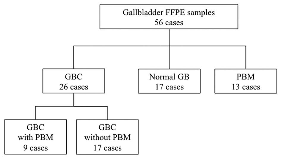
January 1994 and June 2011. There were 17 GBCs without PBM, 9 GBCs
with PBM, and 13 non-cancerous gallbladders with PBM (Fig. 1). Normal gallbladders obtained from
17 patients who underwent pancreatoduodenectomy for intraductal
papillary mucinous neoplasm were used as controls. Formalin-fixed,
paraffin-embedded (FFPE) blocks of gallbladders were sectioned at
10-μm thicknesses, and the epithelia of GBCs, non-cancerous
gallbladders with PBM, and normal gallbladders were obtained.
Cancerous lesions of GBCs were obtained by histological
macrodissection with manual scalpel resection while referring to
hematoxylin and eosin staining. The study was approved by the
Ethics Committee of Kyushu University and was conducted according
to the Ethical Guidelines for Human Genome/Gene Research enacted by
the Japanese government and the Helsinki Declaration.
RNA extraction from FFPE tissues
Total RNA was purified from FFPE tissues of the
gallbladder epithelium as described above using an RNeasy FFPE kit
(Qiagen, Hilden, Germany) in accordance with the manufacturer's
protocol. The RNA quantity was measured with an ND-1000
spectrophotometer (Thermo Fisher Scientific, Wilmington, DE, USA).
Total RNA was checked by a 2100 Bioanalyzer (Agilent Technologies,
Santa Clara, CA, USA) to determine the RNA quality integrity number
and 28s/18s rRNA ratio.
miR-155 expression analysis by real-time
reverse transcription-polymerase chain reaction
Purified RNAs were subjected to miR assay. A
stem-loop quantitative real-time reverse transcription-polymerase
chain reaction (qRT-PCR) was performed with a TaqMan MicroRNA
Reverse Transcription kit and TaqMan Universal PCR Master Mix II
(Applied Biosystems, Foster City, CA, USA) and Chromo4 real-time
PCR detection system (Bio-Rad Laboratories, Hercules, CA, USA) for
each sample according to the manufacturer's instructions. cDNAs
were placed in triplicate onto a plate in a total volume of 5
μl/well. Briefly, for reverse transcription, the reaction mixtures
were incubated at 16°C for 30 min, 42°C for 30 min, 85°C for 5 min,
and then maintained at 4°C. PCR amplification was performed at 95°C
for 10 min, followed by 40 cycles at 94°C for 15 sec and 60°C for
60 sec. The expression levels of specific miRs were normalized to
those of RNA U6 (RNU6B) as the endogenous control.
Cell lines
Human GBC cell lines G-415 (isolated from GBC of a
68-year-old Japanese male), OCUG-1 (isolated from GBC of a
43-year-old Japanese male), and NOZ (isolated from ascites of a
48-year-old Japanese female with GBC) were used in the present
study. The G-415 cell line was purchased from the Riken BioRecourse
Center through the National Bio-Resource Project of the Ministry of
Education, Science, Sports Culture and Technology (Tsukuba, Japan),
and maintained in Roswell Park Memorial Institute (RPMI)-1640
medium supplemented with 10% fetal bovine serum (FBS). OCUG-1 and
NOZ cell lines were purchased from the Japan Health Science
Research Resources Bank (Osaka, Japan), and maintained in
Dulbecco's modified Eagle's medium (DMEM) with 10% FBS and
Williams' E medium with 10% FBS, respectively. Each cell line was
cultured at 37°C in a humidified incubator with 5%
CO2.
Analysis of miR-155 expression levels in
GBC cell lines
To analyze the expression levels of miR-155 in the
GBC cell lines by qRT-PCR, total RNA was extracted from each GBC
cell line using a mirVana miRNA Isolation kit (Applied Biosystems)
according to the manufacturer's instructions. qRT-PCR was performed
using a Chromo4 real-time PCR detection system with TaqMan microRNA
assays (Applied Biosystems) as described above.
Transfections
The cell lines were transfected by electroporation
using an Amaxa Biosystems Nucleofector (Lonza, Basel, Switzerland)
according to the manufacturer's protocol. Briefly, 100 pmol of the
inhibitors or mimics of miR-155 or a negative control miR was added
to 1×10⁶ cells suspended in 100 μl Nucleofector
solution, followed by electroporation. The sequence of the mimic
for miR-155 was 5′-UUAAUGCUAAUC GUGAUAGGGGU-3′. The sequence of the
inhibitor for miR-155 was 5′-UCCCCTUTCUCGUTTUGCUTTUU-3′. Negative
control miR was used as described by Gregory et al(18). Synthesized RNAs were all purchased
from Qiagen. After electroporation, transfected cells were seeded
in 90-mm dishes and cultured for 24 h at 37°C, and then viable
cells were collected and applied to proliferation and invasion
assays. Effects of the miR-155 inhibitors and mimics, and the
negative control on GBC cell lines were evaluated with the TaqMan
microRNA assay.
Proliferation assay
Cell proliferation was assessed by a modified
propidium iodide (PI) assay as previously described (19). Briefly, after 24 h of transfection,
2×10⁴ cells/well were seeded in 24-well plates
containing medium without phenol-red and then incubated at 37°C for
48 and 96 h. At the indicated time, 30 μM PI and 600 μM digitonin
were added to each well. After incubation for 30 min at 37°C, the
fluorescence intensity was measured by an Infinite 200 multi-well
plate reader (Tecan, Männedorf, Switzerland) with 530-nm excitation
and 645-nm emission filters, and the total number of cells was
calculated. Each experiment was performed three times in
triplicate.
Matrigel invasion assay
Cancer cell invasion was assessed by measuring the
movement of cells into an artificial basal membrane in
Matrigel-coated Transwell chambers (Becton-Dickinson, Franklin
Lakes, NJ, USA). Each insert had an 8-μm pore-sized membrane coated
with 20 μg Matrigel. Briefly, 750 μl culture medium was placed into
each well of a 24-well plate, and then 1×10⁵ cells
suspended in 250 μl culture medium were seeded into each insert.
After 16 h of incubation at 37°C with 5% CO2, the
non-invading cells on the surface of the membrane were removed by
scrubbing with a cotton swab. The invading cells on the lower
surface of the membrane were fixed with 70% ethanol and stained
with hematoxylin and eosin. The invading cells in five randomly
selected fields were counted under a light microscope at ×100
magnification. Each experiment was performed three times in
triplicate. Cell growth of GBC cells used in the invasion assays
was not observed after 16 h.
Statistical analysis
The cut-off value of miR-155 expression was set by
the median value of the expression levels, and the lesions were
divided into two groups according to high and low expression levels
of miR-155. The relationship between the expression level of
miR-155 and clinicopathological characteristics, including
prognosis, was assessed. The effect of possible prognostic factors
on survival was also analyzed. Comparison between two groups was
analyzed with the Chi-square test, Mann-Whitney U test, or Cox
regression test. Cumulative survival rates were calculated with the
Kaplan-Meier method and compared with the log-rank test using JMP
version 9.0.2 (SAS Institute, Inc., Cary, NC, USA). Differences
were considered significant when the probability value was
<0.05.
Results
Overexpression of miR-155 in GBCs
The expression level of miR-155 was examined in 26
GBCs (including 9 with PBM), 13 non-cancerous gallbladders with
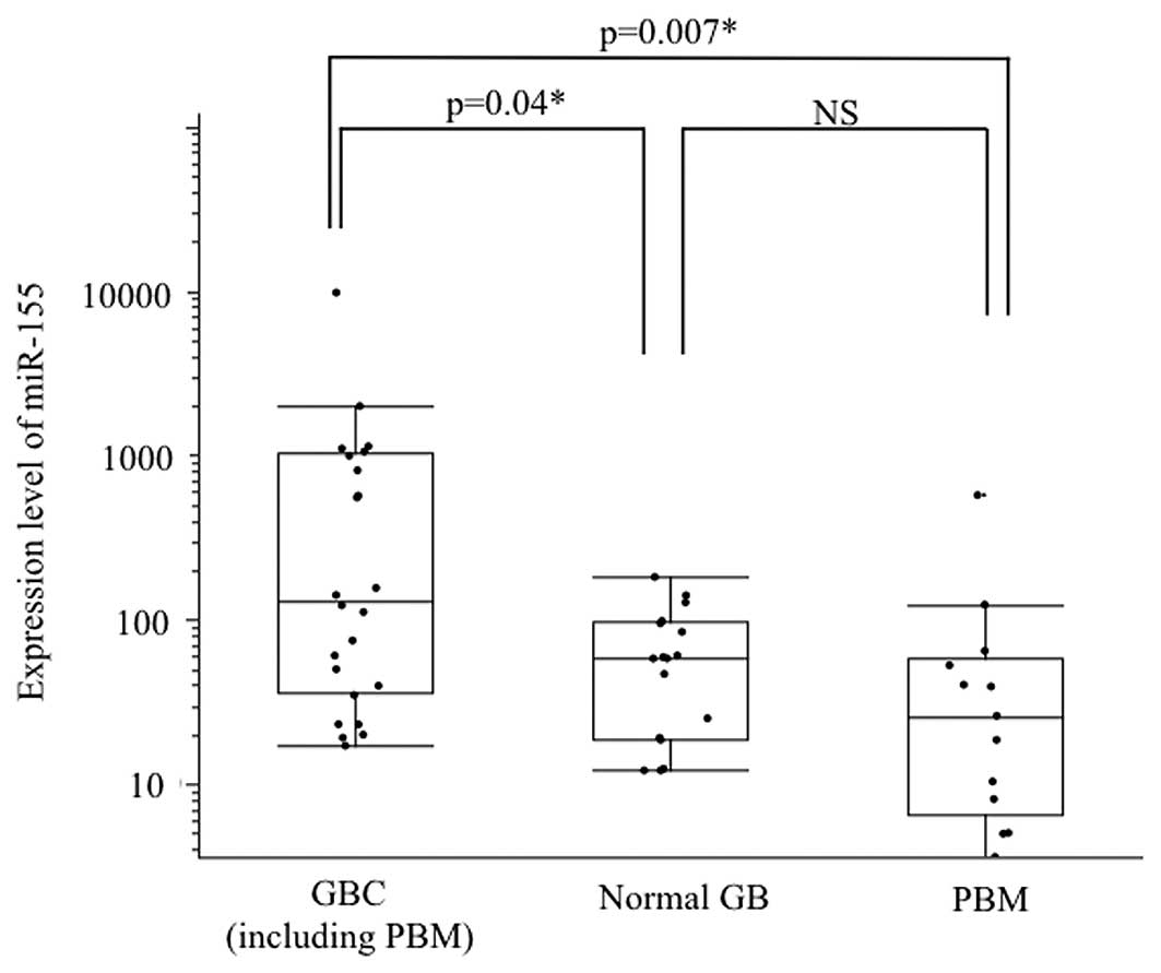
PBM, and 17 normal gallbladders by qRT-PCR (Fig. 1). The expression level of miR-155
was significantly higher in the GBCs when compared with that in the
normal gallbladders, whereas miR-155 was not upregulated in
gallbladders with PBM (Fig. 2).
High expression of miR-155 (>1.50-fold) was more frequently
observed in GBCs (14/26, 53%) than that in normal gallbladders
(4/17, 23%) (P=0.01). There was no significant difference in the
miR-155 expression level between GBCs with PBM (n=9) and GBCs
without PBM (n=17) (P=0.12). There was also no significant
difference in the miR-155 expression level between GBCs with PBM
(n=9) and non-cancerous gallbladders with PBM (n=13) (P=0.08).
Relationship between the expression level
of miR-155 and clinicopathological features in patients with
GBC
High miR-155 expression was significantly associated
with the presence of lymph node metastasis (P=0.03) and vessel
invasion (P=0.007) (Table I). There
was no significant difference in age, gender, presence of PBM, T
categories, UICC stages, lymphatic invasion, or perineural invasion
between high and low levels of miR-155 expression.
 | Table ICorrelation between
clinicopathological features and the miR-155 expression level in
GBCs. |
Table I
Correlation between
clinicopathological features and the miR-155 expression level in
GBCs.
| Clinicopathological
features | Total | High expression | Low expression | P-value |
|---|
| Total no. of GBC
cases | 26 | 13 | 13 | |
| Mean age, years ± SD
(range) | 65±11 (42–85) | 66±11 (45–85) | 64±11 (42–83) | 0.66 |
| Gender, male/female
(%) | 11 (42)/15 (58) | 8/5 | 6/7 | 0.67 |
| Presence of PBM
(%) | 9 (35) | 3 | 6 | 0.97 |
| T category (UICC)
(%) |
| Tis/T1/T2 | 3 (12)/4 (15)/16
(61) | 0/1/11 | 3/3/5 | 0.56 |
| T3/T4 | 1 (4)/2 (8) | 0/2 | 1/0 | |
| N category, N1/N0
(%) | 5 (19)/21(81) | 5/8 | 0/13 | 0.03a |
| UICC stage (%) |
| 0/IA/IB/IIA | 3 (12)/4 (15)/13
(50)/0 (0) | 0/1/8/0 | 3/3/5/0 | 0.11 |
| IIB/III/IV | 4 (15)/2 (8)/0
(0) | 3/2/0 | 1/0/0 | |
| Lymphatic invasion
(%) |
| Positive | 13 (50) | 9 | 4 | 0.11 |
| Negative | 13 (50) | 4 | 9 | |
| Vessel invasion
(%) |
| Positive | 13 (50) | 9 | 4 | 0.007a |
| Negative | 13 (50) | 3 | 10 | |
| Perineural invasion
(%) |
| Positive | 13 (50) | 5 | 8 | 0.11 |
| Negative | 13 (50) | 1 | 12 | |
| Follow-up
period |
| Median ± SD
(months) | 35±33 | 25±19 | 46±40 | 0.29 |
| 5-year DSS
rate | 0.62 | 0.46 | 0.83 | 0.01a |
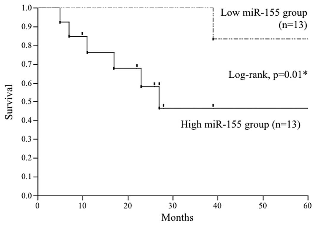
Relationship between the expression level
of miR-155 and prognosis in patients with GBCs
The relationship between the expression level of
miR-155 and the disease-specific survival (DSS) rate after surgery
was examined. DSS was significantly lower for GBC patients with a
high expression level of miR-155 (5-year survival rate, 35%;
median, 25 months) than those with a low miR-155 expression level
(5-year survival rate, 85%; median, 45 months) (P=0.01) (Fig. 3). The presence of lymph node
metastasis (P<0.0001), advanced UICC stage (IIB-IV) (P=0.03) and
positive lymphatic invasion (P=0.005) were also significantly
associated with poor prognoses of GBC patients, whereas the
presence of PBM, T category (Tis-2 vs. T3–4), vessel invasion and
perineural invasion did not affect the prognosis of GBC patients
(Table II). Multivariate analysis
revealed that lymphatic invasion was the only significant
independent factor to predict prognosis (hazard ratio, 9.6;
confidence interval, 1.3–189; P=0.03) (Table III). However, when focusing on
GBCs localized around the gallbladder without lymph node metastasis
(stage 0-IIA of UICC, Tis-T3N0), high miR-155 expression (P=0.02),
lymphatic invasion (P=0.03) and vessel invasion (P=0.02) were
significant predictive factors for the prognoses of GBC patients as
indicated by univariate analysis. Multivariate analysis showed that
a high expression level of miR-155 was the only independent
predictive indicator for poor prognosis of patients with localized
GBCs (hazard ratio, 9.9; confidence interval, 1.10–294; P=0.03)
(Table IV).
| Table IIUnivariate analysis for prognostic
factors of GBC. |
Table II
Univariate analysis for prognostic
factors of GBC.
| Variables | No. | Median survival
(month) | 5-year survival
rate | P-value |
|---|
| miR-155
expression |
| High | 13 | 25 | 0.35 | 0.01a |
| Low | 13 | 45 | 0.85 | |
| Age (years) |
| ≥65 | 13 | 27 | 0.47 | 0.44 |
| <64 | 13 | 33 | 0.67 | |
| Gender |
| Male | 11 | 51 | 0.68 | 0.63 |
| Female | 15 | 19 | 0.48 | |
| PBM |
| Present | 9 | 19 | 0.68 | 0.35 |
| Absent | 17 | 27 | 0.51 | |
| T category |
| Tis-T2 | 23 | 27 | 0.57 | 0.99 |
| T3-T4 | 3 | 17 | 0.50 | |
| Nodal status |
| N0 | 21 | 39 | 0.69 | <0.0001a |
| N1 | 5 | 7 | 0.00 | |
| UICC stage |
| 0-IIA | 20 | 33 | 0.67 | 0.03a |
| IIB-IV | 6 | 12 | 0.20 | |
| Lymphatic
invasion |
| Positive | 13 | 19 | 0.21 | 0.005a |
| Negative | 13 | 81 | 0.90 | |
| Vessel
invasion |
| Positive | 12 | 20 | 0.40 | 0.07 |
| Negative | 14 | 33 | 0.69 | |
| Perineural
invasion |
| Positive | 6 | 15 | 0.30 | 0.26 |
| Negative | 20 | 33 | 0.63 | |
| Table IIIMultivariate analysis for prognostic
factors of GBC. |
Table III
Multivariate analysis for prognostic
factors of GBC.
| Variables | Hazard ratio | 95% CI | P-value |
|---|
| High miR-155
expression | 1.3 | 0.1–10 | 0.77 |
| Nodal status
(N1) | 6.5 | 0.0–14 | 0.45 |
| UICC stage
(IIB-IV) | NA | NA | 0.63 |
| Lymphatic
invasion | 9.6 | 1.3–189 | 0.03a |
| Table IVMultivariate analysis for prognostic
factors of GBCs at stages 0-IA. |
Table IV
Multivariate analysis for prognostic
factors of GBCs at stages 0-IA.
| Variables | Hazard ratio | 95% CI | P-value |
|---|
| High miR-155
expression | 9.9 | 1.10–294 | 0.03a |
| Lymphatic
invasion | 1.4 | 0.24–6.26 | 0.64 |
| Vessel
invasion | 4.4 | 0.29–131 | 0.28 |
Expression levels of miR-155 in GBC cell
lines and the effect of transfection on the expression level of
miR-155
The relative expression levels of miR-155 to RUN6B
expression in G-415, NOZ and OCUG-1 cells were 56.8-, 18.1- and
4.2-fold, respectively (Fig. 4A).
Expression levels of miR-155 in G-415, NOZ, and OCUG-1 cells
transfected with miR-155 inhibitors showed downregulation by 0.12-,
0.35- and 0.17-fold, respectively, when compared with levels in the
negative controls. Expression levels of miR-155 in G-415, NOZ and
OCUG-1 cells transfected with miR-155 mimics were upregulated by
23.6-, 124- and 27,605-fold, respectively, when compared with
levels in the negative controls (Fig.
4B-D).
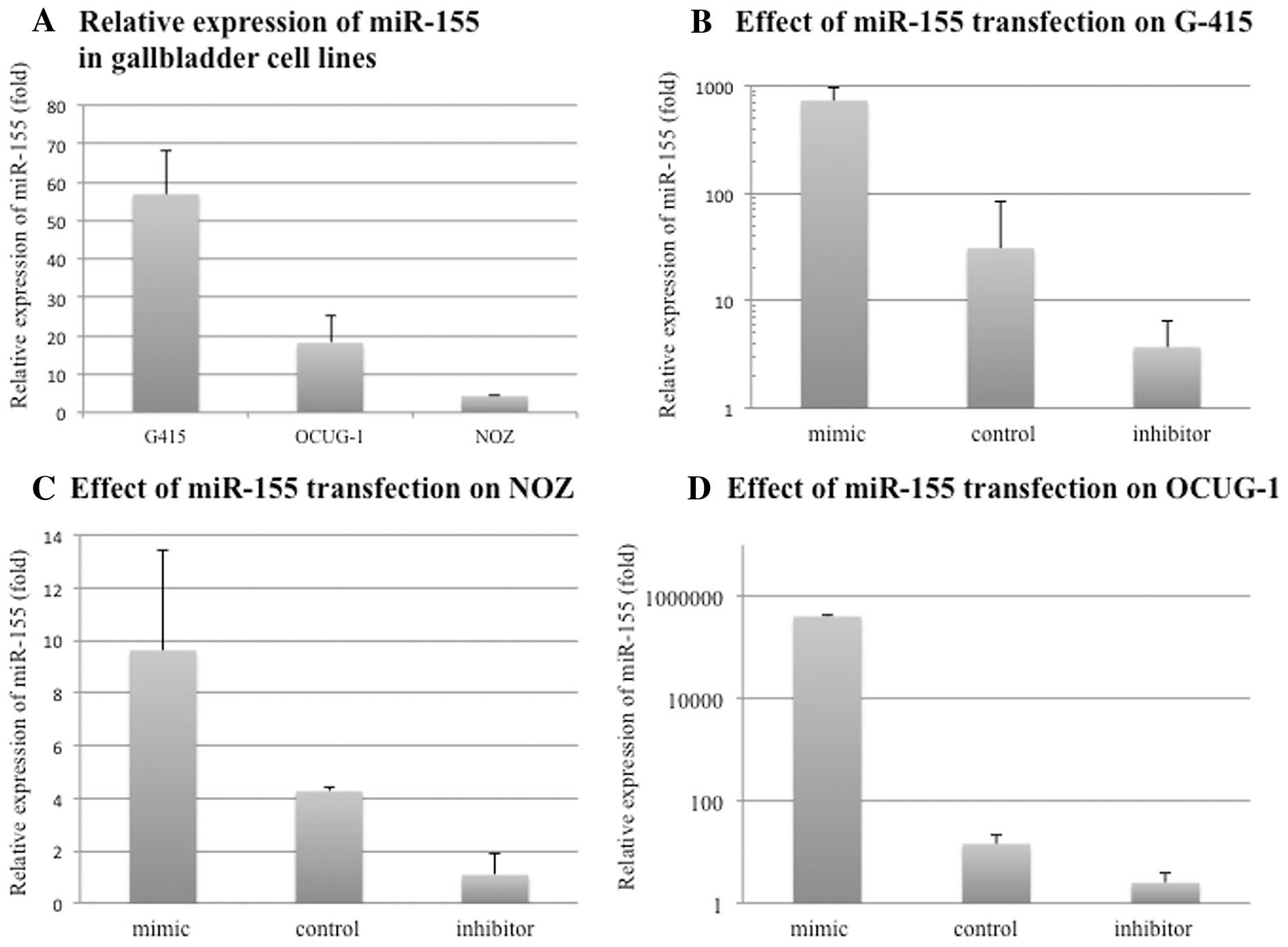 | Figure 4Relative expression of miR-155 in GBC
cell lines and the effect of transfection on the miR-155 expression
level. (A) The expression levels of miR-155 in the GBC lines were
investigated by qRT-PCR. miR-155 expression levels were normalized
to RNU6B expression. Human GBC cell lines, G-415 (isolated from a
GBC of a 68-year-old Japanese male), OCUG-1 (isolated from the GBC
of a 43-year-old Japanese male), and NOZ (isolated from the ascites
of a 48-year-old Japanese female with GBC), were used in the
present study. miR-155 expression levels in G-415, NOZ and OCUG-1
cells were 56.8-, 18.1- and 4.2-fold, respectively. (B-D) Effects
of transfection of miR-155 inhibitors or mimics on each GBC cell
line were assessed by qRT-PCR. Each expression level of miR-155 was
normalized to RNU6B expression. Expression levels of miR-155 in
G-415, NOZ and OCUG-1 cells transfected with miR-155 inhibitors
were downregulated by 0.12-, 0.35- and 0.17-fold, respectively,
when compared with levels in the negative controls. The expression
levels of miR-155 in G-415, NOZ and OCUG-1 cells transfected with
miR-155 mimics were upregulated by 23.6-, 124- and 27,605-fold,
respectively, when compared with levels in the negative controls.
miR; microRNA. |
Aberrant miR-155 expression affects the
proliferation of GBC cell lines
G-415 (P=0.0016) and OCUG-1 (P=0.0071) cells
transfected with miR-155 inhibitors showed significant decreases in
cell proliferation, compared with that in the negative controls. On
the other hand, G-415 (P=0.0009) and OCUG-1 (P=0.0030) cells
transfected with miR-155 mimics showed significant increases in
cell proliferation, compared with that in the negative controls
(Fig. 5A and B). NOZ cells
transfected with miR-155 inhibitors showed decreased cell
proliferation (P=0.0039), whereas those transfected with miR-155
mimics showed no significant differences in cell proliferation
(P=0.17) (Fig. 5C).
Aberrant miR-155 expression affects the
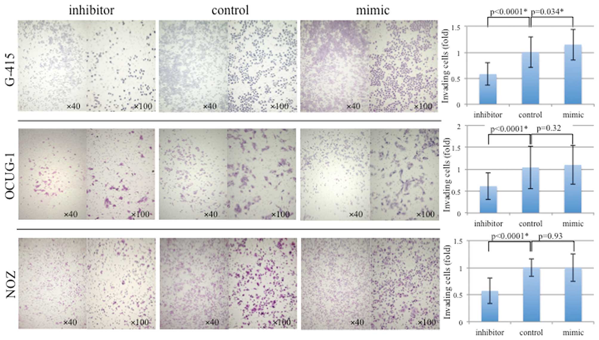
invasion of GBC cell lines
In the invasion assay, the number of invasive cells
among the total number of G-415 cells transfected with miR-155
mimics was significantly higher compared with that of the normal
control (P=0.034), whereas no difference in invasion was observed
in OCUG-1 (P=0.32) and NOZ (P=0.93) cells transfected with mimics.
The numbers of invasive cells among G-415, NOZ and OCUG-1 cells
transfected with miR-155 inhibitors were significantly lower (all
P<0.0001) compared with those of the normal controls (Fig. 6).
Discussion
In the present study, we found that: i) miR-155 is
upregulated in GBCs irrespective of the presence or absence of PBM,
ii)high miR-155 expression is significantly associated with the
presence of lymph node metastasis and vessel invasion, and
indicates a poorer prognosis for GBC patients, when compared with
GBC patients with low miR-155 expression, and iii) upregulation of
miR-155 affects the proliferation and invasion of GBC cells in
vitro. To our knowledge, this is the first report demonstrating
aberrant miR-155 expression and its effects on the function of GBC
cells.
One of the clinical problems during management of
GBCs is that many GBCs are found at an advanced stage with
metastasis at the time of diagnosis, and most early-stage GBCs
within the mucosal layer are diagnosed incidentally after
cholecystectomy for cholecystolithiasis. Because of the difficulty
of the early detection of GBCs, identification of sensitive markers
to diagnose early-stage GBCs is urgently needed. Our present study
was planned based on the hypothesis that assessment of abnormal
regulation of miRs may help us to understand the complicated
mechanism of carcinogenesis and address the clinical problems of
GBC.
Recent reports have analyzed the relationship
between the expression level of miR-155 and the prognosis of
several types of cancers. Shibuya et al(20) showed that high expression of miR-155
in colorectal cancer is associated with a high incidence of lymph
node metastasis and poor prognosis. The relationship between
miR-155 upregulation and advanced stages with poor prognoses has
also been reported in lung cancers (14). Our present study also demonstrated
that the expression level of miR-155 can predict the prognosis of
stage 0-IIA GBCs.
Stable miRs have been recently found in human blood,
despite the presence of RNase, and abnormal regulation of miRs has
been detected in the serum of patients with breast, esophageal and
prostate cancers (21–24). Henegham et al(22) identified upregulated miR-195 and
let-7a in the serum of breast cancer patients while decreased
levels of these miRs after surgery were noted. These circulating
miRs were also correlated with clinicopathological factors such as
nodal and estrogen receptor statuses. miR-155 has been detected in
the serum of patients with diffuse large B-cell lymphoma and is
expected to become a diagnostic marker for this cancer (25,26).
Recently, Shigehara et al(27) demonstrated that miR-9 and miR-145
are present in bile samples, and aberrant expression of these miRs
in bile may be a diagnostic marker for hepatobiliary tract cancers.
Further examination targeting miR-155 in serum and/or bile might
help us to develop diagnostic markers for early detection of GBCs.
In addition, detecting upregulation of miR-155 in the serum and/or
bile of patients with gallbladder disease will be useful to
determine surgical procedures when it is difficult to distinguish
GBC from benign gallbladder diseases such as xanthogranulomatous
cholecystitis.
miRs are also expected to become molecular targets
of cancer therapy (28). Garzon
et al(29) showed that
upregulated miR-155 causes the development of acute lymphocytic
lymphoma and that silencing miR-155 using antisense
oligonucleotides inhibits this process. Xie et al(30) found that ectopic expression of
miR-155 in hepatocellular carcinoma cells enhances in vitro
cell proliferation by targeting the transcriptional regulator,
sex-determining region Y box 6. Our present study also demonstrated
that aberrant expression levels of miR-155 by transfection of
miR-155 inhibitors or mimics, were correlated with proliferation
and the invasiveness of GBC cell lines. In particular, G-415 cells
with an unmodified high expression level of miR-155 showed marked
inhibition or promotion of proliferation and invasion following
transfection of miR-155 inhibitors or mimics. These results are in
good agreement with the hypothesis that miR-155 is a regulator of
proliferation and invasion of GBC cells. The present study suggests
the possibility that modulation of the miR-155 level may be applied
to the treatment of GBCs, particularly for inhibition of cancer
progression such as lymph node invasion.
In contrast to our expectation, PBM did not affect
regulation of the miR-155 level in gallbladder epithelium. This
finding suggests that miR-155 is not involved in the early stage of
multistep carcinogenesis induced by inflammation, the
‘hyperplasia-to-atypical hyperplasia-to-carcinoma sequence’.
Furthermore, it appears that the penetration frequency of miR-155
is too low to be a candidate for a general factor involved in
carcinogenesis. miR-155 may be overexpressed in the process in
which established cancers acquire invasive character.
Evaluation of GBCs as well as gallbladders with PBM
by microarray may reveal the association of miRs with multistep
carcinogenesis under chronic inflammation. miRs are characterized
by their binding to the incomplete complementary sites of their
targets and allowing mismatched G-U base pairing. However, a single
gene target is regulated by several miRs (12). Therefore, it is be necessary to
analyze potential aberrant expression of several miRs at the same
time to elucidate their effects on carcinogenesis and cancer
progression.
In conclusion, high miR-155 expression correlates
with the aggressive behavior of GBCs, and miR-155 may become a
prognostic marker and therapeutic target for GBC.
Acknowledgements
This study was supported by the Japan Society for
the Promotion of Science (JSPS) KAKENHI Grant Number 24592030.
References
|
1
|
Carriaga MT and Henson DE: Liver,
gallbladder, extrahepatic bile ducts, and pancreas. Cancer.
75:171–190. 1995. View Article : Google Scholar : PubMed/NCBI
|
|
2
|
Glauser PM, Strub D, Kaser SA, Mattiello
D, Rieben F and Maurer CA: Incidence, management, and outcome of
incidental gallbladder carcinoma: analysis of the database of the
Swiss Association of Laparoscopic and Thoracoscopic Surgery. Surg
Endosc. 24:2281–2286. 2010. View Article : Google Scholar
|
|
3
|
Gourgiotis S, Kocher HM, Solaini L,
Yarollahi A, Tsiambas E and Salemis NS: Gallbladder cancer. Am J
Surg. 196:252–264. 2008. View Article : Google Scholar
|
|
4
|
Sugiyama Y, Kobori H, Hakamada K, Seito D
and Sasaki M: Altered bile composition in the gallbladder and
common bile duct of patients with anomalous pancreaticobiliary
ductal junction. World J Surg. 24:17–21. 2000. View Article : Google Scholar : PubMed/NCBI
|
|
5
|
Hanada K, Itoh M, Fujii K, et al:
Pathology and cellular kinetics of gallbladder with an anomalous
junction of the pancreaticobiliary duct. Am J Gastroenterol.
91:1007–1011. 1996.PubMed/NCBI
|
|
6
|
Matsubara T, Sakurai Y, Zhi LZ, Miura H,
Ochiai M and Funabiki T: K-ras and p53 gene mutations in
noncancerous biliary lesions of patients with pancreaticobiliary
maljunction. J Hepatobiliary Pancreat Surg. 9:312–321. 2002.
View Article : Google Scholar : PubMed/NCBI
|
|
7
|
Tsuchida A and Itoi T: Carcinogenesis and
chemoprevention of biliary tract cancer in pancreaticobiliary
maljunction. World J Gastrointest Oncol. 2:130–135. 2010.
View Article : Google Scholar : PubMed/NCBI
|
|
8
|
Xiong L, Yang Z, Yang L, Liu J and Miao X:
Expressive levels of MUC1 and MUC5AC and their clinicopathologic
significances in the benign and malignant lesions of gallbladder. J
Surg Oncol. 105:97–103. 2011. View Article : Google Scholar : PubMed/NCBI
|
|
9
|
Shimada K, Yanagisawa J and Nakayama F:
Increased lysophosphatidylcholine and pancreatic enzyme content in
bile of patients with anomalous pancreaticobiliary ductal junction.
Hepatology. 13:438–444. 1991. View Article : Google Scholar
|
|
10
|
Volinia S, Calin GA, Liu CG, et al: A
microRNA expression signature of human solid tumors defines cancer
gene targets. Proc Natl Acad Sci USA. 103:2257–2261. 2006.
View Article : Google Scholar
|
|
11
|
Griffiths-Jones S: miRBase: microRNA
sequences and annotation. Curr Protoc Bioinformatics. Chapter
12(Unit 12): 91–10. 2010. View Article : Google Scholar
|
|
12
|
Esquela-Kerscher A and Slack FJ: Oncomirs
- microRNAs with a role in cancer. Nat Rev Cancer. 6:259–269. 2006.
View Article : Google Scholar
|
|
13
|
Gironella M, Seux M, Xie MJ, et al: Tumor
protein 53-induced nuclear protein 1 expression is repressed by
miR-155, and its restoration inhibits pancreatic tumor development.
Proc Natl Acad Sci USA. 104:16170–16175. 2007. View Article : Google Scholar : PubMed/NCBI
|
|
14
|
Yanaihara N, Caplen N, Bowman E, et al:
Unique microRNA molecular profiles in lung cancer diagnosis and
prognosis. Cancer Cell. 9:189–198. 2006. View Article : Google Scholar : PubMed/NCBI
|
|
15
|
Jiang S, Zhang HW, Lu MH, et al:
MicroRNA-155 functions as an OncomiR in breast cancer by targeting
the suppressor of cytokine signaling 1 gene. Cancer Res.
70:3119–3127. 2010. View Article : Google Scholar : PubMed/NCBI
|
|
16
|
Eis PS, Tam W, Sun L, et al: Accumulation
of miR-155 and BIC RNA in human B cell lymphomas. Proc Natl Acad
Sci USA. 102:3627–3632. 2005. View Article : Google Scholar : PubMed/NCBI
|
|
17
|
Tili E, Michaille JJ, Wernicke D, et al:
Mutator activity induced by microRNA-155 (miR-155) links
inflammation and cancer. Proc Natl Acad Sci USA. 108:4908–4913.
2011. View Article : Google Scholar : PubMed/NCBI
|
|
18
|
Gregory PA, Bert AG, Paterson EL, et al:
The miR-200 family and miR-205 regulate epithelial to mesenchymal
transition by targeting ZEB1 and SIP1. Nat Cell Biol. 10:593–601.
2008. View
Article : Google Scholar : PubMed/NCBI
|
|
19
|
Zhang L, Mizumoto K, Sato N, et al:
Quantitative determination of apoptotic death in cultured human
pancreatic cancer cells by propidium iodide and digitonin. Cancer
Lett. 142:129–137. 1999. View Article : Google Scholar : PubMed/NCBI
|
|
20
|
Shibuya H, Iinuma H, Shimada R, Horiuchi A
and Watanabe T: Clinicopathological and prognostic value of
microRNA-21 and microRNA-155 in colorectal cancer. Oncology.
79:313–320. 2010. View Article : Google Scholar : PubMed/NCBI
|
|
21
|
Zhou SL and Wang LD: Circulating
microRNAs: novel biomarkers for esophageal cancer. World J
Gastroenterol. 16:2348–2354. 2010. View Article : Google Scholar : PubMed/NCBI
|
|
22
|
Heneghan HM, Miller N, Lowery AJ, Sweeney
KJ, Newell J and Kerin MJ: Circulating microRNAs as novel minimally
invasive biomarkers for breast cancer. Ann Surg. 251:499–505. 2010.
View Article : Google Scholar : PubMed/NCBI
|
|
23
|
Mitchell PS, Parkin RK, Kroh EM, et al:
Circulating microRNAs as stable blood-based markers for cancer
detection. Proc Natl Acad Sci USA. 105:10513–10518. 2008.
View Article : Google Scholar : PubMed/NCBI
|
|
24
|
Kosaka N, Iguchi H and Ochiya T:
Circulating microRNA in body fluid: a new potential biomarker for
cancer diagnosis and prognosis. Cancer Sci. 101:2087–2092. 2010.
View Article : Google Scholar : PubMed/NCBI
|
|
25
|
Fang C, Zhu DX, Dong HJ, et al: Serum
microRNAs are promising novel biomarkers for diffuse large B cell
lymphoma. Ann Hematol. 91:553–559. 2011. View Article : Google Scholar : PubMed/NCBI
|
|
26
|
Lawrie CH, Gal S, Dunlop HM, et al:
Detection of elevated levels of tumour-associated microRNAs in
serum of patients with diffuse large B-cell lymphoma. Br J
Haematol. 141:672–675. 2008. View Article : Google Scholar : PubMed/NCBI
|
|
27
|
Shigehara K, Yokomuro S, Ishibashi O, et
al: Real-time PCR-based analysis of the human bile microRNAome
identifies miR-9 as a potential diagnostic biomarker for biliary
tract cancer. PLoS One. 6:e235842011. View Article : Google Scholar
|
|
28
|
Kasinski AL and Slack FJ: MicroRNAs en
route to the clinic: progress in validating and targeting microRNAs
for cancer therapy. Nat Rev Cancer. 11:849–864. 2011. View Article : Google Scholar : PubMed/NCBI
|
|
29
|
Garzon R, Marcucci G and Croce CM:
Targeting microRNAs in cancer: rationale, strategies and
challenges. Nat Rev Drug Discov. 9:775–789. 2010. View Article : Google Scholar : PubMed/NCBI
|
|
30
|
Xie Q, Chen X, Lu F, et al: Aberrant
expression of microRNA 155 may accelerate cell proliferation by
targeting sex-determining region Y box 6 in hepatocellular
carcinoma. Cancer. 118:2431–2442. 2012. View Article : Google Scholar : PubMed/NCBI
|