Introduction
Both genetic and epigenetic aberrations play vital
roles in tumorigenesis and the development of cancer. DNA
methylation is an important epigenetic modification, and aberrant
changes in DNA methylation are prominent features in the
pathogenesis and development of various diseases, including cancer.
Aberrant methylation within the promoter regions of
tumor-suppressor genes has been correlated with their silencing and
with uncontrolled proliferation of tumor cells (1,2).
There are four types of inhibitor of DNA binding
(ID) proteins in mammals, belonging to the helix-ring-helix protein
family, and their coding regions are located on chromosome 6p21-22.
Animal experiments and in vitro cellular assays have shown
that the ID4 protein inhibits the proliferation of tumor cells and
promotes cell apoptosis (3). In
addition, aberrant promoter methylation of the ID4 gene has
been correlated with the pathogenesis and development of
hematological malignancies (4). The
tight junction protein 1 (ZO-1) gene belongs to the membrane
guanylate kinase family, and it encodes a tight junction protein
involved in many signaling pathways that participate in the
regulation of cell proliferation and differentiation (5,6). The
aberrant hypermethylation of the promoter region of ZO-1 was
reported in murine leukemia cell lines, and the concomitant
suppression of the expression of the ZO-1 gene was also
observed. The ZO-1 gene may be a novel oncogene in
hematological malignancies that was identified using restriction
landmark genomic scanning (RLGS) (7). In summary, the alteration of the
methylation pattern on the promoter regions of the ID4 and
ZO-1 genes is a distinctive feature of hematological
malignancies when compared to normal cells (8).
Lymphoma is a hematological malignancy which is
diagnosed based mainly on pathological examination. However, the
diagnosis is not easy, partially because the accuracy of the
pathological examination relies on the experience of the clinical
practitioner and is dependent on subjective impressions,
particularly when the malignant atypical features of the tumor
cells are not apparent, or when the distinction between benign and
malignant hyperplasia is not clear (9,10).
Although auxiliary examinations are currently available, such as
immunohistochemical tests, gene rearrangement analysis, and T cell
receptor (TCR) detection, the diversified pathologic morphologies
of lymphoma tissues often result in difficulties with diagnosis.
Therefore, there is a need for more sensitive and specific
molecular diagnostic methods for the diagnosis, treatment, and
prognostic prediction of lymphoma. The highly sensitive
methylation-specific PCR (MSP) method has been previously applied
to detect methylation of the promoter regions of the ID4 and
ZO-1 genes in the diagnosis of breast cancer (11). One objective of the current study
was to identify the promoter methylation status of the ID4
and ZO-1 genes in the lymph node and bone marrow of lymphoma
patients.
The MSP (12)
technique was utilized in the present study to detect aberrant
alterations in the methylation status of the ID4 and
ZO-1 genes and to evaluate the feasibility of utilizing
these changes as epigenetic markers for lymphoma diagnosis, or for
detection of minimal residual lymphoma after treatment. A
comparison of the methylation statuses of ID4 and
ZO-1 was performed between lymphoma patients and a control
cohort comprised of patients without lymphoma. Meanwhile, the
feasibility of using the ID4 and ZO-1 methylation
status as a molecular indicator for lymphoma diagnosis and
prognosis was also evaluated.
Materials and methods
Patients and tissue samples
Paraffin-embedded lymphoma tissue samples were
obtained from 92 lymphoma patients treated at the PLA General
Hospital (Beijing, China) between May 2006 and October 2009, in
whom the diagnosis was confirmed by pathologic morphological
analysis and immunohistochemical studies. Paraffin-embedded lymph
node samples were also obtained from 10 patients with either
chronic cholecystitis or reactive lymphadenitis, as the control
set. The bone marrow specimens were obtained before treatment from
90 lymphoma patients admitted to our hospital between February 2008
and February 2010, and their clinical information is summarized in
Table I. The therapeutic strategy
was determined according to National Comprehensive Cancer Network
(NCCN) lymphoma treatment guidelines, or hematopoietic stem cell
transplantation was performed if possible. The control samples of
bone marrow tissue were from 8 donors who had donated bone marrow
for hematopoietic stem cell transplantation. Written informed
consent was provided by the patients and their families. The
deadline for the last follow-up for all enrolled patients was
October 31, 2011.
 | Table IClinical information of the
treatment-naive lymphoma patients. |
Table I
Clinical information of the
treatment-naive lymphoma patients.
|
Characteristics | Bone marrow | Lymphoma | P-value |
|---|
| Gender |
| Male/female | 70/20 | 31/20 | 0.03 |
| Age in years, mean
(range) | 44.22±17.56
(13–81) | 47.59±17.61
(17–79) | 0.28 |
| Height in cm, mean
(range) | 170.48±6.93
(155–184) | 167.35±8.10
(150–186) | 0.02 |
| Body weight in kg,
mean (range) | 68.00±12.25
(45–102) | 68.71±11.20
(45.5–89) | 0.73 |
| Systolic pressure
(mmHg) | 120.46±11.53 | 122.45±12.42 | 0.34 |
| Diastolic pressure
(mmHg) | 75.29±8.33 | 75.94±7.78 | 0.65 |
| Heart rate
(beats/min), mean (range) | 81.29±7.84
(60–117) | 77.96±6.30
(62–104) | 0.02 |
| Medical
history |
| Healthy
(yes/no) | 36/54 | 20/31 | 0.93 |
| Familial tumor
history (yes/no) | 11/79 | 6/45 | 0.94 |
| Clinical stage of
lymphoma | | | 0.14 |
| I | 15 | 12 | |
| II | 15 | 15 | |
| III | 16 | 7 | |
| IV | 44 | 17 | |
| Systemic symptoms B
(yes/no) | 37/53 | 23/28 | 0.65 |
| Lymphoma type | | | 0.32 |
| Non-Hodgkin | 78 | 47 | |
| Hodgkin | 12 | 4 | |
| Pathological
type | | | 0.82 |
| B cell | 60 | 37 | |
| T cell | 18 | 10 | |
| Bone marrow
involvement (yes/total) | 15/90 | 6/51 | 0.43 |
The HL-60 and K562 cell lines were used as positive
and negative controls, respectively, for MSP of the ID4 and
ZO-1 genes, and the cells were maintained in the Department
of Hematology, PLA General Hospital. Deionized water was used as
the blank control for MSP.
PCR primers
The methylation-specific primers for the ID4
gene were ID4-MF forward, TCGGAGTTTTCGTTTTCGTT and
ID4-MF reverse, CGATACTACTCACAACCGCG; ID4-UMF
forward, TGTTTGGAGTTTTTGTTTTTGTT and ID4-UMR reverse,
CCCAATACTACTCACAACCACAC. The methylation-specific primers for
ZO-1 were ZO-1-MF forward, AAAATAAATAGGAAGATTCGTACGG
and ZO-1-MR reverse, GAAACTAAACGAAACGAAACGAA;
ZO-1-UMF forward, GGATAAAAATAAATAGGAAGATT TGTATG and
ZO-1-UMR reverse, AACAAAACTAAACA AAACAAAACAA.
Genomic DNA extraction and base
modification
DNA extraction from paraffin-embedded tissues was
performed using a commercial kit (Qiagen, USA), and the genomic DNA
Purification kit (Promega, USA) was utilized for DNA extraction
from the bone marrow. The concentration of extracted DNA was
determined using spectrophotometry. Two micrograms of DNA was
sheared by passing through 26G syringe needles repeatedly after
dilution with water. Then, 5 μl freshly prepared NaOH solution (3
mol/l) was added to the sheared DNA, and the mixture was incubated
at 37°C for 25 min, followed by the addition of 30 μl freshly
prepared hydroquinone solution (10 mmol/l) and 520 μl sodium
bisulfite (3 mol/l) and subsequent incubation at 55°C for 16 h. The
DNA was purified from the above mixture using a DNA gel extraction
kit (Axygen Biosciences, USA) and stored at −20°C.
MSP reactions
The total volume of the reaction was 25 μl, and the
mixture consisted of 4 μg DNA, 2.5 μl 10X Herman’s buffer, 1.25 μl
of 25 mM dNTPs, 0.15 μl Hot Start Taq polymerase, and 3 μl
of each upstream and downstream methylation-specific or
unmethylated DNA-specific PCR primers. The PCR reaction settings
were as follows: pre-denaturation at 94°C for 10 min, followed by
35 cycles of 94°C for 60 sec, 56°C for 40 sec, and 72°C for 50 sec,
and a final elongation for 10 min at 72°C. The HL-60 and K-562
cells were used as positive and negative controls, respectively,
for each trial, and deionized water was used as the blank control.
The products of PCR were separated with 2% agarose DNA
electrophoresis, the resolved DNA was stained with ethidium bromide
and images were recorded with an ultraviolet transmission
analyzer.
The PCR products were recovered and purified from
the agarose gel with the DNA gel extraction kit (Qiagen).
Plasmid construction and DNA
sequencing
The PCR fragments recovered from the agarose gel
were inserted into the pGEM-T Easy vector by T4 DNA ligase-based
cloning, and subsequently transformed into competent E. coli
DH5α. Positive colonies were obtained by antibiotic resistance
screening and blue-white colony screening. One microliter of a
bacterial culture grown overnight was used as the template for PCR.
The PCR-positive colonies were sequenced by Invitrogen (Beijing,
China).
Readout
For the ID4 gene, complete methylation was
indicated by the presence of a 186-bp PCR fragment alone upon
amplification with the methylation-specific primers; completely
unmethylated DNA was indicated by the presence of a 191-bp PCR
fragment alone upon amplification with the unmethylated
DNA-specific primers. Partial methylation was indicated by the
presence of both the 186- and 191-bp PCR fragments after
amplification. Similarly, for the ZO-1 gene, the presence of
a 177-bp or a 179-bp PCR fragment alone indicated complete
methylation or a complete lack of methylation, respectively, while
the presence of both 177- and 179-bp fragments after amplification
was indicative of partial methylation. Since the bone marrow of the
patients consisted of both lymphoma cells and normal cells, partial
methylation and complete methylation were classified as a
methylation-positive readout.
Statistical analysis
Quatitative data are expressed as means ± SD, and
differences between data sets were analyzed using the Student’s
t-test. Categorical data were described as absolute frequencies and
analyzed by the Pearson’s Chi-square test. Cumulative event curves
were plotted using the Kaplan-Meier survival method and differences
between curves were tested for statistical significance using the
log-rank method. P-values <0.05 were considered statistically
significant. All data were analyzed using the SPSS version 12.0
statistical software (SPSS Inc., Chicago, IL, USA).
Results
Methylation status of genes amplified
from the paraffin-embedded lymphoma and bone marrow tissues
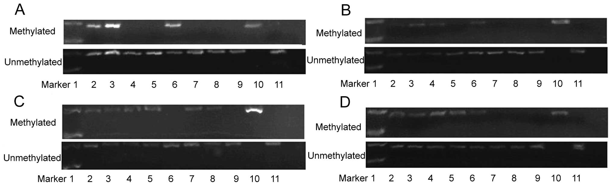
The PCR results showed that 64 out of 92
paraffin-embedded lymphoma tissues were ID4-positive and 57
were ZO-1-positive, respectively, either in the methylated
or unmethylated state, and that 51 were both ID4- and
ZO-1-positive. Eight out of 10 paraffin-embedded lymphoma
tissues from either chronic cholecystitis or reactive lymphadenitis
patients were both positive for completely unmethylated ID4
and ZO-1 genes. All bone marrow tissues of lymphoma patients
were ID4- and ZO-1-positive, for either methylated or
unmethylated DNA.
There were 41 and 43 samples positive for ID4
and ZO-1 gene methylation out of the 51 paraffin-embedded
lymphoma tissues, and the positivity rates were 80.4 and 84.3%,
respectively. Forty-seven samples were positive for either
ID4 or ZO-1 methylation, and the coverage rate for
both genes was 92.2%. The ID4 and ZO-1 genes were
completely unmethylated in the 8 paraffin-embedded lymph node
tissues from either chronic cholecystitis or reactive lymphadenitis
patients.
There were 23 and 13 samples positive for ID4
and ZO-1 gene methylation, respectively, out of 90 bone
marrow samples from lymphoma patients, and the positivity rates
were 25.56 and 14.44%, respectively. The ID4 and ZO-1
genes of the 8 samples from bone marrow donors were completely
unmethylated (Tables II and
III, Fig. 1).
| Table IICorrelation between the ID4
and ZO-1 gene methylation status in paraffin-embedded
tissues and the lymphoma pathological type, stage and
prognosis. |
Table II
Correlation between the ID4
and ZO-1 gene methylation status in paraffin-embedded
tissues and the lymphoma pathological type, stage and
prognosis.
| Tissue samples
Total n | ID4
methylation n (%) | ZO-1
methylation n (%) | P-value
(ID4) | P-value
(ZO-1) |
|---|
| Lymphoma | 51 | 41 (80.4) | 43 (84.3) | | |
| NHL | 47 | 40 (85.1) | 41 (87.2) | 0.004 | 0.05 |
| HD | 4 | 1 (25) | 2 (50) | | |
| Pathological
type |
| B cell | 37 | 31 (83.8) | 31 (83.8) | 0.62 | 0.17 |
| T cell | 10 | 9 (90) | 10 (100) | | |
| Clinical stage |
| I/II | 26 | 22 (84.62) | 20 (76.9) | 0.44 | 0.14 |
| III/IV | 25 | 19 (76) | 23 (92.0) | | |
| IPI |
| 0–2 | 39 | 33 (84.61) | 34 (87.18) | 0.83 | 0.98 |
| 3–5 | 8 | 7 (87.5) | 7 (87.5) | | |
| Table IIICorrelation between the ID4
and ZO-1 gene methylation status in bone marrow samples and
the lymphoma pathological type, stage and prognosis. |
Table III
Correlation between the ID4
and ZO-1 gene methylation status in bone marrow samples and
the lymphoma pathological type, stage and prognosis.
| Bone marrow samples
Total n | ID4
methylation n (%) | ZO-1
methylation n (%) | P-value
(ID4) | P-value
(ZO-1) |
|---|
| Lymphoma | 90 | 23 (25.56) | 13 (14.44) | | |
| NHL | 78 | 21 (26.92) | 12 (15.38) | 0.4483 | 0.5177 |
| HD | 12 | 2 (16.67) | 1 (8.33) | | |
| Pathological
type |
| B cell | 60 | 16 (26.67) | 7 (11.67) | 0.9257 | 0.09659 |
| T cell | 18 | 5 (27.78) | 5 (27.78) | | |
| Clinical stage |
| I/II | 30 | 10 (33.33) | 4 (13.33) | 0.2316 | 0.8321 |
| III/IV | 60 | 13 (21.67) | 9 (15) | | |
| IPI |
| 0–2 | 67 | 16 (23.88) | 7 (10.45) | 0.5340 | 0.06564 |
| 3–5 | 23 | 7 (30.43) | 6 (26.09) | | |
Survival curve
The patients with positive MSP results for
ID4 or ZO-1 isolated from the bone marrow had a lower
survival rate compared to the patients with unmethylated ID4
or ZO-1 genes. A significant difference was observed in the
survival rate between the patients with and without ID4
methylation in the bone marrow (P<0.05). On the contrary, the
methylation status of ID4 and ZO-1 of DNA isolated
from the lymphoma tissue did not show any correlation with the
survival rate (Fig. 2).
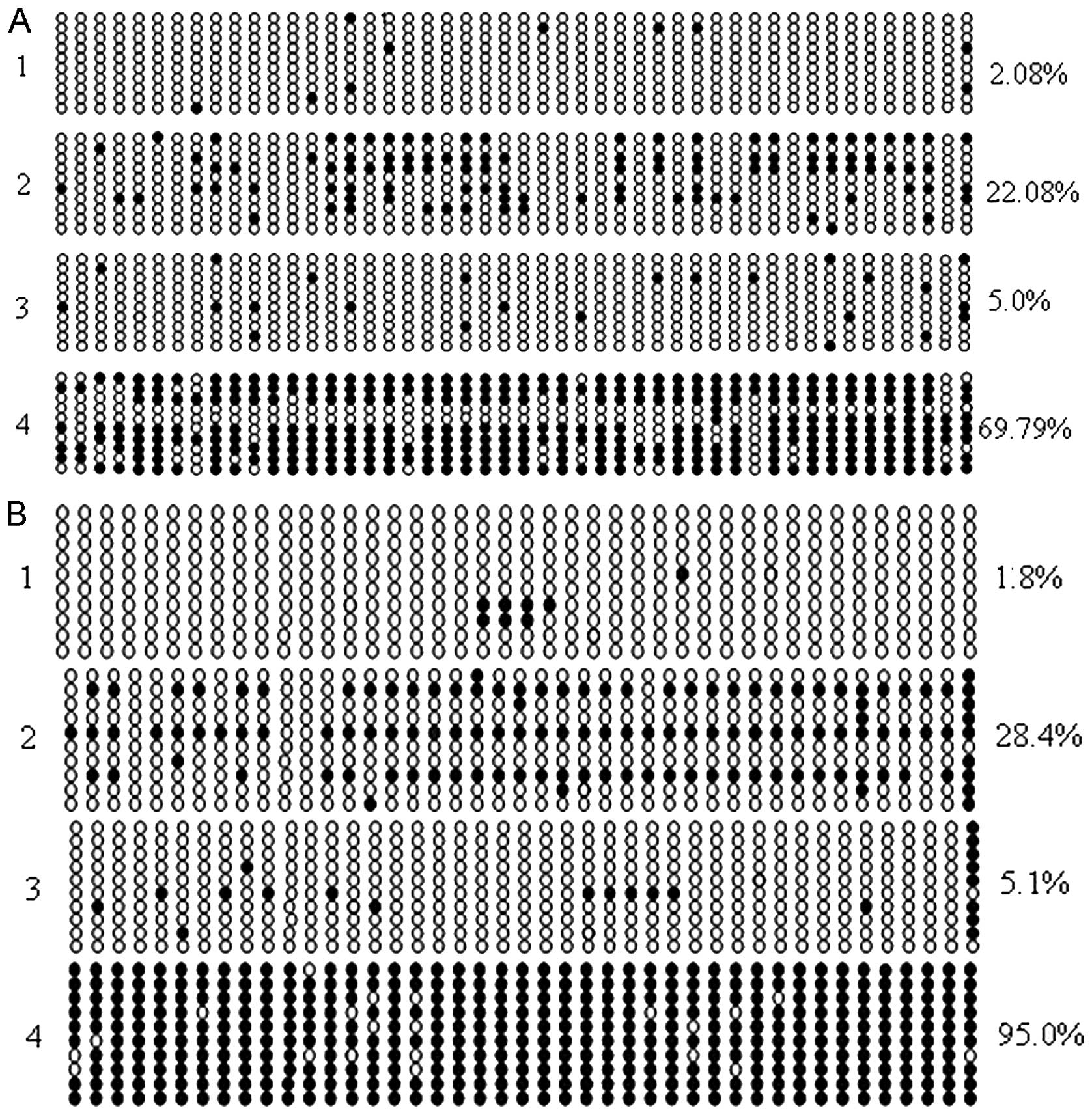
DNA sequencing results
The CpG methylation sites of individual bacterial
clones are shown in Fig. 3. The
total methylation rates of ID4 and ZO-1 from 10
randomly picked colonies were 22.08 and 28.4%, respectively
(Fig. 3). On the contrary, the
total methylation rates of ID4 and ZO-1 were 5.0 and
5.1%, respectively, in the paraffin-embedded tissues of subjects
without lymphoma, indicating that these genes are overwhelmingly
hypomethylated in subjects without lymphoma. The total methylation
rates of ID4 and ZO-1 of 10 colonies from the
paraffin-embedded tissues of lymphoma patients were 69.79 and
95.0%, respectively, which were significantly higher than the rates
in the control subjects without lymphoma.
Discussion
Genetic and epigenetic aberrations are the two major
mechanisms of tumorigenesis. The epigenetic changes are mainly
reflected by alterations in the DNA methylation patterns. For
instance, the hypermethylation of CpG islands in the promoter
region of tumor-suppressor genes and the hypomethylation status of
the entire genome have both been found in tumors (13). Usually, one or several
tumor-suppressor genes are thought to be responsible for
tumorigenesis in certain types of malignancies, and the epigenetic
features of the promoter regions of each tumor-suppressor gene are
unique (14–17). The pathogenesis and development of
lymphoma are closely associated with the methylation status of
various tumor-suppressor genes (18–20).
Successful methylation specific PCR was performed in
51 paraffin-embedded lymphoma samples out of the total 92, and 41
and 43 samples were positive for ID4 and ZO-1
methylation, respectively, with a positivity rate of 80.4 and
84.3%, respectively. Forty-seven samples were positive for either
ID4 or ZO-1 gene methylation, and the coverage rate
for both genes was 92.2%. Importantly, the ID4 and
ZO-1 genes of 8 paraffin-embedded lymphoma tissues from
either chronic cholecystitis or reactive lymphadenitis patients
were completely unmethylated. Therefore, the methylation of
ID4 and ZO-1 promoters in lymphoma patients may be an
epigenetic feature of lymphoma, and the combined detection of the
methylation of ID4 and ZO-1 may be a potential highly
specific, highly sensitive, auxiliary diagnostic approach for
lymphoma.
There are a few factors that may have influenced the
detection rate of MSP in the present study. First of all, the DNA
isolated from the paraffin-embedded samples may hve been degraded
or damaged because of prior chemical and physical treatments, such
as formalin and temperature changes (21,22).
Second, the amount of DNA isolated from the paraffin-embedded
samples was relatively low, and the final concentration was only 50
μg/μl. The quality of DNA was also problematic, since the ratio of
absorption at 260 and 280 nm was usually above 1.9, indicating
contamination with RNA or protein. Moreover, the severe degradation
of DNA along with the prolonged storage of paraffin-embedded
samples may be another reason for the low detection rate of DNA
methylation.
Of the 90 bone marrow samples from the lymphoma
patients, 23 and 13 samples were positive for ID4 and
ZO-1 gene methylation, respectively, and the positivity rate
was 25.56 and 14.44%, respectively. Interestingly, the ID4
and ZO-1 genes of the 8 bone marrow donors were completely
unmethylated. The patients with positive ID4 or ZO-1
gene methylation in bone marrow had lower survival rates compared
to the patients with unmethylated ID4 or ZO-1 genes.
In addition, a significant difference was observed in the survival
rate between the patients with and without ID4 gene
methylation in the bone marrow, indicating that the methylation
status of the ID4 gene in the bone marrow may be associated
with a more favorable prognosis in lymphoma patients. Further
studies in a larger cohort of patients are needed to evaluate the
generalizability and clinical utility of this observation.
In the present study, we found that the MSP-derived
methylation detection rate for the ID4 and ZO-1 genes
was higher in samples from paraffin-embedded lymphoma tissues than
in those from fresh bone marrow. This may be due to the large
amount of lymphoma cells in the paraffin-embedded tissues, and may
be also due to the fact that the bone marrow is not involved during
the early stages of lymphoma. We also found in the present study
that the methylation status of ID4 and ZO-1 in
non-Hodgkin lymphoma patients was higher than that in Hodgkin
lymphoma patients, in DNA samples derived either from the
paraffin-embedded tissue or from fresh bone marrow (Tables II and III). Meanwhile, the stages, the cellular
type of lymphoma, and the International Prognostic Index were not
correlated with the methylation status of the ID4 and
ZO-1 genes (Tables II and
III). Moreover, the survival rate
of the patients with methylated ID4 or ZO-1 in
samples derived from the paraffin-embedded tissues was not
significantly different from that of patients with unmethylated
ID4 and ZO-1 genes, indicating that ID4 and
ZO-1 methylation in the bone marrow is of particular
significance in lymphoma.
In order to analyze the CpG island methylation
status in the promoter regions of the ID4 and ZO-1
genes, bisulfite sequencing was performed for individual clones of
ID4 and ZO-1 amplified from the bone marrow of
healthy individuals and the lymphoma patients without prior
treatment. Similar to the MSP results, the bisulfite sequencing
results showed that the total methylation rates of ID4 and
ZO-1 were 22.08 and 28.4%, respectively. On the contrary,
the total methylation rates of ID4 and ZO-1 were 5.0
and 5.1%, respectively, in paraffin-embedded tissues of subjects
without lymphoma, indicating that these genes are predominantly in
a hypomethylated state in healthy subjects. The total methylation
rates of ID4 and ZO-1 genes of 10 colonies derived
from the paraffin-embedded tissue samples of lymphoma patients were
69.79 and 95.0%, respectively, which were significantly higher than
the rates in the control subjects without lymphoma. The CpG island
methylation status in the promoter regions of the ID4 and
ZO-1 genes was also low in the DNA isolated from bone marrow
of the subjects without lymphoma. Overall, the methylation patterns
of the ZO-1 and ID4 genes were altered in the
lymphoma patients, indicating that the aberration of methylation
patterns of the ZO-1 and ID4 genes may be related to
the pathogenesis and development of lymphoma.
In the present study, we also showed that the
methylation rates of the ID4 and ZO-1 genes were 80.4
and 84.3%, respectively, in samples derived from 51
paraffin-embedded lymphoma tissues. Control samples only contained
completely unmethylated ID4 and ZO-1 DNA, indicating
that the methylation of ID4 and ZO-1 is highly
specific to patients with lymphoma diagnosis. Therefore, the
methylation pattern of the ID4 and ZO-1 genes may be
a potential biomarker of lymphoma (5,22),
which could be used in the early diagnosis of lymphoma. Moreover,
patients with a positive methylation status of ID4 in the
bone marrow had significantly lower survival rates than patients
whose specimens lacked ID4 methylation. These data suggest
that the aberrant methylation status of the ID4 gene
isolated from the bone marrow may be one indicator of poor
prognosis in lymphoma. Further studies will help establish the
clinical value of the methylation status of the ID4 and
ZO-1 genes as diagnostic and prognostic markers in
lymphoma.
Acknowledgements
The research (Identification and Clinical
Significance of Aberrant DNA Methylation in Tumor-Related Gene) was
supported by the National Basic Research Program of China (973
Program) (task 2005CB522408), General Hospital of PLA.
References
|
1
|
Kuang SQ, Bai H, Fang ZH, et al: Aberrant
DNA methylation and epigenetic inactivation of Eph receptor
tyrosine kinases and ephrin ligands in acute lymphoblastic
leukemia. Blood. 115:2412–2419. 2010. View Article : Google Scholar : PubMed/NCBI
|
|
2
|
Whitman SP, Hackanson B, Liyanarachchi S,
et al: DNA hypermethylation and epigenetic silencing of the tumor
suppressor gene, SLC5A8, in acute myeloid leukemia with the MLL
partial tandem duplication. Blood. 112:2013–2016. 2008. View Article : Google Scholar : PubMed/NCBI
|
|
3
|
Hammond NL and Jahoda CA: Id2, Id3, and
Id4 proteins show dynamic changes in expression during vibrissae
follicle development. Dev Dyn. 237:1653–1661. 2008. View Article : Google Scholar : PubMed/NCBI
|
|
4
|
Yu L, Liu C, Vandeusen J, et al: Global
assessment of promoter methylation in a mouse model of cancer
identifies ID4 as a putative tumor-suppressor gene in human
leukemia. Nat Genet. 37:265–274. 2005. View
Article : Google Scholar : PubMed/NCBI
|
|
5
|
Laing JG, Koval M and Steinberg TH:
Association with ZO-1 correlates with plasma membrane partitioning
in truncated connexin45 mutants. J Membr Biol. 207:45–53. 2005.
View Article : Google Scholar : PubMed/NCBI
|
|
6
|
Hoover KB, Liao SY and Bryant PJ: Loss of
the tight junction MAGUK ZO-1 in breast cancer: relationship to
glandular differentiation and loss of heterozygosity. Am J Pathol.
153:1767–1773. 1998. View Article : Google Scholar : PubMed/NCBI
|
|
7
|
Wang C, Tan Y, Wang G, et al: Study on the
relationship between ZO-1 gene expression and the methylation
regulation in acute leukemia. Shandong Med J. 32:0022007.(In
Chinese).
|
|
8
|
Zhao Y, Wang QS, Li HH, et al:
Significance of ID4 promoter methylation in monitoring AML patients
with complete remission. Zhongguo Shi Yan Xue Ye Xue Za Zhi.
16:476–478. 2008.(In Chinese).
|
|
9
|
Butler TM and Fletcher GL: Promoter
analysis of a growth hormone transgene in Atlantic salmon.
Theriogenology. 72:62–71. 2009. View Article : Google Scholar : PubMed/NCBI
|
|
10
|
Papageorgio C, Harrison R, Rahmatpanah FB,
Taylor K, Davis W and Caldwell CW: Algorithmic discovery of
methylation ‘hot spots’ in DNA from lymphoma patients. Cancer
Inform. 6:449–453. 2008.PubMed/NCBI
|
|
11
|
Noetzel E, Veeck J, Niederacher D, et al:
Promoter methylation-associated loss of ID4 expression is a marker
of tumour recurrence in human breast cancer. BMC Cancer. 8:1542008.
View Article : Google Scholar : PubMed/NCBI
|
|
12
|
Herman JG, Graff JR, Myöhänen S, Nelkin BD
and Baylin SB: Methylation-specific PCR: a novel PCR assay for
methylation status of CpG islands. Proc Natl Acad Sci USA.
93:9821–9826. 1996. View Article : Google Scholar : PubMed/NCBI
|
|
13
|
Esteller M: Epigenetics in cancer. N Engl
J Med. 358:1148–1159. 2008. View Article : Google Scholar
|
|
14
|
Krug U, Ganser A and Koeffler HP: Tumor
suppressor genes in normal and malignant hematopoiesis. Oncogene.
21:3475–3495. 2002. View Article : Google Scholar : PubMed/NCBI
|
|
15
|
Miyamoto K and Ushijima T: Diagnostic and
therapeutic applications of epigenetics. Jpn J Clin Oncol.
35:293–301. 2005. View Article : Google Scholar : PubMed/NCBI
|
|
16
|
Paz-Carreira J, Garcia-Rivero A, Losada R,
et al: Prognostic significance of promoter methylation of multiple
genes in germinal center hyperplasia (GCH) and lymphomas of
germinal center origin. Blood. 135:114–122. 2009.
|
|
17
|
Delgado J: Novel epigenetic targets in
lymphoproliferative disorders. Curr Cancer Drug Targets. 8:378–391.
2008. View Article : Google Scholar : PubMed/NCBI
|
|
18
|
Murray PG, Fan Y, Davies G, et al:
Epigenetic silencing of a proapoptotic cell adhesion molecule, the
immunoglobulin superfamily member IGSF4, by promoter CpG
methylation protects Hodgkin lymphoma cells from apoptosis. Am J
Pathol. 177:1480–1490. 2010. View Article : Google Scholar
|
|
19
|
Amara K, Trimeche M, Ziadi S, Laatiri A,
Hachana M and Korbi S: Prognostic significance of aberrant promoter
hypermethylation of CpG islands in patients with diffuse large
B-cell lymphomas. Ann Oncol. 19:1774–1786. 2008. View Article : Google Scholar : PubMed/NCBI
|
|
20
|
Eberth S, Schneider B, Rosenwald A, et al:
Epigenetic regulation of CD44 in Hodgkin and non-Hodgkin lymphoma.
BMC Cancer. 10:5172010. View Article : Google Scholar : PubMed/NCBI
|
|
21
|
Conway K, Edmiston SN, Khondker ZS, et al:
DNA-methylation profiling distinguishes malignant melanomas from
benign nevi. Pigment Cell Melanoma Res. 24:352–360. 2011.
View Article : Google Scholar : PubMed/NCBI
|
|
22
|
Carey JP, Asirvatham AJ, Galm O, Ghogomu
TA and Chaudhary J: Inhibitor of differentiation 4 (Id4) is a
potential tumor suppressor in prostate cancer. BMC Cancer.
9:1732009. View Article : Google Scholar : PubMed/NCBI
|